Patients › Shoulder
روٹیٹر کف کی مرمت
Rotator cuff repair reattaches torn tendons to bone, improving pain and function when conservative treatment fails.
یہ آپریشن کیوں تجویز کیا گیا ہے
ڈاکٹر کیرن ہِرپرا، جو Mater Private Hospital Rockhampton میں بازو اور ہاتھ کے سرجن (upper-limb surgeon) ہیں، آپ کی حالت کے مطابق سب سے کم چیر پھاڑ والے طریقوں سے علاج شروع کرتے ہیں۔ مریضوں کو عام طور پر ان کا فیملی ڈاکٹر (GP) ہمارے کلینک میں بھیجتا ہے؛ اگر کسی فزیوتھراپسٹ نے آپ کو ہم سے ملنے کا مشورہ دیا ہے، تب بھی میڈیکیئر (Medicare) سے رقم کی واپسی (rebate) کا اہل ہونے کے لیے آپ کو اپنے فیملی ڈاکٹر سے ریفرل (referral) لینا ہوگا۔ ہم آپ کی طبی تاریخ لیتے ہیں، آپ کے کندھے کا معائنہ کرتے ہیں، اور جہاں ضرورت ہو وہاں ایکسرے یا اسکین (imaging) کرواتے ہیں تاکہ معلوم ہو سکے کہ مسئلہ کیا ہے۔
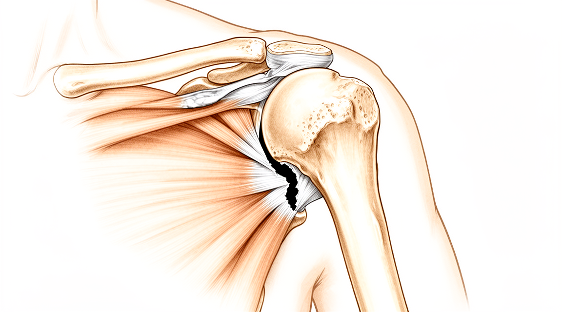
روٹیٹر کف کی مرمت (rotator cuff repair) ایک ایسا آپریشن ہے جس میں کندھے کے پھٹے ہوئے ٹینڈن (tendon)، یعنی وہ مضبوط ڈوری جو پٹھے کو ہڈی سے جوڑتی ہے، کو دوبارہ آپ کے بازو کے اوپری حصے کی ہڈی سے جوڑا جاتا ہے۔ روٹیٹر کف ٹینڈنوں کا وہ گروپ ہے جو آپ کے کندھے کو تھامتا اور حرکت دیتا ہے۔ ہم عام طور پر یہ آپریشن اس وقت تجویز کرتے ہیں جب پھٹاؤ کی وجہ سے ایسا درد ہو جو آپ کو رات کو جگا دیتا ہے، کچھ اٹھاتے وقت یا سر سے اوپر کے کاموں میں درد ہو، یا کمزوری ہو، اور یہ تکلیف آپریشن کے بغیر علاج، جیسے سرگرمیوں میں تبدیلی اور فزیوتھراپی، سے ٹھیک نہ ہوئی ہو۔ گھساؤ سے ہونے والے پرانے پھٹاؤ میں ہم پہلے یہی طریقے آزماتے ہیں۔ اگر پھٹاؤ کسی واضح چوٹ کی وجہ سے ہوا ہو تو ہو سکتا ہے فوراً سرجری کا مشورہ دیا جائے۔
اس کا مقصد درد سے دیرپا نجات اور آپ کے کندھے کی طاقت اور حرکت میں بہتری ہے۔ دردناک پھٹاؤ میں، طویل مدت کے لحاظ سے، سرجری کے ذریعے مرمت سے نمایاں بہتری کا امکان زیادہ ہوتا ہے، اور ہم آپ کے ساتھ مل کر اس پر غور کریں گے اور مشترکہ فیصلہ کریں گے۔
آپریشن سے پہلے
آپ کی مرمت کی منصوبہ بندی کے لیے ہمیں ایکسرے اور اسکین کی ضرورت ہوگی۔ ان میں عام طور پر ایکسرے، اور اکثر آپ کے کندھے کا ایم آر آئی یا الٹراساؤنڈ اسکین شامل ہوتا ہے۔ یہ اسکین دکھاتے ہیں کہ پھٹاؤ کتنا بڑا ہے، ٹینڈن کتنا پیچھے کھنچ گیا ہے، اور پٹھے کی حالت کیسی ہے۔ سرجری سے پہلے کے دنوں میں ہماری ٹیم کی دی گئی ہدایات پر عمل کریں۔ کچھ دوائیں صرف اسی صورت میں بند کریں جب ہم آپ کو کہیں، کیونکہ آپ کے سرجن آپ کو تفصیل بتائیں گے۔ آپ جو کچھ بھی لیتے ہیں، بشمول گولیاں، قطرے اور کریمیں، اس کی لکھی ہوئی فہرست ساتھ لائیں۔ آپریشن کے بعد آپ کو گاڑی میں گھر لے جانے کے لیے کسی کا انتظام کریں، کیونکہ آپ خود گاڑی نہیں چلا سکیں گے۔ ڈھیلے، آرام دہ کپڑے پہنیں جو کندھے کے اوپر سے آسانی سے پہنے جا سکیں۔ آپریشن سے پہلے کے سات گھنٹوں میں نہ کچھ کھائیں اور نہ کچھ پئیں۔ ہم سات گھنٹے اس لیے کہتے ہیں تاکہ اگر آپریشن کی فہرست وقت سے پہلے چل رہی ہو تو آپ کی سرجری کا وقت پہلے کیا جا سکے۔ اگر آپ کو دیگر طبی مسائل ہیں تو ہو سکتا ہے آپریشن کے دن سے پہلے آپ کے خون کے ٹیسٹ ہوں یا بے ہوشی کے ڈاکٹر (anaesthetist) آپ کا معائنہ کریں۔
آپریشن کے دن
آپریشن کے دن آپ ہسپتال کے سرجری کے داخلے کے شعبے میں پہنچیں گے۔ وہاں آپ کا اندراج ہوگا اور آپ کو آپریشن تھیٹر کے لیے تیار کیا جائے گا۔ آپ پہلے وارڈ میں نہیں جائیں گے۔
یہ آپریشن جنرل اینستھیزیا (مکمل بے ہوشی) کے ساتھ ساتھ اعصابی بلاک (regional nerve block)، یعنی ایک انجکشن جو جسم کے ایک حصے کو سُن کر دیتا ہے، کے تحت کیا جاتا ہے۔ بے ہوشی کے ڈاکٹر آپریشن سے پہلے آپ سے ملیں گے اور آپ کو دونوں حصوں کے بارے میں سمجھائیں گے۔
اس کے بعد آپ کو آپریشن تھیٹر لے جایا جائے گا، جہاں آپریشن کیا جاتا ہے۔ آپریشن ختم ہونے کے بعد آپ ریکوری روم میں جاگیں گے۔ وہاں نرسیں آپ کی نگرانی کریں گی جب تک بے ہوشی کی دوا کا اثر ختم ہو رہا ہو۔ جب آپ کی حالت مستحکم ہو جائے گی تو طریقۂ کار اور آپ کی صحت یابی کی رفتار کے مطابق آپ یا تو وارڈ میں منتقل ہوں گے یا گھر چلے جائیں گے۔
آپریشن میں کیا کیا جاتا ہے
یہ دوربین کے ذریعے کیا جانے والا آپریشن (keyhole) ہے۔ آپ کے سرجن کندھے کے اردگرد چند چھوٹے چیرے لگاتے ہیں، جن میں سے ایک پیچھے کی طرف ہوتا ہے، اور جوڑ کے اندر ایک چھوٹے کیمرے کی مدد سے کام کرتے ہیں۔ کیمرا کئی زاویوں سے پھٹاؤ کا واضح منظر دکھاتا ہے، اس لیے مرمت سے پہلے پورے جوڑ کا معائنہ کیا جا سکتا ہے اور پھٹاؤ کی جگہ کو صاف کیا جا سکتا ہے۔
مرمت میں چھوٹے اینکر کی دو قطاریں استعمال ہوتی ہیں جو ہڈی میں اس جگہ لگائے جاتے ہیں جہاں ٹینڈن عام طور پر جڑا ہوتا ہے۔ جوڑ کے قریب والے اینکر نرم سیوچر اینکر (suture anchor)، یعنی ہڈی میں لگایا جانے والا چھوٹا پرزہ جو ٹانکوں کے دھاگے کو تھامتا ہے، ہوتے ہیں، جو سخت پلاسٹک کے بجائے ٹانکوں کے دھاگے کے مواد سے بنے ہوتے ہیں۔ ان کے ٹانکے پھٹے ہوئے ٹینڈن میں سے گزارے جاتے ہیں۔ پھر انہی ٹانکوں کو مزید باہر کی طرف مضبوط طبی معیار کے پلاسٹک سے بنے اینکر کی دوسری قطار تھامتی ہے، اور یہی بیرونی قطار ٹینڈن کو نیچے کھینچ کر ہڈی کے ساتھ چپٹا دباتی ہے۔ دو قطاریں ٹینڈن کو ایک ہی لکیر کے چند نقاط پر پکڑنے کے بجائے گرفت کو ٹینڈن کے ایک وسیع حصے پر پھیلا دیتی ہیں۔
اکثر مرمت کی جگہ پر ٹینڈن کے نیچے ایک حیاتیاتی ڈھانچہ (biological scaffold) رکھا جاتا ہے تاکہ ٹینڈن کو ہڈی پر دوبارہ جڑنے میں مدد ملے۔ آپ اس کے بارے میں مزید ہمارے حصے EnFix حیاتیاتی ڈھانچہ میں پڑھ سکتے ہیں۔
مرمت مکمل ہونے کے بعد چھوٹے چیروں کو ٹانکوں سے بند کر کے زخم کی پٹی سے ڈھانپ دیا جاتا ہے۔ آپ اسی پٹی کے ساتھ گھر جائیں گے، اور ہماری ٹیم آپ کو بتائے گی کہ اسے کب اتارنا ہے۔
آپریشن کے بعد
آپ ریکوری روم میں جاگیں گے، پھر وارڈ میں منتقل ہوں گے۔ آرام کے لیے آپ کا بازو ایک سادہ سلنگ (sling)، یعنی گلے سے لٹکی وہ پٹی جو بازو کو سہارا دیتی ہے، میں رہے گا، اور ورزش اور نہانے دھونے کے لیے اسے اتارا جا سکتا ہے۔ نرسیں آپ کے درد کا جائزہ لیں گی اور آپ کو آرام میں رکھنے کے لیے دوا دیں گی۔ اس آپریشن کے بعد زیادہ تر مریض ایک رات ہسپتال میں رہتے ہیں، البتہ کچھ مریض اسی دن گھر جا سکتے ہیں۔ ہم زخم کی پٹی تقریباً 10 دن تک لگی رہنے دیتے ہیں؛ براہ کرم اس سے پہلے اسے نہ اتاریں، جب تک ہم آپ کو نہ کہیں۔ ہم آپ سے ملاقات کے وقت اسے بدلتے یا اتارتے ہیں۔ پہلے 24 گھنٹے کسی کو آپ کے ساتھ رہنا چاہیے۔ آپ ہماری ٹیم کی ہدایت کے مطابق ہلکی حرکتیں شروع کر سکتے ہیں، اور آپ اسی دن اٹھ کر چلنے پھرنے لگیں گے۔ جب تک آپ سلنگ میں ہیں، آپ ہرگز گاڑی نہ چلائیں۔ جب آپ کے سرجن اجازت دیں، جو عام طور پر چھ ہفتے بعد کے معائنے پر ہوتا ہے، تو آپ دوبارہ گاڑی چلا سکتے ہیں۔ مزید معلومات کے لیے بازو اور ہاتھ کی سرجری کے بعد گاڑی چلانا پڑھیں۔
صحت یابی
پہلے دنوں اور ہفتوں میں آپ کے کندھے میں درد رہے گا اور ہو سکتا ہے اس میں سوجن محسوس ہو۔ یہ آہستہ آہستہ کم ہو جاتا ہے۔ درد کش دوا اور آرام سے مدد ملتی ہے، اور ابتدائی ہفتے گزرنے کے ساتھ زیادہ تر تکلیف کم ہو جاتی ہے۔ درد میں زیادہ تر کمی اور حرکت میں زیادہ تر بہتری پہلے چھ ماہ میں ہوتی ہے، لیکن دو سال تک حالت بہتر ہوتی رہتی ہے۔
آرام کے لیے آپ کا بازو ایک سادہ سلنگ میں رہتا ہے، اور ورزش اور نہانے دھونے کے لیے اسے اتارا جاتا ہے۔ آپ کے فزیوتھراپسٹ پہلے آپ کو ہلکی حرکتیں کروائیں گے، پھر جیسے جیسے ٹینڈن ٹھیک ہوتا جائے، طاقت بڑھانے کی ورزشیں۔ آپ گھر میں فوراً چل پھر سکتے ہیں، لیکن کپڑے پہننے اور چیزیں اٹھا کر لے جانے جیسے کاموں میں آپ کو مدد کی ضرورت ہوگی۔ شروع میں سونا تکلیف دہ ہو سکتا ہے، اور بہت سے لوگوں کو تکیوں کے سہارے اونچا ہو کر آرام کرنا آسان لگتا ہے۔ جیسے جیسے کندھا ٹھیک ہوتا ہے، نیند بتدریج بہتر ہوتی جاتی ہے۔
کچھ مراحل تاریخوں کے بجائے واقعات کی صورت میں آتے ہیں۔ جب آپ کے سرجن آپ کو گاڑی چلانے کی اجازت دے دیں، تو آپ دوبارہ گاڑی چلا سکتے ہیں۔ جب آپ کے فزیوتھراپسٹ آپ کی حرکت اور طاقت سے مطمئن ہوں، تو آپ ہلکی سرگرمیوں پر، اور پھر بھاری سرگرمیوں پر واپس آ سکتے ہیں۔ زیادہ تر لوگ آٹھ ماہ کے اندر کام پر واپس آ جاتے ہیں، البتہ یہ آپ کے کام کی نوعیت پر منحصر ہے۔
صحت یابی ہر شخص میں مختلف ہوتی ہے۔ آپ کی صحت یابی کا دورانیہ مختلف ہو سکتا ہے، اور آپ کے سرجن اور فزیوتھراپسٹ ہر مرحلے پر آپ کی رہنمائی کریں گے۔
کیا مسائل پیدا ہو سکتے ہیں
زیادہ تر مریض ٹھیک رہتے ہیں، لیکن کبھی کبھار مسائل پیدا ہو سکتے ہیں۔ آپ کے سرجن اور ٹیم کسی بھی مسئلے کو جلد پکڑنے کے لیے آپ کی قریب سے نگرانی کرتے ہیں۔
سب سے عام مسئلہ یہ ہے کہ مرمت شدہ ٹینڈن جڑا نہیں رہتا۔ ہو سکتا ہے آپ محسوس کریں کہ کندھے کا درد یا کمزوری واپس آ رہی ہے، بعض اوقات لڑکھڑانے یا کوئی بھاری چیز اٹھانے کے بعد۔ اگر ایسا ہو تو اگلے معائنے پر اپنے سرجن کو بتائیں۔
کندھے کے ٹھیک ہونے کے دوران اکڑاؤ پیدا ہو سکتا ہے۔ آپ کا کندھا جکڑا ہوا محسوس ہو سکتا ہے، اور آپ کو ہاتھ کمر کے پیچھے یا سر کے اوپر لے جانے میں دشواری ہو سکتی ہے۔ زیادہ تر اکڑاؤ فزیوتھراپی سے ٹھیک ہو جاتا ہے، اس لیے انتظار کرنے کے بجائے جلد ہی اپنے فزیوتھراپسٹ یا سرجن سے اس کا ذکر کریں۔
انفیکشن کم ہوتا ہے لیکن اس پر جلد توجہ کی ضرورت ہوتی ہے۔ ایسے گہرے، دھڑکتے ہوئے درد پر نظر رکھیں جو عام درد کش دواؤں سے کم نہ ہو، زخم سے باہر کی طرف پھیلتی ہوئی سرخی، بخار، یا ٹانکوں سے رطوبت کے رسنے پر بھی۔ اسی دن کلینک سے رابطہ کریں، یا اگر آپ کی طبیعت خراب ہو یا سرخی تیزی سے پھیل رہی ہو تو ہسپتال کی ایمرجنسی جائیں۔
رگ میں خون کا لوتھڑا بننا بہت کم ہوتا ہے۔ اس کی علامات پنڈلی کے پچھلے حصے میں اچانک سوجن اور چھونے پر درد ہیں۔ اگر آپ کو ایسا محسوس ہو تو فوراً طبی مدد حاصل کریں۔
ٹینڈن کو تھامنے والے چھوٹے اینکر کبھی کبھار جلن (irritation) پیدا کر سکتے ہیں۔ ہو سکتا ہے آپ کو کلک ہونے یا رگڑ کا احساس ہو، یا ایسا درد ہو جو جاری رہے اور ٹھیک نہ ہو۔ اپنے معائنے پر اس کا ذکر کریں تاکہ آپ کے سرجن اس کا جائزہ لے سکیں۔
کندھے کے قریب کسی عصب میں جلن بہت کم ہوتی ہے۔ اس سے بازو میں نیچے کی طرف جھنجھناہٹ، سُن پن یا عجیب احساسات ہو سکتے ہیں۔ اپنی اگلی ملاقات پر اس کا ذکر کریں۔
کچھ لوگوں کو مرمت کے بعد بھی کندھے میں درد رہتا ہے، چاہے ٹینڈن جڑ چکا ہو۔ اگر درد برقرار رہے تو آپ کے سرجن آپ کا معائنہ کریں گے اور بات کریں گے کہ مزید کون سے طریقے مددگار ہو سکتے ہیں۔
اگر آپ کو دیگر اہم طبی مسائل ہیں، جیسے ذیابیطس (شوگر)، یا آپ کا مدافعتی نظام کمزور ہے، تو منصوبہ بندی کی ملاقات پر ان کا ذکر کریں۔ یہ زخم بھرنے پر اثر ڈال سکتے ہیں، اور سرجری کے بعد ہم ایسے مریضوں کی زیادہ قریب سے نگرانی کرتے ہیں۔
اگر آپ تفصیل جاننا چاہتے ہیں تو اس صفحے پر پیچیدگیوں کی جدول میں ان کی عام شرحیں دی گئی ہیں۔
ہمیں کب فون کریں
اس آپریشن کے بعد زیادہ تر مسائل کم ہوتے ہیں، لیکن کچھ پر جلد توجہ کی ضرورت ہوتی ہے۔ اگر آپ کو بخار ہو، اگر زخم زیادہ سرخ ہو جائے یا اس سے رطوبت رسنے لگے، یا اگر آپ کے کندھے کا درد اچانک بہت بڑھ جائے تو ہمیں فون کریں۔ اگر آپ کو پنڈلی کے پچھلے حصے میں سوجن یا چھونے پر درد محسوس ہو، یا آپ کا سانس پھولنے لگے، تو ہسپتال کی ایمرجنسی جائیں، کیونکہ یہ خون کے لوتھڑے کی علامات ہو سکتی ہیں۔ اگر آپ کا بازو سُن محسوس ہو، اگر آپ اسے حرکت نہ دے سکیں، یا اگر بازو میں نیچے کی طرف جھنجھناہٹ ٹھیک نہ ہو تو فوراً ہمیں فون کریں۔ اگر آپ کی طبیعت خراب ہو اور آپ فکرمند ہوں تو اپنے احساس پر بھروسہ کریں اور طبی مدد حاصل کریں۔ ہم چاہیں گے کہ آپ انتظار کرنے کے بجائے ہم سے رابطہ کریں۔
مزید تفصیل
Advanced reading: the deeper science (optional)
یہ حصہ اس سے زیادہ تفصیل میں جاتا ہے جتنی آپ کو اپنے علاج کے فیصلوں کے لیے ضرورت ہے۔ روٹیٹر کف کی مرمت کے بارے میں یہ اضافی مطالعہ اس لیے فائدہ مند ہے کہ جو عوامل آپ کے نتیجے کی بہترین پیش گوئی کرتے ہیں وہ زیادہ تر سرجری سے متعلق نہیں ہیں، اور ان میں سے دو سب سے مضبوط عوامل ایسے ہیں جن پر آپ آپریشن سے پہلے خود کچھ کر سکتے ہیں۔
تمباکو نوشی صرف زخم کو نہیں، ٹینڈن کو بھی بدل دیتی ہے
رضامندی (consent) کی ہر گفتگو میں تمباکو نوشی کا ذکر ہوتا ہے اور عام طور پر اسے زخم بھرنے کے مسئلے کے طور پر دیکھا جاتا ہے۔ ٹینڈن سے متعلق اعداد و شمار اس سے زیادہ واضح ہیں۔ 73,817 مریضوں کے اعداد و شمار کو یکجا کرنے پر، روٹیٹر کف کی مرمت کے بعد تمباکو نوشی کرنے والوں میں ٹینڈن کے دوبارہ پھٹنے (retear) کی شرح نمایاں طور پر زیادہ تھی، جبکہ زیادہ تر طبی اسکور تمباکو نوشی کرنے والوں اور نہ کرنے والوں میں ملتے جلتے تھے، سوائے کم کانسٹنٹ اسکور (Constant score) کے [1]۔
یہی امتزاج دلچسپ پہلو ہے۔ اسکور بظاہر کافی حد تک ایک جیسے نظر آتے ہیں، لیکن اندر سے مرمت کے ڈھانچے کے طور پر ناکام ہو جانے کا امکان زیادہ ہوتا ہے۔ تمباکو نوشی آپریشن کے بعد کے احساس کو مختلف نہیں بنا رہی؛ یہ مرمت کے جڑے رہنے کا امکان کم کر رہی ہے۔
اس کا نتیجہ یہ ہے کہ ساری زندگی تمباکو نوشی چھوڑنے کی نصیحت سننے کے دوران، سرجری کے آس پاس کے ہفتے سب سے قیمتی موقع ہیں۔ ٹینڈن ہڈی سے دوبارہ جڑنے کی کوشش کر رہا ہوتا ہے، اور یہ عمل خون کی فراہمی پر منحصر ہے۔
سماجی محرومی نتیجے کی اتنی ہی مضبوط پیش گوئی کرتی ہے جتنی جسمانی حیاتیات
یہ وہ نتیجہ ہے جو اس آپریشن کے بارے میں گفتگو کے انداز کو سب سے زیادہ بدل دیتا ہے۔ 102,372 مریضوں کے ایک جائزے میں، صحت کے سماجی عوامل (social determinants of health)، یعنی وہ حالات جن میں لوگ زندگی گزارتے ہیں، نہ کہ ان کے ٹینڈن کی ساخت، روٹیٹر کف کی مرمت کے بعد بدتر طبی نتائج اور مریضوں کے بتائے ہوئے بدتر نتائج سے منسلک تھے، جن میں آپریشن کے بعد زیادہ پیچیدگیاں اور مرمت کی زیادہ ناکامیاں شامل تھیں [2]۔
کف کی مرمت کے بعد بحالی میں کئی مہینے لگتے ہیں، اس کے لیے ملاقاتوں پر حاضر ہونا پڑتا ہے، اور بازو کو استعمال نہ کرنے کی گنجائش ہونی چاہیے۔ یہ صرف فیصلے نہیں بلکہ زندگی کے حالات بھی ہیں۔ جب یہ حالات موجود نہ ہوں تو مرمت کا نتیجہ خراب ہوتا ہے، اور یہ علاج کے راستے کے بارے میں ایک حقیقت ہے، مریض کے بارے میں کوئی رائے نہیں۔ اگر تھراپی تک پہنچنا یا کام سے چھٹی لینا مشکل ہوگا، تو اس بات کی جگہ آپریشن سے پہلے کی گفتگو ہے، جہاں منصوبہ اسی کو سامنے رکھ کر بنایا جا سکتا ہے۔
بعد میں اکڑاؤ عام ہے، اور اس کے خطرے کے عوامل واضح نہیں
مرمت کے بعد حرکت کا کم ہو جانا وہ پیچیدگی ہے جس کی مریض سب سے کم توقع رکھتے ہیں۔ 23,257 مریضوں کے اعداد و شمار کو یکجا کرنے پر، دستیاب شواہد نے مرد ہونے اور زیادہ عمر کو آپریشن کے بعد کندھے کے اکڑاؤ کے خلاف ممکنہ حفاظتی عوامل کے طور پر ظاہر کیا [3]۔
یہ دونوں باتیں عام سوچ کے برعکس ہیں، کیونکہ سمجھا جاتا ہے کہ زیادہ عمر کے کندھے زیادہ آسانی سے اکڑتے ہیں، اور بظاہر یہاں ایسا نہیں ہوتا۔ اس کا عملی فائدہ توقعات طے کرنے میں ہے: کف کی مرمت کروانے والی کم عمر خاتون کو اوسط کی بنیاد پر تسلی دینے کے بجائے یہ بتایا جانا چاہیے کہ اکڑاؤ خاص طور پر اس کے لیے ایک حقیقی امکان ہے، اور یہ کہ ابتدائی حرکت کی ورزشیں اہم ہیں۔
آپ کے فیصلے کے لیے اس کا مطلب
ان میں سے کوئی بات آپریشن کے خلاف دلیل نہیں ہے۔ یہ بتاتی ہے کہ شائع شدہ کامیابی کی شرح ان لوگوں کی اوسط ہے جن کی تمباکو نوشی کی عادت، حالات اور آبادیاتی خصوصیات (عمر، جنس وغیرہ) آپ سے مختلف ہیں، اور یہ کہ آپ کی اپنی شرح بدلی جا سکتی ہے۔ تمباکو نوشی چھوڑنا، اور پہلے سے ایسا انتظام کرنا کہ آپ بحالی کی ملاقاتوں پر حاضر ہو سکیں، ایسے اقدامات ہیں جن کے پیچھے شواہد موجود ہیں، اور یہ بات ان زیادہ تر چیزوں کے بارے میں نہیں کہی جا سکتی جن کی فکر کرنے کو مریضوں سے کہا جاتا ہے۔
حوالہ جات
[1] Fan N, Yuan S, Du P, Wu Q, Li J, Kong X, et al. The effects of smoking on clinical and structural outcomes after rotator cuff repair: a systematic review and meta-analysis. J Shoulder Elbow Surg. 2022;31(3):656-67. https://doi.org/10.1016/j.jse.2021.10.026
[2] Mandalia K, Ames A, Parzick JC, Ives K, Ross G, Shah S. Social determinants of health influence clinical outcomes of patients undergoing rotator cuff repair: a systematic review. J Shoulder Elbow Surg. 2023;32(2):419-34. https://doi.org/10.1016/j.jse.2022.09.007
[3] Stojanov T, Modler L, Müller AM, Aghlmandi S, Appenzeller-Herzog C, Loucas R, et al. Prognostic factors for the occurrence of post-operative shoulder stiffness after arthroscopic rotator cuff repair: a systematic review. BMC Musculoskelet Disord. 2022;23(1). https://doi.org/10.1186/s12891-022-05030-4
Evidence & references
This is the clinical evidence summary written for health professionals. It is technical, and it lists the research this page was built from. You do not need to read it to understand your treatment or to make a decision about it.
Anatomy & Pathophysiology
Anatomy
- The rotator cuff is a complex of four muscles arising from the scapula whose tendons blend with the subjacent capsule as they attach to the humeral tuberosities [8].
- The subscapularis arises from the anterior aspect of the scapula and attaches over much of the lesser tuberosity [8].
- The supraspinatus arises from the fossa superior to the scapular spine, passes beneath the acromion and acromioclavicular joint, and attaches to the superior aspect of the greater tuberosity [8].
- The infraspinatus arises from the fossa below the scapular spine and attaches to the posterolateral aspect of the greater tuberosity [8].
- The teres minor arises from the lower lateral aspect of the scapula and attaches to the lower portion of the greater tuberosity [8].
- The subscapularis is innervated by the upper and lower subscapular nerves [8].
- The supraspinatus is innervated by the suprascapular nerve after it passes through the suprascapular notch [8].
- The infraspinatus is innervated by the suprascapular nerve after it passes through the spinoglenoid notch [8].
- The teres minor is innervated by a branch of the axillary nerve [8].
- Histologic studies describe the rotator cuff tendons as having five distinct layers [8].
- The most superficial layer of the rotator cuff tendon consists of coracohumeral ligament fibers oriented obliquely to the muscle axis [8].
- The second layer of the rotator cuff tendon is composed of large bundles of fibers extending from the supraspinatus tendon over the biceps tendon groove [8].
- The third layer contains smaller, less tightly packed tendon fascicles with less uniform orientation [8].
- The fourth layer is composed of loose connective tissue with thick bands of collagen fibers that merge with the coracohumeral ligament at the anterior edge of the supraspinatus [8].
- The fifth and deepest layer is a continued sheet of collagen fibrils composing the superior joint capsule [8].
- There is significant interdigitation and overlap of the supraspinatus and infraspinatus tendons near the footprint [8].
- The infraspinatus insertion occupies the preponderance of the footprint on the greater tuberosity [8].
- The supraspinatus insertion occupies a smaller portion of the greater tuberosity than previously believed [8].
- The long head of the biceps tendon attaches to the supraglenoid tubercle, runs between the subscapularis and supraspinatus, and exits through the bicipital groove under the transverse humeral ligament [8].
- The coracohumeral ligament and transverse humeral ligament keep the biceps tendon aligned in the groove [8].
- The rotator cable is a thick bundle of fibers running perpendicular to the supraspinatus tendon fibers, connecting the supraspinatus and infraspinatus tendons [9].
- The rotator cable is divided into anterior, middle, and posterior segments [10].
- The anterior segment of the rotator cable forms the lateral part of the rotator interval [10].
- The middle segment of the rotator cable lies under the supraspinatus tendon [10].
- The posterior segment of the rotator cable is covered by the infraspinatus tendon and ends at the insertion region between the infraspinatus and teres minor tendons [10].
- The supraspinatus footprint measures 13 mm in width medial-lateral and 20 mm anteroposterior [9].
- The infraspinatus footprint measures 14 mm in width and 20 mm superoinferior [9].
- A 7 mm medial-lateral tear corresponds to a 50% partial thickness tear of the supraspinatus [9].
- The hypovascular critical zone is located on the articular side of the rotator cuff close to the insertion on the greater tuberosity [9].
- The rotator cuff is a sheet of conjoined tendons closely applied over the shoulder capsule, inserting mainly into the greater tuberosity, with the subscapularis inserting into the lesser tuberosity [5].
- The coracoacromial arch is a fibro-osseous canopy formed by the acromion process posterosuperiorly, the coracoid process anteriorly, and the coracoacromial ligament joining them [5].
- The subacromial bursa separates the rotator cuff tendons from the coracoacromial arch to allow gliding [5].
- The primary passive stabilizers of the glenohumeral joint are the capsule and scapulohumeral ligaments [12].
- The glenohumeral joint capsule is thickest in the inferior pouch at 2.8 mm, 2.4 mm in the anterior portion, and 2.2 mm in the posterior portion [12].
- The superior glenohumeral ligament crosses the rotator interval capsule and lies between the supraspinatus and subscapularis tendons [12].
- The coracohumeral ligament originates at the base of the coracoid, blends into the cuff tendons, and inserts into the greater and lesser tuberosities [12].
- The middle glenohumeral ligament originates anterosuperiorly on the glenoid and inserts midway along the anterior humeral articular surface adjacent to the lesser tuberosity [12].
Pathophysiology
- Rotator cuff tears represent a spectrum of disease progressing from tendinitis to tendinosis, rotator cuff tears, and finally cuff arthropathy [9].
- Intrinsic degeneration involves age-related changes in collagen, proteoglycan, water content, and vascularity, usually starting on the articular side of the supraspinatus and infraspinatus [9].
- Extrinsic injury mechanisms involve chronic impingement on the coracoacromial arch, with tears usually starting on the bursal side of the tendon [9].
- Acute traumatic tears typically occur after a fall or shoulder dislocation in patients younger than 40 years [9].
- Risk factors for rotator cuff tear development include age, smoking, female sex, family history, diabetes, and high cholesterol [9].
- Partial-thickness rotator cuff tears have a limited ability to spontaneously heal [4].
- As many as 53% of partial-thickness rotator cuff tears will progress in tear size [4].
- A portion of partial-thickness rotator cuff tears will progress to full-thickness tears [4].
- Small full-thickness rotator cuff tears and painful partial-thickness tears become 25% to 50% larger within 3 to 4 years [9].
- Larger rotator cuff lesions progress faster than smaller ones [9].
- The prevalence of rotator cuff tears increases with each decade of life after the age of 50 years [23].
- Rotator cuff tear prevalence ranges from 13% in patients in their fifties to 50% in patients aged 80 years or older [23].
- One-quarter of patients above 60 years of age and half of patients above 80 years will have a rotator cuff tear [21].
- Symptom emergence in previously asymptomatic shoulders is linked to tear progression from partial to full thickness, full-thickness tear size worsening, muscle atrophy or fatty infiltration development, and new biceps pathology [23].
- Fatty muscle degeneration is a factor in rotator cuff pathology [18].
- Genetic variations are associated with rotator cuff disease [14].
- Two correlated single nucleotide polymorphisms are associated with full-thickness rotator cuff tears [9].
- The critical shoulder angle is higher in patients with rotator cuff tears compared to asymptomatic individuals [10].
- A critical shoulder angle greater than 38 degrees and an acromial index greater than 0.7 are associated with higher retear rates after arthroscopic rotator cuff repair [11].
- The critical shoulder angle is a static measure that does not change over time [12].
- Calcific tendinitis typically affects patients aged 30 to 60 years and women more commonly than men [22].
- The supraspinatus tendon is most often involved in calcific tendinitis [22].
- Calcific tendinitis involves three main stages: precalcific, calcific, and postcalcific [22].
- The precalcific stage of calcific tendinitis consists of predominantly fibrocartilaginous metaplasia within less vascular areas of the tendon [22].
- The formative phase of the calcific stage involves matrix vesicles uniting to form calcium hydroxyapatite deposits separated by fibrocollagenous tissue [22].
- The resorption phase of calcific tendinitis involves an inflammatory response and is generally the most painful phase [22].
- Cuff tear arthropathy is the final stage of the shoulder impingement syndrome spectrum, characterized by long-term insufficient massive rotator cuff tears and superior migration of the humeral head [13].
- Mechanical factors in cuff tear arthropathy include insufficient cuff, superior migration of the humeral head, instability, eccentric wear of the glenoid, and humeral head deformity [13].
- Nutritional factors in cuff tear arthropathy include hypomobility-induced cartilage atrophy, poor nutrition leading to decreased glycosaminoglycans, dehydration, and subchondral osteoporosis [13].
- Crystalline-induced arthropathy in cuff tear arthropathy involves synovial-based matrix protein degradation destroying rotator cuff tendons and cartilage, with end-stage calcium-phosphate crystal deposition [13].
Classification
- Partial-thickness rotator cuff tears are common in the general population, with an increased incidence with increasing age [4].
- Sher et al. showed an overall prevalence of asymptomatic partial-thickness rotator cuff tears of 20% [4].
- The prevalence of asymptomatic partial-thickness rotator cuff tears increased to 26% in patients older than 60 years [4].
- Partial-thickness rotator cuff tears can be classified as articular-sided, bursal-sided, or intratendinous tears [4].
- Ellman described a classification of partial-thickness rotator cuff tears based on location and depth of tearing noted at the time of shoulder arthroscopy [4].
- Tears are classified as articular-sided (A), bursal-sided (B), or intratendinous (C) [4].
- Tears are grade 1 if involving 3 mm or less of tendon [4].
- Tears are grade 2 if involving 3 to 6 mm of tendon [4].
- Tears are grade 3 if involving more than 6 mm of tendon [4].
- Grade 3 tears represent tears of more than 50% of tendon width based on previous studies noting the width of the supraspinatus footprint [4].
- With MRI evaluation, these tears are classified as low grade or high grade depending on whether they involve less than or more than 50% of the tendon width [4].
Clinical Presentation
History and Symptoms
- Patients with rotator cuff disorders commonly present with pain over the front and lateral aspect of the shoulder [5].
- Weakness on abduction is a presenting symptom associated with supraspinatus involvement, rotator cuff tears, and tendinitis [5].
- Pain over the front of the shoulder is associated with biceps pathology [5].
- Weakness is present if the rotator cuff or biceps tendon has ruptured [5].
- A clear history of trauma resulting in acute pain and weakness strongly suggests an acute rotator cuff tear and warrants expeditious workup [24].
- In cases of chronic rotator cuff disease, patients often describe an insidious onset of lateral and/or anterior shoulder pain associated with overhead activities [24].
- Night pain is a common presenting symptom of chronic rotator cuff disease [24].
- A family or personal history of rotator cuff disease makes the diagnosis more likely [24].
- Patients with full-thickness rotator cuff tears are usually aged over 45 and give a history of refractory shoulder pain with increasing stiffness and weakness [25].
- A full-thickness tear may occur spontaneously after a sprain or jerking injury of the shoulder, resulting in sudden pain and an inability to abduct the arm [25].
- In long-standing cases of partial or complete rotator cuff rupture, secondary osteoarthritis of the shoulder may supervene, resulting in severely restricted movements [25].
Physical Examination
- Basic physical examination for rotator cuff tears consists of assessment of range of motion in adducted and abducted positions, assessment of strength, and examination of associated structures such as the biceps and acromioclavicular joint [24].
- The empty can test has a sensitivity of 71.7% and a specificity of 64.6% for full-thickness supraspinatus tears [24].
- The lift-off and belly-press tests have high specificity but low sensitivity for full-thickness subscapularis tears [24].
- An external rotation lag sign at the side likely indicates a large posterosuperior tear involving the infraspinatus [24].
- A positive hornblower sign suggests a massive posterosuperior cuff tear that prohibits the active positioning of the hand in space [24].
- The painful arc test has a sensitivity of 71% and a specificity of 81% for rotator cuff disease [24].
- The cross-body adduction test has a sensitivity of 75% and a specificity of 61% for rotator cuff disease [24].
- The Hawkins test has a sensitivity of 76% and a specificity of 48% for rotator cuff disease [24].
- The Neer test has a sensitivity of 64–68% and a specificity of 30–61% for rotator cuff disease [24].
- The Yocum test has a sensitivity of 79% and a specificity of 40% for rotator cuff disease [24].
- The passive abduction test has a sensitivity of 74% and a specificity of 10% for rotator cuff disease [24].
- The external rotation lag test has a sensitivity of 47% and a specificity of 94% for full-thickness rotator cuff tears [24].
- The internal rotation lag test has a sensitivity of 97% and a specificity of 83% for full-thickness rotator cuff tears [24].
- The drop arm test has a sensitivity of 24% and a specificity of 93% for rotator cuff disease [24].
- The dropping sign has a sensitivity of 73% and a specificity of 77% for full-thickness rotator cuff tears [24].
- The Gerber (lift-off) test has a sensitivity of 34–68% and a specificity of 50–77% for rotator cuff disease [24].
- The external rotation resistance test has a sensitivity of 63% and a specificity of 75% for rotator cuff disease [24].
- The full can test has a sensitivity of 75% and a specificity of 68% for rotator cuff disease [24].
- The Patte test has a sensitivity of 58% and a specificity of 60% for rotator cuff disease [24].
- The empty can (Jobe) test has a sensitivity of 71% and a specificity of 49% for rotator cuff disease [24].
- The resisted abduction test has a sensitivity of 58% and a specificity of 20% for rotator cuff disease [24].
- A positive Hawkins and Neer test combination has a sensitivity of 78% and a specificity of 50% for rotator cuff disease [24].
- Partial tears may occur within the substance or on the deep surface of the cuff, permitting active abduction with a painful arc [25].
- Wasting of the supraspinatus and infraspinatus muscles is usually present in full-thickness rotator cuff tears [25].
- Testing the biceps may reveal an old tear of the long head of the biceps tendon in patients with full-thickness rotator cuff tears [25].
- Tenderness of the acromioclavicular joint is often present in patients with full-thickness rotator cuff tears [25].
- If active abduction is possible after injecting a local anesthetic into the subacromial space, the tear is likely only partial [25].
- If active abduction remains impossible after injecting a local anesthetic into the subacromial space, a complete tear is likely [25].
- Clinical tests for long head of the biceps tendon pathology, including the O’Brien, Yergason, Speed, and direct palpation tests, have limited specificity [31].
- A history of radiating anterior shoulder pain may inform the examiner of pain generation from the long head of the biceps tendon [31].
Diagnostic Imaging
- The goal of diagnostic imaging for rotator cuff tears is to determine the presence, size, and orientation of the tear, evaluate the healing capacity of the tendon, and assess associated pathology such as long head biceps tendinitis, acromioclavicular joint pathology, and arthrosis [24].
- For full-thickness rotator cuff tears, ultrasonography approaches the sensitivity and specificity of MRI for detecting the presence of a tear with an experienced practitioner [24].
- Ultrasonography is relatively inexpensive and allows for dynamic testing, guided injections, and immediate feedback [24].
- MRI accurately assesses muscle, bone, and cartilage, which has advantages for surgical planning [24].
- MRI continues to be the imaging modality of choice for most providers, with ultrasonography becoming common in certain centers [24].
- Arthroscopic examination of the long head of the biceps tendon is limited to the intra-articular tendon and proximal groove, missing less common distal biceps groove lesions [31].
Investigations
Imaging Modalities and Diagnostic Accuracy
- MRI is indicated in younger, active patients with acute rotator cuff tears and in patients with chronic rotator cuff tears in whom a trial of nonoperative treatment has failed [16].
- MRI allows the surgeon to characterize the location, size, and amount of retraction of the rotator cuff tear [16].
- MRI allows the surgeon to characterize the degree of atrophy and fatty infiltration of the rotator cuff musculature [16].
- MRI is used to define the extent of tear, degree of tear retraction, and presence of muscular atrophy [17].
- MRI is key for evaluating fatty infiltration, although the Goutallier classification was originally based on CT [17].
- Ultrasonography is increasing in popularity as a tool for diagnosis of rotator cuff disease [17].
- Ultrasonography is increasing in popularity as a tool for confirmation of intraarticular or subacromial location of injections [17].
- Ultrasonography is more accurate for full-thickness rotator cuff tears, comparable to MRI [22].
- Radiographic views of the subacromial space such as the supraspinatus outlet view may show a spur on the undersurface of the acromion, causing narrowing of the subacromial space [15].
- Radiographs may demonstrate classic changes within the acromion or coracoacromial ligament, including spurring and calcification [17].
- Radiographs may demonstrate cystic changes within the greater tuberosity [17].
- With chronic rotator cuff disease, superior migration of the humeral head with extensive degenerative change may be present on radiographs [17].
Specific Radiographic and Sonographic Signs
- The tangent sign is defined as failure of the supraspinatus muscle belly to cross a line from the superior border of the coracoid to the superior border of the scapular spine [17].
- The tangent sign correlates with muscle atrophy and fatty infiltration of the supraspinatus [17].
- Patients with the presence of the tangent sign are more likely to have an irreparable rotator cuff tear [17].
- A positive tangent sign predicts the repairability of rotator cuff tears [21].
- Irreparable tears are more likely to occur when the acromiohumeral distance appears shorter than 7 mm on AP radiograph [17].
- Ultrasonography can be used to evaluate fatty degeneration of the rotator cuff muscles [23].
Anatomical and Genetic Risk Factors
- The critical shoulder angle (CSA) is a static measure that did not change over time in a study of 1,552 radiographs [12].
- CSA was higher in the patient group with rotator cuff tears compared to controls [12].
- Patients with CSA greater than 38° and acromial index (AI) greater than 0.7 had higher retear rates after arthroscopic rotator cuff repair [11].
- Patients with CSA greater than 38° and AI greater than 0.7 had similar functional rates compared to control patients [11].
- A study of 147 patients up to 2-year follow-up did not see a difference in functional scores between patients with higher CSA/AI and patients with lower values [13].
- A study of scapular anatomy found statistically significant differences in the critical shoulder angle between asymptomatic individuals, those with osteoarthritis, and patients with full-thickness rotator cuff tears [3].
- Genome-wide association studies have identified single-nucleotide polymorphisms associated with full-thickness rotator cuff tears [3].
- Evidence exists for an inherited predisposition contributing to the risk for rotator cuff disease [2].
Tear Characteristics and Prognostic Indicators
- The quality of the rotator cuff musculature is classified according to the degree of fatty infiltration originally described by Goutallier et al. for CT evaluation and modified by Fuchs et al. for MRI evaluation [16].
- In the Goutallier classification, grade 0 is normal muscle, grade 1 has some fatty streaks, grade 2 has more muscle than fat, grade 3 has equal amounts of muscle and fat, and grade 4 has more fat than muscle [16].
- Goutallier grades 3 and 4 are indications of a long-term chronic rotator cuff tear [16].
- Goutallier grades 3 and 4 have a higher potential for failure when surgery is undertaken and likely are deemed irreparable [16].
- Tears with Goutallier grades III and IV, if accompanied by a tendinous stump of less than 15 mm and a positive tangent sign, have a 90% failure rate [16].
- Larger, more retracted tears greater than 40 mm in length or width are characterized by fatty atrophy [17].
- Larger, more retracted tears greater than 40 mm in length or width are characterized by supraspinatus width of less than 5 mm at the glenoid margin [17].
- Tear progression correlates with presenting tear size [3].
- In a cohort of 122 patients with full or partial rotator cuff tears, 53% increased tear size over a minimum of 6 months [3].
- Symptoms of pain do not correlate with rotator cuff tear severity in a cross-sectional study of 393 patients with symptomatic atraumatic full-thickness rotator cuff tears [3].
- Patient-reported outcomes and pain are not correlated with rotator cuff healing on ultrasonography or MRI [19].
- Postoperative strength is better in individuals with healed rotator cuffs versus those with defects after repair [19].
Treatment
Indications and Patient Stratification
- Neither American Academy of Orthopaedic Surgeons clinical practice guidelines nor Cochrane systematic reviews provide guidance on the management of rotator cuff tears [1].
- Clinical decision-making for the management of rotator cuff tears lacks consensus among orthopedic surgeons [1].
- Patients are generally divided into three categories based on the risk of nonoperative treatment and benefits of surgical intervention: those needing urgent or early operative repair, those who can benefit from a trial of conservative treatment, and those best suited for nonoperative treatment [1].
- Symptomatic rotator cuff injuries affect up to 30% of the population [7].
- The prevalence of full-thickness rotator cuff tears in the aging population is estimated to be as high as 30% [7].
- Surgical repair or reconstruction is generally the treatment of choice for rotator cuff injuries depending on injury factors, activity level, functional status, and occupation [7].
- Alternative options such as arthroplasty can be recommended when tears are irreparable or the patient has concomitant advanced glenohumeral arthrosis [7].
Non-Operative Management
- Treatment of rotator cuff tears begins with nonsurgical measures including activity modification, physical therapy, nonsteroidal anti-inflammatory medications, and corticosteroid injection [7].
- Most partial-thickness rotator cuff tears are best initially managed with nonoperative treatment [4].
- Surgical treatment for partial-thickness rotator cuff tears is indicated for patients in whom nonoperative treatment fails [4].
Partial-Thickness Tear Management
- Partial-thickness rotator cuff tears have a limited ability to spontaneously heal as shown by histological and radiographic studies [4].
- A portion of partial-thickness rotator cuff tears will progress to full-thickness rotator cuff tears [4].
- Tears involving more than 50% of tendon width are best treated with repair [4].
- Tears involving less than 50% of tendon width are best treated with debridement and potential decompression [4].
- A higher failure rate of debridement has been suggested for partial-thickness bursal-sided rotator cuff tears compared to articular-sided rotator cuff tears [4].
- Some surgeons favor repair in even low-grade bursal-sided tears due to the higher failure rate of debridement [4].
- There is no difference in functional outcome scores or re-tear rates between in situ rotator cuff repair and completion to a full-thickness rotator cuff tear with subsequent repair [4].
- For tears with poor-quality tendon remaining and involving more than 80% of tendon thickness, debridement is favored [4].
- Delamination-type tears of the articular side require a transtendinous repair [4].
- Intratendinous tears are repaired side-to-side using arthroscopic technique after the tear is opened and the edge is debrided back slightly to promote local healing [4].
- Extensive debridement is unnecessary for intratendinous tears [4].
Operative Techniques: Arthroscopic
- Arthroscopic repair is the current standard of care for rotator cuff repair [7].
- The superiority of arthroscopic versus open or mini-open repair is still somewhat controversial [7].
- Arthroscopic repair has been shown to have similar outcomes and failure rates compared to other techniques, with decreased short-term pain and more rapid return to activity [7].
- Advances in arthroscopic equipment and increased surgeon familiarity have made arthroscopic surgery the preferred method of rotator cuff repair for many shoulder surgeons [27].
- Arthroscopy allows a more comprehensive assessment of intra-articular pathology and rotator cuff tear configuration by viewing from multiple angles [27].
- Arthroscopy facilitates tendon mobilization through precise releases of adhesions, leading to an improved ability to anatomically reduce the edge and create a tension-free repair [27].
- Arthroscopic repair minimizes injury to the deltoid muscle and preserves the acromial deltoid origin, eliminating the risk of deltoid dehiscence [27].
- A key theoretical benefit of arthroscopic repair is decreased postoperative pain secondary to less soft tissue trauma, which aids in postoperative rehabilitation and earlier resumption of range of motion [27].
- Anatomic footprint restoration is possible with arthroscopic repair using fixation at both the suture-tendon interface and the anchor-bone interface [27].
- Arthroscopic repair is performed with suture anchors in various configurations, including single-row, double-row, and transosseous-equivalent repair techniques [7].
- Controlled laboratory studies have generally shown superiority of double-row techniques over single-row in terms of initial and ultimate failure strength, decreased gap formation, decreased strain and suture cut-through, and improved vascularity in transosseous-equivalent double-row repair [7].
- While biomechanical studies show double-row repair outperforms single-row repair in failure strength, superior clinical results with double-row fixation over single-row fixation are still controversial [27].
Operative Techniques: Open and Mini-Open
- Traditional open repairs, mini-open deltoid split approaches, and arthroscopic repair are described approaches for rotator cuff repair [7].
- Mini-open rotator cuff repair allows direct assessment of the nature of the cuff tear, the quality of the remaining tissue, and the ease with which the tendon edge can be restored to the normal insertion site with the arm in an adducted position [32].
- The transosseous technique for attachment of the tendon to the tuberosity in mini-open repair is simple, expeditious, avoids issues related to suture anchors, and enables the surgeon to find sufficiently strong bone to hold the sutures [32].
- The creation of a trough into which the tendon is inserted increases the surface area of the repair, stimulates local stem cells and growth factors, excludes joint fluid from the repair site, and places sutures laterally so that they do not rub beneath the coracoacromial arch [32].
- In open repair, an anterior acromioplasty is an important part of rotator cuff surgery, and the results of repair without decompression are not as good as the results using the combined procedure [6].
- In open repair, tears usually begin at the supraspinatus insertion and retract into its fossa under the acromioclavicular joint [6].
- Most rotator cuff tears are transverse but also have a longitudinal component, making them oval or triangular [6].
- All but the smallest tears need to be advanced anteriorly and laterally, not just laterally, to restore anatomic position and correct muscle-tendon unit length [6].
- In tears of more than 2 to 3 cm, the infraspinatus tendon is involved as well [6].
- Mobilization in open repair begins posteriorly with the infraspinatus using a blunt probe or a finger to release adhesions inside and outside the joint [6].
- Dissection below the level of the teres minor during mobilization should be avoided to prevent injury to the axillary nerve in the quadrangular space or the suprascapular nerve in the area of the spinoglenoid notch [6].
- If supraspinatus and infraspinatus tendons are retracted so far that adequate length cannot be obtained with tendon mobilization, the capsule is incised at its insertion into the glenoid labrum [6].
- The goals of mobilization in open repair are to obtain tissue of adequate strength, position it anatomically for repair without damage to innervation and without compromise of deltoid function, and decompress the subacromial space to prevent further mechanical impingement on repaired cuff tissue [6].
- The best results in open repair are obtained with the double-row technique, suturing the tendon to bone in a cancellous trough in combination with suture anchor fixation [6].
- Using transosseous tunnels through the greater tuberosity increases the surface area of tendon-to-bone healing and more closely restores the anatomic footprint [6].
- In open repair, sutures are placed 5 to 10 mm from the free edge of the tear using a double loop technique in a horizontal mattress manner [6].
- A 3 mm wide shallow trough is created running the length of the exposed bone of the greater tuberosity to accommodate the thickness of the supraspinatus and infraspinatus tendons [6].
- Two or three rotator cuff suture anchors are placed immediately medial to the trough at a 45-degree angle [6].
- Holes for sutures are drilled 2 to 3 cm distal to the trough and spaced at least 1 to 2 cm apart on the cortical humeral surface [6].
- Most open repairs are done with the shoulder in 0 degrees of abduction [6].
- If the lateral humeral cortex is fractured during tying down of the suture or construction of the suture tunnel, anchors can be used as a salvage procedure [6].
Biological and Mechanical Factors
- The rotator cuff presents special challenges for tendon healing because its tendons are intra-articular and intrasynovial, there is a complex mechanical loading environment, the native fibrocartilaginous insertion to the greater tuberosity is not reconstituted, and there is almost always an element of chronic tendinopathy leading to impaired biological healing capacity on both the tendon and bone side [7].
- Factors known to be associated with failure of rotator cuff repair include muscle degeneration (fatty infiltration and atrophy), tear size, chronicity, advanced age, and other environmental factors [7].
- The rotator cuff tends to fail at or near the tendon-bone junction, and the goal of repair is to restore the tendon to its anatomic footprint to encourage healing [7].
Complications
- Neither the clinical practice guidelines set out by the American Academy of Orthopaedic Surgeons nor the Cochrane systematic reviews provide guidance on the management of rotator cuff tears [1].
References
[1] Rockwood And Matsen S The Shoulder. Fractures, Dislocations, and Acquired Problems of the Shoulder in Children > Indications for Rotator Cuff Repair.
[2] Orthopaedic Knowledge Update Sports Medicine 6. Rotator Cuff Disease > Annotated References.
[3] Orthopaedic Knowledge Update 13 Ebook Without Multimedia. Shoulder Instability, Rotator Cuff Disorders, Muscular Ruptures, Adhesive Capsulitis, Calcific Tendinitis > Annotated References.
[4] Campbell S Operative Orthopaedics 4 Volume Set. ARTHROSCOPIC REPAIR OF POSTERIOR HUMERAL AVULSION OF THE GLENOHUMERAL LIGAMENT > PARTIAL-THICKNESS ROTATOR CUFF TEARS.
[5] Apley And Solomon S Concise System Of Orthopaedics And Trauma. DISORDERS OF THE ROTATOR CUFF.
[6] Campbell S Operative Orthopaedics 4 Volume Set. ANTERIOR CRUCIATE LIGAMENT RECONSTRUCTION WITH BONE-PATELLAR TENDON-BONE GRAFT > OPEN REPAIR OF ROTATOR CUFF TEARS > TECHNIQUE 46.2.
[7] Orthopaedic Basic Science Fifth Edition Print Ebook. Lumbar Spondylosis, Degenerative Disk Disease, and Radiculopathy > Clinical Example: Rotator Cuff Repair.
[8] Rockwood And Matsen S The Shoulder. Fractures, Dislocations, and Acquired Problems of the Shoulder in Children > RELEVANT SHOULDER ANATOMY > Rotator Cuff.
[9] Aaos Comprehensive Orthopaedic Review 3. Rotator Cuff Tears and Cuff Tear Arthropathy > I. Rotator Cuff Tears.
[10] Rockwood And Matsen S The Shoulder. Developmental Anatomy of the Shoulder and Anatomy of the Glenohumeral Joint > Rotator Cuff.
[11] Classifications And Scores Of The Shoulder. THE WESTERN ONTARIO ROTATOR CUFF INDEX (WORC).
[12] Rockwood And Matsen S The Shoulder. Fractures, Dislocations, and Acquired Problems of the Shoulder in Children > Rotator Cuff > Scapulohumeral Ligaments.
[13] Aaos Comprehensive Orthopaedic Review 3. Rotator Cuff Tears and Cuff Tear Arthropathy > II. Cuff Tear Arthropathy.
[14] Campbell S Operative Orthopaedics 4 Volume Set. ANTERIOR CRUCIATE LIGAMENT RECONSTRUCTION WITH BONE-PATELLAR TENDON-BONE GRAFT > DECOMPRESSION AND DEBRIDEMENT OF MASSIVE ROTATOR CUFF TEARS.
[15] A Lange Medical Book Current Diagnosis Treatment In Orthopedics Fifth Edition. 3Sports Medicine > 1. Subacromial Bursitis and Rotator Cuff Tendinosis.
[16] Campbell S Operative Orthopaedics 4 Volume Set. ARTHROSCOPIC REPAIR OF POSTERIOR HUMERAL AVULSION OF THE GLENOHUMERAL LIGAMENT > FULL-THICKNESS ROTATOR CUFF TEARS.
[17] Miller S Review Of Orthopaedics. ROTATOR CUFF DISEASE > 4. Imaging.
[18] Miller S Review Of Orthopaedics. SECTION 16 PATELLAR TRACKING IN TOTAL KNEE ARTHROPLASTY > ROTATOR CUFF DISEASE > 4. Imaging.
[19] Orthopaedic Knowledge Update 13 Ebook Without Multimedia. Shoulder Instability, Rotator Cuff Disorders, Muscular Ruptures, Adhesive Capsulitis, Calcific Tendinitis > Summary.
[21] Rockwood And Matsen S The Shoulder. Fractures, Dislocations, and Acquired Problems of the Shoulder in Children > NATURAL HISTORY OF ROTATOR CUFF PATHOLOGY AND IMPLICATIONS ON SURGICAL INDICATIONS.
[22] Orthopaedic Knowledge Update 13 Ebook Without Multimedia. Shoulder Instability, Rotator Cuff Disorders, Muscular Ruptures, Adhesive Capsulitis, Calcific Tendinitis > Calcific Tendinitis > Pathophysiology.
[23] Orthopaedic Knowledge Update Sports Medicine 6. Rotator Cuff Disease > Natural History and Societal Impact.
[24] Orthopaedic Knowledge Update 13 Ebook Without Multimedia. Shoulder Instability, Rotator Cuff Disorders, Muscular Ruptures, Adhesive Capsulitis, Calcific Tendinitis > Rotator Cuff Tears > Evaluation.
[25] Apley And Solomon S Concise System Of Orthopaedics And Trauma. SECONDARY ARTHROPATHY > TEARS OF THE ROTATOR CUFF.
[27] Rockwood And Matsen S The Shoulder. Fractures, Dislocations, and Acquired Problems of the Shoulder in Children > ARTHROSCOPIC ROTATOR CUFF REPAIR.
[31] Orthopaedic Knowledge Update 13 Ebook Without Multimedia. Shoulder Instability, Rotator Cuff Disorders, Muscular Ruptures, Adhesive Capsulitis, Calcific Tendinitis > Rotator Cuff Tears > Role of the Biceps Tendon.
[32] Rockwood And Matsen S The Shoulder. Fractures, Dislocations, and Acquired Problems of the Shoulder in Children > MINI-OPEN ROTATOR CUFF REPAIR.