Patients › Elbow
اکڑی ہوئی کہنی کو کھولنے کا آپریشن (Arthrolysis)
یہ آپریشن کیوں تجویز کیا گیا ہے
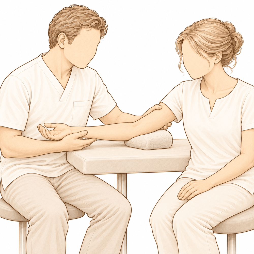
ڈاکٹر کیرن ہِرپرا، جو Mater Private Hospital Rockhampton میں بازو اور ہاتھ کے سرجن (upper-limb surgeon) ہیں، علاج کا آغاز ان کم سے کم تکلیف دہ طریقوں سے کرتے ہیں جو آپ کی حالت کے لیے مناسب ہوں۔ مریضوں کو عام طور پر ان کا فیملی ڈاکٹر (GP) ہمارے کلینک میں بھیجتا ہے؛ اگر کسی فزیوتھراپسٹ نے آپ کو ہمیں دکھانے کا مشورہ دیا ہے، تب بھی میڈیکیئر (Medicare) کی رقم واپسی کا حق دار ہونے کے لیے آپ کو اپنے فیملی ڈاکٹر سے ریفرل (referral) لینا ہوگا۔ آپ کی ملاقات پر ہم آپ کی بیماری کی تاریخ لیتے ہیں، آپ کی کہنی کا معائنہ کرتے ہیں اور ضرورت ہو تو تصویری ٹیسٹوں کا انتظام کرتے ہیں۔ اکڑی ہوئی کہنی اکثر وقت گزرنے اور استعمال کے ساتھ بہتر ہو جاتی ہے، اس لیے ہم عام طور پر پہلے آپریشن کے بغیر علاج آزماتے ہیں۔ اس میں 6 سے 12 ہفتوں کے دوران فزیوتھراپی، اسپلنٹ یا انجکشن شامل ہو سکتے ہیں۔ سرجری پر اس وقت غور کیا جاتا ہے جب اس علاج سے کافی بہتری نہ آئی ہو۔
اکڑی ہوئی کہنی کو کھولنے کا آپریشن، جسے آرتھرولائسس (arthrolysis) بھی کہتے ہیں، ایک ایسا آپریشن ہے جو آپ کی کہنی کے گرد سخت اور زخم کے ریشے سے بھرے ٹشو کو آزاد کرتا ہے تاکہ جوڑ دوبارہ حرکت کر سکے۔ ہم یہ اس وقت تجویز کرتے ہیں جب آپریشن کے بغیر علاج کے بعد بھی اکڑاؤ آپ کو محدود کر رہا ہو۔ یہ اس وقت سب سے بہتر کام کرتا ہے جب جوڑ کی سطح ابھی تک چکنی اور اپنی معمول کی شکل میں ہو۔ مقصد حرکت میں دیرپا اضافہ اور درد میں کمی ہے، تاکہ روزمرہ کے کام آسان ہو جائیں۔
آپریشن سے پہلے
سرجری طے ہو جانے کے بعد کچھ عملی باتوں کا انتظام کرنا ہوتا ہے۔ آپ کو بتایا جائے گا کہ کب کھانا پینا بند کرنا ہے: یہ آپ کے آپریشن سے سات گھنٹے پہلے ہے۔ ہم چھ کے بجائے سات گھنٹے مانگتے ہیں تاکہ اگر آپریشن کی فہرست وقت سے پہلے چل رہی ہو تو ہم آپ کا آپریشن پہلے کر سکیں۔ آپ کا سرجن آپ کو بتائے گا کہ اپنی معمول کی دواؤں میں سے کون سی اور کب بند کرنی ہیں، اور آپ کو ان سب کی مکمل فہرست ساتھ لانی چاہیے۔ کسی ایسے شخص کا انتظام کریں جو بعد میں آپ کو گاڑی میں گھر لے جائے۔ آپریشن کے دن ڈھیلے، آرام دہ کپڑے پہنیں۔ آپریشن کی منصوبہ بندی کے لیے ہمیشہ ایکسرے لیے جاتے ہیں، اور اگر اکڑاؤ میں ہڈی شامل ہو تو سی ٹی اسکین بھی استعمال کیا جا سکتا ہے۔ اگر آپ کو دوسری طبی بیماریاں ہیں تو ہو سکتا ہے آپ کو خون کے ٹیسٹ کروانے پڑیں یا بے ہوشی کے ڈاکٹر (anaesthetist) سے معائنہ کروانا پڑے۔
آپریشن کے دن
آپ ہسپتال کے سرجری کے داخلے کے شعبے میں پہنچتے ہیں، جہاں آپ کا اندراج کیا جاتا ہے اور آپ کو آپریشن تھیٹر کے لیے تیار کیا جاتا ہے۔ وہاں آپ کی ملاقات بے ہوشی کے ڈاکٹر سے ہوتی ہے۔ یہ آپریشن جنرل اینستھیزیا (مکمل بے ہوشی) میں کیا جاتا ہے۔ آپریشن کے بعد درد سے آرام کے لیے کبھی کبھی اعصابی بلاک (nerve block)، یعنی ایک انجکشن جو جسم کے ایک حصے کو سُن کر دیتا ہے، بھی شامل کیا جاتا ہے؛ بے ہوشی کے ڈاکٹر آپریشن کے دن آپ سے اس پر بات کریں گے۔ پھر آپ کو آپریشن تھیٹر میں لے جایا جاتا ہے، جہاں آپریشن کیا جاتا ہے۔
آپ ریکوری روم میں ہوش میں آتے ہیں، جہاں نرسیں اس وقت تک آپ کی نگرانی کرتی ہیں جب تک بے ہوشی کی دوا کا اثر ختم نہ ہو جائے۔ جب آپ کی حالت مستحکم ہو جاتی ہے تو، طریقۂ علاج اور آپ کی صحت یابی کے مطابق، آپ یا تو وارڈ میں چلے جاتے ہیں یا گھر۔
آپریشن میں کیا کیا جاتا ہے
اکڑی ہوئی کہنی کو کھولنے کا ایک سے زیادہ طریقہ ہے، اور آپ کا سرجن وہ طریقہ چنے گا جو آپ کی کہنی کے لیے مناسب ہو۔ کچھ آپریشن دوربین کے ذریعے آپریشن (keyhole) سے کیے جاتے ہیں، جس میں چند چھوٹے چیرے اور ایک پتلا کیمرہ استعمال ہوتا ہے۔ کچھ دوسرے کہنی کی اندرونی یا بیرونی طرف ایک یا دو کھلے چیروں کے ذریعے کیے جاتے ہیں، جو کبھی کبھی تقریباً 3 سے 5 سینٹی میٹر لمبے ہوتے ہیں۔ بہت زیادہ اکڑی ہوئی کہنیوں میں دونوں طرف تک پہنچنے کے لیے کہنی کے پیچھے ایک چیرا استعمال کیا جا سکتا ہے۔
اندر پہنچ کر آپ کا سرجن سخت جوڑ کی جھلی (capsule) کو آزاد کرتا ہے، جو جوڑ کے گرد ٹشو کا غلاف ہے۔ ہڈی کے وہ ابھار (bone spurs) جو حرکت کو روکتے ہیں، تراش دیے جاتے ہیں، اور جوڑ میں تیرتے ہوئے ہڈی یا کارٹلیج (cartilage)، یعنی جوڑ میں ہڈیوں کے سروں پر چکنی، لچکدار تہہ، کے کوئی بھی ڈھیلے ٹکڑے نکال دیے جاتے ہیں۔ اگر کہنی کی اندرونی طرف کا کوئی عصب زخم بھرنے سے بننے والے سخت ریشے (scar tissue) سے دب رہا ہو، تو اسے ٹشو کی اس سرنگ سے آزاد کیا جا سکتا ہے، یا کسی نئی جگہ منتقل کیا جا سکتا ہے جہاں وہ کھنچ نہ سکے۔ اگر خود جوڑ کی سطح بہت زیادہ گھس چکی ہو تو دوسرے راستے موجود ہیں، جیسے ہڈی پر نرم ٹشو کی تہہ لگانا یا جوڑ کی سطحوں کو دھات اور پلاسٹک کے پرزوں سے تبدیل کرنا۔ کبھی کبھی کہنی پر قبضے والا ایک فریم لگایا جاتا ہے تاکہ ٹھیک ہوتے وقت جوڑ کو اچھی پوزیشن میں رکھا جا سکے۔
آخر میں چیروں کو ٹانکوں سے بند کر کے زخم کی پٹی سے ڈھانپ دیا جاتا ہے۔ آپ یہ پٹی تقریباً 10 دن تک لگائے رکھیں گے، جیسا کہ صحت یابی والے حصے میں بتایا گیا ہے۔
آپریشن کے بعد
آپ ریکوری روم میں ہوش میں آتے ہیں، پھر وارڈ میں منتقل ہو جاتے ہیں۔ نرسیں آپ کی کہنی، آپ کا ہاتھ اور آپ کی طبیعت چیک کریں گی۔ درد کش دوا آپ کی ضرورت کے مطابق دی جاتی ہے؛ اگر آپ کی کہنی میں تکلیف ہو تو نرسوں کو بتائیں۔ آپ کی کہنی نرم پٹی میں لپٹی ہوگی، اور ہو سکتا ہے آپ آرام کے لیے اپنا بازو سلنگ (sling)، یعنی گلے سے لٹکی وہ پٹی جو بازو کو سہارا دیتی ہے، میں رکھ کر گھر جائیں۔ آپ جلد ہی بستر سے اٹھ کر چل پھر سکتے ہیں، اور پہلی بار نرسیں آپ کی مدد کریں گی۔ جب تک آپ کی کہنی سنبھل رہی ہو، آپ کا ہاتھ، کلائی اور کندھا آزادی سے حرکت کر سکتے ہیں۔ گھر جانے کے بعد پہلے 24 گھنٹے کسی کو آپ کے ساتھ رہنا چاہیے۔ آپ کی ٹیم آپ کو بتائے گی کہ آپ اسی دن گھر جائیں گے یا ایک رات ہسپتال میں رہیں گے۔ ہم پٹی کو تقریباً 10 دن تک لگا رہنے دیتے ہیں؛ براہ کرم اس سے پہلے اسے نہ اتاریں، جب تک ہم آپ کو ایسا کرنے کو نہ کہیں۔ جب ہم آپ کو دیکھیں گے تو پٹی تبدیل کریں گے یا اتار دیں گے۔
صحت یابی
پہلے چند دنوں اور ہفتوں میں آپ کی کہنی میں درد اور سوجن ہوگی۔ کہنی کھولنے کے آپریشن کے بعد یہ معمول کی بات ہے۔ آرام کرنا، اپنا ہاتھ اونچا رکھنا اور درد کش دوا ہدایت کے مطابق لینا تکلیف کو کم کرے گا۔ سوجن آہستہ آہستہ کم ہوتی ہے، اور جیسے جیسے یہ کم ہوتی ہے، کہنی عام طور پر کم سخت محسوس ہوتی ہے۔
آپ آرام کے لیے اپنا بازو سلنگ میں رکھ کر گھر جائیں گے۔ جب اس سے فائدہ نہ رہے تو آپ اسے استعمال کرنا چھوڑ سکتے ہیں۔ آپ کے ہاتھ، کلائی اور کندھے کو شروع سے ہی آزادی سے حرکت کرتے رہنا چاہیے۔ سرجری کے بعد ہینڈ تھراپی Extend Rehabilitation میں Ruby Doolan کے ساتھ ہوتی ہے۔ وہ آپ کی ورزشوں میں رہنمائی کریں گی اور جس اسپلنٹ کی بھی ضرورت ہو وہ بنائیں گی۔ ورزشیں آپ کی صحت یابی کا بنیادی کام ہیں، اور انہیں ہدایت کے مطابق کرنا کسی بھی دوسری چیز سے زیادہ اہم ہے۔ آپ پٹی تقریباً 10 دن تک لگائے رکھیں گے، اور جب ہم آپ کو دیکھیں گے تو اسے تبدیل کریں گے یا اتار دیں گے۔
روزمرہ کے کام مرحلہ وار واپس آئیں گے۔ شروع میں آپ کو کھانا پکانے، کپڑے پہننے اور چیزیں اٹھا کر لے جانے میں مدد کی ضرورت ہوگی۔ جیسے جیسے حرکت واپس آتی ہے اور سوجن کم ہوتی ہے، آپ آپریشن والے بازو سے زیادہ کام کر سکیں گے۔ جب آپ کا سرجن آپ کو گاڑی چلانے کی اجازت دے دے تو آپ دوبارہ گاڑی چلا سکتے ہیں؛ ہماری رہنمائی بازو اور ہاتھ کی سرجری کے بعد گاڑی چلانا لاگو ہونے والے اصول بتاتی ہے، جن میں یہ بھی شامل ہے کہ جب آپ کا بازو سلنگ میں ہو تو گاڑی نہ چلائیں، اور گاڑی چلانے سے پہلے تیز درد کش دوائیں لینا بند کر چکے ہوں۔
صحت یابی ہر شخص میں مختلف ہوتی ہے۔ آپ کا وقت کا دورانیہ مختلف ہو سکتا ہے، اور آپ کا سرجن اور تھراپسٹ راستے میں آپ کی رہنمائی کریں گے۔
کیا مسائل پیدا ہو سکتے ہیں
زیادہ تر مریض ٹھیک رہتے ہیں، لیکن کبھی کبھار مسائل پیدا ہو سکتے ہیں۔ آپ کا سرجن اور ٹیم آپ کی قریب سے نگرانی کرتے ہیں تاکہ کسی بھی مسئلے کو جلد پکڑا جا سکے۔
کہنی جتنی زیادہ دیر سے اکڑی ہوئی ہو، کھولنے کے آپریشن کے بعد مسائل کا امکان اتنا ہی زیادہ ہوتا ہے۔ اگر آپ کی کہنی لمبے عرصے سے اکڑی ہوئی ہے تو فیصلہ کرنے سے پہلے آپ کا سرجن آپ سے اس پر بات کرے گا۔
کچھ لوگ بعد میں ایک چیز محسوس کرتے ہیں: چھوٹی انگلی اور اس کے ساتھ والی (انگوٹھی والی) انگلی میں نئی جھنجھناہٹ، سُن پن یا کمزوری۔ یہ کہنی کی اندرونی طرف کے عصب کی وجہ سے ہوتا ہے۔ یہ پہلے سے بتانا ہمیشہ ممکن نہیں ہوتا کہ کس کو یہ ہوگا۔ اگر آپ یہ احساسات محسوس کریں تو اپنے اگلے معائنے پر ان کا ذکر کریں، یا اگر یہ اچانک شروع ہوں تو کلینک فون کریں۔
کچھ کہنیاں جنہیں پہلے چوٹ لگ چکی ہو، ان میں بعد میں گہرے انفیکشن کا امکان زیادہ ہوتا ہے۔ زخم پر نظر رکھیں کہ کہیں اس سے باہر کی طرف پھیلتی ہوئی سرخی، گرمی، بڑھتی ہوئی سوجن، یا ایسا گہرا، دھڑکتا ہوا درد تو نہیں جو عام درد کش دواؤں سے کم نہ ہو۔ ہو سکتا ہے آپ کو بخار جیسا محسوس ہو یا عمومی طور پر طبیعت خراب لگے۔ اگر آپ ان میں سے کوئی بھی علامت دیکھیں تو فوراً کلینک فون کریں، یا اگر دفتری اوقات کے بعد کا وقت ہو تو ہسپتال کی ایمرجنسی جائیں۔
اگر آپ کے آپریشن میں کہنی پر قبضے والا فریم لگانا شامل ہو، تو کچھ اضافی باتیں جاننا ضروری ہیں۔ یہ فریم سرجری کے دوران خون کے ضیاع، آپریشن تھیٹر میں گزرنے والے وقت اور ہسپتال میں رہنے کے وقت کو بڑھا دیتا ہے۔ اس سے جوڑ کے ڈھیلا یا غیر مستحکم ہونے کا بھی تھوڑا سا امکان ہوتا ہے۔ آپ کا سرجن وضاحت کرے گا کہ آپ کے ہسپتال میں رہنے اور آپ کی صحت یابی کے لیے اس کا کیا مطلب ہے۔
اگر اس کہنی کے پہلے بھی آپریشن ہو چکے ہیں تو گہرے انفیکشن کا امکان اور بھی زیادہ ہوتا ہے۔ یہی بات اس وقت بھی لاگو ہوتی ہے جب آپریشن خود زیادہ پیچیدہ ہو۔ آپ کا سرجن پہلے سے آپ کے ساتھ ان سب باتوں کا جائزہ لیتا ہے۔
کہنی کی پرانی چوٹیں جوڑ کے کام کرنے کا طریقہ بھی بدل سکتی ہیں۔ زخم کے نشان والی جلد، غلط جگہ پر جڑنے والی ہڈیاں، خراب کارٹلیج، اضافی ہڈی، ڈھیلے یا سخت لیگامنٹ (ligament)، یعنی وہ مضبوط بندھن جو ایک ہڈی کو دوسری ہڈی سے جوڑتے ہیں، دبے ہوئے اعصاب اور سخت ریشے والے پٹھے، یہ سب کہنی کی حرکت کو بدل سکتے ہیں۔ آپ کا سرجن آپ کے آپریشن کی منصوبہ بندی کرتے وقت ان چیزوں کو دیکھے گا۔
اگر آپ تفصیل جاننا چاہتے ہیں تو اس صفحے پر پیچیدگیوں کا جدول عام شرحیں بتاتا ہے۔
ہمیں کب فون کریں
اگر آپ کو بخار ہو، زخم کے گرد سرخی پھیل رہی ہو، زخم سے رطوبت کا اخراج ہو، یا درد مسلسل بڑھتا جا رہا ہو تو ہمیں فون کریں۔ اگر آپ کو اچانک شدید درد ہو، پنڈلی میں سوجن یا درد ہو، یا سانس پھولے تو ہسپتال کی ایمرجنسی جائیں۔ اگر آپ کا ہاتھ یا انگلیاں سُن ہو جائیں یا سفید پڑ جائیں، یا آپ اپنا بازو ہلا نہ سکیں تو فوراً ہمیں فون کریں۔ اگر چھوٹی انگلی اور اس کے ساتھ والی انگلی میں جھنجھناہٹ یا سُن پن اچانک شروع ہو تو اپنے اگلے معائنے کا انتظار کرنے کے بجائے کلینک فون کریں۔
مزید تفصیل
Advanced reading: the deeper science (optional)
یہ حصہ اس سے آگے جاتا ہے جتنا آپ کو اپنے علاج کے فیصلوں کے لیے ضرورت ہے۔ اکڑی ہوئی کہنی کو کھولنے کے بارے میں اضافی مطالعہ اس لیے فائدہ مند ہے کہ شواہد ایک غیر معمولی سمت کی طرف اشارہ کرتے ہیں: زیادہ تر مریض جس انتخاب کے بارے میں پوچھتے ہیں، یعنی دوربین کے ذریعے آپریشن (keyhole) یا کھلا آپریشن، وہ اس سے کم اہم لگتا ہے کہ کتنی سرجری کی جاتی ہے، اور اس آپریشن کا اصل مخالف وہی عمل ہے جس نے شروع میں اکڑاؤ پیدا کیا تھا۔
کھلا آپریشن یا آرتھروسکوپی، یہ اہم سوال نہیں ہے
چوٹ کے بعد کہنی کے اکڑاؤ والے 4,311 مریضوں پر مبنی ایک منظم جائزے (systematic review) اور میٹا اینالیسس (meta-analysis) نے کھلی آرتھرولائسس کا آرتھروسکوپی (دوربین سے جوڑ کے اندر دیکھ کر علاج) والی آرتھرولائسس سے موازنہ کیا اور مجموعی نتائج کو ایک جیسا پایا [1]۔ اس کا نتیجہ یہ تھا کہ سرجن کی مہارت اور مریض کی مخصوص حالت، خود تکنیک سے زیادہ اہم باتیں ہیں [1]۔
کسی منظم جائزے کے لیے یہ غیر معمولی طور پر سیدھی بات ہے، اور اسے جوں کا توں ماننا چاہیے۔ یہ تکنیکی طور پر ایک مشکل آپریشن ہے جو تین بڑے اعصاب کے قریب ایک ایسے جوڑ میں کیا جاتا ہے جس کی گنجائش پہلے ہی اس اکڑاؤ کی وجہ سے کم ہو چکی ہے جس کا علاج کیا جا رہا ہے۔ طریقے سے واقفیت، خود طریقے سے زیادہ کام کر رہی ہے۔
کم سرجری، کم مسائل
798 مریضوں کے ایک پرانے منظم جائزے نے ایک ایسا نتیجہ نکالا جو آج بھی درست ہے: پیچیدگیوں کی تعداد سرجری کے پھیلاؤ کے ساتھ بڑھتی ہوئی لگتی ہے، اور اس سے نکلنے والا مشورہ یہ ہے کہ جتنا ممکن ہو کم چیر پھاڑ سے علاج کیا جائے [2]۔ مصنفین نے اس سفارش کو معتدل درجہ دیا، کیونکہ طبی تحقیق اتنی مضبوط نہیں کہ اس سے کوئی ٹھوس شماریاتی نتیجہ نکالا جا سکے، لیکن سمت ایک جیسی ہے۔
یہی اس آپریشن کے مرکز میں موجود کشمکش ہے۔ زیادہ مکمل کھولنے سے آپریشن کی میز پر زیادہ حرکت حاصل ہوتی ہے، لیکن اس کی قیمت نرم ٹشو کو زیادہ نقصان، جوڑ میں زیادہ خون بہنے، اور زیادہ سوزش کی تحریک کی صورت میں ادا کرنی پڑتی ہے، اور یہی وہ خام مال ہے جس سے اکڑاؤ واپس آتا ہے۔
ہیٹروٹوپک اوسیفیکیشن ہی وہ چیز ہے جسے روکنا ہے
ہیٹروٹوپک اوسیفیکیشن (heterotopic ossification)، یعنی نرم ٹشو میں ہڈی کا بن جانا جسے لچکدار رہنا چاہیے، وہ عمل ہے جس کے ذریعے کھولی گئی کہنی دوبارہ اکڑ جاتی ہے۔ سوزش کم کرنے والی دواؤں سے اس کی روک تھام معمول کا طریقہ ہے، اگرچہ اس کے حق میں شواہد اس عمل کے رواج سے کم ہیں۔
کہنی کی چوٹ کی سرجری کے بعد COX-2 منتخب اور غیر منتخب سوزش کم کرنے والی دواؤں کا موازنہ کرنے والے 622 مریضوں کے ایک میٹا اینالیسس نے پایا کہ دونوں ہیٹروٹوپک اوسیفیکیشن کے خطرے کو مؤثر طریقے سے کم کرتی ہیں، جبکہ الگ الگ براہ راست موازنوں میں دونوں کے درمیان کوئی شماریاتی طور پر نمایاں (statistically significant) فرق نہیں دکھا، اور شواہد کی مجموعی قوت کم تھی [3]۔ لہٰذا: روک تھام فائدہ مند لگتی ہے، دوا کا انتخاب واضح طور پر اہم نہیں، اور یقین محدود ہے۔
پس منظر سے توقعات طے کرنے میں مدد ملتی ہے۔ کہنی کے پورے جوڑ کی تبدیلی (total elbow arthroplasty) کے بعد ہیٹروٹوپک اوسیفیکیشن ایک غیر عام پیچیدگی ہے، اور جہاں یہ بنتی ہے وہاں زیادہ تر مریضوں میں کوئی علامت نہیں ہوتی اور انہیں اس کے لیے سرجری کی ضرورت نہیں ہوتی، یہاں تک کہ طبی تحقیق اس آپریشن کے بعد معمول کے طور پر روک تھام کی حمایت نہیں کرتی [4]۔ کہنی کی ہر سرجری میں یہ خطرہ یکساں نہیں ہے؛ یہ چوٹ اور وسیع پیمانے پر کھولنے کے آپریشنوں میں مرکوز ہے۔
ایک طریقہ جو وہ نہیں کرتا جس کی آپ توقع کریں گے
ٹرینیکسامک ایسڈ (tranexamic acid) ہڈیوں کے بہت سے آپریشنوں میں خون بہنا کم کرتا ہے، اور تازہ کھولی گئی کہنی میں خون بہنا کم کرنے سے لگتا ہے کہ اکڑاؤ بھی کم ہونا چاہیے۔ 660 مریضوں کے ایک میٹا اینالیسس نے پایا کہ ٹرینیکسامک ایسڈ کھلی کہنی کی آرتھرولائسس کے دوران واقعی خون کی مقدار کم کر سکتا ہے، لیکن اس نے حرکت کی آخری حد یا درد کے اسکور پر کوئی اثر نہیں ڈالا [5]۔
یہ اس بات کی ایک صاف مثال ہے کہ ایک معقول لگنے والا طریقۂ کار اس نتیجے میں تبدیل نہیں ہو پاتا جس کی کسی کو واقعی پروا ہے، اور یہ یاد دہانی ہے کہ "کم خون بہنا" ایک متبادل پیمانہ ہے، نتیجہ نہیں۔
آپ کا نتیجہ دراصل کس چیز سے طے ہوتا ہے
اس طبی تحقیق میں کوئی بھی بات کسی تکنیکی شارٹ کٹ کی طرف اشارہ نہیں کرتی۔ مستقل اشارے یہ ہیں کہ آپریشن ضرورت سے بڑا نہیں ہونا چاہیے، کہ ہیٹروٹوپک اوسیفیکیشن کے خلاف روک تھام کی دوا لینا فائدہ مند ہے، اور کہ آپریشن تھیٹر میں حاصل کی گئی حرکت صرف اس کے بعد ہونے والی بحالی کے ذریعے ہی برقرار رہتی ہے۔ کہنی غیر معمولی حد تک اکڑنے پر آمادہ رہتی ہے؛ کھولنے کے آپریشن کے بعد کے مہینے کم از کم اتنے ہی اہم ہیں جتنا خود آپریشن۔
حوالہ جات
[1] Khorram R, Ghayyad K, Vafadar R, Borazjani R, Nezameslami A, Huffman GR, et al. Surgical treatments of post-traumatic elbow stiffness: a systematic review and meta-analysis. J Shoulder Elbow Surg. 2026;35(1):387-407. https://doi.org/10.1016/j.jse.2025.05.004
[2] Kodde IF, van Rijn J, van den Bekerom MP, Eygendaal D. Surgical treatment of post-traumatic elbow stiffness: a systematic review. J Shoulder Elbow Surg. 2013;22(4):574-80. https://doi.org/10.1016/j.jse.2012.11.010
[3] Ahmad A, Khorram R, Ghayyad K, Amin V, Kachooei AR, Huffman GR, et al. Postoperative nonsteroidal anti-inflammatory drug prophylaxis for elbow heterotopic ossification: a systematic review and meta-analysis comparing COX-2 selective and nonselective inhibitors. JSES Rev Rep Tech. 2026;6(2):100628. https://doi.org/10.1016/j.xrrt.2025.100628
[4] Liu EY, Hildebrand A, Horner NS, Athwal GS, Khan M, Alolabi B. Heterotopic ossification after total elbow arthroplasty: a systematic review. J Shoulder Elbow Surg. 2019;28(3):587-95. https://doi.org/10.1016/j.jse.2018.10.003
[5] Nejat MH, Khayami A, Daliri M, Ebrahimzadeh MH, Sadeghi M, Moradi A. Does tranexamic acid diminish hemorrhage and pain in open elbow arthrolysis? A systematic review and meta-analysis. BMC Musculoskelet Disord. 2023;24(1). https://doi.org/10.1186/s12891-023-06835-7
Evidence & references
This is the clinical evidence summary written for health professionals. It is technical, and it lists the research this page was built from. You do not need to read it to understand your treatment or to make a decision about it.
Overview
- In the setting of failed nonsurgical treatment of the stiff elbow, surgical release can yield sustained improvement of range of motion and patient quality of life [1].
- Treatment choices for elbow stiffness must consider non-surgical management and various surgical options including arthroscopic or open capsular release, arthroplasty, and elbow replacement [2].
- Arthroscopic capsular release of the elbow is effective for restoring a functional arc of motion in the short term in most patients with extrinsic contractures [3].
- Arthroscopic elbow contracture release can improve function and range of motion, though outcomes may vary based on preoperative patient characteristics [4].
- Current literature provides state-of-the-art guidance on the management regarding prevention, evaluation, and treatment of elbow stiffness [5].
- Patients with stiff elbows who underwent arthroscopic arthrolysis achieved satisfactory clinical outcomes very early postoperatively [7].
- Current treatment options for post-traumatic elbow stiffness range from conservative to surgical, with varying rates of success, invasiveness, and complications [8].
- A multinational initiative provides the first comprehensive clinical practice guideline for open arthrolysis in adult posttraumatic elbow stiffness [9].
- A subset of pediatric patients with persistent stiffness following medial epicondyle fractures may benefit from additional interventions, including intensive therapy, transposition of the ulnar nerve, and open capsular release [13].
- In most cases, the current use of arthroscopy by experienced surgeons will produce results superior to those of open release given the proper indications [14].
- The use of a hinged external fixator in open arthrolysis for posttraumatic elbow stiffness may result in short-term improvements in flexion-extension range of motion [16].
- The use of a hinged external fixator in open arthrolysis for posttraumatic elbow stiffness is accompanied by increased blood loss, longer operative time, extended hospitalization, and higher costs [16].
- The dual mediolateral mini-open technique allows for a safe and effective release of stiff elbows through small incisions of 3–5 cm in length [18].
Anatomy & Pathophysiology
Bony Anatomy
- The elbow is a trocho-ginglymoid joint consisting of medial and lateral articulations that provide bony stability [28].
- The medial articulation involves the trochlea and the ulna within the greater sigmoid notch, forming the ulnohumeral hinged portion [28].
- The ulnohumeral articulation features highly congruent anatomy through almost 180° of articular contact, except for a bare area of the greater sigmoid notch devoid of cartilage [28].
- The coronoid process has medial and lateral facets that buttress the trochlea anteriorly [28].
- The sublime tubercle is located just distal and medial to the coronoid and serves as the attachment site for the anterior bundle of the medial ulnar collateral ligament [28].
- The lateral articulation involves the capitellum and radial head, forming the radiocapitellar joint [28].
- The radius is held in close approximation to the ulna at the proximal radioulnar joint by the annular ligament [28].
- The radial head is a concave elliptical structure covered with articular cartilage along the radiocapitellar joint and approximately 270° of the articular margin [28].
- The distal humeral articulation is angled 30° from the longitudinal axis [28].
- The axis of rotation is angulated 5° to 7° in the coronal plane relative to the epicondylar axis, with the medial side more distal than the lateral side [28].
- The ulna medially bends approximately 8° at 8 cm from the tip of the olecranon [28].
- The articulation to the tip of the coronoid is approximately 30° from the long axis of the ulna in the sagittal plane [28].
Ligaments and Soft Tissue
- Elbow stability is determined by primary stabilizers (ulnohumeral articulation, MUCL, LUCL complex) and secondary stabilizers (radiocapitellar articulation, common flexor tendon, common extensor tendon, joint capsule) [25].
- The medial ulnar collateral ligament is the primary valgus stabilizer of the elbow [29].
- The anterior bundle of the medial ulnar collateral ligament is the most important component for stability [29].
- The posterior bundle of the medial ulnar collateral ligament becomes taut at flexion beyond 120 degrees [29].
- The lateral ulnar collateral ligament serves as the posterolateral stabilizer of the elbow [29].
- Osborne’s ligament stabilizes the ulnar nerve in the cubital tunnel [29].
- The ligament of Struthers is a variant anatomy arising from the supracondylar process to attach to the medial epicondyle and is a potential site of median nerve compression [29].
- The joint capsule allows maximum distension at approximately 70 to 80 degrees of flexion [29].
- The anterior capsule attaches at a point approximately 6 mm distal to the tip of the coronoid [29].
- Tensile forces are present at the medial elbow, while compressive forces are present at the lateral elbow [29].
Muscles
- The brachialis is the strongest elbow flexor and attaches to the coronoid 11 mm distal to the tip [29].
- The biceps brachii inserts at the ulnar margin of the radial tuberosity and acts as a powerful supinator of the forearm [29].
- The triceps brachii is the primary elbow extensor and inserts on the olecranon process [29].
- The mobile wad consists of the brachioradialis, extensor carpi radialis longus, and extensor carpi radialis brevis [29].
- The flexor-pronator mass includes the pronator teres, flexor carpi radialis, palmaris longus, flexor carpi ulnaris, and flexor digitorum superficialis [29].
Pathophysiology and Functional Range
- The normal elbow has a range of motion from 0° to 140° from extension to flexion [25].
- The normal elbow has a range of motion of 75° in pronation and 85° in supination [25].
- A functional arc of motion for the elbow is 100° for flexion and extension [25].
- A functional arc of motion for the elbow is 100° for forearm rotation [25].
- Successful intervention for a stiff elbow depends on the quality of the joint and the extent of soft tissue damage [11].
- The functional arc of motion serves as a frame of reference rather than an absolute goal for stiff elbow treatment [11].
- Treatment of the stiff elbow requires a thorough understanding of normal anatomy and etiological factors to develop effective strategies [10].
Classification
- The S.T.I.F. classification system highlights the aetiology of elbow stiffness [51].
- The S.T.I.F. classification system provides a framework to better understand the natural history of elbow stiffness [51].
- The S.T.I.F. classification system provides a framework to direct surgical management of elbow stiffness [51].
- The S.T.I.F. classification system provides a framework to predict clinical outcomes for elbow stiffness [51].
- Influencing factors must be taken into consideration for proper surgical indication and prognosis when using the S.T.I.F. classification system [51].
- The S.T.I.F. classification system is beneficial for research because identifying homogenous patterns of stiffness allows for more appropriate comparison between different treatment options [51].
- Based on the S.T.I.F. classification system, a treatment algorithm can be proposed that includes the severity of the disorders as a guide to surgical treatment [51].
- Based on the S.T.I.F. classification system, a treatment algorithm can be proposed that includes the severity of the disorders as a guide to the preferred operative approach (open or scope) [51].
- The prevalent types of stiffness identified by the S.T.I.F. classification system could be applied in the future to Artificial Intelligence to help in diagnosing the cause of elbow stiffness [51].
Clinical Presentation
- Elbow stiffness is a challenging problem with no ideal management solution [6].
- Elbow contracture is challenging to treat, and therefore prevention is of paramount importance [17].
- Restoration of joint motion in the posttraumatic stiff elbow can be a difficult, time-consuming, and costly challenge [15].
- Successful intervention for stiff elbow depends on the quality of the joint and the extent of soft tissue damage [11].
- The functional arc of motion serves as a frame of reference rather than an absolute goal for stiff elbow intervention [11].
- Conservative treatment is indicated for patients that have elbow stiffness due to heterotopic ossification for less than six months [22].
- Current non-surgical treatment measures for elbow stiffness due to heterotopic ossification include physical therapy and manipulation under anesthesia in order to restore the range of motion of the involved joint [22].
- Non-surgical options for heterotopic ossification have limited effects and can be used mainly in cases that cause a small limitation of range of motion [22].
- Surgical options are necessary if nonoperative management fails to restore the elbow function and range of motion after 6 months in patients with heterotopic ossification [22].
- Surgical excision of ectopic bone and contracture release, if present, are the most common surgical options in elbow heterotopic ossification management [22].
- Arthroscopic elbow contracture release can improve function and range of motion, although outcomes may vary based on preoperative patient characteristics [4].
- Although both open and arthroscopic techniques may produce satisfactory results, the current use of arthroscopy by experienced surgeons will produce results superior to those of open release given the proper indications [14].
- Operative management of pediatric elbow contractures is effective [23].
- Patients undergoing operative management of elbow stiffness secondary to heterotopic ossification maintained substantial reductions in pain, improvement in elbow range of motion, and increased overall elbow function [12].
- Treatment of bony encasement of the ulnar nerve secondary to heterotopic ossification of the elbow leads to superior range of motion, improved or resolved ulnar neuropathy, and good to excellent long-term functional outcomes [24].
- Delayed-onset anterior interosseous nerve palsy can develop one week after open elbow contracture release, likely due to a stretch injury [26].
Investigations
History and Physical Examination
- The history for elbow stiffness evaluation includes the duration of the contracture, initial injury, previous surgical procedures, trials of splinting/therapy/injections, surgical complications, and patient work/life demands [34].
- Physical examination must assess the function of the shoulder, wrist, and hand [34].
- The soft tissue surrounding the elbow should be examined for previous skin incisions, grafts, eschar, or infection [34].
- Active and passive flexion, extension, supination, and pronation should be evaluated using a goniometer for accurate measurement [34].
- The contralateral elbow should be examined for comparison during range of motion assessment [34].
- If the elbow has less than 90° to 100° of flexion, the posterior bundle of the medial collateral ligament is contracted and must be released to restore flexion [34].
- Pain assessment during the mid-arc or at terminal ends of motion helps distinguish intrinsic disease from contracture, as mid-arc pain is more common with intrinsic disease and may not improve with contracture release alone [34].
- The ulnar nerve is of utmost importance in the neurovascular examination due to its anatomic proximity to the elbow [34].
- Electromyography and nerve conduction velocity studies should be performed if there is any question about neurologic dysfunction [34].
- An assessment for ulnar nerve subluxation should be performed, as subluxation is a relative contraindication for arthroscopic procedures due to the risk of iatrogenic nerve injury [34].
- The surgeon must verify if the ulnar nerve has been transposed if there is a history of prior surgical procedures [34].
- Successful intervention for stiff elbow depends on the quality of the joint and the extent of soft tissue damage, with the functional arc of motion serving as a frame of reference rather than an absolute goal [11].
Imaging
- Radiographs should always be obtained for the evaluation of elbow stiffness [34].
- Standard radiographic views include AP, lateral, and oblique views, with serial radiography used as follow-up when heterotopic ossification is present [34].
- Primary bony landmarks assessed on radiographs include the ulnohumeral joint, coronoid process, radial head, capitellum, radiocapitellar joint, olecranon tip, coronoid/olecranon fossae, and trochlear ridge [34].
- CT is helpful for assessing malunion architecture and the location and pattern of osteophytes or loose bodies [34].
- Three-dimensional CT is used to check for heterotopic ossification [34].
- CT is not necessary when the stiffness is entirely soft-tissue related, but is beneficial if any joint incongruity or abnormal bony anatomy is present [34].
- MRI can be used to evaluate ligaments and tendons but is rarely indicated for elbow stiffness [34].
- The normal elbow has a range of motion from 0° to 140° from extension to flexion and 75° and 85° in pronation and supination respectively [25].
- A functional arc in each plane is 100° for flexion and extension and forearm rotation [25].
- Elbow stability is determined by primary stabilizers (ulnohumeral articulation, MUCL, LUCL) and secondary stabilizers (radiocapitellar articulation, common flexor tendon, common extensor tendon, joint capsule) [25].
Treatment
General Principles and Indications
- Elbow stiffness is a challenging problem with no ideal management solution; however, functional improvements can be achieved with both nonsurgical and surgical strategies [6].
Arthroscopic Release
- Arthroscopic elbow contracture release can improve function and range of motion; however, outcomes may vary based on preoperative patient characteristics [4].
- Although both open and arthroscopic techniques may produce satisfactory results, the authors believe that in most cases the current use of arthroscopy by experienced surgeons will produce results superior to those of open release given the proper indications [14].
- Additional peripheral nerve block combined with a postoperative nerve block catheter in arthroscopic arthrolysis in cases of elbow stiffness may be an opportunity to enhance postoperative outcomes by achieving better functional ROM, perhaps through reduced postoperative pain [48].
Open Release and Techniques
- This multinational initiative provides the first comprehensive clinical practice guideline for open arthrolysis in adult posttraumatic elbow stiffness [9].
- The use of a hinged external fixator in open arthrolysis for posttraumatic elbow stiffness may result in short-term improvements in flexion-extension range of motion but is accompanied by increased blood loss, longer operative time, extended hospitalization, and higher costs [16].
- The most common reason for a failed contracture release is a failure on the part of the surgeon to abandon an orthodox method in favor of a less assured technique or to use a single anatomic approach to the joint when the potential gain in motion is not achieved at the time of surgery [40].
- The operative contracture release requires a progressive, adaptive surgical approach, and the surgeon must safely increase the exposure in order to excise the offending fibrosis on nearly all aspects of the joint, anteroposterior and mediolateral [40].
- If an uncomplicated, simple dislocation has caused a 30-degree flexion contracture, arthroscopic capsular excision may be sufficient [40].
- The best care for a given patient with a contracture is the most minimal necessary surgery, including minimal anatomic exposure, and rehabilitation to achieve the desired result [40].
- A patient with a range of 40 to 105 degrees and heterotopic ossification will usually require exposure of the capsule for complete excision from both the medial and lateral approaches but not necessarily hinged fixation [40].
- In a circumstance with more massive heterotopic ossification, hinged external fixation may be necessary to stabilize the elbow postoperatively and permit immediate motion with intermittent passive stretch [40].
- The surgeon should be prepared to extend the exposure or approach the joint from an additional direction if full passive motion is not achieved on the operating table [40].
- In older patients with suspected cartilage damage or avascular bone, total elbow replacement should be available and discussed before surgery [40].
Outcomes and Complications
- Overall, patients maintained substantial reductions in pain, improvement in elbow range of motion, and increased overall elbow function after operative management of elbow stiffness secondary to heterotopic ossification [12].
- The gain in range of motion for open arthrolysis was 51° [19].
- The gain in range of motion for arthroscopic arthrolysis was 40° [19].
- The gain in range of motion for open arthrolysis with external fixation was 88° [19].
- The gain in range of motion for open arthrolysis with distraction arthroplasty was 56° [19].
- The average percentage of complications for open arthrolysis was 23% [19].
- The average percentage of complications for arthroscopic arthrolysis was 5% [19].
- The average percentage of complications for open arthrolysis with external fixation was 73% [19].
- The average percentage of complications for open arthrolysis with distraction arthroplasty was 58% [19].
Complications
- The use of a hinged external fixator in open arthrolysis is accompanied by increased blood loss [16].
- The use of a hinged external fixator in open arthrolysis is accompanied by longer operative time [16].
- The use of a hinged external fixator in open arthrolysis is accompanied by extended hospitalization [16].
- The use of a hinged external fixator in open arthrolysis is accompanied by higher costs [16].
- Current treatment options for post-traumatic elbow stiffness have varying rates of complications [8].
Recovery
- Surgical release of the stiff elbow can yield sustained improvement of range of motion and patient quality of life in the setting of failed nonsurgical treatment [1].
- Arthroscopic capsular release is effective for restoring a functional arc of motion in the short term in most patients with extrinsic contractures [3].
- Functional improvements can be achieved with both nonsurgical and surgical strategies for elbow stiffness [6].
- Treatment for bony encasement of the ulnar nerve secondary to heterotopic ossification of the elbow leads to superior range of motion, improved or resolved ulnar neuropathy, and good to excellent long-term functional outcomes [24].
Key Evidence
- [L5] In the setting of failed nonsurgical treatment of the stiff elbow, surgical release can yield sustained improvement of ROM and patient quality of life. [1] (10.5435/jaaos-d-14-00051)
- [L5] Treatment choices must consider non-surgical management and various surgical options including arthroscopic or open capsular release, arthroplasty, and elbow replacement. [2] (10.1016/j.jisako.2023.10.009)
- [L5] Arthroscopic capsular release of the elbow is effective for restoring a functional arc of motion in the short term in most patients with extrinsic contractures. [3] (10.5435/00124635-201105000-00004)
- [L4] Arthroscopic elbow contracture release can improve function and range of motion; however, outcomes may vary based on preoperative patient characteristics. [4] (10.1016/j.jseint.2026.101621)
- [L4] This paper reviews the current literature and provides state-of-the-art guidance on the management regarding prevention, evaluation, and treatment of elbow stiffness. [5] (10.1530/eor-23-0039)
- [L4] Elbow stiffness is a challenging problem with no ideal management solution; however, functional improvements can be achieved with both nonsurgical and surgical strategies, and recent advancements in biology and pathology may lead to future breakthroughs in prevention and treatment. [6] (10.1016/j.jhsa.2013.06.007)
- [L1] Patients with stiff elbows who underwent arthroscopic arthrolysis achieved satisfactory clinical outcomes very early postoperatively. [7] (10.1016/j.jse.2024.06.009)
- [L5] Current treatment options for post-traumatic elbow stiffness range from conservative to surgical, with varying rates of success, invasiveness, and complications. [8] (10.1177/1758573218793903)
- [L5] This multinational initiative provides the first comprehensive clinical practice guideline for open arthrolysis in adult posttraumatic elbow stiffness. [9] (10.1016/j.jse.2025.07.015)
- [L5] Treatment of the stiff elbow requires a thorough understanding of normal anatomy and etiological factors to develop effective strategies. [10] (10.1016/j.jisako.2023.10.006)
- [L5] Successful intervention for stiff elbow depends on the quality of the joint and the extent of soft tissue damage, with the functional arc of motion serving as a frame of reference rather than an absolute goal. [11] (10.1016/j.jisako.2023.09.002)
- [L4] Overall, patients maintained substantial reductions in pain, improvement in elbow range of motion, and increased overall elbow function. [12] (10.1016/j.jse.2024.11.019)
- [L4] A subset of pediatric patients with persistent stiffness following medial epicondyle fractures may benefit from additional interventions, including intensive therapy, transposition of the ulnar nerve, and open capsular release. [13] (10.1016/j.jhsg.2023.07.002)
- [L5] Although both open and arthroscopic techniques may produce satisfactory results, the authors believe that in most cases the current use of arthroscopy by experienced surgeons will produce results superior to those of open release given the proper indications. [14] (10.1016/j.jse.2010.11.029)
- [L4] Restoration of joint motion in the posttraumatic stiff elbow can be a difficult, time-consuming, and costly challenge. [15] (10.1016/j.jhsa.2007.09.015)
- [L3] The use of a hinged external fixator in open arthrolysis for posttraumatic elbow stiffness may result in short-term improvements in flexion-extension range of motion but is accompanied by increased blood loss, longer operative time, extended hospitalization, and higher costs. [16] (10.1186/s12891-024-08167-6)
- [L5] Elbow contracture is challenging to treat, and therefore prevention is of paramount importance. [17] (10.1016/j.jhsa.2009.02.020)
- [L4] The dual mediolateral mini-open technique allows for a safe and effective release of stiff elbows through small incisions of 3–5 cm in length. [18] (10.1186/s13018-025-06288-9)
- [L4] [19] (10.1016/j.jse.2012.11.010)
- [L4] [22] (10.3390/life13122358)
- [L1] Operative management of pediatric elbow contractures is effective. [23] (10.1016/j.jhsa.2024.01.010)
- [L4] This treatment approach leads to superior range of motion, improved or resolved ulnar neuropathy, and good to excellent long-term functional outcomes. [24] (10.1016/j.jse.2023.12.003)
- [Case_report] The authors describe a case of delayed-onset anterior interosseous nerve palsy developing one week after open elbow contracture release, likely due to a stretch injury. [26] (10.5397/cise.2022.00899)
- [L3] Additional peripheral nerve block combined with a postoperative nerve block catheter in arthroscopic arthrolysis in cases of elbow stiffness may be an opportunity to enhance postoperative outcomes by achieving better functional ROM, perhaps through reduced postoperative pain. [48] (10.1016/j.jseint.2024.10.009)
- [L5] [51] (10.1016/j.jisako.2023.10.011)
References
[1] Open Surgical Release for Contractures of the Elbow. Journal of the American Academy of Orthopaedic Surgeons. 2015. DOI: 10.5435/jaaos-d-14-00051
[2] Elbow stiffness: Arthritis and heterotopic ossification. Journal of ISAKOS. 2024. DOI: 10.1016/j.jisako.2023.10.009
[3] Arthroscopic Management of the Stiff Elbow. American Academy of Orthopaedic Surgeon. 2011. DOI: 10.5435/00124635-201105000-00004
[4] Preoperative risk factors associated with patient outcomes following arthroscopic elbow contracture release. JSES International. 2026. DOI: 10.1016/j.jseint.2026.101621
[5] Management of the stiff elbow: a literature review. EFORT Open Reviews. 2023. DOI: 10.1530/eor-23-0039
[6] Prevention and Treatment of Elbow Stiffness: A 5-Year Update. The Journal of Hand Surgery. 2013. DOI: 10.1016/j.jhsa.2013.06.007
[7] Does tranexamic acid reduce elbow swelling and improve early function following arthroscopic arthrolysis? A double-blind randomized controlled trial. Journal of Shoulder and Elbow Surgery. 2024. DOI: 10.1016/j.jse.2024.06.009
[8] Post-traumatic elbow stiffness: Pathogenesis and current treatments. Shoulder & Elbow. 2018. DOI: 10.1177/1758573218793903
[9] Clinical guideline on the open arthrolysis for post-traumatic elbow stiffness in adult patients. Journal of Shoulder and Elbow Surgery. 2026. DOI: 10.1016/j.jse.2025.07.015
[10] The examination and treatment of soft tissue contracture of the elbow. Journal of ISAKOS. 2024. DOI: 10.1016/j.jisako.2023.10.006
[11] Elbow stiffness: Interview with professor Bernard Morrey. Journal of ISAKOS. 2024. DOI: 10.1016/j.jisako.2023.09.002
[12] Assessing long-term outcomes after operative management of elbow stiffness secondary to heterotopic ossification. Journal of Shoulder and Elbow Surgery. 2025. DOI: 10.1016/j.jse.2024.11.019
[13] Outcomes of Therapy and Ulnar Nerve Transposition for Elbow Stiffness After Pediatric Medial Epicondyle Fractures. Journal of Hand Surgery Global Online. 2023. DOI: 10.1016/j.jhsg.2023.07.002
[14] Arthroscopic management of the post-traumatic stiff elbow. Journal of Shoulder and Elbow Surgery. 2011. DOI: 10.1016/j.jse.2010.11.029
[15] The Posttraumatic Stiff Elbow: A Review of the Literature. The Journal of Hand Surgery. 2007. DOI: 10.1016/j.jhsa.2007.09.015
[16] Comparative study of open elbow arthrolysis with and without hinge external fixation for the treatment of post-traumatic elbow stiffness. BMC Musculoskeletal Disorders. 2024. DOI: 10.1186/s12891-024-08167-6
[17] Prevention and Treatment of Elbow Stiffness. The Journal of Hand Surgery. 2009. DOI: 10.1016/j.jhsa.2009.02.020
[18] Dual mediolateral mini-open technique for the release of elbow contracture. Journal of Orthopaedic Surgery and Research. 2025. DOI: 10.1186/s13018-025-06288-9
[19] Surgical treatment of post-traumatic elbow stiffness: a systematic review. Journal of Shoulder and Elbow Surgery. 2013. DOI: 10.1016/j.jse.2012.11.010
[22] Heterotopic Ossification around the Elbow Revisited. Life. 2023. DOI: 10.3390/life13122358
[23] Clinical Outcomes Following Surgical Management of Post-Traumatic Elbow Contractures in the Pediatric Age Group: A Meta-Analysis and Systematic Review. The Journal of Hand Surgery. 2025. DOI: 10.1016/j.jhsa.2024.01.010
[24] Bony encasement of the ulnar nerve secondary to heterotopic ossification of the elbow: an evaluation of long-term outcomes. Journal of Shoulder and Elbow Surgery. 2024. DOI: 10.1016/j.jse.2023.12.003
[25] Orthopaedic Knowledge Update 13 Ebook Without Multimedia. Anatomy, Biomechanics, Physical Examination, and Imaging of the Elbow > Summary and Conclusions.
[26] Anterior interosseous nerve palsy in the early postoperative period after open capsular release for elbow stiffness: a case report. Clinics in Shoulder and Elbow. 2023. DOI: 10.5397/cise.2022.00899
[28] Orthopaedic Knowledge Update 13 Ebook Without Multimedia. Anatomy, Biomechanics, Physical Examination, and Imaging of the Elbow > Anatomy > Bony Anatomy.
[29] Miller S Review Of Orthopaedics. SECTION 16 PATELLAR TRACKING IN TOTAL KNEE ARTHROPLASTY > TABLE 2.3 Shoulder Spaces.
[34] Aaos Comprehensive Orthopaedic Review 3. Elbow Stiffness* > IV. Evaluation.
[40] Green S Operative Hand Surgery. Classifying the Contracture and Matching the Operative Plan.
[48] A comparative analysis of short-term results in range of motion following arthroscopic arthrolysis with vs. without peripheral nerve block in cases of elbow stiffness. JSES International. 2025. DOI: 10.1016/j.jseint.2024.10.009
[51] Classification of elbow stiffness. Journal of ISAKOS. 2024. DOI: 10.1016/j.jisako.2023.10.011