Patients › Elbow
کیوبیٹل ٹنل ریلیز
Cubital tunnel release relieves ulnar nerve compression—addressing numbness/weakness in the ring & little fingers.
یہ آپریشن کیوں تجویز کیا گیا ہے
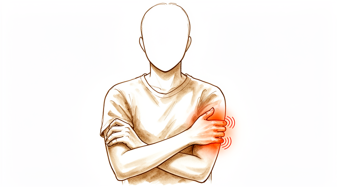
ڈاکٹر کیرن ہِرپرا، جو Mater Private Hospital Rockhampton میں بازو اور ہاتھ کے سرجن (upper-limb surgeon) ہیں، آپ کے مسئلے کے لحاظ سے سب سے کم چیر پھاڑ والے طریقوں سے شروع کرتے ہیں۔ مریضوں کو عام طور پر ان کے فیملی ڈاکٹر (GP) ہمارے کلینک بھیجتے ہیں؛ اگر کسی فزیوتھراپسٹ نے آپ کو ہمیں دکھانے کا مشورہ دیا ہے تو بھی میڈیکیئر (Medicare) کی رقم واپسی کا حق دار ہونے کے لیے آپ کو اپنے فیملی ڈاکٹر سے ریفرل (referral) لینا ہوگا۔ آپ کی اپوائنٹمنٹ پر ہم آپ کی طبی تاریخ لیتے ہیں، آپ کے بازو کا معائنہ کرتے ہیں، اور ضرورت ہو تو امیجنگ یا اعصاب کے ٹیسٹ کا انتظام کرتے ہیں۔ کیوبیٹل ٹنل سنڈروم (cubital tunnel syndrome) آپ کی کہنی کے اندرونی طرف ایک عصب پر دباؤ کا نام ہے۔ اس دباؤ سے آپ کی چھوٹی انگلی اور ہاتھ کے اس طرف والی انگوٹھی کی انگلی میں سُن پن یا جھنجھناہٹ ہوتی ہے، اور جب آپ کی کہنی لمبے وقت تک مڑی رہے تو یہ بڑھ سکتی ہے۔ اس طرح کے پرانے مسئلے میں ہم عام طور پر پہلے آپریشن کے بغیر علاج آزماتے ہیں: سرگرمیوں میں تبدیلی، فزیوتھراپی یا ہینڈ تھراپی، اور اسپلنٹ کا استعمال۔ جب ان اقدامات سے آپ کو کافی بہتری نہ ملے تو ہم سرجری پر غور کرتے ہیں۔
اس آپریشن کو کیوبیٹل ٹنل ریلیز (release) کہتے ہیں، یعنی دباؤ ختم کرنے کا آپریشن۔ یہ عصب پر دباؤ کم کرتا ہے تاکہ آپ کی علامات ٹھیک ہو سکیں۔ یہ سرجری کروانے والے 90 فیصد سے زیادہ لوگ ٹھیک ہو جاتے ہیں یا ان میں بہتری آتی ہے۔ ہم ان باتوں پر بات کریں گے جو آپ کے لیے اہم ہیں اور آپ کے ساتھ مل کر فیصلہ کریں گے کہ آیا یہ اگلا صحیح قدم ہے۔
آپریشن سے پہلے
سرجری کی تاریخ طے ہونے کے بعد ہم آپ کو واضح ہدایات دیں گے۔ آپریشن سے سات گھنٹے پہلے آپ کو کھانا پینا بند کرنا ہوگا۔ ہم چھ کے بجائے سات گھنٹے اس لیے کہتے ہیں تاکہ اگر آپریشن کی فہرست وقت سے پہلے چل رہی ہو تو آپ کی سرجری کا وقت پہلے کیا جا سکے۔ ہو سکتا ہے کچھ دوائیں سرجری سے پہلے عارضی طور پر روکنی پڑیں، اور آپ کے سرجن آپ کو بتائیں گے کہ کون سی اور کب۔ جو کچھ بھی آپ لیتے ہیں اس کی فہرست ساتھ لائیں، جس میں گولیاں، انجکشن اور کریمیں شامل ہوں۔ بعد میں گاڑی چلا کر آپ کو گھر لے جانے کے لیے کسی کا انتظام کریں، کیونکہ آپ خود گاڑی نہیں چلا سکیں گے۔ ڈھیلے، آرام دہ کپڑے پہنیں جن کی آستین آسانی سے اتر سکے۔ ہو سکتا ہے آپ کے آپریشن کی منصوبہ بندی میں مدد کے لیے ایکسرے، الٹراساؤنڈ یا ایم آر آئی اسکین جیسی امیجنگ کا انتظام کیا گیا ہو۔ اگر آپ کو کوئی اور بیماری ہے تو ہو سکتا ہے آپ کو خون کے ٹیسٹ یا بے ہوشی کے ڈاکٹر (anaesthetist) سے معائنے کی ضرورت ہو، لیکن زیادہ تر لوگوں کو اس کی ضرورت نہیں ہوتی۔
آپریشن کے دن
آپ ہسپتال کے سرجری کے داخلے کے شعبے میں پہنچتے ہیں، جہاں آپ کا اندراج ہوتا ہے اور آپ کو آپریشن تھیٹر کے لیے تیار کیا جاتا ہے۔ آپ کی ملاقات بے ہوشی کے ڈاکٹر سے ہوگی، جو آپریشن کے دوران آپ کے آرام اور حفاظت کا خیال رکھتے ہیں۔ یہ آپریشن جنرل اینستھیزیا (مکمل بے ہوشی) میں کیا جاتا ہے۔ آپریشن کے دوران آپ مکمل طور پر بے ہوش ہوں گے۔ ہو سکتا ہے کچھ مریضوں کو آپریشن کے بعد درد میں آرام کے لیے ریجنل اعصابی بلاک (nerve block)، یعنی ایک انجکشن جو جسم کے ایک حصے کو سُن کر دیتا ہے، بھی دیا جائے؛ بے ہوشی کے ڈاکٹر آپ کے انفرادی حالات کی بنیاد پر آپریشن کے دن اس کا فیصلہ کرتے ہیں۔ پھر آپ کو آپریشن تھیٹر لے جایا جاتا ہے، جہاں آپریشن کیا جاتا ہے۔
آپ ریکوری روم میں جاگیں گے، جہاں بے ہوشی کی دوا کا اثر ختم ہونے تک نرسیں آپ کی نگرانی کرتی ہیں۔ جب آپ کی حالت مستحکم ہو جائے تو آپریشن کی نوعیت اور آپ کی صحت یابی کے حساب سے آپ یا تو وارڈ میں چلے جاتے ہیں یا گھر۔ بہت سے لوگ اسی دن گھر چلے جاتے ہیں۔ آپ کے جانے سے پہلے ہم آپ کو بتائیں گے کہ اپنے بازو کی دیکھ بھال کیسے کرنی ہے اور آنے والے دنوں میں کیا توقع رکھنی ہے۔
آپریشن میں کیا کیا جاتا ہے
اس آپریشن کو کیوبیٹل ٹنل ریلیز کہتے ہیں۔ آپ کے سرجن آپ کی کہنی کے اندرونی طرف، اس ہڈی کے ابھار کے بالکل پیچھے جو آپ وہاں محسوس کر سکتے ہیں، ایک چیرا لگاتے ہیں۔ اس چیرے کے ذریعے آپ کے سرجن النر عصب (ulnar nerve) کو ڈھونڈتے ہیں، یعنی وہ عصب جو دبا ہوا ہے اور آپ کی چھوٹی انگلی اور انگوٹھی والی انگلی میں جھنجھناہٹ پیدا کر رہا ہے۔ یہ عصب کہنی کے قریب بافتوں سے بنی ایک سرنگ میں سے گزرتا ہے، کچھ اس طرح جیسے کوئی تار ایک تنگ نالی میں سے گزرتی ہے۔ آپ کے سرجن اس سرنگ کو کھول دیتے ہیں تاکہ عصب مزید دبا ہوا نہ رہے۔ اسے ڈی کمپریشن (decompression) کہتے ہیں، جس کا مطلب ہے عصب پر سے دباؤ ہٹانا۔
کبھی کبھی عصب کو کہنی کے اگلے حصے میں ایک نئی جگہ پر منتقل کرنے کی بھی ضرورت ہوتی ہے تاکہ جب آپ بازو موڑیں تو وہ دباؤ سے محفوظ رہے۔ آپ کے سرجن آپریشن کے دوران فیصلہ کریں گے کہ آیا صرف سرنگ کھولنا کافی ہے، یا عصب کو منتقل کرنا آپ کے لیے بہتر رہے گا۔ یہ انتخاب آپ کے معائنے اور اس بات پر منحصر ہے کہ آپ کے سرجن کو آپریشن میں کیا ملتا ہے۔
جب عصب آزاد ہو جائے تو آپ کے سرجن چیرے کو بند کر دیتے ہیں۔ پہلے زخم پر ایک باریک، خود چپکنے والی جالی رکھی جاتی ہے جو جلد کے کناروں کو آپس میں ملائے رکھتی ہے۔ پھر جالی کے اوپر جلد کا ایک مائع گوند لگایا جاتا ہے، جو سوکھ کر سب کچھ ایک ساتھ بند کر دیتا ہے۔ یہ تقریباً ایک سے دو ہفتے تک لگا رہتا ہے، پھر خود ہی اٹھ کر اتر جاتا ہے، اس لیے کچھ نکالنا نہیں پڑتا۔
آپریشن کے بعد
آپ ریکوری روم میں جاگیں گے، جہاں بے ہوشی کی دوا کا اثر ختم ہونے تک نرسیں آپ کی نگرانی کرتی ہیں۔ یہ ایک دن کا آپریشن (day case) ہے، اس لیے آپ اسی دن گھر چلے جائیں گے۔ درد عام طور پر ہلکے سے درمیانے درجے کا ہوتا ہے اور عام درد کش دوا سے قابو میں رکھا جا سکتا ہے؛ آپ کے جانے سے پہلے ہم بتائیں گے کہ کیا لینا ہے۔ آپ کے بازو کے زخم پر ایک نرم پٹی ہوگی، اور آپ آرام کے مطابق اپنی انگلیاں، ہاتھ اور کندھا ہلا سکتے ہیں۔ کوئی پلستر یا بریس نہیں ہوتا، اور جب تک ہم مشورہ نہ دیں آپ کو سلنگ (sling)، یعنی گلے سے لٹکی وہ پٹی جو بازو کو سہارا دیتی ہے، کی ضرورت نہیں ہوگی۔ براہ کرم گھر پہنچنے کے بعد پہلے 24 گھنٹے کسی کو اپنے ساتھ رہنے کا انتظام کریں۔ ہم یہ پٹی تقریباً 10 دن تک لگی رہنے دیتے ہیں؛ براہ کرم اس سے پہلے اسے نہ اتاریں، جب تک ہم آپ کو ایسا کرنے کو نہ کہیں۔ جب ہم آپ کو دیکھتے ہیں تو اسے بدل دیتے ہیں یا اتار دیتے ہیں۔
صحت یابی
زیادہ تر لوگ دیکھتے ہیں کہ سرجری کے بعد ان کی چھوٹی انگلی اور انگوٹھی والی انگلی کی جھنجھناہٹ جلد کم ہو جاتی ہے۔ بہت سے لوگ بتاتے ہیں کہ چند ہی دنوں میں ان کی نیند بہتر ہو گئی، کیونکہ رات کی تکلیف جلد کم ہو جاتی ہے۔ اگر آپ کی علامات شدید تھیں تو سُن پن یا کمزوری کو ختم ہونے میں زیادہ وقت لگ سکتا ہے۔ احساس آہستہ آہستہ واپس آ سکتا ہے، اور اس میں ہفتے، مہینے یا اس سے بھی زیادہ لگ سکتے ہیں، اس پر منحصر ہے کہ عصب کتنا دبا ہوا تھا۔ شدید علامات والے زیادہ تر لوگوں میں بھی بہتری آتی ہے، چاہے صحت یابی آہستہ ہو۔
آپ کے بازو پر ایک نرم پٹی ہوگی، جسے ہم تقریباً 10 دن تک لگی رہنے دیتے ہیں۔ آپ آرام کے مطابق اپنی انگلیاں، ہاتھ اور کندھا ہلا سکتے ہیں۔ زخم کو خشک رکھیں اور گھر جانے سے پہلے ہماری دی ہوئی دیکھ بھال کی ہدایات پر عمل کریں۔ سرجری کے بعد ہینڈ تھراپی Extend Rehabilitation میں Ruby Doolan کے ساتھ ہوتی ہے۔ آپ کے ہینڈ تھراپسٹ (ہاتھ کی بحالی کے ماہر) آپ کی ورزشوں میں رہنمائی کریں گے اور ضرورت ہو تو آپ کے لیے اسپلنٹ بنائیں گے۔ ہلکی حرکت سے عصب کو آسانی سے سرکنے میں مدد ملتی ہے اور آپ کی کہنی اکڑنے سے بچی رہتی ہے۔
جیسے ہی آرام محسوس ہو، آپ گھر پر روزمرہ کے زیادہ تر کام کر سکتے ہیں، ضرورت ہو تو اپنا دوسرا بازو استعمال کریں۔ جب پٹی اتر جائے اور زخم ٹھیک ہو جائے تو آپ ہلکی سرگرمیوں پر واپس آ سکتے ہیں۔ عام طور پر آپ اس وقت گاڑی چلا سکتے ہیں جب اسپلنٹ (اگر لگا ہو) اتر چکا ہو اور آپ بغیر درد کے اسٹیئرنگ پکڑ سکیں اور تیزی سے ردعمل دے سکیں؛ ہمارا صفحہ بازو اور ہاتھ کی سرجری کے بعد گاڑی چلانا دیکھیں۔ کام اور کھیل پر واپسی اس بات پر منحصر ہے کہ آپ کے کام اور مشغلوں میں کیا شامل ہے، اور اوپر کی جدول میں عام طور پر لگنے والا وقت دیا گیا ہے۔
صحت یابی ہر شخص میں مختلف ہوتی ہے۔ آپ کا وقت مختلف ہو سکتا ہے، اور ہم ہر قدم پر آپ کی رہنمائی کریں گے۔
کیا مسائل پیدا ہو سکتے ہیں
زیادہ تر مریض ٹھیک رہتے ہیں، لیکن کبھی کبھار مسائل پیدا ہو سکتے ہیں۔ آپ کے سرجن اور ٹیم آپ کی قریب سے نگرانی کرتے ہیں تاکہ کسی بھی مسئلے کو جلد پکڑا جا سکے۔
کبھی کبھی اصل علامات ٹھیک نہیں ہوتیں، یا کچھ عرصہ آرام کے بعد واپس آ جاتی ہیں۔ آپ دیکھ سکتے ہیں کہ آپ کی چھوٹی انگلی اور انگوٹھی والی انگلی میں جھنجھناہٹ یا سُن پن واپس آ رہا ہے، یا کہنی کے اندرونی طرف دکھن ہے جو ختم نہیں ہوتی۔ اگر ایسا ہو تو اپنے اگلے معائنے پر اس کا ذکر کریں۔ عصب کو دوبارہ آزاد کرنے کے لیے مزید سرجری ممکن ہے، اور بہت سے لوگوں کو اس سے پھر بھی بہتری ملتی ہے، اگرچہ اس کے نتائج پہلے آپریشن کی نسبت کم قابلِ پیش گوئی ہوتے ہیں۔
زخم میں انفیکشن ہو سکتا ہے۔ چیرے کے اردگرد بڑھتے ہوئے درد، اس سے باہر کی طرف پھیلتی سرخی، سوجن، یا زخم سے رطوبت کے اخراج پر نظر رکھیں۔ اگر آپ کو ان میں سے کوئی علامت نظر آئے تو بلا تاخیر کلینک سے رابطہ کریں۔ انفیکشن کا علاج عام طور پر اینٹی بائیوٹکس اور زخم کی دیکھ بھال سے کیا جاتا ہے، اس لیے جلد معائنہ اہم ہے۔
کہنی کے اندرونی طرف کے قریب سے گزرنے والے ایک چھوٹے عصب میں سرجری کے دوران جلن (irritation) پیدا ہو سکتی ہے۔ اس سے بازو کے نچلے حصے کے اندرونی طرف سُن پن کا ایک حصہ یا چھونے پر درد والی جگہ رہ سکتی ہے، یا کبھی کبھار ایک چھوٹی تکلیف دہ گانٹھ بن سکتی ہے جہاں عصب زخم بھرنے سے بننے والے سخت ریشے (scar tissue) میں بھر گیا ہو۔ اگر آپ کو اس جگہ نیا سُن پن یا چھونے پر درد محسوس ہو تو اپنی معائنے کی اپوائنٹمنٹ پر اس کا ذکر کریں۔
کبھی کبھار آپریشن کے بعد خود النر عصب غیر مستحکم ہو سکتا ہے، اور کہنی موڑتے وقت ہڈی کے ابھار کے اوپر سے آگے پیچھے کھسکتا ہے۔ آپ کو کہنی کے اندرونی طرف ایک جھٹکا سا محسوس یا نظر آ سکتا ہے۔ اگر آپ کو ایسا محسوس ہو تو اپنے سرجن کو بتائیں۔
تمباکو نوشی آپ کی صحت یابی پر اثر ڈال سکتی ہے، اس لیے سرجری سے پہلے ہم سے اس کا ذکر کرنا بہتر ہے۔ اسی کہنی کا پہلے کبھی فریکچر (ہڈی کا ٹوٹنا) یا جوڑ کا اپنی جگہ سے نکل جانا (dislocation) بھی مزید سرجری کا امکان بڑھا سکتا ہے، جس پر ہم آپ کے آپریشن کی منصوبہ بندی کرتے وقت بات کریں گے۔
اگر آپ تفصیلات جاننا چاہتے ہیں تو اس صفحے پر پیچیدگیوں کی جدول میں عام شرحیں درج ہیں۔
ہمیں کب فون کریں
زیادہ تر مسائل جلد سامنے آ جاتے ہیں، اور ہم ان کے بارے میں دیر سے سننے کے بجائے جلد سننا پسند کریں گے۔ اگر آپ کو بخار ہو، زخم کے اردگرد سرخی پھیل رہی ہو، یا زخم سے رطوبت کا اخراج ہو تو ہمیں فون کریں۔ اگر آپ کا درد کم ہونے کے بجائے بڑھتا ہی جا رہا ہو، یا آپ کے ہاتھ میں نیا سُن پن ظاہر ہو تو بھی ہمیں فون کریں۔ اگر آپ کی پنڈلی کے پچھلے حصے میں سوجن یا درد ہو، سانس پھولے، بازو میں اچانک شدید درد ہو، یا آپ اپنا بازو بالکل حرکت نہ دے سکیں تو ہسپتال کی ایمرجنسی جائیں۔
اس مسئلے کے بارے میں مزید کہاں پڑھیں
یہ صفحہ خود آپریشن کے بارے میں ہے۔ جس مسئلے کا یہ علاج کرتا ہے، اس کی تفصیل، بشمول اس کے کہ شواہد کیا بتاتے ہیں کہ سرجری کب مدد کرتی ہے اور کب نہیں، کیوبیٹل ٹنل سنڈروم والے صفحے پر دی گئی ہے۔
Evidence & references
This is the clinical evidence summary written for health professionals. It is technical, and it lists the research this page was built from. You do not need to read it to understand your treatment or to make a decision about it.
Anatomy & Pathophysiology
Epidemiology & Clinical Presentation
- Cubital tunnel syndrome is the second most common entrapment neuropathy of the upper extremity behind carpal tunnel syndrome [1].
- The incidence of cubital tunnel syndrome is reported to be nearly 21 cases per 100,000 people per year [1].
- The prevalence of cubital tunnel syndrome in the United States population is between 1.8% and 5.9% [9].
- Pain is not a common symptom of cubital tunnel syndrome, but an aching pain localized to the elbow or proximal forearm may be reported [1].
- Patients typically present with worsening sensory numbness of the hand and digits in an ulnar distribution [1].
- Clinical progression may involve motor weakness of the hypothenar musculature and clawing of the hand due to loss of intrinsic musculature [1].
- Dysesthesias in the small finger and ulnar side of the ring finger are exacerbated by prolonged elbow flexion [8].
- Advanced clinical findings include intrinsic muscle weakness of the hand, hypothenar wasting, and resulting functional impairment [8].
- Patients with cubital tunnel release often have a history of trauma to the anatomic site of the cubital tunnel [9].
- Male gender is a risk factor for cubital tunnel syndrome [9].
Anatomical & Biomechanical Factors
- The ulnar nerve’s posterior location and superficial course make it susceptible to irritation, compression, and traction with elbow motion [8].
- Elbow flexion diminishes the volume of the cubital tunnel and elongates the nerve [8].
- Both compression and nerve tension can contribute to cubital tunnel syndrome [8].
- Ulnar nerve compression may stem from space-occupying lesions [8].
- Compression may occur proximally at the ligament of Struthers (medial intermuscular septum) [8].
- Compression may occur distally from fascial bands between the ulnar and humeral heads of the flexor carpi ulnaris [8].
- Compression may occur about the roof of the cubital tunnel in patients with an anconeus epitrochlearis [8].
- Subluxation or dislocation of the ulnar nerve during elbow flexion can be palpated and may be associated with increased symptoms [1].
- Ulnar nerve mobility may be associated with dislocation of the medial head of the triceps [1].
Diagnostic Localization
- The dorsal ulnar cutaneous nerve innervates the ulnar aspect of the dorsum of the hand and arises from the ulnar nerve approximately 6 cm proximal to the wrist [1].
- Diminished sensation in the dorsal ulnar cutaneous nerve territory localizes the lesion proximal to this branch, likely within the cubital tunnel [1].
- Preserved dorsal sensation may localize the lesion to Guyon canal in the wrist [1].
- Ultrasonography sensitivity for diagnosing ulnar neuropathy at the elbow ranges from 46% to 100% [1].
- Ultrasonography specificity for diagnosing ulnar neuropathy at the elbow ranges from 43% to 97% [1].
- MRI sensitivity for diagnosing ulnar neuropathy at the elbow is as high as 95% [1].
- MRI specificity for diagnosing ulnar neuropathy at the elbow is 80% [1].
- MRI can demonstrate enlarged, T2-hyperintense nerve lesions proximal to a point of compression with distal muscle atrophy [1].
- Ultrasonography can demonstrate hypoechoic, enlarged nerve fascicles [1].
- Electrodiagnostic studies are critical in the differential diagnosis and localization of the level of compression [7].
- If sensory loss far exceeds motor complaints, a sensory neuropathy should be considered [7].
- If motor complaints exceed sensory complaints, compression in the Guyon canal should be considered, especially if proximal extrinsic hand muscles are normal and there is no thenar atrophy [7].
- If there is additional thenar atrophy, radiculopathy or compression in the thoracic outlet should be considered [7].
- If extrinsic motors are equally involved, brachial plexus neuritis or upper motor neuron disease may be considered if deep tendon reflexes are increased [7].
Clinical Presentation
- Cubital tunnel syndrome is the second most common peripheral nerve entrapment disorder of the upper extremity after carpal tunnel syndrome [8].
- Pain is not a common symptom of cubital tunnel syndrome, but an aching pain localized to the elbow or proximal forearm may be reported by patients [1].
- Clinical manifestations include dysesthesias in the small finger and ulnar side of the ring finger exacerbated by prolonged elbow flexion [8].
- Patients may report numbness and tingling in the ulnar one and one-half digits of the affected upper extremity [8].
- Patients may report pain and numbness along the ulnar forearm and elbow [8].
- The disease progresses to involve motor weakness of the hypothenar musculature [1].
- Advanced findings include intrinsic muscle weakness of the hand, hypothenar wasting, and resulting functional impairment [8].
- Later in the disease process, patients may complain of grip weakness and hand atrophy [8].
- Clawing of the hand due to loss of intrinsic musculature is a typical presentation feature [1].
- In mild-to-moderate cubital tunnel syndrome, patients are instructed to avoid prolonged elbow flexion for sleeping [9].
- The severity of cubital tunnel syndrome is divided into three categories: mild dysfunction (intermittent paresthesias and subjective weakness), moderate dysfunction (intermittent paresthesias and measurable weakness), and severe dysfunction (persistent paresthesias and measurable weakness) [9].
- A detailed ulnar nerve examination should assess the presence or absence of sensation in the distribution of the dorsal ulnar cutaneous nerve [1].
- If sensation is diminished in the dorsal ulnar cutaneous nerve territory, the localization is proximal to this nerve and likely within the cubital tunnel [1].
- With preserved dorsal sensation, the lesion may be localized to Guyon canal in the wrist [1].
- Identification of dislocating structures is important as it may affect surgical decision making [1].
- Many conditions can mimic cubital tunnel syndrome, including amyotrophic lateral sclerosis and Pancoast tumors [7].
- If there is additional thenar atrophy, radiculopathy or rarely compression in the thoracic outlet with the very rare Gilliatt “true” neurogenic thoracic outlet syndrome should be considered [7].
- If extrinsic motors are equally involved, brachial plexus neuritis can be considered or upper motor neuron disease if the deep tendon reflexes are increased [7].
- Electrodiagnostic studies by an experienced examiner are critical in the differential diagnosis and localization of the level of compression [7].
- EMG/NCS may be ordered to confirm the extent of compression as well as sites, including identifying additional sites of compression such as above the elbow in the cervical spine [8].
- Electrophysiology is helpful to ensure proper diagnosis and targeted treatment [1].
Investigations
- Diagnosis of cubital tunnel syndrome is made by clinical history and physical examination, with adjunct electrophysiology and imaging as needed [1].
- Diminished sensation in the dorsal ulnar cutaneous nerve territory localizes the lesion proximal to this branch and likely within the cubital tunnel [1].
- Ultrasonography demonstrates hypoechoic, enlarged nerve fascicles in ulnar neuropathy at the elbow [1].
- Ultrasonography sensitivity for diagnosing ulnar neuropathy at the elbow is reported between 46% and 100% [1].
- Ultrasonography specificity for diagnosing ulnar neuropathy at the elbow is reported between 43% and 97% [1].
- MRI demonstrates enlarged, T2-hyperintense nerve lesions proximal to a point of compression with distal muscle atrophy in ulnar neuropathy at the elbow [1].
- MRI and ultrasonography can be used to evaluate recurrent or persistent symptoms after surgery [1].
Treatment
Non-Operative Management
- Conservative management for cubital tunnel syndrome includes activity modification, anti-inflammatory medications, and therapy [1].
- Many cases of cubital tunnel syndrome require surgical intervention despite attempts at conservative management [1].
Surgical Techniques: In Situ Decompression
- In situ decompression is a commonly utilized technique for ulnar nerve surgery within the cubital tunnel [1].
- Techniques for in situ decompression vary, with some surgeons unroofing the cubital tunnel and others performing circumferential neurolysis from the distal third of the arm (arcade of Struthers) through the cubital tunnel to distal structures (Osborne fascia) [1].
- The reported success rate for in situ decompression is 65.3% to 94.1% [1].
- The complication rate for in situ decompression is reported to be 3% [1].
- The rate of secondary surgery for in situ decompression is 2.5% [1].
- In patients with ulnar neuropathy and no evidence of active subluxation, in situ decompression is feasible and cost-effective [1].
- A recent trend favors performing in situ decompression rather than ulnar nerve transposition due to similar outcomes but higher complication and revision surgery rates associated with transposition [1].
Surgical Techniques: Anterior Transposition
- Anterior transposition is a commonly utilized technique for ulnar nerve surgery within the cubital tunnel [1].
- Anterior transposition can be performed subcutaneously, intramuscularly, or in a submuscular fashion [1].
- The reported success rate for subcutaneous transposition is 77.7% to 94% [1].
- Complication rates for anterior transposition are reported to be up to 14%, likely due to the more extensive dissection required [1].
- The rate of secondary surgery for anterior transposition is 11.1% [1].
- Anterior transposition involves more mobilization of the nerve and more extensive degrees of dissection of the soft tissues around the elbow compared to in situ decompression [1].
- Anterior transposition should be considered in patients where ulnar nerve subluxation or dislocation is apparent and reproducible during elbow flexion on physical examination [1].
- Structures that may tether the nerve distally after transposition include branches of the medial antebrachial cutaneous nerve, vascular branches from the ulnar artery, Osborne fascia, ulnar motor branches to the flexor carpi ulnaris, the distal intermuscular septum, the flexor-pronator muscle origin, and the investing fascia of the flexor digitorum superficialis overlying the ulnar nerve [1].
Surgical Techniques: Endoscopic Release
- Endoscopic techniques are being increasingly utilized for cubital tunnel release [1].
- Outcomes for endoscopic cubital tunnel release have been similar to in situ decompression for symptom relief and return to work [1].
- Patients undergoing endoscopic cubital tunnel release report higher scar satisfaction compared to other techniques [1].
- For endoscopic cubital tunnel release, the patient is placed supine with the shoulder abducted and externally rotated, and the arm on an arm table [4].
- A tourniquet is placed high on the brachium to avoid interfering with the surgical release during endoscopic cubital tunnel release [4].
- The incision for endoscopic cubital tunnel release is 2 cm long, made through the skin over the cubital tunnel just posterior to the medial epicondyle [4].
- During endoscopic cubital tunnel release, the dissection is carried down to the medial epicondyle while protecting superficial nerves and avoiding violation of the deep fascia during initial exposure [4].
- The ulnar nerve is identified by palpation directly posterior to the medial epicondyle, and an incision is made through the roof of the canal [4].
- A spatula is used to open the space between the ulnar nerve and the roof of the canal (fascia) during endoscopic release [4].
- A trocar/cannula is placed into the canal, with an attached retractor sliding on the external surface of the fascia to elevate superficial nerves [4].
- The roof of the canal (fascia) is divided with a blade along the superior slot of the cannula during endoscopic release [4].
Surgical Techniques: Open Decompression with Epicondylectomy
- An 8-cm skin incision is made along the course of the ulnar nerve centered over the posterior aspect of the medial epicondyle for open decompression with epicondylectomy [4].
- The medial epicondyle is exposed subperiosteally, incising the common flexor-pronator origin while protecting the ulnar collateral ligament [4].
- The entire medial epicondyle and a portion of the supracondylar ridge are removed with an osteotome or rongeur to release the insertion of the medial intermuscular septum [4].
- The medial intermuscular septum is exposed and excised proximally to the insertion of the coracobrachialis muscle to release the arcade of Struthers [4].
- A bone rasp is used to ensure that no bony ridges remain in the area of the osteotomy [4].
- The periosteum is reattached to the common flexor-pronator tendon to separate the raw cancellous surface from the ulnar nerve [4].
- The ulnar nerve is allowed to seek its own position adjacent to the medial humeral condyle after decompression [4].
- Postoperative care involves protecting the wound in a soft bulky dressing and allowing early range of motion as tolerated [4].
Comparative Outcomes and Complications
- A Cochrane review demonstrated no difference in symptom severity scores at 6 and 12 months follow-up between in situ decompression and anterior transposition techniques [1].
- There was insufficient evidence in the Cochrane review to recommend a best treatment between in situ decompression and anterior transposition [1].
- Complications from both in situ decompression and anterior transposition include ulnar instability, infection, and medial antebrachial cutaneous nerve injury [1].
- Cohorts reporting success rates for in situ decompression and subcutaneous transposition are small and subject to significant publication bias [1].
Revision Surgery
- Recurrent or persistent symptoms after cubital tunnel surgery can be difficult to treat [1].
- In patients with recurrent symptoms, 77% can experience either motor and/or sensory improvement after revision cubital tunnel decompression [1].
- 23% of patients with recurrent symptoms achieve complete recovery from symptoms at final follow-up after revision cubital tunnel decompression [1].
- Improvement in recurrent symptoms after revision surgery may be due to pain relief even when neurologic findings do not improve [1].
Complications
- Complications from in situ decompression and anterior transposition include ulnar instability, infection, and medial antebrachial cutaneous nerve injury [1].
- The reported complication rate for in situ decompression is 3% [1].
- The reported complication rate for anterior transposition is up to 14% [1].
- Higher complication rates for anterior transposition are likely due to the more extensive dissection required to complete the nerve transposition [1].
- Rates of secondary surgery were higher in patients undergoing anterior transposition compared to in situ release (11.1% vs 2.5%) [1].
- In patients with recurrent symptoms after surgery, 77% can experience either motor and/or sensory improvement after revision cubital tunnel decompression [1].
- In patients with recurrent symptoms after surgery, 23% achieve complete recovery from symptoms at final follow-up after revision cubital tunnel decompression [1].
References
[1] Orthopaedic Knowledge Update 13 Ebook Without Multimedia. Neuropathies, Vascular Conditions: Buerger’s, Raynaud’s; Degenerative Conditions > Upper Extremity Neuropathies > Cubital Tunnel Syndrome.
[4] Campbell S Operative Orthopaedics 4 Volume Set. COMPRESSIVE NEUROPATHIES OF THE HAND, FOREARM, AND ELBOW > ENDOSCOPIC CUBITAL TUNNEL RELEASE.
[7] Green S Operative Hand Surgery. Median Nerve Compression at the Elbow and Forearm > Recurrent Cubital Tunnel Syndrome.
[8] Orthopaedic Knowledge Update 13 Ebook Without Multimedia. Elbow Degenerative Conditions and Nerve Disorders > Nerve Disorders > Cubital Tunnel Syndrome.
[9] Campbell S Operative Orthopaedics 4 Volume Set. COMPRESSIVE NEUROPATHIES OF THE HAND, FOREARM, AND ELBOW > CUBITAL TUNNEL SYNDROME AND TARDY ULNAR NERVE PALSY.