Patients › Wrist
کلائی کا گینگلیئن سسٹ (Wrist ganglia)
Wrist ganglia are common, fluid-filled lumps – often painless – and this page covers observation, aspiration, and excision.
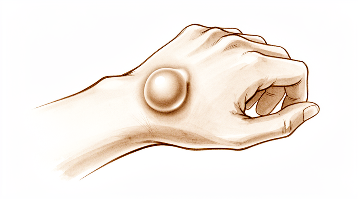
آپ کو کیا محسوس ہوتا ہے
کلائی کا گینگلیئن سسٹ (ganglion) سیال سے بھری ایک گانٹھ ہے جو کلائی کے کسی جوڑ سے یا کسی ٹینڈن (tendon)، یعنی وہ مضبوط ڈوری جو پٹھے کو ہڈی سے جوڑتی ہے، کے غلاف سے بنتی ہے۔ یہ ایسی رسولی نہیں جو کسی نقصان دہ چیز میں بدل جائے۔ بہت سے لوگ اسے سب سے پہلے کلائی کی پشت پر ایک ہموار ابھار کی صورت میں دیکھتے ہیں، اگرچہ یہ ہتھیلی والی طرف بھی ظاہر ہو سکتی ہے۔ یہ سخت محسوس ہو سکتی ہے، اور وقت کے ساتھ اس کا سائز بدل سکتا ہے۔
گانٹھ میں خود اکثر درد نہیں ہوتا۔ جب درد ہو تو یہ عموماً کلائی میں گہرائی میں، گانٹھ کے قریب ہوتا ہے، اور اکثر سرگرمی سے بڑھ جاتا ہے۔ ایسے کام جن میں کلائی مڑے یا اس پر وزن پڑے، جیسے ہاتھوں کے زور پر کرسی سے اٹھنا، سودے کے تھیلے اٹھا کر چلنا، یا بھاری پین پکڑنا، اس میں دکھن پیدا کر سکتے ہیں۔ کچھ لوگوں کو کلائی معمول سے زیادہ کمزور یا اکڑی ہوئی محسوس ہوتی ہے، جس سے ٹائپ کرنا، لکھنا یا مرتبان کھولنا مشکل ہو سکتا ہے۔
چونکہ گانٹھ کلائی کے اعصاب کے قریب ہوتی ہے، اس لیے گینگلیئن کبھی کبھی ان پر دباؤ ڈال سکتا ہے۔ اس سے ہاتھ میں جھنجھناہٹ، سُن پن یا کمزوری ہو سکتی ہے۔ اگر آپ کو اعصاب کی ان میں سے کوئی بھی علامت محسوس ہو تو بہتر ہے کہ کلائی کا معائنہ دیر کرنے کے بجائے جلد کروائیں، کیونکہ عصب کو اس دباؤ سے نجات کی ضرورت ہوتی ہے۔
اگر گانٹھ یا درد ٹھیک نہیں ہو رہا، ہفتوں کے دوران بدتر ہو رہا ہے، یا آپ کو اپنا ہاتھ استعمال کرنے یا کام کرنے سے روک رہا ہے، تو اپنے فیملی ڈاکٹر (GP) سے ملیں یا ماہر سے معائنہ کروانے کا کہیں۔
دراصل کیا ہو رہا ہے
گینگلیئن آپ کی کلائی کے جوڑ کی اندرونی جھلی میں موجود ایک چھوٹے، یک طرفہ والو سے بنتا ہے۔ وہ سیال جو عام طور پر جوڑ کو چکنا رکھتا ہے، اس والو کے ذریعے باہر دھکیلا جاتا ہے لیکن واپس نہیں جا سکتا۔ یہ جلد کے نیچے ایک تھیلی میں جمع ہو جاتا ہے، اور یہی تھیلی وہ گانٹھ ہے جسے آپ دیکھ اور محسوس کر سکتے ہیں۔ گانٹھ ایک پتلی ڈنڈی کے ذریعے جوڑ سے جڑی ہوتی ہے، اسی لیے جب جوڑ زیادہ یا کم سیال بناتا ہے تو گانٹھ کا سائز بدل سکتا ہے۔
زیادہ تر گینگلیئن اسکیفولیونیٹ لیگامنٹ (scapholunate ligament) سے بنتے ہیں، جو ٹشو کی ایک چھوٹی پٹی ہے جو کلائی کی دو چھوٹی ہڈیوں کو آپس میں جوڑے رکھتی ہے۔ یہ ان کے درمیان کچھ کچھ گسکٹ (gasket، دو پرزوں کے درمیان لگی سیل) کی طرح کام کرتا ہے۔ اس پٹی کے ارد گرد کی جھلی میں جلن (irritation) پیدا ہو جاتی ہے، اکثر ہلکی موچ یا بار بار کے کھنچاؤ کے بعد، اور سیال وہیں سے رستا ہے۔ اس سے یہ بھی سمجھ آتا ہے کہ دکھن جلد پر ہونے کے بجائے کلائی میں گہرائی میں کیوں ہوتی ہے: مسئلہ خود جوڑ میں ہے، گانٹھ میں نہیں۔
گانٹھ کی جگہ آپ کی باقی علامات کی وضاحت کرتی ہے۔ کلائی کی پشت پر یہ عموماً بالکل اسی لیگامنٹ کے اوپر ہوتی ہے، ان ٹینڈنوں کے قریب جو آپ کی انگلیوں کو سیدھا کرتے ہیں۔ ہتھیلی والی طرف یہ اکثر اس بڑی شریان اور ان اعصاب کے قریب بنتی ہے جو آپ کے ہاتھ میں جاتے ہیں، اسی لیے جب سسٹ ان پر دباؤ ڈالتا ہے تو جھنجھناہٹ یا سُن پن ہو سکتا ہے۔ کچھ گانٹھیں چھوٹی اور چھپی ہوئی ہوتی ہیں، جو صرف اس وقت ظاہر ہوتی ہیں جب کلائی آگے کی طرف مڑی ہو، پھر بھی وہ گہرا درد پیدا کر سکتی ہیں۔
یہ جاننا مفید ہے کہ گینگلیئن عام ہیں۔ بہت سے لوگوں میں یہ بغیر کسی علامت کے ہوتے ہیں، اور بغیر درد والی کلائیوں کے اسکین میں یہ آپ کی توقع سے زیادہ بار نظر آتے ہیں۔ گینگلیئن ہونے کا مطلب یہ نہیں کہ خود آپ کی کلائی کے جوڑ کی بناوٹ میں کوئی خرابی ہے۔
ہم اس کے لیے کیا کر سکتے ہیں
ڈاکٹر کیرن ہِرپرا، جو Mater Private Hospital Rockhampton میں بازو اور ہاتھ کے سرجن (upper-limb surgeon) ہیں، آپ کی حالت کے مطابق سب سے کم چیر پھاڑ والے طریقوں سے آغاز کرتے ہیں۔ مریضوں کو عام طور پر ان کے فیملی ڈاکٹر (GP) ریفرل (referral) دے کر ہمارے کلینک بھیجتے ہیں؛ اگر کسی فزیوتھراپسٹ نے آپ کو ہمیں دکھانے کا مشورہ دیا ہے، تب بھی میڈیکیئر (Medicare) سے رقم کی واپسی (rebate) کا اہل ہونے کے لیے آپ کو اپنے فیملی ڈاکٹر سے ریفرل لینا ہوگا۔ کلینک میں معائنہ، جس میں آپ کی تکلیف کی تفصیل، جسمانی معائنہ اور ضرورت ہو تو تصویری جانچ (imaging) شامل ہیں، تشخیص طے کرتا ہے۔
چونکہ گینگلیئن بے ضرر ہوتا ہے، اس لیے کچھ نہ کرنا ایک معقول پہلا قدم ہے۔ بہت سے گینگلیئن خود ہی ختم ہو جاتے ہیں۔ کلائی کے 28 سے 58 فیصد گینگلیئن کسی علاج کے بغیر ٹھیک ہو جاتے ہیں۔ 10 سال سے کم عمر بچوں میں گینگلیئن عموماً کلائی کی ہتھیلی والی طرف ہوتے ہیں، اور ان میں سے 69 سے 79 فیصد 12 سے 18 ماہ کے اندر خود ہی سکڑ کر ختم ہو جاتے ہیں۔ انتظار کرنا اور نظر رکھنا، کبھی کبھی آرام کے لیے اسپلنٹ (splint) کے ساتھ، ہاتھ یا کلائی کے گینگلیئن والے زیادہ تر بچوں کے لیے مناسب ہے۔
اگر گانٹھ تکلیف دہ ہو تو ہم اسے سوئی سے خالی کر سکتے ہیں۔ اسے سوئی سے سیال نکالنا (aspiration) کہتے ہیں۔ سیال کو کھینچ کر نکالنا، جب ایک، دو یا تین بار کیا جائے، 85 فیصد تک گینگلیئن کو ختم کر دیتا ہے۔ اس کے بعد اکثر کچھ سیال واپس آ جاتا ہے، کیونکہ سیال نکالنے سے گانٹھ تو خالی ہو جاتی ہے لیکن وہ والو باقی رہتا ہے جس نے اسے بنایا تھا۔ اس کے باوجود، سوئی سے سیال نکالنے اور گانٹھ کے بارے میں سادہ مشورے کے بعد 74 فیصد لوگ مطمئن ہوتے ہیں۔ پہلے سیال نکالنے سے آپریشن پہلے قدم کے بجائے بعد کا ایک انتخاب رہتا ہے۔ کلائی کی پشت پر چھپا ہوا گینگلیئن، یعنی وہ جو گہرا درد پیدا کرے لیکن نظر نہ آئے، عموماً پہلے سرجری کے بغیر سنبھالا جاتا ہے، جس میں کلائی کو ساکت رکھا جاتا ہے اور جوڑ کی اندرونی جھلی میں اسٹیرائیڈ (کورٹیسون) کے انجکشن لگائے جاتے ہیں۔
جب ان اقدامات سے کافی آرام نہ ملا ہو تو سرجری سے گینگلیئن کو نکالا جا سکتا ہے۔ آپریشن میں گانٹھ اور اس کی ڈنڈی، یعنی کلائی کے جوڑ سے اس کا پتلا رابطہ، نکال دی جاتی ہے، کیونکہ ڈنڈی کو پیچھے چھوڑ دینے سے گانٹھ کے واپس آنے کا امکان بڑھ جاتا ہے۔ یہ کھلے آپریشن کے طور پر یا چھوٹے سوراخوں (portals) کے ذریعے دوربین کے ذریعے آپریشن (keyhole) سے کیا جا سکتا ہے۔ ہم آپ سے بات کریں گے کہ آپ کی کلائی کے لیے کون سا طریقہ مناسب ہے، اور آپ ہمارے ساتھ مل کر فیصلہ کرتے ہیں کہ آیا سرجری آپ کے لیے فائدہ مند ہے۔
کیا توقع رکھیں
کلائی کے بہت سے گینگلیئن ایک جیسے رہنے کے بجائے وقت کے ساتھ بدلتے رہتے ہیں۔ یہ سکڑ سکتے ہیں، پھول سکتے ہیں، یا آتے جاتے رہ سکتے ہیں۔ کچھ خود ہی ٹھیک ہو جاتے ہیں: کلائی کے تقریباً 40 فیصد گینگلیئن پہلی بار معائنے کے بعد پہلے 6 سال میں چھوٹے ہو جاتے ہیں، اور کلائی کی پشت کے 39 سے 42 فیصد گینگلیئن کسی بھی علاج کے بغیر ختم ہو جاتے ہیں۔ چھوٹے بچوں میں ہاتھ اور کلائی کے زیادہ تر سسٹ خود ہی ٹھیک ہو جاتے ہیں، عموماً ایک سال کے اندر۔ اگر آپ کی گانٹھ زیادہ تکلیف نہیں دے رہی تو انتظار کرنا ایک معقول انتخاب ہے، اور اگر آپ اسے ویسے ہی چھوڑ دیں تو یہ کسی نقصان دہ چیز میں نہیں بدلے گی۔
علاج امکانات کو بدل دیتا ہے۔ سوئی سے گانٹھ کا سیال نکالنے سے یہ خالی ہو جاتی ہے، لیکن سیال نکالنے کے بعد زیادہ تر گینگلیئن واپس آ جاتے ہیں، کیونکہ وہ والو جس نے گانٹھ بنائی تھی، اب بھی موجود ہوتا ہے۔ وہ سرجری جو گانٹھ اور اس کی ڈنڈی کو نکال دیتی ہے، اس میں گانٹھ کے دوبارہ ہونے کا امکان بہت کم ہوتا ہے: سرجری کے بعد کلائی کے تقریباً 10 فیصد گینگلیئن واپس آتے ہیں، اور کلائی کی پشت کے گینگلیئن کو کھلے چیرے سے نکالنے کے بعد 3 سے 12 فیصد واپس آتے ہیں۔ سرجری کروانے والے زیادہ تر لوگوں کی علامات ٹھیک ہو جاتی ہیں۔ تاہم بعد میں کچھ درد باقی رہ سکتا ہے، خاص طور پر اگر آپ کے کام یا کھیل میں کلائی کو زور سے پیچھے کی طرف موڑنا پڑتا ہو۔
اگر آپ سرجری کا راستہ اختیار کریں تو صحت یابی عموماً سیدھی سادی ہوتی ہے۔ شروع میں آرام کے لیے آپ کے ہاتھ پر ایک موٹی پٹی باندھی جاتی ہے اور اسے اونچا رکھا جاتا ہے، اور کلائی کی حرکت جلد شروع ہو جاتی ہے، ہتھیلی والی طرف کے گینگلیئن کی سرجری کے بعد پہلے 2 ہفتوں کے اندر۔ کلائی کو جلد حرکت دینا اہم ہے: اکڑاؤ عام نہیں، لیکن اگر جلد حرکت کی حوصلہ افزائی نہ کی جائے تو ہو سکتا ہے۔ سرجری کے بعد ہینڈ تھراپی (ہاتھ کی بحالی کا علاج) Extend Rehabilitation کی Ruby Doolan کے ساتھ ہوتی ہے، اور وہ آپ کی ورزشوں کی رہنمائی کریں گی اور آپ کو جس اسپلنٹ کی بھی ضرورت ہو، وہ بنائیں گی۔ ہتھیلی والی طرف دوربین کے ذریعے سرجری کے بعد، جہاں گانٹھ تھی وہاں 3 ہفتے تک ایک سخت جگہ یا ایسی جگہ رہ سکتی ہے جسے چھونے پر درد ہو، اور ہدایت کے مطابق اس جگہ کی مالش کرنے سے یہ ٹھیک ہونے میں مدد ملتی ہے۔ زخم کے نشان ہر آپریشن کا حصہ ہیں، اور کلائی کی لکیروں کے قریب خمدار چیرے عموماً زیادہ اچھے دکھنے والے نشان کے ساتھ بھرتے ہیں۔
اگر علاج کی گئی گانٹھ یا آپ کی علامات ٹھیک نہیں ہو رہیں، یا ہفتوں کے دوران بدتر ہو رہی ہیں، تو اپنے فیملی ڈاکٹر سے ملیں یا ماہر سے معائنہ کروانے کا کہیں۔
ڈاکٹر کو کب دکھائیں
کلائی کے زیادہ تر گینگلیئن بے ضرر ہوتے ہیں، اور بہت سے خود ہی ٹھیک ہو جاتے ہیں، اس لیے صرف گانٹھ کا ہونا ایمرجنسی نہیں ہے۔ اگر گانٹھ یا درد ہفتوں میں ٹھیک نہیں ہو رہا، بدتر ہو رہا ہے، یا آپ کو کام کرنے یا اپنا ہاتھ استعمال کرنے سے روک رہا ہے، تو اپنے فیملی ڈاکٹر سے ملیں یا ماہر سے معائنہ کروانے کا کہیں۔ اگر آپ کے ہاتھ میں جھنجھناہٹ، سُن پن یا کمزوری ہو تو جلد معائنہ کروانے کا کہیں، کیونکہ اس سے لگتا ہے کہ سسٹ کسی عصب پر دباؤ ڈال رہا ہے۔ اگر آپ کا ہاتھ یا انگلیاں سفید پڑ جائیں، ٹھنڈی ہو جائیں، یا سفید یا نیلی ہو جائیں، یا اگر کسی چوٹ کے بعد اچانک ہاتھ میں احساس یا حرکت ختم ہو جائے، تو ہسپتال کی ایمرجنسی جائیں۔ گینگلیئن میں ایسا شاذ و نادر ہی ہوتا ہے، لیکن ان کے لیے اسی دن علاج ضروری ہے۔
مزید تفصیل
Advanced reading: the deeper science (optional)
یہ حصہ اس سے زیادہ تفصیل میں جاتا ہے جتنی آپ کو اپنے علاج کے فیصلوں کے لیے ضرورت ہے۔ کلائی کے گینگلیئن کے بارے میں یہ اضافی مطالعہ اس لیے مفید ہے کہ اصل انتخاب واقعی گانٹھ کے دوبارہ ہونے کا خطرہ قبول کرنے اور آپریشن قبول کرنے کے درمیان ہے، اور اس سودے کے پیچھے اعداد و شمار اتنے واضح ہیں کہ ان کی بنیاد پر فیصلہ کیا جا سکے۔
سوئی سے خالی کرنے کے بعد گانٹھ واپس آ جاتی ہے؛ کاٹ کر نکالنے کے بعد اتنی کثرت سے نہیں
دو فعال علاج یہ ہیں: سسٹ کے سیال کو سوئی سے کھینچ کر نکالنا (aspiration)، اور سسٹ کو اس کی ڈنڈی سمیت سرجری سے نکالنا (excision)۔ 2,239 مریضوں کے اعداد و شمار کو ملا کر دیکھا گیا تو کھلے چیرے سے سرجری کے ذریعے نکالنے میں گانٹھ کے دوبارہ ہونے کا امکان سوئی سے خالی کرنے کے مقابلے میں نمایاں طور پر کم ہے۔ دوربین کے ذریعے نکالنے (arthroscopic excision) کے نتائج حوصلہ افزا رہے ہیں، لیکن موازنہ کرنے والے ٹرائلز کا ڈیٹا محدود ہے اور ان سے اس کا بہتر ہونا ثابت نہیں ہوا [1]۔
اس فرق کی وجہ اس کے طریقۂ کار میں ہے۔ گینگلیئن سیال کی کوئی آزاد تیرتی تھیلی نہیں ہے؛ یہ ایک ڈنڈی کے ذریعے نیچے کے جوڑ کی جھلی (capsule) سے جڑا ہوتا ہے، اور جوڑ سیال بناتا رہتا ہے۔ سوئی سے خالی کرنے میں ذخیرہ تو خالی ہو جاتا ہے لیکن سیال کا منبع باقی رہتا ہے، اس لیے گانٹھ کا دوبارہ بھر جانا علاج کی ناکامی نہیں بلکہ ایک عام نتیجہ ہے۔ کاٹ کر نکالنے کا مقصد یہ ہوتا ہے کہ ڈنڈی کو اس کی جڑ تک ڈھونڈ کر نکال دیا جائے۔
دوربین سے اور کھلے چیرے سے نکالنے کے نتائج ایک جیسے ہیں
جہاں کاٹ کر نکالنے کا فیصلہ کیا جائے، وہاں دونوں طریقوں کا براہِ راست موازنہ کیا گیا ہے۔ 910 مریضوں میں دوربین کے ذریعے اور کھلے چیرے والے طریقوں کے نتائج گانٹھ کے دوبارہ ہونے اور پیچیدگیوں دونوں کے لحاظ سے ملتے جلتے تھے، اور مصنفین نے معیاری اور مناسب حد تک بڑی تحقیقات کی ضرورت پر زور دیا [2]۔
اس سے فیصلہ گانٹھ کے دوبارہ ہونے کے خطرے کے بجائے دوسری باتوں پر آ جاتا ہے: زخم کا نشان، سرجن کی کسی طریقے سے واقفیت، اور یہ کہ آیا جوڑ کے اندر کسی اور خرابی کا اسی وقت جائزہ لینا ضروری ہے۔
آپریشن کے بعد کیا کیا جائے، اس پر کوئی اتفاق نہیں، اور شاید اس سے فرق بھی نہ پڑے
ایک چھوٹی سی، دیانت دارانہ بات۔ ہاتھ کے سرجنوں کے ایک منظم جائزے (systematic review) اور سروے سے پتا چلا کہ کلائی کی پشت کے گینگلیئن کو نکالنے کے بعد کلائی کو ساکن رکھا جائے یا نہیں، اس پر سرجنوں کی رائے بٹی ہوئی ہے، اور ہاتھ کی کارکردگی کے نتائج کے لحاظ سے کوئی ٹھوس ڈیٹا یہ ظاہر نہیں کرتا کہ کوئی ایک طریقہ بہتر ہے [3]۔
یہ جاننا مفید ہے کہ اس معاملے میں مختلف سرجنوں کی ہدایات کا مختلف ہونا حقیقی غیر یقینی صورتحال کی عکاسی کرتا ہے، اس بات کی نہیں کہ ان میں سے کوئی ایک غلط ہے۔
اکثر سب سے مضبوط دلیل کچھ نہ کرنے کے حق میں ہوتی ہے
اوپر کی کسی بات سے یہ ثابت نہیں ہوتا کہ گینگلیئن کا علاج ضرور کیا جانا چاہیے۔ یہ بے ضرر سسٹ ہیں۔ ان کا سائز اکثر گھٹتا بڑھتا رہتا ہے، اور ان میں سے ایک حصہ کسی علاج کے بغیر ختم ہو جاتا ہے۔ یہ کسی اور چیز میں تبدیل نہیں ہوتے۔
اس سے فیصلے کا زاویہ بدل جاتا ہے۔ علاج کی وجوہات درد، کلائی کی حرکت یا گرفت میں رکاوٹ، کسی قریبی عصب پر دباؤ، یا ایسا سائز ہیں جو واقعی اس شخص کو پریشان کرتا ہو، نہ کہ محض گانٹھ کا موجود ہونا۔ چونکہ سوئی سے خالی کرنے کے بعد گانٹھ کے دوبارہ ہونے کی شرح قابلِ ذکر ہے اور کاٹ کر نکالنے میں جوڑ کی جھلی پر آپریشن کے خطرات ہوتے ہیں، اس لیے انتظار کرنا اور نظر رکھنا پہلے قدم کے طور پر ایک جائز موقف ہے، اور اسے کچھ نہ کرنے کی ناکامی سمجھنے کے بجائے واضح طور پر بیان کرنا چاہیے۔
اس میں استثنا وہ گینگلیئن ہے جو اعصابی علامات پیدا کر رہا ہو، یعنی سُن پن، کمزوری، یا ہاتھ میں پھیلنے والا درد۔ اس صورت میں سسٹ کسی ایسے حصے کو دبا رہا ہوتا ہے جو اس دباؤ کو غیر معینہ مدت تک برداشت نہیں کر سکتا، اور انتظار کرنا اور نظر رکھنا اب کم خطرے والا راستہ نہیں رہتا۔
حوالہ جات
[1] Head L, Gencarelli JR, Allen M, Boyd KU. Wrist ganglion treatment: systematic review and meta-analysis. J Hand Surg Am. 2015;40(3):546-553.e8. https://doi.org/10.1016/j.jhsa.2014.12.014
[2] Crawford C, Keswani A, Lovy AJ, Levy I, Lutz K, Kim J, et al. Arthroscopic versus open excision of dorsal ganglion cysts: a systematic review and meta-analysis. J Hand Surg Eur Vol. 2017;43(6):659-64. https://doi.org/10.1177/1753193417734428
[3] Wong CR, Karpinski M, Hatchell AC, McRae MH, Murphy J, McRae MC. Immobilization of the wrist after dorsal wrist ganglion excision: a systematic review and survey. Hand (N Y). 2021;18(2):254-63. https://doi.org/10.1177/15589447211014631
Evidence & references
This is the clinical evidence summary written for health professionals. It is technical, and it lists the research this page was built from. You do not need to read it to understand your treatment or to make a decision about it.
Overview
Epidemiology and Natural History
- The dorsal wrist ganglion is the prototype of all hand and wrist ganglions, accounting for 60% to 70% of all hand and wrist ganglions [1].
- Volar wrist ganglia are the second most common ganglion of the hand and wrist, accounting for 18% to 20% of cases [11].
- In an asymptomatic population, 51% of 103 wrists scanned demonstrated ganglia on MRI [7].
- In an asymptomatic population evaluated by MRI, volar wrist ganglia are more common than dorsal wrist ganglia [2].
- Neither surgical excision nor aspiration provides a clear long-term benefit over the natural history of the dorsal wrist ganglion [6].
- Pediatric ganglions of the hand have a greater rate of resolution than ganglions of the wrist [4].
Clinical Presentation and Diagnosis
- The main cyst of a dorsal wrist ganglion is usually located directly over the scapholunate ligament [1].
- Dorsal wrist ganglions may occur anywhere between the extensor tendons and can be connected to the scapholunate ligament through an elongated pedicle [1].
- Careful preoperative palpation of a dorsal wrist ganglion with digital compression often reveals its extent and the direction of the pedicle [1].
- Transillumination or aspiration confirms the diagnosis of a wrist ganglion preoperatively [1].
- In patients with recalcitrant dorsal wrist pain in the area of the scapholunate interval, occult dorsal wrist ganglions had a prevalence of 11 out of 14 [3].
- Routine wrist radiography is not cost-effective in the evaluation and treatment decision-making process for patients with a wrist ganglion due to a low prevalence of therapeutically significant findings [5].
- A systematic X-ray should be performed for painful dorsal wrist ganglia [13].
- Preoperative x-rays should be obtained as part of the routine workup for a dorsal wrist ganglion [22].
- Volar wrist ganglions may appear small clinically but can be surprisingly extensive at surgery, with multiloculated cysts extending under the thenar muscles, along the flexor carpi radialis tendon, into the carpal canal, and under the first extensor compartment [11].
- The Allen test should be performed routinely to assess the patency of the radial and ulnar arteries in patients with volar wrist ganglions [11].
Operative Considerations
- Failure to identify and excise the pedicle attachment to the scapholunate ligament increases the likelihood of recurrence for dorsal wrist ganglions [1].
- A small, mucin-filled duct is invariably seen piercing the transverse fibers of the scapholunate ligament, connecting the underlying scapholunate joint with the main cyst [1].
- The radial artery is frequently intimately attached to the wall of a volar wrist ganglion and may be completely encircled by the ganglion [11].
- Some authors recommend leaving a portion of the volar ganglion cyst wall attached to the radial artery to avoid arterial injury [11].
- The best indication for arthroscopic treatment of a volar wrist ganglion is a sessile type arising from the radiocarpal joint and centered on the proximal wrist crease [12].
- Mobile volar ganglia with a long pedicle are poor candidates for arthroscopic surgery [12].
- Open excision of dorsal wrist ganglia leads to a lower recurrence rate than arthroscopic excision [25].
- Routine midcarpal joint exploration during arthroscopic excision of dorsal wrist ganglions appeared to reduce recurrence at 1 year without negatively impacting patient outcomes [35].
- Arthroscopic resection of dorsal wrist ganglia has low morbidity, with unnoticeable scars and wrist mobility and strength close to normal by three months [10].
- Arthroscopic resection of radiocarpal volar ganglia is a reasonable alternative to open excision with decreased postoperative morbidity, easy technique, and a high rate of good results [56].
- Arthroscopic ganglionectomy through an intrafocal cystic portal is a safe and efficacious option for the treatment of painful wrist ganglia [39].
- Surgical excision of primary wrist ganglia significantly reduces patient symptoms with low recurrence rates and high patient satisfaction [18].
- Open surgical excision offers a significantly lower chance of recurrence compared with aspiration in the treatment of wrist ganglions [59].
- Female patients who have preoperative pain around dorsal wrist ganglia are the most likely to have residual pain after surgery [8].
- Patients whose occupation or activities require forceful wrist extension should be counseled on the considerable risk of residual pain and functional limitations that may occur after open dorsal wrist ganglion excision [26].
- Hand surgeons are divided regarding the need to immobilize the wrist after dorsal wrist ganglion excision [63].
Pediatric Management
- There is no consensus within the literature regarding the best management of pediatric wrist ganglia, and no single treatment modality confers a particular advantage or disadvantage over another [14].
- Surgical excision of pediatric hand and wrist ganglions is indicated for those that are symptomatic, do not resolve after approximately 2 months of observation and/or splinting, or recur [31].
Anatomy & Pathophysiology
Dorsal Wrist Ganglion
- Dorsal wrist ganglions may occur anywhere else between the extensor tendons, connected to the scapholunate ligament through an elongated pedicle [1].
- Failure to identify and excise the pedicle attachment to the scapholunate ligament increases the likelihood of recurrence [1].
- Although ganglions have been reported in other carpal joints, they are rare, and attachments to the scapholunate joint must be ruled out before a dissection is considered complete [1].
- Dorsal wrist ganglions can be confused with carpal bosses, which are osteoarthritic spurs or prominences that develop at the base of the second or third carpometacarpal joints [23].
- A small ganglion is associated with a carpal boss in 30% of cases [23].
- Unlike protruding dorsal ganglions, smaller, occult dorsal ganglions are easily overlooked and can often only be palpated with the involved wrist in marked volar flexion [64].
- An occult ganglion may be the cause of unexplained wrist pain and is disproportionately tender [64].
- Dorsal ganglions occasionally occur in association with an underlying scapholunate diastasis [64].
- Dorsal prominence of the proximal pole of the scaphoid secondary to intercarpal instability may be confused with a painful occult ganglion [64].
- Chronic tenosynovitis of the extensor tendons can be confused with a dorsal ganglion but is distinguished by the diffuse nature of the swelling and the puckering seen with digital extension, known as the tuck sign [64].
- Ganglions arising on or within extensor tendons typically occur over the metacarpals and are distinguished by their proximal motion with the fingers in extension [125].
- Two anomalous muscles, the anomalous extensor indicis proprius and the extensor digitorum brevis manus, may be found in the area of the dorsal wrist ganglion and can complicate management [151].
Volar Wrist Ganglion
- The volar wrist ganglion is the second most common ganglion of the hand and wrist, accounting for 18% to 20% of cases [11].
- The majority of volar ganglions occur either directly over the distal edge of the radius or slightly more distally over the scaphoid tubercle [11].
- Volar ganglions arising from the distal edge of the radius originate from the capsular and ligamentous fibers of the radiocarpal joint and occur under the volar wrist crease between the flexor carpi radialis and abductor pollicis longus tendons [11].
- The main cyst of a volar ganglion may be intertwined with bifurcating branches of the radial artery [11].
- Another type of volar ganglion arises from the capsule of the scaphotrapezial joint [11].
- Volar ganglions may appear small clinically but can be surprisingly extensive at surgery, with multiloculated cysts extending under the thenar muscles, along the flexor carpi radialis tendon, into the carpal canal, and under the first extensor compartment [11].
- The radial artery is frequently intimately attached to the wall of a volar ganglion and may even be completely encircled by the ganglion [11].
- The radioscaphocapitate and long radiolunate ligament interval may be the site of origin for the volar wrist ganglion [27].
- Ganglia arising from the interval between the radioscapocapitate (RSC) and long radiolunate (LRL) ligament appear at the more lateral aspect of the distal radius on arthrogram [12].
- Ganglia arising from the interval between the long radiolunate (LRL) and short radiolunate (SRL) ligaments appear at a more central position of the distal radius on arthrogram [12].
- Ganglia arising from the midcarpal joint or scaphotrapeziotrapezoid (STT) joint will not be revealed by an arthrogram of the radiocarpal joint unless an interosseous ligament at the proximal carpal row is ruptured [12].
Intraneural Ganglion
- Involvement of the peripheral nerves of the upper extremity by intraneural ganglion is rare [42].
- The ulnar nerve is the most commonly involved nerve in the upper extremity for intraneural ganglions [42].
- Most patients with intraneural ganglions are aged 30 to 50 years, though occurrence in children has also been reported [42].
- Most patients with intraneural ganglions present with a painless mass, while some may present with symptoms of nerve irritation or entrapment neuropathy [42].
- The articular (synovial) theory has gained worldwide acceptance as a scientifically proven explanation for the formation of intraneural ganglions [42].
Wrist Anatomy & Biomechanics
- The wrist is the anatomic region between the forearm and the hand, including the distal radioulnar, radiocarpal, and ulnocarpal joints and the eight carpal bones [69].
- The eight carpal bones include the scaphoid, lunate, triquetrum, and pisiform in the proximal row and the trapezium, trapezoid, capitate, and hamate in the distal row [69].
- The radiocarpal joints are formed by the articulation of the distal radius with the scaphoid and lunate through their respective concave facets on the distal radius and the triquetrum on the triangular fibrocartilage [69].
- The distal concave articular surfaces of the proximal carpal row form the midcarpal articulations with the distal row [69].
- The triangular fibrocartilage complex (TFCC) attaches to the base of the ulnar styloid and separates the hyaline cartilage–covered ulnar head from the styloid [69].
- The scapholunate interosseous ligament is C-shaped in the sagittal plane, with the dorsal third being the thickest and strongest portion [70].
- The volar portion of the lunotriquetral ligament is the thickest [70].
- The proximal carpal row has no muscular or tendinous attachments and is an intercalary segment [70].
- With axial loading through the neutral wrist, approximately 80% of forces are transmitted through the distal radius (60% scaphoid facet, 40% lunate facet) and 20% through the distal ulna [70].
- With wrist flexion, 60% of the motion is midcarpal and 40% is radiocarpal [70].
- With wrist extension, 33% of the motion is midcarpal and 66% is radiocarpal [70].
- The wrist can essentially be considered to be a two-joint system linking the hand to the forearm around the highly mobile bones of the proximal carpal row [71].
- The joints within the proximal carpal row govern 35% of the wrist extension-flexion arc and 33% of the wrist radial-ulnar deviation arc [94].
- Wrist ligaments co-ordinate the positioning of the bones in the mid-range of carpal motions and restrict further motion in extreme positions of the wrist joint [97].
- The terminal branches of the radial, ulnar, and anterior interosseous arteries provide extraosseous blood supply to the carpus through three dorsal and three palmar transverse arterial arches with longitudinal connections [72].
- The dorsal intercarpal arch is the largest of the dorsal arches and supplies the distal carpal row and, through anastomoses with the radiocarpal arch, the lunate and triquetrum [72].
- The deep palmar arch at the level of the metacarpal bases is consistent and communicates with the dorsal basal metacarpal arch and the palmar metacarpal arteries [72].
- The distal radial articular surface has a double obliquity of 12–15 degrees in the lateral view and 15–20 degrees in the anteroposterior view [78].
- The carpus is more stable in flexion than extension due to its anterior concavity [78].
- The stability of the carpal bones relies largely on interosseous ligaments and bony configuration rather than tendon insertions, with the exception of the pisiform [78].
- The scaphoid presents a long axis inclined by 45 degrees to the long axis of the radius [78].
- The lunate sits on the capitate and has anterior and posterior horns, with a line drawn between them lying perpendicular to the long axis of the wrist in neutral position [78].
- The ulnar head sits proximal to the distal radius and has only an indirect effect on stability of the wrist [78].
- Extension of the wrist is dependent on the extensor carpi radialis longus (ECRL), extensor carpi radialis brevis (ECRB), and extensor carpi ulnaris (ECU) [79].
- The ECRB is the primary wrist extensor because it has the greatest tension and the most favorable moment arm [79].
- The ECU is an extensor of the wrist in supination and primarily causes ulnar deviation of the wrist in pronation [79].
- The moment arms for extension of the wrist are 16.30 mm for the ECRB and 12.50 mm for the ECRL [79].
- The moment arm for extension of the wrist by the ECU is 6.3 mm in supination and becomes zero when the wrist is in complete pronation [79].
- The most frequently used wrist movements are not in the axis of flexion–extension but in semipronation, with an oblique axis between the ECRL/ECRB and the flexor carpi ulnaris [79].
- The superficial branch of the radial nerve averages 16 mm (range 5 to 22 mm) from the 3-4 arthroscopic portal [27].
- The dorsal sensory branch of the ulnar nerve averages 8 mm (range 0 to 14 mm) from the 6R arthroscopic portal [27].
- The 1-2 arthroscopic portal carries a high risk of injury to the superficial branch of the radial nerve [27].
- The 6U arthroscopic portal carries a high risk of injury to the dorsal sensory branch of the ulnar nerve [27].
- The ideal working portal for arthroscopic excision of a dorsal wrist ganglion is the 3-4 portal with visualization from the 4-5 or 6R portals [27].
Pathophysiology & Diagnostic Associations
- Preoperative x-rays should be obtained as part of the routine workup for a dorsal wrist ganglion to rule out interosseous components or instability [22].
- MRI is useful in confirming the diagnosis of an occult dorsal ganglion and differentiating pain related to scapholunate ligament injury [64].
- Excising a dorsal ganglion alone might not alleviate all the patient’s preoperative pain if other causes of wrist pain and tenderness, especially directly over the scapholunate ligament, are present [64].
- An occult dorsal ganglion is best initially treated conservatively by immobilization and steroid injections directly into the dorsal capsule if other causes of wrist pain are excluded [64].
- Excision of the posterior interosseous nerve at the level of the radiocarpal joint may help alleviate pain and add to the patient’s postoperative comfort in cases of dorsal ganglion [64].
- Arthroscopic resection was an effective treatment method for well-selected volar wrist ganglion arising from the radiocarpal joint in the long run [20].
- Abnormal synovitis at the interligamentous sulcus is a hallmark of a volar ganglion from the radiocarpal joint [47].
- Pressing the ganglion externally should demonstrate bulging of the origin of the ganglion as viewed intraarticularly during arthroscopy [47].
- The complex nature of the wrist has plagued clinicians and hampered the ability to formulate concise, yet thorough, algorithmic approaches to evaluation and treatment [45].
- A wrist joint should be considered biomechanically unstable when it is not able to bear loads and does not exhibit normal kinematics throughout its arc of motion [67].
- The longitudinal 'columnar' concept of wrist kinematics does not fit in with many of the recent findings related to carpal instabilities [68].
- Traction (distraction) changes normal carpal kinematics and motion of the wrist combined with traction induces abnormal patterns of motion [73].
- Distal radial fractures that result in a loss of normal palmar tilt lead to progressive load on the ulnocarpal and radioscaphoid articulations [103].
- The most significant effect on radiocarpal joint contact characteristics occurs with a depression of the scaphoid side of the joint [110].
- Findings of abnormalities in the scapholunate joint were observed in 10 of 16 wrists with painful dorsal ganglia based on arthroscopic assessment [149].
- In most cases of dorsal wrist ganglion, there is a mild chronic 'sprain' of the scapholunate ligament which does not give rise to the well-recognised radiological and clinical features associated with scapholunate instability [149].
- Only one of 16 wrist radiographs showed radiological scapholunate widening in a study of painful dorsal ganglia [149].
- The anatomical configuration of ulnar nerve compression by a synovial cyst should be investigated if clinical signs do not improve following surgical decompression of the ulnar nerve at the wrist [32].
Classification
- In a selected patient group with recalcitrant dorsal wrist pain in the area of the scapholunate interval, occult dorsal wrist ganglions had a high prevalence of 11 of 14 [3].
- There is a significant prevalence of asymptomatic wrist ganglia, with 51% of 103 wrists scanned demonstrating ganglia [7].
- In a series of 543 ganglions of the hand and wrist, 57% occurred over the dorsal aspect of the wrist [65].
- In a series of 543 ganglions of the hand and wrist, 17% occurred over the volar aspect of the wrist [65].
- In a series of 543 ganglions of the hand and wrist, 26% occurred on the fingers [65].
- In a series of 543 ganglions of the hand and wrist, 7% were mucous cysts [65].
- Approximately 65% of anterior wrist ganglia arise from the radioscaphoid/scapholunate interval [48].
- Approximately 34% of anterior wrist ganglia arise from the scaphotrapezial joint [48].
- Ganglions in pediatric populations most commonly affect the dorsal wrist [28].
- In a longitudinal follow-up of 14 children less than 10 years old, 79% of all hand and wrist cysts spontaneously resolved, with the majority resolving within a year [37].
- The incidence of dorsal wrist ganglia was higher in the military population compared with the civilian population [40].
- In a series of 543 ganglions, 80% of patients were between twenty and fifty years of age [65].
- In a series of 543 ganglions, there were 363 females and 180 males, an almost 2 to 1 ratio [65].
- In a series of 543 ganglions, only one-fourth of patients complained of pain when first seen [65].
- In a series of 543 ganglions, the left and right hands were equally involved (242/257) [65].
- In a series of 543 ganglions, 90% were less than two centimeters in diameter [65].
- In a series of 543 ganglions, 50% were between 0.5 and 2.0 centimeters in diameter [65].
- In a series of 543 ganglions, the majority had been present for less than two years when patients sought attention [65].
- In a series of 543 ganglions, specific injury related to the onset of the ganglion occurred in only fifty patients [65].
- Ganglions may arise from a variety of additional sites over the dorsal wrist capsule, particularly in the region of the capitate, although a scapholunate origin is usual [15].
- Ganglions arising from wrist joints can present as painless masses in the center of the palm without signs or symptoms of median or ulnar nerve compression [17].
- The ulnar nerve is the most commonly involved nerve in the upper extremity by intraneural ganglion [42].
- Most patients with intraneural ganglions are aged 30 to 50 years, but occurrence in children has also been reported [42].
- Most patients with intraneural ganglions present with a painless mass, though some may present with symptoms of nerve irritation or entrapment neuropathy [42].
Clinical Presentation
Dorsal Wrist Ganglion
- The dorsal wrist ganglion accounts for 60% to 70% of all hand and wrist ganglions [1].
- Dorsal wrist ganglions may occur anywhere else between the extensor tendons, connected to the scapholunate ligament by an elongated pedicle [1].
- Transillumination or aspiration confirms the diagnosis of a dorsal wrist ganglion preoperatively [1].
- Review of preoperative radiographs is wise to rule out an interosseous component in dorsal wrist ganglions [1].
- MRI scans provide relatively good reliability in establishing the diagnosis of an occult dorsal wrist ganglion, with a sensitivity of 83% when using intra-operative findings as the standard [38].
- Female patients who have preoperative pain around dorsal wrist ganglia were the most likely to have residual pain after surgery [8].
- Although a scapholunate origin is usual, ganglia may also arise from a variety of additional sites over the dorsal wrist capsule, particularly in the region of the capitate [15, 16].
- A dorsal wrist ganglion can present preoperatively as an anterior wrist ganglion [19].
- The incidence of dorsal wrist ganglia was higher in the military compared with the civilian population [40].
Volar Wrist Ganglion
- The volar wrist ganglion is the second most common ganglion of the hand and wrist, accounting for 18% to 20% [11].
- Volar ganglions arising from the distal edge of the radius occur under the volar wrist crease between the flexor carpi radialis and abductor pollicis longus tendons [11].
- Volar ganglions arising from the scaphotrapezial joint capsule are a distinct type from those arising from the radiocarpal joint [11].
- Although volar ganglions may appear small clinically, they can be surprisingly extensive at surgery, with multiloculated cysts extending under the thenar muscles, along the flexor carpi radialis tendon, into the carpal canal, and under the first extensor compartment [11].
- Extensions of volar ganglions can often be appreciated preoperatively by careful palpation and digital compression [11].
- Unlike previous surgical and pathological series, an MRI study showed volar wrist ganglia are more common than dorsal wrist ganglia in an asymptomatic population [2].
- Wrist joint ganglions can present as painless masses in the center of the palm without signs or symptoms of median or ulnar nerve compression [17].
General Characteristics and Epidemiology
- There is a significant prevalence of asymptomatic wrist ganglia, with 51% of the 103 wrists scanned demonstrating ganglia [7].
- Ganglions in pediatric populations, which most commonly affect the dorsal wrist, demonstrate a female predilection [28].
- In children aged <10 years, ganglions mainly occur on the volar wrist [33].
- In children aged <10 years, 69% to 79% of ganglions display spontaneous regression within a span of 12-18 months [33].
- In a cohort of 14 children less than 10 years old, 79% of all cysts spontaneously resolved, the majority within a year [37].
- Ganglia are the most common benign soft tissue tumours of the hand [44].
- The majority of ganglia resolve spontaneously over 1 to 2 years [44].
- In a study of 50 patients with ganglia, 38% were concerned about the cosmetic appearance and 28% were concerned that their ganglion was a malignant growth [44].
- A minority of patients sought advice and treatment for ganglia because of pain [44].
Differential Diagnosis and Imaging
- The study concludes that routinely performing wrist radiography is not cost-effective in the evaluation and treatment decision-making process for patients with a wrist ganglion due to a low prevalence of therapeutically significant findings [5].
- It is suggested that preoperative x-rays should be obtained as part of the routine workup for a dorsal wrist ganglion [22].
- Sonography can localize occult ganglia [66].
- Preoperative ultrasound scanning helps to confirm the articular origin and nature of the cystic mass for volar wrist ganglions [12].
Investigations
Clinical Examination and Physical Diagnosis
- Careful preoperative palpation of the cyst with digital compression often reveals its extent and the direction of the pedicle [1].
- Transillumination or aspiration confirms the diagnosis preoperatively [1].
- A firm, bony, nonmobile, tender mass visible and palpable at the base of the carpometacarpal joints, especially when the wrist is flexed, is characteristic of a carpal boss [23].
- Dorsal wrist ganglions can be confused with carpal bosses [23].
Radiography
- Review of the patient's preoperative radiographs to rule out an interosseous component is wise [1].
- The mass of a carpal boss is best visualized radiologically with the hand in 30 to 40 degrees of supination and 20 to 30 degrees of ulnar deviation ("carpal boss view") [23].
Magnetic Resonance Imaging (MRI)
- In a selected patient group with recalcitrant dorsal wrist pain in the area of the scapholunate interval, occult dorsal wrist ganglions had a high prevalence (11 of 14) [3].
- MRI is an excellent diagnostic modality to evaluate rapidly growing upper extremity masses and distinguish ganglions from malignant processes [109].
- Convolutional neural networks (CNNs) can detect ganglion cysts in wrist MRI [136].
- Unlike previous surgical and pathological series, an MRI study of an asymptomatic population showed volar wrist ganglia are more common than dorsal wrist ganglia [2].
Arthrography and Arthroscopy
- A wrist arthrogram can be performed immediately prior to arthroscopic intervention or as a separate investigative procedure for volar wrist ganglions [12].
- Intraoperative arthrogram of a ganglion arising from the radiocarpal joint involves injecting 3 to 5 cc of nonionic contrast solution admixed with 2% lidocaine into the radiocarpal joint at the 3-4 portal site under fluoroscopic guidance [12].
- The relative position of the contrast-filling stalk on arthrogram can aid the identification of the true stalk during the actual arthroscopic procedure [12].
- Ganglia arising from the midcarpal joint or scaphotrapeziotrapezoid (STT) joint will not be revealed by arthrogram of the radiocarpal joint unless an interosseous ligament at the proximal carpal row is ruptured and creates a free communication between the radiocarpal and midcarpal joints [12].
- Arthroscopy is not the gold standard for the diagnosis of carpal ganglia because the majority of the cyst is found either subcutaneously or at the point of the capsular attachment to the proximal carpal row, external to the wrist joint [62].
- Sonography-assisted arthroscopic resection is a safer and more reliable method for treating volar wrist ganglia [21].
Treatment
Non-Operative Management
- The spontaneous resolution rate for wrist ganglia is reported as 28% to 58% [106].
- Aspiration treatment of ganglion cysts of the wrist and hand can safely remove 85% of these tumors if one, two, or three separate treatments are administered [95].
- Aspiration with or without cortisone injection has an average success rate of 35% to 50% [106].
- In a study of 50 patients, 74% were satisfied with aspiration of the ganglion and general advice [44].
- Although aspiration with triamcinolone acetonide injection plus wrist immobilization is an alternative method, surgery was the most successful form of treatment when considering the cure rate of dorsal wrist ganglion [84].
- In children aged <10 years, ganglions mainly occur on the volar wrist and can be treated expectantly, with 69% to 79% displaying spontaneous regression within a span of 12-18 months [33].
- While observation and/or splinting will likely be helpful in resolution of a majority of pediatric hand and wrist ganglions, surgical excision is indicated for those that are symptomatic, do not resolve after approximately 2 months of observation and/or splinting, or recur [31].
- An occult dorsal ganglion is best initially treated conservatively by immobilization and steroid injections directly into the dorsal capsule, which can also aid in diagnosis [64].
Operative Management: Dorsal Wrist Ganglion
- Failure to identify the pedicle and excise its attachment to the scapholunate ligament increases the likelihood of recurrence [1].
- Most dorsal ganglions can be approached through a transverse incision over the proximal carpal row [1].
- The diagnosis of ganglion cyst should be made before commitment to a transverse incision because this type of incision is not readily incorporated into a limb-sparing incision in the event of a subsequent diagnosis of a malignant soft tissue tumor [1].
- Typically, a dorsal ganglion appears between the extensor pollicis longus and extensor digitorum communis tendons [1].
- The main cyst and its pedicle are mobilized down to the underlying joint capsule [1].
- With the wrist in volar flexion, the joint capsule is opened along the border of the radius and scaphoid's proximal pole [1].
- The capsular incision is extended more laterally if any capsular ducts, which can be identified by small amounts of mucin drainage, are encountered during the dissection [1].
- The ganglion and its capsular attachments are tangentially excised off the scapholunate ligament [1].
- A small, mucin-filled duct is invariably seen piercing the transverse fibers of the scapholunate ligament [1].
- Synovial and capsular attachments along the distal margin of the scapholunate ligament are excised to give an unobstructed view of the head and neck of the capitate [1].
- The key to successful open treatment is excision of the ganglion stalk based at the scapholunate ligament and its associated dorsal-capsular attachments [106].
- Open ganglionectomy creates a defect in the capsule of 1 to 1.5 cm, which is left open [106].
- The rate of recurrence for open ganglionectomy can be as high as 40% [106].
- Stiffness has been reported from prolonged immobilization following open ganglionectomy [106].
- Excision of the posterior interosseous nerve at the level of the radiocarpal joint may help alleviate the pain and add to the patient’s postoperative comfort in cases of occult dorsal ganglion [64].
- Excising the ganglion alone might not alleviate all the patient’s preoperative pain if an occult dorsal ganglion is associated with underlying scapholunate diastasis [64].
- Dorsal ganglions occasionally occur in association with an underlying scapholunate diastasis, and they may be blamed for the carpal instability after their excision [64].
- In the treatment of dorsal wrist ganglion, surgery was the most successful form of treatment when considering the cure rate compared to aspiration with triamcinolone acetonide injection plus wrist immobilization [84].
- The quality of care would not be compromised by abandoning the practice of routine submission of surgical specimens for pathological examination after excision of the ganglion cyst in patients with the clinical diagnosis of wrist ganglion cyst [137].
- Following a dorsal incision distal to Lister’s tubercle, the extensor retinaculum between the tendons of the second and fourth extensor compartments is incised, the ganglion identified and its root followed to the dorsal capsule, and a small arthrotomy is performed over the scapholunate ligament [131].
- Complete excision of the ganglion along with its capsule is performed with local synovectomy [131].
- The capsule is either fenestrated or sutured, depending on the surgeon [131].
- After skin closure, a compressive dressing is applied and left in place for 3 days [131].
- Active mobilization of the wrist starts after the dressing is changed by the hand therapist [131].
- Sutures are removed 10 to 14 days postoperatively [131].
- Splints are not used on a regular basis after dorsal wrist ganglion excision [131].
- After 3 weeks, patients start with a stabilizing and strength programme under the guidance of the hand therapist [131].
Operative Management: Volar Wrist Ganglion
- The main cyst of a volar ganglion arising from the radiocarpal joint may be intertwined with bifurcating branches of the radial artery [11].
- The Allen test should be performed routinely and ulnar artery occlusion excluded before volar ganglion excision [11].
- The surgeon must be aware of the importance of preserving the radial artery, particularly in patients with a radial-dominant circulation [11].
- Longitudinal incisions allow for optimal visualization during volar ganglion excision [11].
- The radial artery is frequently intimately attached to the wall of the volar ganglion and may even be completely encircled by the ganglion [11].
- Loupe magnification aids in the dissection of the radial artery from the volar ganglion [11].
- The pedicle is traced to the volar joint capsule, usually the scaphotrapezial or radiocarpal ligament [11].
- The joint is opened and explored and the ganglion attachments are excised approximately 3 ± 4 mm [11].
- Once the ganglion has been excised, the surrounding tissues can be compressed digitally to rule out further mucin-filled pockets [11].
- Capsular closure is unnecessary and only delays early mobilization after volar ganglion excision [11].
- Motion of the wrist should begin within the first 2 weeks after volar ganglion surgery [11].
- Unexpected branches of the radial sensory or lateral antebrachial cutaneous nerves may be injured during volar ganglion excision and lead to troublesome neuromas [11].
- Extensions of the routine incision into the carpal canal must avoid injury to the palmar cutaneous branch of the median nerve [11].
- Injuries to the radial artery can be repaired microscopically [11].
- Some authors recommend leaving a portion of the cyst wall attached to the radial artery to avoid arterial injury [11].
- Stiffness of the wrist is less common than with dorsal ganglions but can occur if early motion is not encouraged [11].
- Curved incisions appear to consistently provide more attractive scars, especially near the volar wrist creases [11].
- Arthroscopic resection of radiocarpal volar ganglia is a reasonable alternative to open excision with decreased postoperative morbidity, easy technique, and high rate of good results [56].
- The best indication for arthroscopic volar wrist ganglionotomy is a sessile type of volar wrist ganglion arising from the radiocarpal joint and centered on the proximal wrist crease [12].
- Mobile ganglia with long pedicle are poor candidates for arthroscopic surgery [12].
- Preoperative ultrasound scanning helps to confirm the articular origin and nature of the cystic mass for volar ganglions [12].
- Arthroscopic drainage of a ganglion from the STT joint is feasible with the addition of new portals [12].
- A wrist arthrogram can be performed immediately prior to the arthroscopic intervention or as a separate investigative procedure to confirm the connection of the ganglion with the wrist joint [12].
- Ganglia arising from the interval between the RSC and the LRL ligament will show up at the more lateral aspect of the distal radius during arthrogram [12].
- Ganglia arising from the interval between the LRL and SRL ligaments will show up at a more central position of the distal radius during arthrogram [12].
- Ganglia arising from the midcarpal joint or STT joint will not be revealed by arthrogram of the radiocarpal joint unless one of the interosseous ligaments at the proximal carpal row is ruptured [12].
- For a right-handed surgeon operating on the right wrist, the scope entry site is typically 1-2 and the working portal is 3-4 [12].
- The author prefers to perform arthroscopic volar wrist ganglionotomy under PSLA without the use of a tourniquet to monitor any possible iatrogenic damage to the radial artery [12].
- Pressing the ganglion externally should demonstrate bulging of the origin of the ganglion as viewed intraarticularly [47].
- The ligament interval with abnormal synovitis should be shaved, but not the ligament itself, to abolish the abnormal check-valve of the ganglion [47].
- The cyst wall should not be shaved during arthroscopic volar ganglionotomy as the radial artery is at risk [47].
- The tip of the shaver should be palpated through the volar surface of the wrist to avoid accidental damage to the flexor tendons and radial artery [47].
- If the surgery is performed under PSLA without tourniquet, the radial pulse is checked intermittently to ensure its integrity [47].
- Overaggressive shaving may cause injury to the volar radiocarpal ligaments [47].
- Attempted shaving of the cyst wall may cause radial artery damage and bleeding [47].
- Inadequate instruction to the patient on massaging the ganglionotomy site at the postoperative period may lead to persistence of pseudorecurrence of the ganglion [47].
- No stitch is required for wound closure to optimize the scar appearance after arthroscopic volar ganglionotomy [47].
- Compressive bandaging is applied for 2 days to reduce saline swelling [47].
- The patient should be informed about the possibility of pseudorecurrence phenomenon of up to 3 weeks postoperative and be instructed to do daily regular massage at the ganglionotomy site [47].
- Further shaving of the stalk until a capsular defect of 5 to 10 mm is made, with an aim to abolish the pathologic valve phenomenon [47].
- One should avoid passing the shaver too anteriorly to endanger the superficial branch of the radial artery during STT ganglion decompression [47].
- The decompression of a ganglion arising from the STT interval is generally more difficult compared with the standard volar ganglion [47].
- A small arthroscope of 1.9 mm and small shaver of 2 mm are mandatory for STT interval ganglion decompression [47].
Operative Management: Arthroscopic Dorsal Wrist Ganglion
- Arthroscopic excision of dorsal wrist ganglia leads to a higher recurrence rate than open excision [25].
- The outcomes, recurrence, and complications rates after 4 years of follow-up support the use of arthroscopy as a treatment for dorsal wrist ganglion [29].
- The proposed classification of ganglia helps minimize the area of resection required in arthroscopic treatment [36].
- Arthroscopic treatment of a dorsal wrist ganglion is a good alternative to open surgery, though it is a difficult procedure requiring adequate experience [99].
- Arthroscopic resection of wrist ganglia is a safe technique allowing a satisfactory resection of the cyst and the adjoining joint capsule [101].
- Advancements in wrist arthroscopy have lowered the recurrence rates of dorsal wrist ganglions [124].
- The superficial branch of the radial nerve averages 16 mm (5 to 22 mm) from the 3-4 portal [27].
- The dorsal sensory branch of the ulnar nerve averages 8 mm (0 to 14 mm) from the 6R portal [27].
- The 1-2 portal carries a high risk of injury to the superficial branch of the radial nerve [27].
- The 6U portal carries a high risk of injury to the dorsal sensory branch of the ulnar nerve [27].
- Arthroscopy of the wrist typically is safe, with minor and transient complications [27].
- Nerve injury is a complication related to portal placement or suture of the TFCC, typically affecting the dorsal sensory branch of the radial or ulnar nerve [27].
- Infection is an uncommon complication of wrist arthroscopy [27].
- ECU tendinitis may be related to portal placement or the suture knot after TFCC repair [27].
- Tendon injury may result from improper portal placement [27].
- Metacarpophalangeal joint pain is typically caused by overdistraction and is transient [27].
- Wrist stiffness is an uncommon complication of uncertain etiology [27].
- The author prefers arthroscopic dorsal wrist ganglionotomy to be performed under PSLA as it minimizes the anesthetic risk to the patient [127].
- Tourniquet is not essential for arthroscopic dorsal wrist ganglionotomy as bleeding is rarely a problem [127].
- A 1.9 mm arthroscope is preferred to improve scar appearance in arthroscopic dorsal wrist ganglionotomy [127].
- Two portals are made at 1-2 and 6R for arthroscopic dorsal wrist ganglionotomy [127].
- Lidocaine is infiltrated around the stalk of the ganglion near 3-
Complications
Dorsal Wrist Ganglion Excision
- Patients whose occupation or activities require forceful wrist extension should be counseled on the considerable risk of residual pain and functional limitations after open dorsal wrist ganglion excision [26].
- Female patients with preoperative pain around dorsal wrist ganglia are the most likely to have residual pain after surgery [8].
- Surgical intervention for wrist ganglions has about a 10% recurrence rate, leaves scars, and carries some risk for adverse events [114].
Volar Wrist Ganglion Excision
- Unexpected branches of the radial sensory or lateral antebrachial cutaneous nerves may be injured during volar wrist ganglion excision, leading to troublesome neuromas [11].
- Injuries to the radial artery during volar wrist ganglion excision can be repaired microscopically [11].
- Wrist stiffness is less common with volar ganglion excision than with dorsal ganglion excision but can occur if early motion is not encouraged [11].
- Unpleasant scars are not an uncommon problem following volar wrist ganglion excision, particularly on the curved volar aspect of the wrist [11].
- Operation-related complications after arthroscopic volar wrist ganglionectomy are associated with the anatomical location of the ganglion being distal to the bifurcation of the radial artery and concurrently penetrated up to the superficial fascia layer [120].
Arthroscopic Complications
- Nerve injury during wrist arthroscopy is related to portal placement or suture of the TFCC and typically affects the dorsal sensory branch of the radial or ulnar nerve [27].
- Improper portal placement during wrist arthroscopy may result in tendon injury [27].
- Wrist stiffness is an uncommon complication of wrist arthroscopy of uncertain etiology [27].
- Overaggressive shaving during arthroscopic volar wrist ganglionotomy may cause injury to the volar radiocarpal ligaments [47].
- Attempted shaving of the cyst wall during arthroscopic volar wrist ganglionotomy may cause radial artery damage and bleeding [47].
- Inadequate instruction to the patient on massaging the ganglionotomy site postoperatively may lead to persistence of pseudorecurrence of the ganglion [47].
General and Pediatric Considerations
- In children aged <10 years, 69% to 79% of volar wrist ganglions display spontaneous regression within a span of 12-18 months [33].
Recovery
Postoperative Care and Mobilization
- A bulky bandage and elevation of the hand are used to ensure early postoperative comfort following volar wrist ganglion excision [11].
- Wrist motion should begin within the first 2 weeks after volar wrist ganglion surgery [11].
- Capsular closure is unnecessary after volar wrist ganglion excision and only delays early mobilization [11].
- Wrist mobility and strength are close to normal by three months following arthroscopic resection of dorsal wrist ganglia [10].
Outcomes and Functional Recovery
- At 8-month follow-up, a patient with a dorsal wrist ganglion associated with extensor digitorum brevis manus remained pain free with no ganglion recurrence or functional limitations [118].
Complications and Adverse Events
- Stiffness of the wrist is less common than with dorsal ganglions but can occur if early motion is not encouraged after volar wrist ganglion excision [11].
- Unexpected branches of the radial sensory or lateral antebrachial cutaneous nerves may be injured during volar wrist ganglion excision and lead to troublesome neuromas [11].
- Unpleasant scars are not an uncommon problem after volar wrist ganglion excision, particularly on the curved volar aspect of the wrist [11].
- Arthroscopic resection of dorsal wrist ganglia has low morbidity, with unnoticeable scars [10].
Key Evidence
- [L4] Unlike previous surgical and pathological series, our study showed volar wrist ganglia are more common than dorsal wrist ganglia. [2] (10.1197/j.jht.2003.10.037)
- [L4] In our selected patient group (recalcitrant dorsal wrist pain in the area of the scapholunate interval), occult dorsal wrist ganglions had a high prevalence (11 of 14). [3] (10.1016/s0363-5023(05)80288-6)
- [L4] Pediatric ganglions of the hand have a greater rate of resolution than ganglions of the wrist. [4] (10.1016/j.jhsa.2023.07.002)
- [L4] The study concludes that routinely performing wrist radiography is not cost-effective in the evaluation and treatment decision-making process for patients with a wrist ganglion due to a low prevalence of therapeutically significant findings. [5] (10.1007/s11552-007-9032-8)
- [L2] Neither surgical excision nor aspiration provides a clear long-term benefit over the natural history of the dorsal wrist ganglion. [6] (10.1016/j.jhse.2007.05.007)
- [L4] There is a significant prevalence of asymptomatic wrist ganglia, with 51% of the 103 wrists scanned demonstrating ganglia. [7] (10.1016/j.jhsb.2005.02.012)
- [L4] Female patients who have preoperative pain around dorsal wrist ganglia were the most likely to have residual pain after surgery. [8] (10.1016/j.arthro.2013.04.002)
- [L4] Arthroscopic resection of dorsal wrist ganglia has low morbidity, with unnoticeable scars and wrist mobility and strength close to normal by three months. [10] (10.1016/j.main.2006.07.006)
- [L4] A systematic X-ray should be performed for painful dorsal wrist ganglia. [13] (10.1016/j.main.2003.12.005)
- [L4] There is no consensus within the literature regarding the best management of pediatric wrist ganglia, and no single treatment modality confers a particular advantage or disadvantage over another. [14] (10.1177/1558944720966716)
- [L4] Although a scapholunate origin is usual, ganglia may also arise from a variety of additional sites over the dorsal wrist capsule, particularly in the region of the capitate. [15] (10.1016/0266-7681_88_90135-0)
- [L4] Although a scapholunate origin is usual, ganglia may also arise from a variety of additional sites over the dorsal wrist capsule, particularly in the region of the capitate. [16] (10.1016/0266-7681(88)90135-0)
- [L4] We report 2 cases of ganglions arising from wrist joints that presented as painless masses in the center of the palm without signs or symptoms of median or ulnar nerve compression. [17] (10.1016/j.jhsa.2003.11.006)
- [L4] Surgical excision of primary wrist ganglia significantly reduces patient symptoms with low recurrence rates and high patient satisfaction. [18] (10.1177/1753193411434376)
- [L5] The case demonstrates that a dorsal wrist ganglion can present preoperatively as an anterior wrist ganglion. [19] (10.1016/0363-5023(94)90096-5)
- [L4] Arthroscopic resection was an effective treatment method for well-selected volar wrist ganglion arising from the radiocarpal joint in long run. [20] (10.1016/j.main.2006.07.028)
- [Paper] This method is safer and more reliable for treating volar wrist ganglia. [21] (10.1016/j.eats.2011.12.007)
- [L5] It is suggested that preoperative x-rays should be obtained as part of the routine workup for a dorsal wrist ganglion. [22] (10.1016/s0363-5023(83)80096-3)
- [L3] This study suggests that open excision of dorsal wrist ganglia leads to a lower recurrence rate than does arthroscopic excision. [25] (10.1177/15589447211003184)
- [L4] Patients whose occupation or activities require forceful wrist extension should be counseled on the considerable risk of residual pain and functional limitations that may occur after open dorsal wrist ganglion excision. [26] (10.1016/j.jhsa.2015.05.030)
- [L2] Ganglions in pediatric populations, which most commonly affect the dorsal wrist, demonstrate a female predilection. [28] (10.1016/j.jhsa.2021.02.026)
- [L4] The outcomes, recurrence, and complications rates after 4 years of follow-up presented in this study support the use of arthroscopy as a treatment for dorsal wrist ganglion. [29] (10.1177/1558944717743601)
- [L4] While observation and/or splinting will likely be helpful in resolution of a majority of pediatric hand and wrist ganglions, surgical excision is indicated for those that are symptomatic, do not resolve after approximately 2 months of observation and/or splinting, or recur. [31] (10.1007/s11552-008-9122-2)
- [L5] This anatomical configuration should be investigated if clinical signs do not improve following surgical decompression of the ulnar nerve at the wrist. [32] (10.1016/s1297-3203(02)00123-3)
- [L4] In children aged <10 years, ganglions mainly occur on the volar wrist and can be treated expectantly, with 69% to 79% displaying spontaneous regression within a span of 12-18 months. [33] (10.1016/j.jhsa.2021.12.015)
- [L3] Routine midcarpal joint exploration during arthroscopic excision of dorsal wrist ganglions appeared to reduce recurrence at 1 year without negatively impacting patient outcomes. [35] (10.1177/17531934251405730)
- [L4] The proposed classification of ganglia helps minimize the area of resection required. [36] (10.1054/jhsb.2001.0620)
- [L4] [37] (10.1053/jhsu.2001.26141)
- [L3] MRI scans provide relatively good reliability in establishing the diagnosis of an occult dorsal wrist ganglion, with a sensitivity of 83% when using intra-operative findings as the standard. [38] (10.1177/1753193408092041)
- [L4] Arthroscopic ganglionectomy through an intrafocal cystic portal is a safe and efficacious option for the treatment of painful wrist ganglia. [39] (10.1016/j.arthro.2009.08.021)
- [L3] The incidence of dorsal wrist ganglia was higher in the military compared with the civilian population. [40] (10.1016/j.jhsg.2020.08.001)
- [L4] [42] (10.1016/j.jhsa.2015.05.025)
- [L4] [44] (10.1054/jhsb.2000.0504)
- [Paper] The complex nature of the wrist has plagued us clinically and hampered our ability to formulate concise, yet thorough, algorithmic approaches to evaluation and treatment. [45] (10.1016/s0894-1130(96)80065-2)
- [L4] This study demonstrates that approximately 65% of anterior wrist ganglia arise from the radioscaphoid/scapholunate interval, with 34% arising from the scaphotrapezial joint. [48] (10.1016/0363-5023(92)90358-v)
- [L1] The arthroscopic resection of radiocarpal volar ganglia is a reasonable alternative to open excision with decreased postoperative morbidity, easy technique, and high rate of good results. [56] (10.1080/02844310802210897)
- [L1] Open surgical excision offers significantly lower chance of recurrence compared with aspiration in the treatment of wrist ganglions. [59] (10.1016/j.jhsa.2014.12.014)
- [L5] They argue that arthroscopy is not the gold standard for the diagnosis of carpal ganglia because the majority of the cyst is found either subcutaneously or at the point of the capsular attachment to the proximal carpal row, external to the wrist joint. [62] (10.1016/j.jhsa.2004.06.002)
- [L2] The systematic review and survey of Canadian hand surgeons reveal that hand surgeons are divided regarding the need to immobilize the wrist after dorsal wrist ganglion excision. [63] (10.1177/15589447211014631)
- [L4] [65] (10.2106/00004623-197254070-00009)
- [L5] A wrist joint should be considered biomechanically unstable when it is not able to bear loads and does not exhibit normal kinematics throughout its arc of motion. [67] (10.1053/jhsu.1999.0866)
- [L5] The longitudinal 'columnar' concept of wrist kinematics does not fit in with many of the recent findings related to carpal instabilities. [68] (10.1016/s0363-5023(83)80025-2)
- [L5] Traction (distraction) changes normal carpal kinematics and motion of the wrist combined with traction induces abnormal patterns of motion. [73] (10.1053/jhsu.1999.jhsu24a0113)
- [Paper] Although aspiration with triamcinolone acetonide injection plus wrist immobilization is an alternative method, surgery was the most successful form of treatment when considering the cure rate of dorsal wrist ganglion. [84] (10.1007/s12593-011-0039-6)
- [L5] The joints within the proximal carpal row govern 35% of the wrist extension-flexion arc and 33% of the wrist radial-ulnar deviation arc. [94] (10.1016/0363-5023(90)90101-v)
- [L2] Aspiration treatment of ganglion cysts of the wrist and hand can safely remove 85% of these tumors if one, two or three separate treatments are administered. [95] (10.1016/s0363-5023(87)80221-6)
- [L5] Wrist ligaments co-ordinate the positioning of the bones in the mid-range of carpal motions, and restrict further motion in extreme positions of the wrist joint. [97] (10.1016/0266-7681(93)90015-8)
- [L4] Arthroscopic treatment of a dorsal wrist ganglion is a good alternative to open surgery, though it is a difficult procedure requiring adequate experience. [99] (10.1054/jhsb.1999.0290)
- [L4] Arthroscopic resection of wrist ganglia is a safe technique allowing a satisfactory resection of the cyst and the adjoining joint capsule. [101] (10.1016/s0363-5023(03)80308-8)
- [L5] Distal radial fractures that result in a loss of normal palmar tilt lead to progressive load on the ulnocarpal and radioscaphoid articulations. [103] (10.1016/s0363-5023(87)80202-2)
- [L4] [106] (10.1016/s0749-0712(21)00020-2)
- [L4] MRI is an excellent diagnostic modality to evaluate rapidly growing upper extremity masses and distinguish ganglions from malignant processes. [109] (10.1007/s11552-007-9083-x)
- [L5] The most significant effect on radiocarpal joint contact characteristics occurs with a depression of the scaphoid side of the joint. [110] (10.1016/s0363-5023(96)80003-7)
- [L5] Current best evidence suggests that about 40% of lesions decrease over the first 6 years after evaluation by a hand surgeon, that most ganglions recur after aspiration, and that surgical intervention has about a 10% recurrence rate, leaves scars, and has some risk for adverse events. [114] (10.1016/j.jhsa.2010.11.048)
- [L5] At 8-month follow-up, the patient remained pain free with no ganglion recurrence or functional limitations. [118] (10.1142/s0218810417200222)
- [L3] The operation-related complications after arthroscopic volar wrist ganglionectomy are associated with its anatomical location: distal to the bifurcation of the radial artery and concurrently penetrated up to the superficial fascia layer. [120] (10.1186/s12891-025-08766-x)
- [L2] [131] (10.1177/17531934231153029)
- [L4] CNNs can detect ganglion cysts in wrist MRI. [136] (10.1186/s12891-025-09011-1)
- [L4] In patients with the clinical diagnosis of wrist ganglion cyst, the quality of care would not be compromised by abandoning the practice of routine submission of surgical specimens for pathological examination after excision of the ganglion cyst. [137] (10.1016/s0363-5023(10)60107-4)
- [L4] [149] (10.1080/028443101750523267)
- [L5] [151] (10.1016/s0363-5023(79)80111-2)
References
[1] Green S Operative Hand Surgery. BOX 59.1 Ganglions of the Hand and Wrist > Operative Treatment > Dorsal Wrist Ganglion.
[2] The prevalence of wrist ganglia in an asymptomatic population: magnetic resonance imaging evaluation. Journal of Hand Therapy. 2004. DOI: 10.1197/j.jht.2003.10.037
[3] Evaluating dorsal wrist pain: MRI diagnosis of occult dorsal wrist ganglion. The Journal of Hand Surgery. 1995. DOI: 10.1016/s0363-5023(05)80288-6
[4] Natural History of Pediatric Hand and Wrist Ganglion Cysts: Longitudinal Follow-Up of a Prospective, Dual-Center Cohort. The Journal of Hand Surgery. 2023. DOI: 10.1016/j.jhsa.2023.07.002
[5] The Use of Routine Wrist Radiography is Not Useful in the Evaluation of Patients with a Ganglion Cyst of the Wrist. HAND. 2007. DOI: 10.1007/s11552-007-9032-8
[6] The Natural History of Untreated Dorsal Wrist Ganglia and Patient Reported Outcome 6 Years after Intervention. Journal of Hand Surgery (European Volume). 2007. DOI: 10.1016/j.jhse.2007.05.007
[7] The Prevalence of Wrist Ganglia in an Asymptomatic Population: Magnetic Resonance Evaluation. Journal of Hand Surgery. 2005. DOI: 10.1016/j.jhsb.2005.02.012
[8] Arthroscopic Excision of Dorsal Wrist Ganglion: Factors Related to Recurrence and Postoperative Residual Pain. Arthroscopy. 2013. DOI: 10.1016/j.arthro.2013.04.002
[10] Traitement arthroscopique des kystes synoviaux dorsaux du poignet. Chirurgie de la Main. 2006. DOI: 10.1016/j.main.2006.07.006
[11] Green S Operative Hand Surgery. BOX 59.1 Ganglions of the Hand and Wrist > Operative Treatment > Volar Wrist Ganglion.
[12] Green S Operative Hand Surgery. AUTHOR'S PREFERRED METHOD OF TREATMENT: ARTHROSCOPIC PARTIAL WRIST FUSION > Volar Wrist Ganglion.
[13] Les kystes synoviaux intra-osseux du semi-lunaire : problèmes diagnostiques. Chirurgie de la Main. 2004. DOI: 10.1016/j.main.2003.12.005
[14] Wrist Ganglion Cysts in Children: An Update and Review of the Literature. HAND. 2022. DOI: 10.1177/1558944720966716
[15] The Treatment of Dorsal Wrist Ganglia by Radical Excision. Journal of Hand Surgery. 1988. DOI: 10.1016/0266-7681_88_90135-0
[16] The treatment of dorsal wrist ganglia by radical excision. The Journal of Hand Surgery: Journal of the British Society for Surgery of the Hand. 1988. DOI: 10.1016/0266-7681(88)90135-0
[17] Wrist joint ganglion presenting as a painless mass in the palm: report of 2 cases. The Journal of Hand Surgery. 2004. DOI: 10.1016/j.jhsa.2003.11.006
[18] Patient outcomes following wrist ganglion excision surgery. Journal of Hand Surgery (European Volume). 2012. DOI: 10.1177/1753193411434376
[19] Dorsal wrist ganglion presenting as anterior wrist ganglion. The Journal of Hand Surgery. 1994. DOI: 10.1016/0363-5023(94)90096-5
[20] Résection des kystes synoviaux palmaires par arthroscopie. Chirurgie de la Main. 2006. DOI: 10.1016/j.main.2006.07.028
[21] Sonography‐Assisted Arthroscopic Resection of Volar Wrist Ganglia: A New Technique. Arthroscopy Techniques. 2012. DOI: 10.1016/j.eats.2011.12.007
[22] Rotatory subluxation of the scaphoid after excision of dorsal carpal ganglion and wrist manipulation—A case report. The Journal of Hand Surgery. 1983. DOI: 10.1016/s0363-5023(83)80096-3
[23] Green S Operative Hand Surgery. BOX 59.1 Ganglions of the Hand and Wrist > Carpmetacarpal Boss.
[25] Recurrence Rates of Dorsal Wrist Ganglion Cysts After Arthroscopic Versus Open Surgical Excision: A Retrospective Comparison. HAND. 2021. DOI: 10.1177/15589447211003184
[26] Outcomes of Open Dorsal Wrist Ganglion Excision in Active-Duty Military Personnel. The Journal of Hand Surgery. 2015. DOI: 10.1016/j.jhsa.2015.05.030
[27] Aaos Comprehensive Orthopaedic Review 3. Wrist Arthroscopy > V. Complications.
[28] Clinical Presentation and Characteristics of Hand and Wrist Ganglion Cysts in Children. The Journal of Hand Surgery. 2021. DOI: 10.1016/j.jhsa.2021.02.026
[29] Arthroscopic Resection of Dorsal Wrist Ganglion: Results and Rate of Recurrence Over a Minimum Follow-up of 4 Years. HAND. 2017. DOI: 10.1177/1558944717743601
[31] Pediatric Ganglion Cysts of the Hand and Wrist: An Epidemiologic Analysis. HAND. 2008. DOI: 10.1007/s11552-008-9122-2
[32] Compression de la branche profonde du nerf ulnaire par un kyste synovial. À propos d'un cas. Chirurgie de la Main. 2002. DOI: 10.1016/s1297-3203(02)00123-3
[33] Pediatric Ganglions of the Hand and Wrist: A Review of Current Literature. The Journal of Hand Surgery. 2022. DOI: 10.1016/j.jhsa.2021.12.015
[35] Arthroscopic resection of dorsal wrist ganglions with or without midcarpal exploration. Journal of Hand Surgery (European Volume). 2025. DOI: 10.1177/17531934251405730
[36] Arthroscopic Diagnosis and Treatment of Dorsal Wrist Ganglion. Journal of Hand Surgery. 2001. DOI: 10.1054/jhsb.2001.0620
[37] Longitudinal observation of pediatric hand and wrist ganglia. The Journal of Hand Surgery. 2001. DOI: 10.1053/jhsu.2001.26141
[38] Magnetic Resonance Imaging in the Diagnosis of Occult Dorsal Wrist Ganglions. Journal of Hand Surgery (European Volume). 2008. DOI: 10.1177/1753193408092041
[39] Arthroscopic Ganglionectomy Through an Intrafocal Cystic Portal for Wrist Ganglia. Arthroscopy. 2010. DOI: 10.1016/j.arthro.2009.08.021
[40] Epidemiology of Symptomatic Dorsal Wrist Ganglia in Active Duty Military and Civilian Populations. Journal of Hand Surgery Global Online. 2020. DOI: 10.1016/j.jhsg.2020.08.001
[42] Intraneural Ganglions of the Hand and Wrist. The Journal of Hand Surgery. 2015. DOI: 10.1016/j.jhsa.2015.05.025
[44] Ganglia: The Patient’s Perception. Journal of Hand Surgery. 2000. DOI: 10.1054/jhsb.2000.0504
[45] The Mysterious Wrist. Journal of Hand Therapy. 1996. DOI: 10.1016/s0894-1130(96)80065-2
[47] Green S Operative Hand Surgery. AUTHOR'S PREFERRED METHOD OF TREATMENT: ARTHROSCOPIC PARTIAL WRIST FUSION > Arthroscopic Volar Wrist Ganglionotomy.
[48] Anterior wrist ganglia from the scaphotrapezial joint. The Journal of Hand Surgery. 1992. DOI: 10.1016/0363-5023(92)90358-v
[56] Articular ganglia of the volar aspect of the wrist: Arthroscopic resection compared with open excision. A prospective randomised study. Scandinavian Journal of Plastic and Reconstructive Surgery and Hand Surgery. 2008. DOI: 10.1080/02844310802210897
[59] Wrist Ganglion Treatment: Systematic Review and Meta-Analysis. The Journal of Hand Surgery. 2015. DOI: 10.1016/j.jhsa.2014.12.014
[62] Arthroscopic ganglionectomy?. The Journal of Hand Surgery. 2004. DOI: 10.1016/j.jhsa.2004.06.002
[63] Immobilization of the Wrist After Dorsal Wrist Ganglion Excision: A Systematic Review and Survey of Current Practice. HAND. 2021. DOI: 10.1177/15589447211014631
[64] Green S Operative Hand Surgery. BOX 59.1 Ganglions of the Hand and Wrist > Operative Treatment > Occult Dorsal Carpal Ganglion.
[65] Ganglions of the Wrist and Hand. The Journal of Bone & Joint Surgery. 1972. DOI: 10.2106/00004623-197254070-00009
[66] Exam Of The Hand Wrist 2Ed. Special techniques for imaging the hand and wrist.
[67] Position Statement: Definition of Carpal Instability. The Journal of Hand Surgery. 1999. DOI: 10.1053/jhsu.1999.0866
[68] Management of chronic rotary subluxation of the scaphoid by scapho-trapezio-trapezoid arthrodesis. The Journal of Hand Surgery. 1983. DOI: 10.1016/s0363-5023(83)80025-2
[69] Campbell S Operative Orthopaedics 4 Volume Set. NERVE INJURIES AT THE LEVEL OF THE HAND AND WRIST > ANATOMY.
[70] Aaos Comprehensive Orthopaedic Review 3. Anatomy of the Hand and Wrist > VII. The Wrist.
[71] Green S Operative Hand Surgery. WRIST BIOMECHANICS > Carpal Kinematics.
[72] Campbell S Operative Orthopaedics 4 Volume Set. NERVE INJURIES AT THE LEVEL OF THE HAND AND WRIST > CIRCULATION.
[73] The effects of wrist distraction on carpal kinematics. The Journal of Hand Surgery. 1999. DOI: 10.1053/jhsu.1999.jhsu24a0113
[78] Exam Of The Hand Wrist 2Ed. 1.2 SKELETON OF THE WRIST.
[79] Exam Of The Hand Wrist 2Ed. Extension of the wrist > Muscles of wrist extension.
[84] Surgical Excision Versus Aspiration Combined with Intralesional Triamcinolone Acetonide Injection Plus Wrist Immobilization Therapy in the Treatment of Dorsal Wrist Ganglion; A Randomized Controlled Trial. Journal of Hand and Microsurgery. 2011. DOI: 10.1007/s12593-011-0039-6
[94] Segmental motion of the proximal carpal row: Their global effect on the wrist motion. The Journal of Hand Surgery. 1990. DOI: 10.1016/0363-5023(90)90101-v
[95] Management of ganglion cysts of the hand by simple aspiration. The Journal of Hand Surgery. 1987. DOI: 10.1016/s0363-5023(87)80221-6
[97] Kinematic Analysis of Relative Motion within the Proximal Carpal Row. Journal of Hand Surgery. 1993. DOI: 10.1016/0266-7681(93)90015-8
[99] Arthroscopic Resection of Dorsal Wrist Ganglia and Treatment of Recurrences. Journal of Hand Surgery. 2000. DOI: 10.1054/jhsb.1999.0290
[101] Arthroscopic resection of wrist ganglia. The Journal of Hand Surgery. 2003. DOI: 10.1016/s0363-5023(03)80308-8
[103] A biomechanical study of distal radial fractures. The Journal of Hand Surgery. 1987. DOI: 10.1016/s0363-5023(87)80202-2
[106] ARTHROSCOPIC RESECTION OF DORSAL GANGLION OF THE WRIST. Hand Clinics. 1995. DOI: 10.1016/s0749-0712(21)00020-2
[109] Compression Neuropathy of the Radial Nerve Due to Ganglion Cysts. HAND. 2008. DOI: 10.1007/s11552-007-9083-x
[110] Effects of intra-articular distal radius depression on wrist joint contact characteristics. The Journal of Hand Surgery. 1996. DOI: 10.1016/s0363-5023(96)80003-7
[114] Wrist Ganglions. The Journal of Hand Surgery. 2011. DOI: 10.1016/j.jhsa.2010.11.048
[118] Dorsal Wrist Ganglion Associated with Extensor Digitorum Brevis Manus: Case Report and Review of the Literature. The Journal of Hand Surgery (Asian-Pacific Volume). 2017. DOI: 10.1142/s0218810417200222
[120] Anatomical location of volar wrist ganglion in preoperative MRI is a risk factor for operation-related complications after arthroscopic ganglionectomy. BMC Musculoskeletal Disorders. 2025. DOI: 10.1186/s12891-025-08766-x
[124] Orthopaedic Knowledge Update Sports Medicine 6. Hand and Wrist Injuries > Summary.
[125] Green S Operative Hand Surgery. BOX 59.1 Ganglions of the Hand and Wrist > Carpmetacarpal Boss > Ganglions of Extensor Tendons.
[127] Green S Operative Hand Surgery. AUTHOR'S PREFERRED METHOD OF TREATMENT: ARTHROSCOPIC PARTIAL WRIST FUSION > Arthroscopic Wrist Synovectomy > Dorsal Wrist Ganglion.
[131] Factors associated with self-reported pain and hand function following dorsal wrist ganglion excision. Journal of Hand Surgery (European Volume). 2023. DOI: 10.1177/17531934231153029
[136] Automated detection of wrist ganglia in MRI using convolutional neural networks. BMC Musculoskeletal Disorders. 2025. DOI: 10.1186/s12891-025-09011-1
[137] Necessity of Routine Pathological Examination following Surgical Excision of Wrist Ganglions. The Journal of Hand Surgery. 2010. DOI: 10.1016/s0363-5023(10)60107-4
[149] Arthroscopic findings in patients with painful wrist ganglia. Scandinavian Journal of Plastic and Reconstructive Surgery and Hand Surgery. 2001. DOI: 10.1080/028443101750523267
[151] The dorsal ganglion with anomalous muscles. The Journal of Hand Surgery. 1979. DOI: 10.1016/s0363-5023(79)80111-2