Patients › Elbow
کہنی کے پاس بائسیپس ٹینڈن کا پھٹنا (Distal Biceps Rupture)
Distal biceps rupture causes sudden elbow pain, bruising, and weakness—often needing surgical repair.
آپ کو کیا محسوس ہوتا ہے
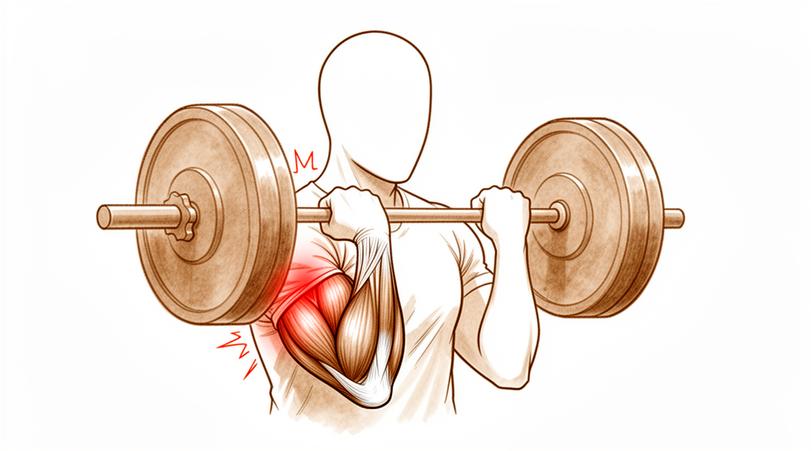
کہنی کے پاس بائسیپس ٹینڈن کا پھٹنا (distal biceps rupture) مہینوں میں نہیں بلکہ ایک ہی لمحے میں ہوتا ہے۔ یہ عموماً 90 ڈگری پر مڑے ہوئے بازو پر اچانک اور غیر متوقع طور پر سیدھا کرنے والی قوت سے ہوتا ہے، جیسے کسی گرتی ہوئی بھاری چیز کو تھامنا، یا کسی چیز کو زور سے پکڑے رکھنا جبکہ آپ کا بازو کھنچ کر سیدھا ہو رہا ہو۔ بہت سے لوگ چوٹ لگتے وقت کہنی کے سامنے کچھ چٹخنے کو محسوس کرتے یا اس کی آواز سنتے ہیں۔ عام طور پر ٹینڈن (tendon)، یعنی وہ مضبوط ڈوری جو پٹھے کو ہڈی سے جوڑتی ہے، بازو کے نچلے حصے (forearm) کی ایک ہڈی پر اپنے جڑنے کی جگہ سے پھٹ کر الگ ہو جاتا ہے۔ ہو سکتا ہے آپ دیکھیں کہ پٹھے کی شکل بدل گئی ہے، کیونکہ وہ کھنچ کر بازو میں اوپر چلا جاتا ہے اور درمیان کے قریب اکٹھا ہو جاتا ہے۔
شروع ہی سے آپ کی کہنی کا سامنے والا حصہ غالباً دردناک اور سوجا ہوا ہوگا۔ نیل پڑنا عام ہے اور اگلے ایک دو دن میں کہنی اور بازو کے نچلے حصے کے ارد گرد ظاہر ہو سکتا ہے۔ بازو کو موڑنے اور گھمانے میں درد ہوتا ہے، اس لیے ہو سکتا ہے آپ کیتلی اٹھانے، دروازے کا ہینڈل گھمانے، یا گاڑی کا اسٹیئرنگ مضبوطی سے پکڑنے سے بچنے لگیں۔ مزاحمت کے خلاف ہتھیلی کو اوپر کی طرف گھمانا، جیسے پیچ کس استعمال کرنا، اکثر سب سے مشکل حرکت ہوتی ہے۔ کچھ لوگ پھر بھی کہنی موڑ سکتے ہیں کیونکہ دوسرے پٹھے اس میں مدد کرتے ہیں، جس کی وجہ سے چوٹ اپنی اصل شدت سے کم محسوس ہو سکتی ہے۔
پہلے دنوں اور ہفتوں میں درد اکثر حرکت سے بڑھ جاتا ہے اور رات کو دکھن ہو سکتی ہے۔ جیسے جیسے جسم پھٹے ہوئے ٹینڈن کے ارد گرد بھرنا شروع کرتا ہے، یہ آہستہ آہستہ کم ہو جاتا ہے۔ سوجن اور نیل دو ایک ہفتوں میں کم ہو جاتے ہیں، لیکن طاقت کی کمی خود بخود بحال نہیں ہوتی، کیونکہ ٹینڈن ہڈی سے کھنچ کر الگ ہو چکا ہے اور خود اپنی اصل جگہ پر دوبارہ نہیں جڑ سکتا۔
چند خطرے کی علامات پر فوری کارروائی ضروری ہے۔ اگر آپ کا ہاتھ یا انگلیاں سفید پڑنے لگیں، ٹھنڈی ہو جائیں، بالکل سفید یا نیلی ہو جائیں، یا چوٹ کے بعد اچانک ہاتھ میں احساس یا حرکت ختم ہو جائے، تو اسی دن ہسپتال کی ایمرجنسی جائیں۔ اگر درد کم نہ ہو رہا ہو، ہفتوں کے دوران بڑھ رہا ہو، یا آپ کو رات کو جگا دیتا ہو، تو اپنے فیملی ڈاکٹر (GP) سے ملیں یا ماہر سے معائنہ کروانے کا کہیں۔
دراصل کیا ہو رہا ہے
بائسیپس (biceps) پٹھا آپ کے بازو کے اوپری حصے کے سامنے سے نیچے کی طرف جاتا ہے اور کہنی سے ذرا پہلے ایک مضبوط ڈوری، یعنی ٹینڈن، میں بدل جاتا ہے۔ یہ ٹینڈن بازو کے نچلے حصے کی ایک ہڈی، ریڈیس (radius)، پر ایک چھوٹے سے ابھار سے جڑا ہوتا ہے۔ اس کا کام بازو کے نچلے حصے کو گھمانا ہے تاکہ آپ کی ہتھیلی اوپر کی طرف مڑ جائے، اور کہنی موڑنے میں مدد کرنا ہے۔
جب ٹینڈن پھٹتا ہے تو وہ ہڈی کے اس ابھار سے الگ ہو کر سکڑتا ہوا بازو میں اوپر چلا جاتا ہے۔ پٹھا اب بھی صحت مند ہوتا ہے، لیکن وہ اس ہڈی سے جڑا نہیں رہتا جسے وہ حرکت دیتا ہے، اس لیے اس کی کھینچنے کی طاقت کسی کام نہیں آتی۔ ہتھیلی کو اوپر کی طرف گھمانا وہ حرکت ہے جو سب سے زیادہ متاثر ہوتی ہے، کیونکہ کوئی اور پٹھا یہ کام نہیں سنبھال سکتا۔ کہنی موڑنے کی صلاحیت عموماً بہتر طور پر بحال ہو جاتی ہے، کیونکہ بائسیپس کے نیچے موجود ایک دوسرا پٹھا اس میں مدد کرتا ہے۔
ایک رسی کا تصور کریں جو ایک چرخی (winch) سے بندھی ہو۔ اگر رسی چرخی کے ڈرم سے ٹوٹ کر الگ ہو جائے تو چرخی کی موٹر چلتی رہتی ہے، لیکن کچھ نہیں گھومتا۔ رسی خود بخود واپس آ کر دوبارہ بندھ نہیں جاتی۔ اسے دوبارہ جوڑنا پڑتا ہے۔
اگر اسے ایسے ہی چھوڑ دیا جائے تو پھٹا ہوا سرا ہڈی پر اپنی اصل جگہ دوبارہ نہیں جڑتا۔ اس کے بجائے خلا زخم بھرنے سے بننے والے سخت ریشے (scar tissue) سے بھر جاتا ہے، جو چیزوں کو ایک ساتھ تو رکھتا ہے لیکن طاقت بحال نہیں کرتا۔ سرجری ٹینڈن کے سرے کو اس کی اصل جڑنے کی جگہ پر دوبارہ جوڑ دیتی ہے تاکہ ٹینڈن اور ہڈی کا ملاپ دوبارہ مضبوطی سے بھر سکے۔
یہ پھٹاؤ عموماً چالیس سے ساٹھ کے پیٹے کی عمر کے مردوں میں اس بازو میں ہوتا ہے جسے وہ زیادہ استعمال کرتے ہیں، اکثر کسی ایک بھاری کوشش کے دوران جب بازو مڑا ہوا ہو اور ہتھیلی اوپر کی طرف ہو، جیسے وزن اٹھاتے ہوئے۔ ٹینڈن تقریباً ہمیشہ اپنی لمبائی میں چرنے کے بجائے ہڈی سے بالکل الگ ہو جاتا ہے، اگرچہ ایسے جزوی پھٹاؤ بھی ہو سکتے ہیں جن میں ٹینڈن کا کچھ حصہ جڑا رہتا ہے۔
ہم اس کے لیے کیا کر سکتے ہیں
ڈاکٹر کیرن ہِرپرا، جو Mater Private Hospital Rockhampton میں بازو اور ہاتھ کے سرجن (upper-limb surgeon) ہیں، علاج کو آپ کی مخصوص چوٹ کے مطابق طے کرتے ہیں۔ کہنی کے پاس بائسیپس کے کچھ پھٹاؤ کا علاج آپریشن کے بغیر کیا جا سکتا ہے، اور کچھ کے لیے ان کے ہونے کے کچھ ہی عرصے بعد سرجری کی ضرورت ہوتی ہے، اس لیے بروقت معائنہ اہم ہے۔ مریضوں کو عام طور پر ان کے فیملی ڈاکٹر (GP) ہمارے کلینک بھیجتے ہیں؛ اگر کسی فزیوتھراپسٹ نے آپ کو ہمیں دکھانے کا مشورہ دیا ہے، تب بھی میڈیکیئر (Medicare) سے رقم کی واپسی (rebate) کا اہل ہونے کے لیے آپ کو اپنے فیملی ڈاکٹر سے ریفرل (referral) کی ضرورت ہوگی۔ کلینک میں ہم آپ کی طبی تاریخ لیتے ہیں، آپ کی دونوں کہنیوں کا معائنہ کرتے ہیں، اور جہاں مددگار ہو وہاں امیجنگ کا انتظام کرتے ہیں۔ عام طور پر پہلا معائنہ ایکسرے ہوتا ہے، اور ایم آر آئی اسکین یہ تصدیق کر سکتا ہے کہ ٹینڈن پھٹا ہے یا نہیں اور کتنی بری طرح۔
مکمل پھٹاؤ میں، جہاں ٹینڈن ہڈی سے پوری طرح الگ ہو چکا ہو، شروع ہی سے سرجری تجویز کی جاتی ہے۔ ان لوگوں کے لیے یہ عموماً صحیح انتخاب ہے جن کے کام یا کھیل میں بازو کے نچلے حصے کو زور سے گھمانے کی ضرورت ہوتی ہے، جیسے مزدور اور کھلاڑی۔ آپریشن ٹینڈن کو اس کی اصل جڑنے کی جگہ پر دوبارہ جوڑ دیتا ہے تاکہ طاقت اور کارکردگی واپس آ سکے۔ ایک چھوٹے بٹن کے ذریعے جوڑنے (button fixation) سے کی گئی مرمت تقریباً معمول کی طاقت اور کارکردگی بحال کر دیتی ہے، اور اس سرجری میں سنگین پیچیدگیوں کی شرح کم ہے۔ مرمت کے بعد زیادہ تر معمولی مسائل سُن پن کے ایسے حصے ہوتے ہیں جو وقت کے ساتھ ٹھیک ہو جاتے ہیں۔ مرمت چوٹ کے جلد بعد سب سے بہتر کام کرتی ہے، بہتر یہ ہے کہ تقریباً 3 ہفتوں کے اندر، کیونکہ ٹینڈن سکڑ کر بازو میں اوپر چلا جاتا ہے اور جس جگہ سے وہ الگ ہوا تھا وہاں سخت ریشہ بن جاتا ہے۔ تاخیر سے مرمت کبھی کبھار ممکن ہوتی ہے، لیکن یہ کہیں زیادہ مشکل ہوتی ہے اور لمبائی پوری کرنے کے لیے ٹینڈن کے گرافٹ (graft، ٹینڈن کی کمی پوری کرنے کے لیے لگایا جانے والا ٹکڑا) کی ضرورت پڑ سکتی ہے۔
جزوی پھٹاؤ میں، جہاں ٹینڈن کا کچھ حصہ اب بھی جڑا ہوا ہو، آپریشن کے بغیر علاج بعض اوقات ایک معقول انتخاب ہوتا ہے۔ اس میں بازو کو سلنگ (sling)، یعنی گلے سے لٹکی وہ پٹی جو بازو کو سہارا دیتی ہے، یا اسپلنٹ میں آرام دینا، بھرنے کے دوران پھٹاؤ کی حفاظت کرنا، اور اپنے ہینڈ تھراپسٹ (ہاتھ کی بحالی کے ماہر) کے ساتھ مرحلہ وار حرکت کی طرف واپسی شامل ہے۔ اس راستے میں سرجری سے بچنے کے بدلے کچھ طاقت چھوڑنی پڑتی ہے، اور یہ سودا ایک حقیقی فیصلہ ہے جو ہم آپ کے ساتھ مل کر کریں گے۔ اگر ہتھیلی کو اوپر کی طرف گھمانے میں کمزوری آپ کے کام یا روزمرہ زندگی کے لیے قابلِ قبول نہ ہو، تو سرجری آپ کے لیے زیادہ موزوں ہو سکتی ہے۔
آپ جو بھی راستہ اختیار کریں، ابتدائی ہفتوں کی بنیادی باتیں ایک جیسی ہوتی ہیں۔ درد پر قابو، بھرنے کے دوران بازو کی حفاظت، اور صحیح مرحلے پر ہینڈ تھراپی۔ سرجری کے بعد آپ کے ہینڈ تھراپسٹ آپ کی بحالی کی رہنمائی کرتے ہیں اور آپ کو جس اسپلنٹ کی ضرورت ہو وہ بناتے ہیں؛ آپریشن کے جلد بعد Extend Rehabilitation میں Ruby Doolan کے ساتھ بحالی شروع ہو جاتی ہے، اور ابتدائی ہلکی حرکت اچھی طرح برداشت ہو جاتی ہے۔
کیا توقع رکھیں
کہنی کے پاس بائسیپس ٹینڈن کے پھٹنے کے بعد صحت یابی آہستہ آہستہ ہوتی ہے، یہ کوئی فوری حل نہیں۔ ٹینڈن کو ہڈی سے دوبارہ جڑنے کے لیے وقت چاہیے، اور طاقت دنوں کے بجائے مہینوں میں آہستہ آہستہ واپس آتی ہے۔ پہلے ہفتوں میں توجہ بھرنے کے دوران بازو کی حفاظت پر ہوتی ہے، اور درد اور سوجن تقریباً اسی طرح کم ہوتے ہیں جیسے چوٹ کے بعد کے ابتدائی دنوں میں ہوئے تھے۔
اگر آپ کی سرجری ہوتی ہے، تو زیادہ تر لوگ اپنے کام کی ذمہ داریوں میں کسی تبدیلی کے بغیر کام پر واپس آ جاتے ہیں، اور کام پر واپسی کا اوسط وقت تقریباً 14 ہفتے ہے۔ کھیل میں زیادہ وقت لگتا ہے۔ تقریباً 95 فیصد کھلاڑی کھیل میں واپس آتے ہیں، اور 82 فیصد چوٹ سے پہلے والی مقابلے کی سطح پر واپس پہنچتے ہیں، کھیل کی نوعیت کے مطابق تقریباً 40 ہفتوں میں۔ بازو کو موڑنے اور گھمانے کی طاقت تقریباً معمول کی سطح تک واپس آ سکتی ہے۔ کچھ لوگ چوٹ سے پہلے کے بازو کے مقابلے میں تھوڑا سا فرق محسوس کرتے ہیں، خاص طور پر بار بار گھمانے میں۔
اگر آپ کے پھٹاؤ کا علاج سرجری کے بغیر کیا جائے، تو بھرنے کے دوران بازو کو سلنگ یا اسپلنٹ میں آرام دیا جاتا ہے، اور آپ اپنے ہینڈ تھراپسٹ کے ساتھ مرحلہ وار حرکت دوبارہ بحال کرتے ہیں۔ اس راستے میں ہتھیلی کو اوپر کی طرف گھمانے میں کچھ مستقل کمزوری رہ جاتی ہے، اور یہی وہ سودا ہے جس پر اس صفحے میں پہلے بات کی گئی ہے۔
زیادہ تر لوگ اچھی طرح صحت یاب ہو جاتے ہیں، لیکن یہ جاننا ایمانداری کی بات ہے کہ کیا مسائل پیدا ہو سکتے ہیں۔ مرمت کے بعد بنیادی خطرات یہ ہیں: عصب کی چوٹ، جو اکثر سُن پن کا ایک حصہ ہوتی ہے جو وقت کے ساتھ ٹھیک ہو جاتا ہے، کہنی میں اکڑاؤ، اور مرمت کا دوبارہ پھٹ جانا۔ مرمت کے دوبارہ پھٹنے کا امکان کم ہے، 1.5 فیصد، اور جب ایسا ہوتا ہے تو عموماً سرجری کے 3 ہفتوں کے اندر ہوتا ہے۔ کبھی کبھی بازو کے نچلے حصے کے آر پار نئی ہڈی بن سکتی ہے اور گھمانے کو محدود کر سکتی ہے، اگرچہ ایسا ہونے پر بھی طاقت برقرار رہ سکتی ہے اور کوئی قابلِ پیمائش فرق نہیں ہوتا۔
چند علامات پر فوری کارروائی ضروری ہے۔ اگر آپ کا ہاتھ یا انگلیاں سفید پڑنے لگیں، ٹھنڈی ہو جائیں، بالکل سفید یا نیلی ہو جائیں، یا چوٹ کے بعد اچانک ہاتھ میں احساس یا حرکت ختم ہو جائے، تو اسی دن ہسپتال کی ایمرجنسی جائیں۔ اگر درد یا اکڑاؤ کم نہ ہو رہا ہو، ہفتوں کے دوران بڑھ رہا ہو، یا آپ کو اپنا بازو استعمال کرنے سے روکتا ہو، تو اپنے فیملی ڈاکٹر سے ملیں یا ماہر سے معائنہ کروانے کا کہیں۔
ڈاکٹر کو کب دکھائیں
اگر آپ کا بازو واضح طور پر بگڑی ہوئی شکل میں نظر آئے، چوٹ کی جگہ پر کھلا زخم ہو، یا آپ بازو بالکل استعمال نہ کر سکیں، تو فوری طبی امداد حاصل کریں۔ اگر آپ کا ہاتھ یا انگلیاں سفید پڑنے لگیں، ٹھنڈی ہو جائیں، بالکل سفید یا نیلی ہو جائیں، یا چوٹ کے بعد اچانک ہاتھ میں احساس یا حرکت ختم ہو جائے، تو اسی دن ہسپتال کی ایمرجنسی جائیں۔ ان علامات کا ذکر اس صفحے میں پہلے کیا گیا ہے۔
باقی ہر چیز کے لیے پہلے اپنے فیملی ڈاکٹر سے رجوع کریں۔ اگر درد کم نہ ہو رہا ہو، یا بھرنے کے عمل کے ساتھ ساتھ سوجن، حرکت یا طاقت میں ہفتہ بہ ہفتہ بہتری نہ آ رہی ہو، تو اپنے فیملی ڈاکٹر سے ملیں یا ماہر سے معائنہ کروانے کا کہیں۔ اگر کہنی کے سامنے اچانک کچھ چٹخے اور ساتھ ہی پٹھے کی شکل بدل جائے، تو اسے جلد دکھانا بہتر ہے، کیونکہ مرمت چوٹ کے تقریباً 3 ہفتوں کے اندر سب سے بہتر کام کرتی ہے۔
مزید تفصیل
Advanced reading: the deeper science (optional)
یہ حصہ آپ کے اپنے علاج کے فیصلوں کے لیے ضروری معلومات سے آگے جاتا ہے۔ کہنی کے پاس بائسیپس ٹینڈن کا پھٹنا اضافی مطالعے کے لائق ہے کیونکہ یہ اس سائٹ پر ان چند چوٹوں میں سے ایک ہے جہاں سرجری واضح طور پر وہ چیز بحال کرتی ہے جو سرجری کے بغیر بھرنے سے بحال نہیں ہوتی، جس کی وجہ سے فائدے اور نقصان کا توازن غیر معمولی طور پر ٹھوس ہو جاتا ہے، اور پیچیدگیوں کے اعداد و شمار غیر معمولی طور پر اہم۔
یہاں آپریشن واقعی بازو کی کارکردگی بدل دیتا ہے
اس سائٹ پر زیادہ تر موازنوں کا نتیجہ "کوئی نمایاں فرق نہیں" ہوتا ہے۔ یہ موازنہ ایسا نہیں۔ 2,481 مریضوں کے اعداد و شمار کو یکجا کرنے پر، آپریشن سے علاج نے آپریشن کے بغیر علاج کے مقابلے میں کہنی اور بازو کے نچلے حصے کی بہتر طاقت اور برداشت دی، اور ساتھ ہی بازو کی کارکردگی کے پیمانوں (DASH اور Mayo Elbow Performance scores) پر بہتر اسکور بھی [1]۔
اس کی وجہ اعداد و شمار نہیں بلکہ جسم کی بناوٹ ہے۔ نچلا بائسیپس بازو کے نچلے حصے کو گھمانے والا سب سے اہم پٹھا (supinator) ہے، یعنی وہ پٹھا جو مزاحمت کے خلاف ہتھیلی کو اوپر کی طرف گھماتا ہے۔ جب ٹینڈن ریڈیس (radius)، یعنی بازو کے نچلے حصے کی انگوٹھے والی طرف کی ہڈی، سے الگ ہوتا ہے تو وہ سکڑ کر بازو میں اوپر چلا جاتا ہے، اور پٹھا یہ گردش پیدا نہیں کر سکتا، چاہے اس کے ارد گرد اور کچھ بھی بھر جائے۔ کہنی کو موڑنے کا کام کچھ حد تک ایک اور پٹھا، بریکیئلس (brachialis)، سنبھال لیتا ہے، اس لیے کہنی موڑنے کی صلاحیت کافی حد تک بحال ہو جاتی ہے؛ ہتھیلی کو اوپر گھمانے کے لیے ایسا کوئی متبادل پٹھا نہیں ہے۔
یہی وجہ ہے کہ یہ کمی کسی ایک بار کی پوری کوشش کے بجائے سب سے زیادہ واضح طور پر برداشت میں ظاہر ہوتی ہے۔ بار بار گھمانے کے کام، جیسے پیچ کس، دروازے کا ہینڈل، یا لمبے سفر میں گاڑی کا اسٹیئرنگ، وہ جگہ ہیں جہاں یہ فرق محسوس ہوتا ہے۔
پیچیدگیوں کی شرح فیصلے کا دوسرا حصہ ہے
ایک آپریشن جو قابلِ اعتماد طریقے سے طاقت بحال کرتا ہے، پھر بھی ایک آپریشن ہے، اور اس آپریشن کے خطرات اچھی طرح معلوم ہیں۔ سب سے بڑے تجزیے میں، جس میں 3,091 مریض شامل تھے، بڑی پیچیدگیوں کی شرح 4.6 فیصد تھی [2]۔
جو پیچیدگیاں ہوتی ہیں ان میں سے زیادہ تر معمولی اور عارضی ہوتی ہیں؛ ان میں سب سے عام بازو کے نچلے حصے کی پشت پر سُن پن ہے، جو ایک چھوٹے حسی عصب کے کھنچنے سے ہوتا ہے، اور عموماً ٹھیک ہو جاتا ہے۔ بڑی پیچیدگیاں وہ ہیں جو آپریشن کے طریقے کے انتخاب پر اثر ڈالتی ہیں: عصب کی مستقل چوٹ، غیر معمولی جگہ پر ہڈی کا بننا (heterotopic ossification، یعنی بازو کے نچلے حصے کے آر پار نئی ہڈی کا بن جانا جو گردش کو محدود کر دیتی ہے)، اور ٹینڈن کا دوبارہ پھٹ جانا۔
دو طریقے، اور ان کے درمیان توازن حقیقی ہے
مرمت یا تو کہنی کے سامنے ایک چیرے کے ذریعے کی جاتی ہے یا دو چھوٹے چیروں کے ذریعے، ایک سامنے اور ایک پیچھے۔ دونوں طریقے مختلف سمتوں سے ایک ہی ہڈی تک پہنچتے ہیں، اور ہر ایک ایک مختلف خطرے سے بچاتا ہے۔
2,622 مریضوں میں عملی کارکردگی کے اسکور میں کوئی نمایاں فرق نہیں تھا۔ ایک چیرے والے طریقے سے کہنی موڑنے اور ہتھیلی نیچے کی طرف گھمانے (pronation) کی حرکت کی حد زیادہ رہی اور غیر معمولی جگہ پر ہڈی بننے اور دوبارہ آپریشن کا خطرہ کم رہا، جبکہ دو چیروں والا طریقہ دوسرے پیمانوں پر بہتر رہا [3]۔ 2,833 مریضوں پر مشتمل 2025 کے ایک میٹا اینالیسس (meta-analysis) میں بھی یہی تصویر سامنے آئی: ایک چیرے والا طریقہ بہتر معروضی کارکردگی اور کم ساختی پیچیدگیوں سے منسلک تھا، جبکہ دو چیروں والا طریقہ بعض حسی اعصاب کی چوٹ کے کم خطرے سے [4]۔
دونوں کو ساتھ ملا کر دیکھیں تو انتخاب سامنے والے عصب اور پیچھے والی ہڈی کے درمیان ہے۔ کوئی بھی طریقہ غلط نہیں، اور دونوں تحقیقات کے مصنفین کہتے ہیں کہ اس سوال کو طے شدہ سمجھنے سے پہلے زیادہ معیاری رینڈمائزڈ ٹرائلوں (randomised trials) کی ضرورت ہے۔
یہاں وقت دوسری جگہوں کے مقابلے میں زیادہ اہم کیوں ہے
چونکہ ٹینڈن سکڑ کر پیچھے ہٹ جاتا ہے اور جو جگہ وہ گھیرتا تھا وہاں زخم بھرنے سے بننے والا سخت ریشہ (scar tissue) بن جاتا ہے، اس لیے تاخیر سے دوبارہ جوڑنا مشکل ہو جاتا ہے، اور جہاں براہ راست مرمت ممکن نہ رہے وہاں گرافٹ (graft، ٹینڈن کی کمی پوری کرنے کے لیے لگایا جانے والا ٹکڑا) سے تعمیر نو ضروری ہو جاتی ہے۔ یہی عملی وجہ ہے کہ کہنی کے پاس بائسیپس ٹینڈن کے پھٹنے کا علاج زیادہ تر دوسری ٹینڈن کی چوٹوں کے مقابلے میں زیادہ جلدی سے کیا جاتا ہے، اور اسی لیے اگر کہنی کے سامنے اچانک درد کے ساتھ کچھ چٹخے، اور پٹھے کی شکل میں واضح تبدیلی نظر آئے، تو انتظار کر کے دیکھنے کے بجائے جلد معائنہ کروانا بہتر ہے۔
آپریشن کے بغیر علاج اب بھی ایک جائز انتخاب ہے، خاص طور پر ان بازوؤں کے لیے جن سے کم مشقت لی جاتی ہے اور ان لوگوں کے لیے جنہیں بار بار زور سے ہتھیلی اوپر گھمانے کی ضرورت نہیں ہوتی۔ یہ سوچ سمجھ کر ایک قابلِ پیمائش کمی کو قبول کرنے کا فیصلہ ہے، نہ کہ اسی نتیجے کا ایک کمتر روپ۔
حوالہ جات
[1] Looney AM, Day J, Bodendorfer BM, Wang D, Fryar CM, Murphy JP, et al. Operative vs. nonoperative treatment of distal biceps ruptures: a systematic review and meta-analysis. J Shoulder Elbow Surg. 2022;31(4):e169-e189. https://doi.org/10.1016/j.jse.2021.12.001
[2] Amarasooriya M, Bain GI, Roper T, Bryant K, Iqbal K, Phadnis J. Complications after distal biceps tendon repair: a systematic review. Am J Sports Med. 2020;48(12):3103-11. https://doi.org/10.1177/0363546519899933
[3] Castioni D, Mercurio M, Fanelli D, Cosentino O, Gasparini G, Galasso O. Single- versus double-incision technique for the treatment of distal biceps tendon rupture: a systematic review and meta-analysis of comparative studies. Bone Joint J. 2020;102-B(12):1608-17. https://doi.org/10.1302/0301-620X.102B12.BJJ-2020-0822.R2
[4] Awad G, Boutros M, Hajj Youssef M, Elhassan B. Comparing single-incision and double-incision techniques in distal biceps tendon repair: a systematic review and meta-analysis. Shoulder Elbow. 2025. https://doi.org/10.1177/17585732251399838
Evidence & references
This is the clinical evidence summary written for health professionals. It is technical, and it lists the research this page was built from. You do not need to read it to understand your treatment or to make a decision about it.
Overview
- Distal biceps tendon injuries typically occur in middle-aged, higher-demand patients who often require surgical reconstruction to optimize outcome and performance [24].
- Surgical repair of distal biceps tendon ruptures has an overall low rate of serious complications regardless of approach or technique [8, 9, 11, 12].
- Distal biceps repair is associated with a 7.5% major complication rate and a 4.5% reoperation rate [7].
- A systematic review indicates a major complication rate of 4.6% after distal biceps repair [31].
- Patients should be counseled that 1 in 5 patients will have a minor complication and 1 in 20 patients will have a major complication after surgery on the distal biceps tendon [14].
- The overall frequency of reported complications is higher for single-incision distal biceps repair than for double-incision repair [1].
- Nerve injury is the most common complication following distal biceps tendon repair [38].
- The early complication rate following partial distal biceps tendon repair was 20.5% [23].
- Surgical treatment of chronic distal biceps injuries yields favorable objective and subjective outcomes [3].
- Although there may be a slightly higher immediate complication rate for chronic distal biceps ruptures, functional outcomes remain comparable with those seen in the patient population with acute distal biceps [6].
- Patients who underwent distal biceps reconstruction with a graft had similar failure rates, reoperation rates, and final range of motion compared to primary repair [18].
- Even in the setting of a distal biceps reconstruction with graft augmentation, patients can expect low complications and good functional results [10].
- Surgical repair of the distal biceps insertion reliably regains both supination and flexion strength in appropriate patients [55].
- Athletes sustaining distal biceps tendon rupture have a high postoperative return to sport rate, independently of selected surgical technique or rehabilitation program [50].
- Surgical management of distal biceps tears was a predictor of improved patient-reported outcomes only for patients with complete tears [16].
Anatomy & Pathophysiology
Anatomy
- The biceps brachii is the primary supinator and a secondary flexor of the elbow [52].
- The short head of the biceps muscle arises from the coracoid process of the scapula [52].
- The long head of the biceps originates from the supraglenoid tubercle of the scapula and the glenoid labrum [52].
- The long head merges with the short head to form the distal biceps tendon and its medial extension, the lacertus fibrosus [52].
- The distal biceps tendon rotates 90 degrees such that the medial side of the tendon attaches to the distal portion of the radial tuberosity [52].
- The distal biceps tendon is a paratenon-covered extrasynovial tendon with three zones of blood supply [120].
- Zone 1 of the distal biceps tendon blood supply receives vessels from the brachial artery via branches extending past the musculotendinous junction to supply the bulk of the tendon [120].
- Zone 3 of the distal biceps tendon blood supply receives contributions from the posterior interosseous recurrent artery and is limited to the tendon enthesis on the radial tuberosity [120].
- Zone 2 of the distal biceps tendon is covered in a significantly thinner paratenon and receives vessels from both Zone 1 and Zone 3 sources through its extratendinous paratenon cover [120].
- The junction among the three zones of blood supply in the distal biceps tendon may be a transition area where tendon repair mechanisms are limited [120].
- The narrow passage between the lateral ulnar border and the radial tuberosity decreases by roughly 50% in pronation as compared with supination [120].
- The brachialis tendon is an intramuscular tendon at the level of the antecubital fossa and cannot be hooked away from an underlying muscle belly [37].
- The normal lacertus fibrosus often remains intact even with biceps rupture and must be discriminated from the biceps tendon during examination [34].
Pathophysiology
- Distal biceps injuries frequently occur in middle-aged males after an eccentric load to the elbow in flexion [17].
- Distal biceps tendon ruptures typically occur in the dominant arm of males during the fifth to sixth decade of life [37].
- The incidence of distal biceps tendon ruptures is 1.2 per 100,000 persons per year [37].
- Distal biceps tendon ruptures are associated with a traumatic event causing a sudden, eccentric load to a flexed and supinated forearm [37].
- Common risk factors for distal biceps tendon rupture include anabolic steroid use, body building, and smoking [37].
- Distal biceps tendon ruptures were most commonly observed in weightlifting with a slightly flexed or isometrically extended elbow and forearm supination [98].
- In a series of competitive strength athletes, the most common strength sport activities leading to distal biceps tendon rupture were deadlifting (20%) and biceps curls (13%) [119].
- Outside of sporting activities, the most common mechanisms of injury for distal biceps tendon rupture were lifting (34%) or carrying (11%) a heavy object and falling onto the outstretched arm (13%) [119].
- A hypovascular zone near the tendon insertion on the tuberosity can limit repair mechanisms in this area [52].
- A 50% narrowing of the space available for the tendon between the radius and ulna occurs in pronation as opposed to supination [52].
- Preexisting inflammatory or degenerative changes involving the distal biceps tendon predispose to rupture from the radial tuberosity [120].
- The junction among the three zones of blood supply in the distal biceps tendon is more prone to attritional injury and rupture [120].
- Rupture of the distal insertion of the biceps tendon necessarily completely eliminates the function of the muscle [52].
- Distal biceps tendon rupture has been found to result in a 30–50% decrease in supination endurance and strength [52].
- Distal biceps tendon rupture has been found to result in an approximate 20% decrease in flexion strength [52].
- Partial distal biceps tendon tears encompass a spectrum of disease from tendinopathic micro-tears to near-full-thickness rupture from the radial tuberosity [2].
- The mechanism of full-thickness distal biceps tendon tears is well reported with a defined episode of injury, deformity, pain, and weakness [2].
- Partial distal biceps tendon tears may have a variable presentation with either an insidious or acute traumatic history, pain, mechanical symptoms, and subtle examination findings [2].
- The condition of partial distal biceps tendon tears may remain undiagnosed or mismanaged, leading to a delay in treatment [2].
- The insertion site of the distal biceps tendon on the radial tuberosity undergoes degenerative changes that begin with partial tearing of the tendon [40].
- Degenerative changes at the distal biceps insertion can be painful but also clearly can be asymptomatic [40].
- Patients with distal biceps ruptures often show signs of significant chronic degeneration with partial tearing before the acute rupture [40].
- Many patients with distal biceps ruptures deny having any symptoms referable to their distal biceps insertion before the acute event that caused them to present for treatment [40].
- Partial biceps tendon tears may result from an acute event or tearing in conjunction with chronic degeneration [40].
Classification
- Partial distal biceps tendon tears encompass a spectrum of disease ranging from tendinopathic micro-tears to near-full-thickness rupture from the radial tuberosity [2].
- Full-thickness distal biceps tendon tear patients tend to present acutely [2].
- Recommended management algorithms for partial distal biceps tendon tears typically focus on patient factors and a tear percentage of 50% as a guide for conservative treatment versus repair [2].
- The '50%-rule' on a 2-dimensional MRI measurement is likely an over-simplified parameter for a complex three-dimensional structure with known anatomical variants [2].
- Isolated short head ruptures are a rare and distinct form of distal biceps tear that present with consistent clinical findings that can aid in diagnosis [19].
- The incidence of women sustaining a distal biceps tendon tear is 3.2% [20].
- Partial tears are statistically more common than complete ruptures in women sustaining a distal biceps tendon tear [20].
- Injury to the distal biceps typically occurs in middle aged, higher demand patients who often require surgical reconstruction to optimize outcome and performance [24].
- Distal biceps tendon ruptures occur at a rate of 1.2 ruptures per 100,000 persons per year [47].
- Distal biceps tendon ruptures account for approximately 3% of all biceps tendon injuries [47].
- Distal biceps tendon ruptures remain a predominately male injury, possibly due to the larger cross-sectional area of biceps in men [47].
- Distal biceps tendon injuries are most likely traumatic events arising when sudden extension load is applied to a flexed, supinated arm [47].
- The aetiology of biceps tendon ruptures is most likely multifactorial, involving a variety of mechanical, degenerative and hypovascular factors [47].
- Seiler et al. discovered three vascular zones in the tendon and reported an approximately 2 cm zone of hypovascularity between the proximal and distal zones [47].
- A more than 50% decrease in the cross sectional area available for the tendon from full supination to full pronation leads to an increased risk for rupture [47].
- Kannus and colleagues reported degenerative tendinopathy in ruptured tendons, based on histopathologic findings [47].
- Smokers have a 7.5 times higher risk of a total rupture compared with non smokers [47].
- Smoking along with anabolic steroids may be related to tendon degeneration and rupture [47].
- There was a 7-fold increase in the incidence of distal biceps tendon rupture surgery in Finnish men during 1997 to 2016 [25].
- There was a 28-fold increase in the incidence of distal biceps tendon rupture surgery in Swedish men during 1997 to 2016 [25].
Clinical Presentation
Epidemiology and Risk Factors
- Distal biceps tendon ruptures typically occur in the dominant arm of males during the fifth to sixth decade of life at a rate of 1.2 per 100,000 persons per year [37].
- The vast majority of complete distal biceps tendon ruptures occurs in men between 40 and 60 years of age [46].
- The dominant limb is involved in 52% of complete distal biceps tendon rupture cases [46].
- An 8% cumulative incidence of bilateral biceps tendon ruptures has been reported [46].
- The incidence of women sustaining a distal biceps tendon tear is 3.2%, with partial tears being statistically more common than complete ruptures in women [20].
- In women, distal biceps tendon ruptures are described as having a more gradual onset of symptoms and a higher incidence of partial tears [46].
- There was a 7-fold increase in the incidence of distal biceps tendon rupture surgery in Finnish men between 1997 and 2016 [25].
- There was a 28-fold increase in the incidence of distal biceps tendon rupture surgery in Swedish men between 1997 and 2016 [25].
History and Mechanism
- Patients with full-thickness distal biceps tendon tears tend to present acutely [2].
- Distal biceps tendon ruptures are associated with a traumatic event which causes a sudden, eccentric load to a flexed and supinated forearm [37].
- A patient typically relates a history of lifting an object or a forced extension of a flexed and loaded forearm [40].
- The patient may recall an audible pop over the anterior elbow [37].
- Patients with chronic and slowly degenerative partial tears often complain of nonspecific elbow pain that is worsened with daily activities [40].
Physical Examination Findings
- A patient with biceps pathologic conditions exhibits pain and weakness in flexion and supination [34].
- Ecchymoses in the antecubital fossa may be present in patients with biceps pathologic conditions [34].
- An abnormal muscle contour of the biceps may be observed in patients with biceps pathologic conditions [34].
- Inspection of the soft tissues tends to demonstrate swelling of the soft tissues in the antecubital fossa [37].
- Retraction of the muscle belly with a visible bulge, known as a reverse Popeye deformity, is a clinical finding supporting the diagnosis of distal biceps rupture [37].
- Weakness in flexion and supination is a clinical finding supporting the diagnosis of distal biceps rupture [37].
- Bruising is a clinical finding supporting the diagnosis of distal biceps rupture [37].
- If seen a few days after the injury episode, ecchymosis is commonly present over the elbow and proximal forearm [40].
- Patients often point out the asymmetry and the "Pop-eye" deformity in the biceps muscle belly [40].
- In partial tears, the biceps may still be palpable [40].
- Patients with chronic partial tears often point to the anterior middle aspect of the antecubital fossa but do not specifically point to the posterolateral aspect of the elbow or the area of the radial tuberosity [40].
- Pain elicited by deep palpation of the radial tuberosity from the posterolateral aspect while passively pronating the forearm is a good indication of tendinopathy involving the insertion of the distal biceps [40].
- Isolated short head ruptures present with consistent clinical findings that can aid in diagnosis [19].
Diagnostic Tests
- The hook test involves having the patient's elbow in 90° of flexion and full supination, then hooking a finger around the lateral edge of the distal biceps tendon as it spans the antecubital fossa [37].
- The hook test has proven 100% sensitivity and specificity for making a prompt diagnosis of distal biceps rupture [37].
- The hook test allows for the distinction between complete avulsion, which results in an abnormal hook test, and partial avulsion, which yields a painful hook test [37].
- O'Driscoll hook test sensitivity is not 100% but remains a useful clinical test to assess patients with suspected distal biceps tendon tears [81].
- A negative O'Driscoll hook test does not exclude rupture [81].
- The evidence-based diagnostic algorithm using the combination of the Flexion Initiation Test (FIT) and hook test demonstrates high accuracy for the diagnosis of both complete and high-grade partial distal biceps tendon tears [33].
- Missed injuries have been attributed to the examiner palpating an intact brachialis tendon, which is an intramuscular tendon at the level of the antecubital fossa and cannot be hooked away from an underlying muscle belly [37].
- Occasionally, some fibers or a fascial sleeve may remain intact, giving an appearance of an intact tendon [34].
- The examiner must discriminate between the normal lacertus fibrosus, which often remains intact even with biceps rupture, and the biceps tendon [34].
Imaging
- Plain film radiographs are most commonly normal and do not show pathologic changes in distal biceps ruptures [34].
- Biceps ruptures typically do not involve bony ruptures, and minimal changes are seen on standard radiographs of the elbow [34].
- MRI may show rupture but is unnecessary in most cases and occasionally may be read as falsely negative [34].
- Some authors suggest a 100% correlation between MRI findings and intraoperative findings for distal biceps ruptures [34].
- MRI did not affect the operative plan in the setting of rupture of a previously repaired ruptured distal biceps tendon [34].
- MRI is useful to exclude alternative diagnoses and to evaluate the extent of suspected partial rupture [34].
- MRI is useful in cases where the history suggests a biceps rupture but the clinical examination is unclear [34].
- MRI tells the surgeon the expected location of the distal tendon end and whether it has retracted proximally [34].
- MRI findings suggestive of complete rupture include absence of the tendon insertion or a fluid-filled sheath [34].
- Partial ruptures on MRI are typically seen as high signal intensity, fluid within the tendon sheath, or thinning or thickening of the tendon distally [34].
- The FABS view (shoulder abducted over the head, elbow in 90 degrees of flexion, forearm in supination) has been suggested to improve visualization of the distal biceps insertion into the radial tuberosity [34].
- Ultrasound is a lower-cost diagnostic tool that is usually unnecessary but has been shown to be accurate in the diagnosis of complete or partial tears [34].
- Ultrasound findings suggestive of complete rupture include tendon absence, fluid, and mass in the antecubital fossa [34].
- Incomplete rupture on ultrasound may be represented by a focal hypoechogenic area or thinning of the tendon [34].
- Advanced imaging with MRI or ultrasonography is helpful in cases of partial avulsions as it can assess the biceps insertion [37].
- The finding that distal biceps MRI signal changes consistent with tendinopathy are common even in asymptomatic elbows reduces the probability that symptoms correlate with pathology on imaging [42].
- The prevalence of distal biceps tendon signal changes on MRI in asymptomatic patients is very low [43].
Investigations
Clinical Examination
- The hook test has proven 100% sensitivity and specificity for making a prompt diagnosis of distal biceps tendon rupture [37].
- Missed injuries have been attributed to the examiner palpating an intact brachialis tendon, which is an intramuscular tendon at the level of the antecubital fossa and cannot be hooked away from an underlying muscle belly as in the case of an intact distal biceps tendon [37].
- On clinical examination, a patient with biceps pathologic conditions exhibits pain and weakness in flexion and supination and sometimes ecchymoses in the antecubital fossa or an abnormal muscle contour of the biceps [34].
- With the elbow at 90 degrees of flexion, the examiner may ask the patient to rotate the pronated forearm into supination; normally, the examiner can hook the tip of his or her thumb around the biceps tendon in the cubital fossa, but with distal avulsion of the tendon, this may be impossible [34].
- Deep palpation of the radial tuberosity from the posterolateral aspect while passively pronating the forearm sometimes elicits pain, which is a very good indication of tendinopathy involving the insertion of the distal biceps [40].
Imaging
- Plain film radiographs should be obtained but are most commonly normal and do not show pathologic changes, as biceps ruptures typically do not involve bony ruptures [34].
- MRI may show rupture, but it is unnecessary in most cases and occasionally may be read as falsely negative [34].
- Some authors suggest a 100% correlation between MRI findings and intraoperative findings for distal biceps rupture [34].
- If obtained, the surgeon should view the MRI images themselves, as the radiologic interpretation is just an interpretation [34].
- In the setting of rupture of a previously repaired ruptured distal biceps tendon, MRI did not affect the operative plan [34].
- MRI may be useful to exclude alternative diagnoses and to evaluate the extent of suspected partial rupture [34].
- MRI may be useful in cases in which the history suggests a biceps rupture but the clinical examination is unclear [34].
- Positioning the prone patient with the shoulder abducted over the head and the elbow in 90 degrees of flexion and the forearm in supination (the FABS view) has been suggested to improve visualization of the distal biceps insertion into the radial tuberosity [34].
- Obtaining an MRI view of the elbow during flexion, abduction, and supination allows for optimal evaluation of the tendon along its long axis and minimizes error by reducing volume averaging [37].
Treatment
Non-Operative Management
- Nonoperative management of complete distal biceps tendon ruptures does not restore function to the elbow [45].
- Conservative treatment is a viable option for very low-demand patients who have concerns about problems connected to surgery [86].
- For partial distal biceps tears, nonoperative treatment is recommended initially if less than 50% of the tendon is involved [86].
- Patients with partial distal biceps tears should be treated nonoperatively for the first 3 to 6 months [87].
- Patients with partial distal biceps tears are encouraged to avoid heavy lifting and use the arm primarily for activities of daily living only [87].
- Strengthening exercises and physical therapy are unnecessary for patients with partial biceps tendon ruptures [87].
- In a cohort study, 82% of participants with partial distal biceps tears had failed non-surgical treatment [86].
- In a study by Bauer et al., 55.7% of patients who tried a nonoperative course for partial distal biceps tears ultimately underwent surgery [36].
- High-need patients, as defined by occupation, were more likely to report ideal recovery if they underwent surgery compared to those who did not [36].
- An MRI-diagnosed tear of greater than 50% was a predictor of needing surgery for partial distal biceps tears [36].
- A meta-analysis showed the superiority of surgical management over the nonoperative approach for distal biceps tendon detachment, with superior flexion and supination strength and better patient-reported outcomes [73].
- In two case reports of nonoperative management for distal biceps tendon rupture, flexion and supination strength returned to normal limits when compared with the opposite upper extremity [22].
Operative Management
- Surgical reinsertion of the biceps tendon to the radial tuberosity is considered the treatment of choice for most complete ruptures [112].
- Although there may be a slightly higher immediate complication rate for chronic distal biceps repairs, functional outcomes remain comparable with those seen in the patient population with acute distal biceps [6].
- Patients undergoing distal biceps reconstruction with a graft had similar failure rates, reoperation rates, and final range of motion compared to primary repair [18].
- Patients can be consulted that direct repair in the acute setting is preferred; however, even in the setting of a distal biceps reconstruction with graft augmentation, they can expect low complications and good functional results [10].
- Distal biceps tendon repair surgery can be safely and successfully executed with either a single- or double-incision approach [48].
- Single anterior incision with suture anchor repair may be utilized for repair of ruptures of the distal biceps tendon with good clinical and functional outcomes and minimal loss of range of motion and strength [80].
- Bioabsorbable interference screw fixation of distal biceps ruptures through a single anterior incision is a safe and successful technique [15].
- Anatomic reattachment of the distal biceps tendon is a successful operative treatment option [29].
- In chronic biceps tendon rupture, anatomical reinsertion is the desired surgical approach due to a high unsatisfactory results rate of 40% in non-anatomical insertion [86].
- Morrey et al. suggest performing primary distal biceps tendon repairs in up to 90 degrees of flexion [86].
- Operative treatment for significant partial distal biceps tears that do not improve after 6 months of nonoperative treatment involves taking down the remaining fibers of the distal biceps and repair to the insertion site [87].
- Operative management for significant partial distal biceps tears has been found to be satisfactory with restoration of function and resolution of symptoms through a single posterior incision [87].
- Patients who present with a classic history of a “pop” in the arm with resisted motion and sudden onset of pain should be treated as having complete biceps tears regardless of MRI findings suggestive of “partial tear” [87].
- The average time to surgery for the operative group in a study of distal biceps tendon ruptures was 9 days, ranging from 3 days to 4 weeks [69].
Complications and Outcomes
- Complication rates after distal biceps repair are low [5].
- Distal biceps repair is associated with a 7.5% major complication rate and 4.5% reoperation rate [7].
- Workers' compensation patients who underwent distal biceps tendon repair took longer to return to work and had worse DASH scores than non-workers' compensation patients [56].
Complications
- The short-term clinical influence of biceps complications on shoulder outcome is very limited [4].
- Although there may be a slightly higher immediate complication rate, the functional outcomes remain comparable with those seen in the patient population with acute distal biceps [6].
- The study examined clinical and surgical outcomes for distal biceps tendon repairs in a large cohort, finding that anterior single incision techniques were used in the majority of cases with varying fixation methods [13].
- This is a safe and successful technique for the management of distal biceps tendon ruptures [15].
- Patients that underwent distal biceps reconstruction with a graft had similar failure rates, reoperation rates, and final range of motion compared to primary repair [18].
- Minor complications were common after distal biceps tendon repair; however, most were sensory nerve injuries that resolved with time [26].
- This is the largest analysis of complications after distal biceps repair, indicating a major complication rate of 4.6% [31].
- Complication rates after distal biceps tendon repair performed by newly trained surgeons were similar to those previously reported in large cohort studies, with nerve injury as the most common complication [38].
Complication Rates and Outcomes
Specific Complications and Risk Factors
- Timing significantly impacts surgical technique selection in distal biceps tendon ruptures, with direct repair favored for acute injuries and high flexion angle repair or allograft reconstruction for chronic cases [39].
Functional Recovery and Strength
- Distal biceps repair/reconstruction can restore near-normal flexion and supination strength [108].
Key Evidence
- [L1] The overall frequency of reported complications is higher for single-incision distal biceps repair than for double-incision repair. [1] (10.1177/2325967116668137)
- [L5] [2] (10.1177/17585732241245054)
- [L1] Surgical treatment of chronic distal biceps injuries yields favorable objective and subjective outcomes. [3] (10.1016/j.xrrt.2022.02.007)
- [L3] Nevertheless, the short-term clinical influence of biceps complications on shoulder outcome is very limited. [4] (10.1177/2325967121s00362)
- [L3] Complication rates after distal biceps repair are low. [5] (10.5397/cise.2021.00472)
- [L4] Although there may be a slightly higher immediate complication rate, the functional outcomes remain comparable with those seen in the patient population with acute distal biceps. [6] (10.1177/23259671211065772)
- [L3] Distal biceps repair is associated with a 7.5% major complication rate and 4.5% reoperation rate. [7] (10.1016/j.jse.2018.06.028)
- [L3] Surgical repair of distal biceps tendon ruptures has an overall low rate of serious complications regardless of approach or technique. [8] (10.1016/j.jse.2017.06.021)
- [L3] Surgical repair of distal biceps tendon ruptures has an overall low rate of serious complications regardless of approach or technique. [9] (10.1016/j.jse.2017.06.020)
- [L3] Patients can be consulted that direct repair in the acute setting is preferred; however, even in the setting of a distal biceps reconstruction with graft augmentation, they can expect low complications and good functional results. [10] (10.1016/j.jseint.2020.10.023)
- [L3] The surgical repair of distal biceps tendon ruptures has an overall low rate of serious complications, regardless of approach or technique. [11] (10.1177/0363546517720200)
- [L3] The surgical repair of distal biceps tendon ruptures has an overall low rate of serious complications, regardless of approach or technique. [12] (10.1016/j.arthro.2017.08.065)
- [L3] The study examined clinical and surgical outcomes for distal biceps tendon repairs in a large cohort, finding that anterior single incision techniques were used in the majority of cases with varying fixation methods. [13] (10.1016/j.jse.2016.12.053)
- [L4] Patients should be counseled that 1 in 5 patients will have a minor complication and 1 in 20 patients will have a major complication after surgery on the distal biceps tendon. [14] (10.1016/j.jse.2016.02.032)
- [Paper] This is a safe and successful technique for the management of distal biceps tendon ruptures. [15] (10.1007/s00402-009-0974-x)
- [L2] Surgical management of distal biceps tears was a predictor of improved patient-reported outcomes only for patients with complete tears. [16] (10.1016/j.jhsa.2025.12.027)
- [Paper] Distal biceps injuries frequently occur in middle-aged males after an eccentric load to the elbow in flexion. [17] (10.1016/j.injury.2025.112556)
- [L3] Patients that underwent distal biceps reconstruction with a graft had similar failure rates, reoperation rates, and final range of motion compared to primary repair. [18] (10.1016/j.jse.2020.01.062)
- [L4] Isolated short head ruptures are a rare and distinct form of distal biceps tear that present with consistent clinical findings that can aid in diagnosis. [19] (10.1016/j.jse.2020.04.038)
- [L4] The incidence of women sustaining a distal biceps tendon tear is 3.2%, with partial tears being statistically more common than complete ruptures. [20] (10.1016/j.jse.2014.02.006)
- [L5] In both patients, flexion and supination strength returned to normal limits when compared with the opposite upper extremity. [22] (10.1016/j.jht.2020.03.013)
- [L4] The early complication rate following partial distal biceps tendon repair was 20.5%. [23] (10.1016/j.jhsa.2025.04.012)
- [Paper] Injury to the distal biceps typically occurs in middle aged, higher demand patients who often require surgical reconstruction to optimize outcome and performance. [24] (10.1016/j.csm.2020.02.004)
- [L4] There was a 7-fold and a 28-fold increase in the incidence of distal biceps tendon rupture surgery in Finnish and Swedish men during 1997 to 2016. [25] (10.1016/j.jhsa.2020.07.024)
- [L4] Minor complications were common after distal biceps tendon repair; however, most were sensory nerve injuries that resolved with time. [26] (10.1016/j.jhsa.2012.06.022)
- [Case_report] Anatomic reattachment of the distal biceps tendon is a successful operative treatment option. [29] (10.1186/s12891-020-03304-3)
- [L2] This is the largest analysis of complications after distal biceps repair, indicating a major complication rate of 4.6%. [31] (10.1177/0363546519899933)
- [L4] The evidence-based diagnostic algorithm using the combination of the FIT and hook test demonstrates high accuracy for the diagnosis of both complete and high-grade partial distal biceps tendon tears. [33] (10.1016/j.asmr.2021.01.010)
- [L4] [36] (10.1016/j.jse.2017.12.010)
- [L4] Complication rates after distal biceps tendon repair performed by newly trained surgeons were similar to those previously reported in large cohort studies, with nerve injury as the most common complication. [38] (10.1016/j.jse.2022.09.014)
- [L3] Timing significantly impacts surgical technique selection in distal biceps tendon ruptures, with direct repair favored for acute injuries and high flexion angle repair or allograft reconstruction for chronic cases. [39] (10.1016/j.jse.2025.07.027)
- [L4] The finding that distal biceps MRI signal changes consistent with tendinopathy are common even in asymptomatic elbows reduces the probability that symptoms correlate with pathology on imaging. [42] (10.5397/cise.2023.00164)
- [L3] The prevalence of distal biceps tendon signal changes on MRI in asymptomatic patients is very low. [43] (10.1016/j.jhsa.2022.01.020)
- [L5] Nonoperative management does not restore function to the elbow. [45] (10.1007/s11552-013-9551-4)
- [L4] [46] (10.1302/2058-5241.6.200145)
- [Paper] [47] (10.1016/j.injury.2013.01.003)
- [L4] Distal biceps tendon repair surgery can be safely and successfully executed with either a single- or double-incision approach. [48] (10.1016/j.jse.2010.11.009)
- [L4] Athletes sustaining distal biceps tendon rupture have a high postoperative return to sport rate, independently of selected surgical technique or rehabilitation program. [50] (10.1016/j.jse.2022.02.027)
- [L4] [52] (10.1097/00130911-199812000-00005)
- [L4] Surgical repair of the distal biceps insertion reliably regains both supination and flexion strength in appropriate patients. [55] (10.5435/00124635-201003000-00003)
- [L3] WC patients who underwent distal biceps tendon repair took longer to return to work and had worse DASH scores than non-WC patients. [56] (10.1016/j.jse.2012.11.011)
- [L4] [69] (10.1016/s1058-2746(00)90059-4)
- [L1] The results of this meta-analysis showed the superiority of surgical management over the nonoperative approach for distal biceps tendon detachment, with superior flexion and supination strength and better patient-reported outcomes. [73] (10.1177/23259671211037311)
- [L4] Single anterior incision with suture anchor repair may be utilized for repair of ruptures of the distal biceps tendon with good clinical and functional outcomes and minimal loss of range of motion and strength. [80] (10.1097/bco.0000000000000529)
- [L3] O'Driscoll hook test sensitivity is not 100% but remains a useful clinical test to assess patients with suspected distal biceps tendon tears; a negative test does not exclude rupture. [81] (10.1177/1758573219847146)
- [L5] [86] (10.1530/eor-23-0035)
- [L4] Distal biceps tendon ruptures were most commonly observed in weightlifting with a slightly flexed or isometrically extended elbow and forearm supination. [98] (10.1016/j.jse.2023.02.061)
- [L5] Distal biceps repair/reconstruction can restore near-normal flexion and supination strength. [108] (10.1016/j.jhsa.2019.09.014)
- [Paper] Surgical reinsertion of the biceps tendon to the radial tuberosity is considered the treatment of choice for most complete ruptures. [112] (10.1097/bte.0000000000000011)
- [L4] [119] (10.1177/23259671251322700)
- [L5] [120] (10.1016/s1058-2746(05)80044-8)
References
[1] Complications of Distal Biceps Tendon Repair. Orthopaedic Journal of Sports Medicine. 2016. DOI: 10.1177/2325967116668137
[2] Current concepts in the aetiology, assessment and management of partial distal biceps tendon tears. Shoulder & Elbow. 2024. DOI: 10.1177/17585732241245054
[3] Outcomes and complications after different surgical techniques for the treatment of chronic distal biceps tendon ruptures: a systematic review and quantitative synthesis. JSES Reviews, Reports, and Techniques. 2022. DOI: 10.1016/j.xrrt.2022.02.007
[4] Biceps-related complications after tenotomy associated with arthroscopic rotator cuff repair: risk factors and clinical impact. Orthopaedic Journal of Sports Medicine. 2022. DOI: 10.1177/2325967121s00362
[5] Determining the incidence and risk factors for short-term complications following distal biceps tendon repair. Clinics in Shoulder and Elbow. 2022. DOI: 10.5397/cise.2021.00472
[6] Surgical Results of Chronic Distal Biceps Ruptures: A Systematic Review. Orthopaedic Journal of Sports Medicine. 2022. DOI: 10.1177/23259671211065772
[7] Major complications after distal biceps tendon repairs: retrospective cohort analysis of 970 cases. Journal of Shoulder and Elbow Surgery. 2018. DOI: 10.1016/j.jse.2018.06.028
[8] Analysis of 784 surgically treated distal biceps tendon ruptures. Journal of Shoulder and Elbow Surgery. 2017. DOI: 10.1016/j.jse.2017.06.021
[9] The distal biceps deficient arm, “the short arc syndrome”. Journal of Shoulder and Elbow Surgery. 2017. DOI: 10.1016/j.jse.2017.06.020
[10] Outcomes of chronic distal biceps reconstruction with tendon grafting: a matched comparison with primary repair. JSES International. 2021. DOI: 10.1016/j.jseint.2020.10.023
[11] Surgical Treatment of Distal Biceps Tendon Ruptures: An Analysis of Complications in 784 Surgical Repairs. The American Journal of Sports Medicine. 2017. DOI: 10.1177/0363546517720200
[12] Paper #76: All‐Endoscopic Distal Biceps Repair: Cadaveric Portal Safety Analysis and Technical Feasibility Using 2 Fixation Techniques. Arthroscopy. 2017. DOI: 10.1016/j.arthro.2017.08.065
[13] The distal biceps deficient arm, “the short arc syndrome”. Journal of Shoulder and Elbow Surgery. 2017. DOI: 10.1016/j.jse.2016.12.053
[14] Factors associated with adverse events after distal biceps tendon repair or reconstruction. Journal of Shoulder and Elbow Surgery. 2016. DOI: 10.1016/j.jse.2016.02.032
[15] Bioabsorbable interference screw fixation of distal biceps ruptures through a single anterior incision: a single-surgeon case series and review of the literature. Archives of Orthopaedic and Trauma Surgery. 2009. DOI: 10.1007/s00402-009-0974-x
[16] Thirty-Six Percent Conversion to Surgery Rate for Nonsurgical Treatment of Complete and Partial Distal Biceps Tears. The Journal of Hand Surgery. 2026. DOI: 10.1016/j.jhsa.2025.12.027
[17] Distal biceps injuries: an overview. Injury. 2025. DOI: 10.1016/j.injury.2025.112556
[18] Chronic Distal Biceps Repair with Tendon Grafting: A Matched Comparison to Primary Repair. Journal of Shoulder and Elbow Surgery. 2020. DOI: 10.1016/j.jse.2020.01.062
[19] Distal biceps short head tears: repair, reconstruction, and systematic review. Journal of Shoulder and Elbow Surgery. 2020. DOI: 10.1016/j.jse.2020.04.038
[20] Re-rupture rate of primarily repaired distal biceps tendon injuries. Journal of Shoulder and Elbow Surgery. 2014. DOI: 10.1016/j.jse.2014.02.006
[22] Distal biceps tendon rupture: Is surgery the best course of treatment? Two case reports. Journal of Hand Therapy. 2021. DOI: 10.1016/j.jht.2020.03.013
[23] Early Postoperative Complications Following Partial Distal Biceps Tendon Surgical Repair. The Journal of Hand Surgery. 2026. DOI: 10.1016/j.jhsa.2025.04.012
[24] Distal Biceps Injuries. Clinics in Sports Medicine. 2020. DOI: 10.1016/j.csm.2020.02.004
[25] Distal Biceps Tendon Rupture Surgery: Changing Incidence in Finnish and Swedish Men Between 1997 and 2016. The Journal of Hand Surgery. 2020. DOI: 10.1016/j.jhsa.2020.07.024
[26] Complications Following Distal Biceps Repair. The Journal of Hand Surgery. 2012. DOI: 10.1016/j.jhsa.2012.06.022
[29] No functional differences in anatomic reconstruction with one vs. two suture anchors after non-simultaneous bilateral distal biceps brachii tendon rupture: a case report and review of the literature. BMC Musculoskeletal Disorders. 2020. DOI: 10.1186/s12891-020-03304-3
[31] Complications After Distal Biceps Tendon Repair: A Systematic Review. The American Journal of Sports Medicine. 2020. DOI: 10.1177/0363546519899933
[33] The Flexion Initiation Test and an Evidence‐Based Diagnostic Algorithm for Distal Biceps Tendon Tears. Arthroscopy, Sports Medicine, and Rehabilitation. 2021. DOI: 10.1016/j.asmr.2021.01.010
[34] Green S Operative Hand Surgery. DISTAL BICEPS RUPTURE > Diagnosis.
[36] Is nonoperative management of partial distal biceps tears really successful?. Journal of Shoulder and Elbow Surgery. 2018. DOI: 10.1016/j.jse.2017.12.010
[37] Orthopaedic Knowledge Update 13 Ebook Without Multimedia. Tendinopathy, Elbow Ligament Reconstruction, and Throwing Injuries > Tendinopathy > Distal Biceps Injury.
[38] Trends and complications of distal biceps tendon repair among American Board of Orthopaedic Surgery part II oral examination candidates. Journal of Shoulder and Elbow Surgery. 2023. DOI: 10.1016/j.jse.2022.09.014
[39] Effect of time from injury to surgery on surgical technique and complication rate in distal biceps tendon repair. Journal of Shoulder and Elbow Surgery. 2026. DOI: 10.1016/j.jse.2025.07.027
[40] Green S Operative Hand Surgery. DISTAL BICEPS RUPTURE.
[42] Prevalence of incidental distal biceps signal changes on magnetic resonance imaging. Clinics in Shoulder and Elbow. 2023. DOI: 10.5397/cise.2023.00164
[43] Evaluation of MRI Signal Changes of the Distal Biceps Tendon in Asymptomatic Patients. The Journal of Hand Surgery. 2022. DOI: 10.1016/j.jhsa.2022.01.020
[45] Treatment of Chronic Biceps Tendon Ruptures. HAND. 2013. DOI: 10.1007/s11552-013-9551-4
[46] Acute complete and partial distal biceps tendon ruptures: what have we learned? A review. EFORT Open Reviews. 2021. DOI: 10.1302/2058-5241.6.200145
[47] Distal biceps and triceps ruptures. Injury. 2013. DOI: 10.1016/j.injury.2013.01.003
[48] Controversies in the surgical treatment of distal biceps tendon ruptures: single versus double-incision repairs. Journal of Shoulder and Elbow Surgery. 2011. DOI: 10.1016/j.jse.2010.11.009
[50] Systematic review of distal biceps tendon rupture in athletes: treatment and rehabilitation. Journal of Shoulder and Elbow Surgery. 2022. DOI: 10.1016/j.jse.2022.02.027
[52] Single Incision Repair of Distal Biceps Tendon Rupture. Techniques in Hand & Upper Extremity Surgery. 1998. DOI: 10.1097/00130911-199812000-00005
[55] Surgical Treatment of Distal Biceps Rupture. American Academy of Orthopaedic Surgeon. 2010. DOI: 10.5435/00124635-201003000-00003
[56] Outcomes after distal biceps repair in patients with workers' compensation claims. Journal of Shoulder and Elbow Surgery. 2013. DOI: 10.1016/j.jse.2012.11.011
[69] Repair of distal biceps brachii tendon ruptures. Journal of Shoulder and Elbow Surgery. 2000. DOI: 10.1016/s1058-2746(00)90059-4
[73] Operative Versus Nonoperative Management for Distal Biceps Brachii Tendon Lesions: A Systematic Review and Meta-analysis. Orthopaedic Journal of Sports Medicine. 2021. DOI: 10.1177/23259671211037311
[80] Clinical outcomes of single-incision suture anchor repair of distal biceps tendon rupture. Current Orthopaedic Practice. 2017. DOI: 10.1097/bco.0000000000000529
[81] Distal biceps hook test – Sensitivity in acute and chronic tears and ability to predict the need for graft reconstruction. Shoulder & Elbow. 2019. DOI: 10.1177/1758573219847146
[86] Distal biceps tendon rupture: a comprehensive overview. EFORT Open Reviews. 2023. DOI: 10.1530/eor-23-0035
[87] Green S Operative Hand Surgery. AUTHORS’ PREFERRED METHOD OF TREATMENT: PARTIAL BICEPS TEARS.
[98] Distal Biceps Tendon Ruptures Occur With The Almost Extended Elbow And Supinated Forearm – An Online Video Analytic Study. Journal of Shoulder and Elbow Surgery. 2023. DOI: 10.1016/j.jse.2023.02.061
[108] Distal Biceps Tendon Repair and Reconstruction. The Journal of Hand Surgery. 2020. DOI: 10.1016/j.jhsa.2019.09.014
[112] Repair and Rehabilitation of Distal Biceps Ruptures. Techniques in Shoulder & Elbow Surgery. 2014. DOI: 10.1097/bte.0000000000000011
[119] Distal Biceps Tendon Repair in Competitive Strength Athletes: A Retrospective Series of 183 Athletes. Orthopaedic Journal of Sports Medicine. 2025. DOI: 10.1177/23259671251322700
[120] The distal biceps tendon. Journal of Shoulder and Elbow Surgery. 1995. DOI: 10.1016/s1058-2746(05)80044-8