یہ کیا ہے
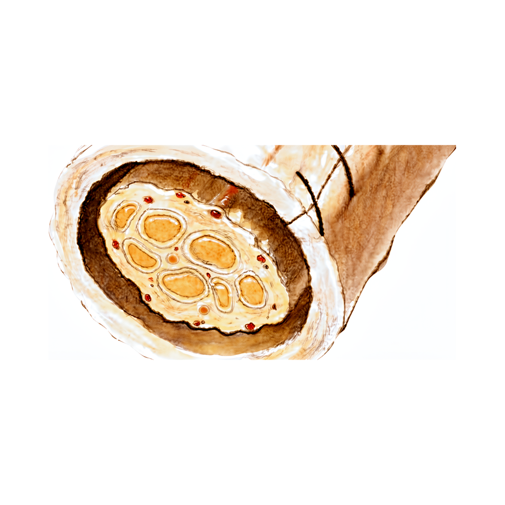
ریمپلر (Remplir) کولیجن سے بنا ایک نرم غلاف ہے، جو آپ کے جسم میں پایا جانے والا ایک قدرتی پروٹین ہے۔ اسے سرجری میں کسی خراب پیریفرل عصب (peripheral nerve) کو ٹھیک ہونے میں مدد دینے کے لیے استعمال کیا جاتا ہے۔ پیریفرل اعصاب وہ اعصاب ہیں جو آپ کی حرام مغز (spinal cord) سے نکل کر آپ کے بازوؤں، ہاتھوں اور ٹانگوں تک جاتے ہیں۔ جب ان میں سے کسی عصب کو چوٹ لگتی ہے تو خود کو ٹھیک کرنے کے دوران اسے حفاظت کی ضرورت ہوتی ہے۔
ریمپلر زخمی عصب کے گرد ایک حفاظتی آستین کی طرح کام کرتا ہے۔ آپ کا سرجن آپریشن کے دوران اسے مرمت شدہ عصب کے اوپر رکھتا ہے۔ یہ غلاف عصب کو بچاتا ہے اور نئے اعصابی ریشوں کے اس میں سے گزر کر بڑھنے کے لیے اچھا ماحول بناتا ہے۔ پھر یہ اپنا کام کر کے غائب ہو جاتا ہے۔ ریمپلر 3 سے 6 ماہ کے اندر گھل جاتا ہے، اس لیے جب صحت یابی شروع ہو جاتی ہے تو آپ کے جسم میں کوئی مصنوعی چیز باقی نہیں رہتی۔
یہ کس چیز سے بنا ہے
ریمپلر ایک حیاتیاتی مادہ ہے، پلاسٹک یا دھات کا امپلانٹ نہیں۔ یہ کولیجن سے بنا ہے، جو ایک قدرتی پروٹین ہے اور آپ کے جسم میں پہلے سے موجود ہے۔ کولیجن جانوروں کے ٹشو سے تیار کیا جاتا ہے اور آپریشن تھیٹر تک پہنچنے سے پہلے اسے جراثیم سے پاک کیا جاتا ہے۔
چونکہ یہ حیاتیاتی مادے سے آتا ہے، اس لیے آپ کا جسم اسے کوئی اجنبی چیز نہیں بلکہ ایک مانوس چیز سمجھتا ہے۔ یہ غلاف ہمیشہ آپ کے جسم میں نہیں رہتا۔ یہ 3 سے 6 ماہ کے اندر گھل جاتا ہے، اس لیے جب عصب ٹھیک ہونا شروع ہو جاتا ہے تو مرمت کی جگہ پر کوئی مصنوعی چیز باقی نہیں رہتی۔
آپ کا سرجن اسے کیسے استعمال کرتا ہے
ریمپلر آپریشن کے دوران اس وقت رکھا جاتا ہے جب آپ کا سرجن زخمی عصب کی مرمت کر چکا ہو۔ یہ غلاف ایک نرم آستین کی طرح مرمت کی جگہ کے گرد لپیٹا جاتا ہے۔ یہ عصب اور ارد گرد کے ٹشو کے درمیان رہتا ہے، تاکہ زخم بھرنے سے بننے والا سخت ریشہ (scar tissue) اندر گھس کر ٹھیک ہوتے ہوئے عصب کو دبا نہ سکے۔ کولیجن کا یہ مادہ نرم اور لچکدار ہوتا ہے، اس لیے یہ عصب کی شکل میں ڈھل جاتا ہے اور بغیر کسی پیچ یا پرزے کے اپنی جگہ پر رہتا ہے۔
غلاف اپنی جگہ پر رکھ دیے جانے کے بعد اپنا کام شروع کر دیتا ہے۔ یہ عصب کی حفاظت کرتا ہے جبکہ نئے اعصابی ریشے مرمت کی جگہ سے گزر کر بڑھتے ہیں۔ یہ عصب کے گرد ایک نم اور پرسکون جگہ بھی بناتا ہے، جو وہ ماحول ہے جس کی اعصاب کو دوبارہ بننے کے لیے ضرورت ہوتی ہے۔ وقت کے ساتھ آپ کا جسم اس غلاف کو جذب کر لیتا ہے اور اس کی جگہ آپ کا اپنا ٹشو لے لیتا ہے۔ کوئی مصنوعی چیز باقی نہیں رہتی۔
آپ کا سرجن اعصاب کی مختلف قسم کی سرجریوں میں Remplir استعمال کر سکتا ہے، جہاں بھی کسی مرمت شدہ پیریفرل عصب کو ٹھیک ہونے کے دوران حفاظت کی ضرورت ہو۔ یہ اکیلا علاج نہیں ہے۔ یہ ایک بڑے آپریشن کا حصہ ہے جس میں خود عصب کی مرمت کی جاتی ہے۔
کیا توقع رکھیں
یہ غلاف بذاتِ خود آپ کی صحت یابی کو نہیں بدلتا۔ یہ 3 سے 6 ماہ کے اندر خود ہی گھل جاتا ہے، اس لیے نہ اسے نکالنا پڑتا ہے اور نہ کوئی اضافی طریقۂ علاج کروانا پڑتا ہے۔ عصب کے ٹھیک ہونے کے ساتھ ساتھ آپ کا جسم اسے جذب کر لیتا ہے اور اس کی جگہ آپ کا اپنا ٹشو لے لیتا ہے۔
اعصابی غلاف کے ساتھ اور اس کے بغیر کی گئی سرجری کا موازنہ کرنے والی تحقیق میں اس بات میں کوئی فرق نہیں پایا گیا کہ اعصاب کیسے دوبارہ بڑھے، وہ پٹھوں سے دوبارہ کیسے جڑے، یا مریض کتنی اچھی طرح صحت یاب ہوئے [1]۔ سادہ الفاظ میں، یہ غلاف عصب کے ٹھیک ہونے کے دوران اس کی حفاظت کے لیے ہے، ٹھیک ہونے کے عمل کو تیز کرنے یا بدلنے کے لیے نہیں۔ اسی وجہ سے آپ کی صحت یابی اسی راستے پر چلتی ہے جس پر وہ اصل آپریشن چلتا ہے جس میں آپ کے عصب کی مرمت کی گئی۔ آپ کا سرجن آپ کو بتائے گا کہ اس آپریشن سے کیا توقع رکھیں، جس میں بعد کے معائنے اور کوئی بھی ورزشیں شامل ہیں۔
ایک بات جاننا فائدہ مند ہے: چونکہ یہ غلاف کولیجن سے بنا ہے، اس لیے آپ کا جسم اسے کوئی اجنبی چیز نہیں بلکہ اپنا حصہ سمجھتا ہے۔ کولیجن سے بنی اعصابی نالیوں (nerve conduits) پر تحقیقات میں پایا گیا کہ جب جسم نے انہیں نئی شکل دی تو وہ ایک غلاف میں لپٹی ہوئی سی (encapsulation) دکھائی دینے لگیں [2]۔ یہ ٹھیک ہونے کے عمل کا ایک معمول کا حصہ ہے، کوئی پیچیدگی نہیں۔
شواہد کیا کہتے ہیں
اعصابی غلاف پر تحقیق ابھی بڑھ رہی ہے۔ غلاف کے ساتھ اور غلاف کے بغیر سرجری کا موازنہ کرنے والی تحقیقات میں اس بات میں کوئی فرق نہیں پایا گیا کہ اعصاب کیسے دوبارہ بڑھے، وہ پٹھوں سے دوبارہ کیسے جڑے، یا مریض کتنی اچھی طرح صحت یاب ہوئے [1]۔ سادہ الفاظ میں، یہ غلاف عصب کے ٹھیک ہونے کے دوران اس کی حفاظت کرتا ہے۔ یہ ٹھیک ہونے کے عمل کو تیز نہیں کرتا اور نہ نتیجہ بدلتا ہے۔
کولیجن کی اعصابی نالیوں کے ابتدائی نتائج اعصاب کی مرمت کے دوسرے شائع شدہ نتائج کے مقابلے میں اچھے ہیں [3]۔ اسی قسم کے کولیجن آلے پر کی گئی ایک تحقیق میں، جسے بازو کے زخمی اعصاب کی مرمت کے لیے استعمال کیا گیا، حرکت کی بحالی رپورٹ کی گئی [4]۔ چونکہ یہ غلاف آپ کے اپنے جسم سے لیے گئے عصب کے بجائے کولیجن سے بنا ہے، اس لیے یہ ان مسائل سے بچاتا ہے جو اُس دوسرے زخم سے پیدا ہو سکتے ہیں جہاں سے عصب کا پیوند (nerve graft) لیا جاتا ہے [5]۔
سچی تصویر یہ ہے: شواہد ابھی ابتدائی ہیں، اور لمبی مدت کی تحقیقات کی ضرورت ہے۔ اب تک یہ معلوم ہے کہ یہ غلاف اپنا حفاظتی کام کرتا ہے اور منصوبے کے مطابق گھل جاتا ہے۔ یہاں یقینی نتائج کا کوئی دعویٰ نہیں کیا جاتا۔ آپ کا سرجن آپ سے بات کرے گا کہ آپ کی خاص چوٹ اور آپریشن کے لیے شواہد کا کیا مطلب ہے۔
حوالہ جات
[1] The Effects of a Porcine Extracellular Matrix Nerve Wrap as an Adjunct to Primary Epineurial Repair. The Journal of Hand Surgery. 2021. DOI: 10.1016/j.jhsa.2020.11.023
[2] A Comparative Study of Porcine Small Intestine Submucosa and Cross-Linked Bovine Type I Collagen as a Nerve Conduit. Journal of Hand Surgery Global Online. 2021. DOI: 10.1016/j.jhsg.2021.06.006
[3] Early Clinical Experience With Collagen Nerve Tubes in Digital Nerve Repair. The Journal of Hand Surgery. 2008. DOI: 10.1016/j.jhsa.2008.03.015
[4] Reconstruction of Upper Extremity Peripheral Nerve Injuries Using an Epineurial-Like Collagen Device — A Prospective Clinical Study. J Reconstr Microsurg Open. 2024. DOI: 10.1055/s-0044-1785213
[5] Nerve Conduits: An Update on Tubular Nerve Repair and Reconstruction. The Journal of Hand Surgery. 2013. DOI: 10.1016/j.jhsa.2013.02.034
Evidence & references
This is the clinical evidence summary written for health professionals. It is technical, and it lists the research this page was built from. You do not need to read it to understand your treatment or to make a decision about it.
Anatomy & Pathophysiology
General Hand Architecture
- The hand is an organ designed to obtain information and an organ of execution [2].
- The hand functions efficiently only if the proximal joints of the limb are stable and yet mobile [2].
- The hand moves within a large volume of space with the shoulder as the apex [2].
- Distal to the elbow, the wrist and forearm form a single physiological unit that places the hand in a position for grasping [2].
- The hand consists of 19 bones, 17 articulations, and 19 muscles situated entirely within the hand [2].
- The hand contains approximately the same number of tendons activated by the forearm muscles as it has intrinsic muscles [2].
- The dorsal aspect of the hand is convex and the palmar or volar aspect is concave [2].
- The palmar surface is the functional surface of the hand, while the dorsal surface is aesthetically important [2].
- When fingers are extended and separated, their tips lie on the circumference of a circle whose center is the head of the third metacarpal [2].
Cutaneous Units and Innervation
- Functional cutaneous units in the hand are similar to those described in the face [3].
- One dorsal cutaneous unit extends from the wrist to the proximal interphalangeal joints of the fingers and the interphalangeal joint of the thumb [3].
- The dorsal covering of the interphalangeal articulations forms a unique cutaneous unit characterized by considerable excess of skin when digits are in extension [3].
- The palm forms a cutaneous unit extending from the distal transverse crease of the wrist up to the transverse crease at the base of the digits [3].
- The oppositional crease of the thumb subdivides the palmar integument into two separate zones [3].
- The skin of the radial portion of the palm covers the thenar eminence and is relatively well vascularized and mobile [3].
- The skin of the ulnar and distal portion of the palm covers the hypothenar eminence where skin mobility is poor [3].
- The central triangular part of the palm has fixed, poorly vascularized skin that covers almost directly the superficial palmar aponeurosis [3].
- When a digit is completely flexed, the integument of adjacent phalanges comes into contact in the zones of the flexion creases, establishing areas of cutaneous contact in the form of a diamond [3].
- The sides of the diamond-shaped cutaneous contact zones do not undergo variations in length during flexion and extension movements [3].
- Incisions made along the lines of the diamond-shaped cutaneous contact zones present a minimal chance of retraction [3].
- The web spaces are formed from the union of two nonsymmetrical cutaneous surfaces [3].
- The dorsal slope of the web space has a gradual incline and its supple skin is not adherent to the subjacent region [3].
- The palmar surface of the web space is flat and precipitously interrupted, with skin densely adherent to the commissural skeleton [3].
- The commissural skeleton is formed by the interdigital palmar ligament between the fingers and by the distal transverse ligament at the level of the thumb web [3].
Intrinsic Musculature
- There are seven interosseous muscles in the hand, consisting of four dorsal and three volar muscles [4].
- The dorsal interossei are abductors [4].
- The anatomic axis of the hand coincides with the axis of the third metacarpal [4].
- The dorsal interossei lie to the radial side of the index and middle fingers and the ulnar side of the middle and ring fingers [4].
- The little finger is abducted by the abductor digiti quinti [4].
- The volar interossei are adductors [4].
- The volar interossei lie to the ulnar side of the index finger and the radial side of the ring and little fingers [4].
- The middle finger has two dorsal interossei and no volar interossei because the central axis of the hand lies within it [4].
- Each dorsal interosseous muscle, with the exception of the third, has two muscle heads [4].
- The superficial head of the dorsal interosseous muscles arises most dorsally from the shaft of the contiguous metacarpals [4].
- The superficial head of the dorsal interosseous muscles is inserted deeply by a medial tendon onto the lateral tubercle of the base of the proximal phalanx [4].
- The superficial head of the dorsal interosseous muscles abducts and weakly flexes the proximal phalanx [4].
- The deep head of each dorsal interosseous muscle forms a lateral tendon, or lateral band, at the level of the MP joint [4].
- The deep head of the dorsal interosseous muscles flexes and weakly abducts the proximal phalanx while extending the middle and distal phalanges [4].
- Transverse fibers arch dorsally from each lateral band to join each other over the dorsum of the finger, flexing the proximal phalanx [4].
- Oblique fibers from the lateral bands sweep over the distal third of the proximal phalanx to insert onto the lateral tubercles at the base of the middle phalanx [4].
- The oblique fibers extend the middle phalanx at the PIP joint [4].
- The lateral bands are joined by the lateral slips of the extensor tendon to form the conjoined lateral band [4].
- The two conjoined lateral bands to each finger unite at the distal third of the middle phalanx to form the terminal tendon [4].
- The terminal tendon inserts at the base of the distal phalanx to extend it [4].
- The flexor digiti quinti brevis is structurally and functionally similar to the deep head of the dorsal interossei, forming the ulnar lateral band of the little finger [4].
- Each volar interosseous muscle has only one muscle head and none of them insert onto the proximal phalanx [4].
- The volar interossei form the ulnar lateral band of the index finger and the radial lateral band of the ring and little fingers [4].
- The abductor digiti quinti arises from the fifth metacarpal and inserts onto the ulnar lateral tubercle at the base of the proximal phalanx of the little finger [4].
- The flexor digiti quinti brevis arises from the fifth metacarpal and forms the ulnar lateral band [4].
- The opponens digiti quinti arises from the pisohamate ligament and the hook of the hamate [4].
- The opponens digiti quinti inserts onto the ulnar side of the diaphysis of the fifth metacarpal, which it flexes and supinates [4].
Metacarpal Arch and Stability
- The metacarpal arch is endowed with a great deal of adaptability because of the mobility of the peripheral metacarpals [7].
- The peripheral metacarpals form the sides of the cup or palmar gutter and can deepen the concavity as they approach each other [7].
- The thumb metacarpal is independent and articulates with the trapezium [7].
- The middle metacarpals are united to the carpus by the intrinsic interlocking encasement of the bones themselves [7].
- The index metacarpal is the most firmly fixed [7].
- The ring metacarpal has about 10 degrees of mobility in flexion and extension [7].
- The fifth metacarpal is semi-independent and articulates with the hamate [7].
- The fifth metacarpal has a range of flexion–extension of approximately 20 degrees [7].
- The second to fifth metacarpals are bound together by various fibrous structures, the most distal of which is the deep transverse intermetacarpal ligament [7].
- The deep transverse intermetacarpal ligament is better named the interglenoid ligament because it ties together the anterior glenoid ligaments of the metacarpophalangeal articulations [7].
- The volar plates are interconnected by the transverse interglenoid ligament [7].
- The stability of the metacarpophalangeal joints is essential to the support of the longitudinal arch as well as of the transverse metacarpal arch [7].
- The keystones of the longitudinal arches are the metacarpophalangeal articulations [7].
- The thick anterior glenoid capsules, known as volar plates, prevent hyperextension at the metacarpophalangeal joints [7].
Vascular Anatomy of the Thumb
- The arteries of the thumb vary in both size and number, making surgical reconstruction delicate [8].
- The palmar aspect of the thumb can be schematized into three segments defined by the metacarpophalangeal and interphalangeal flexion creases [8].
- In the classical layout, the princeps pollicis artery crosses the first intermetacarpal space and runs along the ulnar side of the first metacarpal bone [8].
- The princeps pollicis artery emerges onto the subcutaneous palmar tissue at the level of the cutaneous flexion crease of the metacarpophalangeal joint [8].
- The princeps pollicis artery divides into two terminal rami, the collateral palmar arteries of the thumb [8].
- The collateral palmar arteries run along the digital tunnel symmetrically and are of equal caliber [8].
- An arcade located deep in the flexor tendon joins together the two arteries at the level of the distal metaphysis of the first phalanx [8].
- Only 15% of anatomical dissections fall into the category of the classical description of palmar arteries of the thumb [8].
- In the second segment of the thumb, the two arteries run alongside the flexor tendon and behind the collateral nerves [8].
- In the second segment, the main artery is the ulnar collateral artery [8].
- The subtendinous anastomosis situated at the level of the neck of the first phalanx acts as a moderator between the two arteries [8].
- In the pulp segment, the two arteries are of similar size and run through the thick fatty subcutaneous padding [8].
- In the pulp segment, the arteries cross over and convert into the ends of the digital nerves at the level of the median axis [8].
- The posterior area of the thumb is vascularized by two arteries which originate from the palmar arteries at the level of the first metacarpal [8].
- At the level of the neck of the first phalanx, an anastomosis can be found which originates from the palmar arteries [8].
Surgical Considerations and Nerve Anatomy
- Distal palmar incisions are transverse, while proximal palmar incisions tend to be more longitudinal with the distal end curving radially [9].
- An incision of any desired length can be made across the palm provided that the underlying digital nerves and other vital structures are protected [9].
- After skin and underlying fat are incised, the fat is dissected from the palmar fascia and carried with the skin flaps [9].
- In the distal palm, structures lying between the metacarpal heads are not protected by the palmar fascia [9].
- The superficial volar neurovascular arch should be protected when deeper exposure is required [9].
- Incisions in the more proximal palm should parallel the thenar crease [9].
- When extended proximal to the wrist, incisions should not cross the flexor wrist creases at a right angle [9].
- The most important structure in the thenar area is the recurrent branch (motor) of the median nerve [9].
- Anatomic studies have shown that there is no single longitudinal incision in the proximal palm that completely avoids the palmar cutaneous branches of the median and ulnar nerves [9].
- The radial side of the thumb is more accessible for midlateral incisions [9].
- Care should be taken to avoid the dorsal branch of the superficial radial nerve to the radial side of the thumb during midlateral incisions [9].
Investigations
- Clinical evaluation of the injured or dysfunctional hand and wrist requires a systematic method to approach the physical examination due to the high density of structures in a small space [1].
- Patients often have difficulty accurately describing their symptoms and may incorrectly attribute pathology to a perceived deficit, whether real or imagined [1].
- Diagnostic tests such as imaging and serum laboratory studies are useful in determining pathologic processes but can be expensive, time consuming, and often nonspecific [1].
- A careful physical examination is essential to direct care and future testing if indicated [1].
- An 8-MHz Doppler tone assessment may be used to identify superficially displaced neurovascular bundles when Dupuytren cords lie beneath soft fleshy prominences [11].
- False-negatives are possible with 8-MHz Doppler tone assessment for identifying neurovascular bundles [11].
- Doppler imaging is a promising improvement over handheld Doppler assessment, but higher resolution imaging technology is needed [11].
- MR assessment of Dupuytren’s disease is hindered by the resolution of current equipment, orientation issues due to multiplanar deformities of the fingers, and lack of intraoperative availability [11].
- MRI is probably most useful in identifying additional pathology such as flexor tendon bowstringing [11].
- MRI may be helpful in providing a quantitative noninvasive measure of cellularity of affected areas, which is an index of biologic activity [11].
- The potential of MRI as a staging tool based on cellularity measurement has not been investigated yet on a large scale [11].
- The skeleton of the hand and wrist consists of 27 bones, of which 19 are long bones [12].
- The skeleton is divided into five rays, each ray making up a polyarticulated chain comprising the metacarpals and phalanges [12].
- The base of each metacarpal articulates with the distal row of the carpus [12].
- The carpus articulates with the skeleton of the forearm through its proximal row [12].
- The radioulnocarpal articulation has two axes of movement to which is added a third—pronation and supination from the forearm [12].
- The wrist has three axes of movement, permitting the hand to be positioned in any spatial configuration and allowing it to be placed as needed for grasping [12].
- The radial ray or first ray is the shortest and is made up of only three bones—a metacarpal and two phalanges [12].
- The trapezium is clearly angled out in front of the carpal plane so that the first metacarpal makes an angle of about 45 degrees with the second metacarpal in the sagittal plane [12].
- The thumb metacarpal is the shortest and the index metacarpal is by far the longest [12].
- The proximal and middle phalanges of the long and ring fingers are longer than those of the index finger [12].
- The transverse axis of the palm, which corresponds to the metacarpophalangeal articulations, is not perpendicular to the longitudinal axis, represented by the median ray [12].
- The transverse axis is oblique, more distal at the metacarpophalangeal joint of the index finger and more proximal at the fifth metacarpophalangeal joint [12].
- The transverse axis forms an acute angle of approximately 75 degrees with the longitudinal axis [12].
- The epiphyseal plates are located at the proximal ends of the phalanges and the first metacarpal [12].
- The epiphyseal plates are located at the distal ends of the other metacarpals [12].
References
[1] Orthopaedic Knowledge Update 13 Ebook Without Multimedia. Anatomy, Evaluation, Clinical Examination, and Imaging > Evaluation and Clinical Examination: Current Concepts.
[2] Exam Of The Hand Wrist 2Ed. INTRODUCTION.
[3] Exam Of The Hand Wrist 2Ed. Functional cutaneous units.
[4] Green S Operative Hand Surgery. Interosseous and Hypothenar Muscles.
[7] Exam Of The Hand Wrist 2Ed. The arches of the hand > The metacarpal arch.
[8] Exam Of The Hand Wrist 2Ed. Techniques of investigation of the arterial supply by J P Melki > Vascularization of the thumb > Palmar aspect.
[9] Campbell S Operative Orthopaedics 4 Volume Set. RESULTS OF SUTURE OF THE SCIATIC NERVE > PALMAR INCISIONS.
[11] Dupuytren S Disease And Related Hyperproliferative Disorders. 54. The Future of Dupuytren’s Research and Treatment > 54.4 Mechanical Measurements and Procedures > 54.4.4 Imaging.
[12] Exam Of The Hand Wrist 2Ed. 1.1 SKELETON OF THE HAND > The osseous skeleton.