Patients › Shoulder
ریورس شولڈر آرتھروپلاسٹی (کندھے کے جوڑ کی ریورس تبدیلی)
Reverse shoulder replacement for severe rotator cuff tears and arthritis—when a traditional replacement isn’t ideal.
یہ آپریشن کیوں تجویز کیا گیا ہے
بازو اور ہاتھ کے سرجن (upper-limb surgeon) ڈاکٹر کیرن ہِرپرا، جو Mater Private Hospital Rockhampton میں کام کرتے ہیں، آپ کی حالت کے مطابق سب سے کم چیر پھاڑ والے طریقوں سے علاج شروع کرتے ہیں۔ مریضوں کو عام طور پر ان کے فیملی ڈاکٹر (GP) ہمارے کلینک میں بھیجتے ہیں؛ اگر کسی فزیوتھراپسٹ نے آپ کو ہمارے پاس آنے کا مشورہ دیا ہے، تب بھی میڈیکیئر (Medicare) کی رقم کی واپسی (rebate) کا حقدار ہونے کے لیے آپ کو اپنے فیملی ڈاکٹر سے ریفرل (referral) لینا ہوگا۔ ہم آپ سے آپ کی بیماری کی تاریخ پر بات کر کے، آپ کے کندھے کا معائنہ کر کے، اور جہاں ضرورت ہو ایکسرے یا اسکین کا انتظام کر کے آپ کا جائزہ لیتے ہیں۔
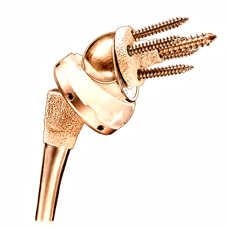
ریورس شولڈر ریپلیسمنٹ (reverse shoulder replacement) جوڑ کی تبدیلی کا ایک ایسا آپریشن ہے جس میں کندھے کے عام گیند اور پیالے (ball-and-socket) والے ڈھانچے کو الٹ دیا جاتا ہے، یعنی گیند وہاں لگتی ہے جہاں پیالہ تھا اور پیالہ وہاں لگتا ہے جہاں گیند تھی۔ ہم عام طور پر یہ آپریشن اس وقت تجویز کرتے ہیں جب روٹیٹر کف (rotator cuff)، یعنی ٹینڈنوں کا وہ گروپ جو کندھے کو تھامتا اور گھماتا ہے، بری طرح گھس چکا ہو یا پھٹ چکا ہو، یا جب کندھے کے دوسرے مسائل بغیر آپریشن کے علاج، جیسے سرگرمیوں میں تبدیلی، فزیوتھراپی، اسپلنٹ یا انجکشن، سے ٹھیک نہ ہوئے ہوں۔ کچھ چوٹوں میں فوراً سرجری تجویز کی جا سکتی ہے۔ اس آپریشن کا مقصد درد کم کرنا اور آپ کے کندھے کی حرکت اور کارکردگی کو بہتر بنانا ہے۔ اس کے طبی فوائد 10 سال تک برقرار رہتے ہیں۔ ہم آپ کے ساتھ اس کے فوائد اور خطرات پر بات کریں گے، اور یہ فیصلہ آپ ہمارے ساتھ مل کر کریں گے۔
آپریشن سے پہلے
جب آپ اور آپ کے سرجن ریورس شولڈر ریپلیسمنٹ پر متفق ہو جائیں، تو ہم آپ کے آپریشن کی منصوبہ بندی کے لیے ضروری ٹیسٹوں کا انتظام کریں گے۔ اس میں عام طور پر آپ کے کندھے کے ایکسرے شامل ہوتے ہیں، اور کبھی کبھی سی ٹی اسکین بھی، جو ایک تفصیلی اسکین ہے جو ہڈی کی سہ جہتی (تھری ڈی) تصویر بناتا ہے۔ یہ تصاویر ہمیں نئے جوڑ کے پرزوں کی جگہ طے کرنے اور آپریشن کے دن سے پہلے آپ کے کندھے کی احتیاط سے پیمائش کرنے میں مدد دیتی ہیں۔ آپ سے آپ کی عمومی صحت کے بارے میں بھی پوچھا جائے گا، جس میں آپ کے کندھے کے پچھلے آپریشن، اسٹیرائیڈ جیسی دوائیں، اور ہڈیوں کی کمزوری (osteoporosis)، یعنی ہڈیوں کا پتلا ہو جانا، جیسی بیماریاں شامل ہیں۔ براہ کرم اپنی موجودہ دواؤں کی فہرست اپنی ملاقات پر ساتھ لائیں۔ اگر آپ کچھ خاص دوائیں لیتے ہیں تو آپ کے سرجن آپ سے سرجری سے پہلے تھوڑے عرصے کے لیے انہیں بند کرنے کو کہہ سکتے ہیں۔ آپ کو آپریشن سے پہلے سات گھنٹے تک کچھ نہ کھانا ہوگا اور نہ پینا ہوگا؛ ہم معمول سے تھوڑا زیادہ وقت کا کہتے ہیں تاکہ اگر آپریشن کی فہرست وقت سے پہلے چل رہی ہو تو آپ کو پہلے بلایا جا سکے۔ کسی ایسے شخص کا انتظام کریں جو بعد میں آپ کو گاڑی میں گھر لے جائے، اور ڈھیلے، آرام دہ کپڑے پہنیں۔ اگر آپ کو دوسری طبی بیماریاں ہیں تو آپ کو خون کے ٹیسٹ یا بے ہوشی کے ڈاکٹر (anaesthetist)، یعنی وہ ڈاکٹر جو بے ہوشی کی دوا دیتا ہے، سے معائنے کی ضرورت پڑ سکتی ہے۔
آپریشن کے دن
آپریشن کے دن آپ ہسپتال کے سرجری کے مریضوں کے داخلے کے شعبے میں آتے ہیں۔ وہاں آپ کا اندراج کیا جاتا ہے اور آپ کو آپریشن تھیٹر کے لیے تیار کیا جاتا ہے۔ آپ پہلے کسی وارڈ میں نہیں جاتے۔
یہ آپریشن جنرل اینستھیزیا (مکمل بے ہوشی) کے ساتھ ریجنل اعصابی بلاک (nerve block)، یعنی ایک انجکشن جو جسم کے ایک حصے کو سُن کر دیتا ہے، ملا کر کیا جاتا ہے۔ بے ہوشی کے ڈاکٹر آپریشن سے پہلے آپ سے ملیں گے اور دونوں حصوں کے بارے میں آپ کو سمجھائیں گے۔
پھر آپ کو آپریشن تھیٹر میں لے جایا جاتا ہے، جہاں آپریشن کیا جاتا ہے۔ آپریشن مکمل ہونے پر آپ ریکوری روم میں جاگتے ہیں۔ وہاں نرسیں آپ کی نگرانی کرتی ہیں جب تک بے ہوشی کی دوا کا اثر ختم ہوتا ہے۔ جب آپ کی حالت مستحکم ہو جائے تو آپ کو وارڈ میں منتقل کر دیا جاتا ہے۔
آپریشن میں کیا کیا جاتا ہے
آپ کے سرجن کندھے کے جوڑ تک پہنچنے کے لیے آپریشن والی جگہ کے اوپر ایک ہی چیرا لگاتے ہیں۔ جوڑ کی گھسی ہوئی سطحیں نکال دی جاتی ہیں اور ان کی جگہ دھات اور پلاسٹک کے نئے پرزے لگا دیے جاتے ہیں۔ عام گیند اور پیالے والا ڈھانچہ الٹ دیا جاتا ہے، یعنی گیند وہاں لگتی ہے جہاں پیالہ تھا اور پیالہ وہاں لگتا ہے جہاں گیند تھی۔ اسی وجہ سے اسے "ریورس" (الٹی) تبدیلی کہا جاتا ہے۔ نئے پرزے اس طرح بنائے گئے ہیں کہ کندھے کو اوپر سے ڈھانپنے والا مضبوط پٹھا، ڈیلٹائیڈ (deltoid)، گھسے ہوئے یا پھٹے ہوئے روٹیٹر کف کے ٹینڈنوں کی جگہ بازو کو اٹھا سکے۔
آپریشن کے دوران آپ کے سرجن کندھے کے ٹینڈنوں (tendon)، یعنی ان مضبوط ڈوریوں جو پٹھوں کو ہڈی سے جوڑتی ہیں، اور دوسری نرم بافتوں کے ارد گرد احتیاط سے کام کرتے ہیں۔ اگر ان میں سے کچھ بافتیں اچھی حالت میں ہوں تو نیا امپلانٹ (جسم میں لگایا گیا مصنوعی پرزہ) اپنی جگہ لگنے کے بعد انہیں ٹانکوں سے واپس اس پر جوڑ دیا جاتا ہے۔ نئے پرزوں کی جگہ اور سائز آپریشن کے دن سے پہلے آپ کے ایکسرے اور اسکینوں سے بنائے گئے منصوبے کے مطابق طے کیے جاتے ہیں۔
جب نیا جوڑ اپنی جگہ لگ جائے تو زخم بند کر دیا جاتا ہے۔ پہلے بند زخم پر ایک باریک، خود چپکنے والی جالی رکھی جاتی ہے جو جلد کے کناروں کو آپس میں ملا کر رکھتی ہے۔ پھر اس جالی کے اوپر جلد پر لگانے والا مائع گوند لگایا جاتا ہے، جو جم کر پورے حصے کو بند کر دیتا ہے۔ یہ تقریباً ایک سے دو ہفتے تک لگا رہتا ہے اور پھر خود ہی اٹھ کر اتر جاتا ہے، اس لیے کچھ بھی نکالنے کی ضرورت نہیں پڑتی۔
آپریشن کے بعد
آپ ریکوری روم میں جاگیں گے، اور جب آپ کی حالت مستحکم ہو جائے گی تو آپ کو وارڈ میں منتقل کر دیا جائے گا۔ وہاں نرسیں آپ کی دیکھ بھال کرتی ہیں اور آپ کو آرام میں رکھنے کے لیے دوا دیتی ہیں۔ آپ کا بازو سہارے کے لیے ایک سادہ سلنگ (sling)، یعنی گلے سے لٹکی وہ پٹی جو بازو کو سہارا دیتی ہے، میں رہتا ہے؛ ورزش اور نہانے دھونے کے وقت اسے اتار دیا جاتا ہے۔ زیادہ تر مریض اس آپریشن کے بعد ایک یا دو راتیں ہسپتال میں رہتے ہیں۔ ہم زخم کی پٹی تقریباً 10 دن تک لگی رہنے دیتے ہیں؛ براہ کرم اس سے پہلے اسے نہ اتاریں، سوائے اس کے کہ ہم آپ کو ایسا کرنے کو کہیں۔ جب ہم آپ کو دیکھیں گے تو ہم اسے بدل دیں گے یا اتار دیں گے۔ ایک فزیوتھراپسٹ عام طور پر آپ کے گھر جانے سے پہلے آپ کو دیکھے گا تاکہ ہلکی حرکتیں شروع کروائی جا سکیں۔ براہ کرم گھر پہنچنے کے بعد پہلے 24 گھنٹوں کے لیے کسی ایسے شخص کا انتظام کریں جو آپ کے ساتھ رہے۔
صحت یابی
گھر پر پہلے چند دن آرام اور سکون کے ہوتے ہیں۔ آپ کے کندھے میں درد اور سوجن ہوگی، اور سلنگ ورزش کے اوقات کے درمیان بازو کو سہارا دینے کے لیے ہے۔ عام درد کش دوا، ہدایت کے مطابق لینے سے، عام طور پر درد کو قابلِ برداشت رکھتی ہے۔ ہفتے گزرنے کے ساتھ تکلیف بتدریج کم ہوتی جاتی ہے، اور زیادہ تر لوگ دیکھتے ہیں کہ سوجن اکڑاؤ سے پہلے ختم ہو جاتی ہے۔
آپ کے فزیوتھراپسٹ آپ کی ورزشوں میں رہنمائی کریں گے، جو ہلکی حرکتوں سے شروع ہو کر آپ کے کندھے کی گنجائش کے مطابق بڑھتی جائیں گی۔ ان سیشنوں اور نہانے دھونے کے لیے سلنگ اتار دی جاتی ہے۔ جب تک آپریشن والا بازو ٹھیک ہو رہا ہو، آپ اپنے دوسرے ہاتھ سے گھر کے ہلکے کام کر سکیں گے۔ شروع میں سونا مشکل ہو سکتا ہے؛ بہت سے لوگوں کو کرسی پر ٹیک لگا کر یا تکیوں کے سہارے آرام کرنا آسان لگتا ہے، جب تک لیٹنا دوبارہ آرام دہ محسوس نہ ہونے لگے۔
صحت یابی کے اہم مراحل تاریخوں کے بجائے واقعات کی صورت میں آتے ہیں۔ جب آپ کے سرجن آپ کو گاڑی چلانے کی اجازت دے دیں، جو عام طور پر چھ ہفتے والے معائنے پر ہوتا ہے، تو آپ دوبارہ گاڑی چلا سکتے ہیں؛ ہماری الگ گائیڈ بازو اور ہاتھ کی سرجری کے بعد گاڑی چلانا میں اس کی تفصیل موجود ہے۔ جیسے جیسے حرکت واپس آتی ہے، روزمرہ کے کام جیسے کپڑے پہننا اور ہاتھ بڑھا کر چیزیں پکڑنا آسان ہو جاتے ہیں۔ جب آپ کے فزیوتھراپسٹ آپ کی طاقت سے مطمئن ہوں، تو آپ ان سرگرمیوں کی طرف واپس جا سکتے ہیں جن سے آپ پہلے لطف اٹھاتے تھے، اور زیادہ تر لوگ جو اس آپریشن سے پہلے متحرک تھے، بعد میں ان سرگرمیوں کی طرف واپس آ جاتے ہیں۔
صحت یابی ہر شخص میں مختلف ہوتی ہے۔ آپ کا وقت مختلف ہو سکتا ہے، اور آپ کے سرجن اور فزیوتھراپسٹ ہر مرحلے پر آپ کی رہنمائی کریں گے۔
کیا مسائل پیدا ہو سکتے ہیں
زیادہ تر مریض ٹھیک رہتے ہیں، لیکن کبھی کبھار مسائل پیدا ہو سکتے ہیں۔ آپ کے سرجن اور ٹیم کسی بھی مسئلے کو جلد پکڑنے کے لیے آپ کی قریب سے نگرانی کرتے ہیں۔
کبھی کبھی نیا جوڑ اپنی جگہ سے نکل سکتا ہے۔ آپ کو اچانک کھٹ کی آواز یا جھٹکا محسوس ہو سکتا ہے، جس کے بعد درد ہو اور کندھا معمول کے مطابق حرکت نہ کرے۔ اگر ایسا ہو تو فوراً کلینک سے رابطہ کریں یا ہسپتال کی ایمرجنسی جائیں۔
نئے جوڑ کے ارد گرد انفیکشن ہو سکتا ہے۔ ان علامات پر نظر رکھیں: گہرا، ٹیس مارتا درد جو عام درد کش دواؤں سے کم نہ ہو، زخم سے باہر کی طرف پھیلتی ہوئی سرخی، یا بخار۔ اگر آپ ان میں سے کوئی بھی علامت دیکھیں تو جلد کلینک کو بتائیں۔ انفیکشن کا علاج مرحلہ وار کیا جاتا ہے، اور کندھے کی تبدیلی کے ارد گرد زیادہ تر انفیکشن اس طریقے سے ختم کیے جا سکتے ہیں۔
نئے پرزے وقت کے ساتھ ڈھیلے ہو سکتے ہیں۔ یہ عام طور پر ایسے درد کی صورت میں ظاہر ہوتا ہے جو واپس آ جائے یا بڑھ جائے، کبھی کبھی کلک کی آواز یا رگڑ کے احساس کے ساتھ۔ اپنے اگلے معائنے پر اس کا ذکر کریں تاکہ ایکسرے سے اس کی جانچ کی جا سکے۔
اعصاب کندھے کے قریب سے گزرتے ہیں، اور سرجری کے دوران ان پر چوٹ لگ سکتی ہے یا ان میں جلن (irritation) پیدا ہو سکتی ہے۔ آپ کو بازو میں سُن پن، جھنجھناہٹ یا کمزوری محسوس ہو سکتی ہے۔ اکثر یہ خود ہی ٹھیک ہو جاتا ہے، لیکن اپنے معائنے پر اس کا ذکر کریں تاکہ اس پر نظر رکھی جا سکے۔
نئے جوڑ کے ارد گرد کی ہڈی میں، یا کندھے کے سب سے اوپر ہڈی کے ابھرے ہوئے سرے میں، چھوٹے فریکچر (ہڈی کا ٹوٹنا) ہو سکتے ہیں۔ آپ کو تیز درد محسوس ہوگا، اکثر گرنے یا زور سے دھکا دینے کے بعد۔ اگر ایسا ہو تو کلینک سے رابطہ کریں۔
کندھے کی سرجری کے بعد کبھی کبھار خون کا لوتھڑا بن سکتا ہے، جس سے عام طور پر پنڈلی کے پچھلے حصے میں اچانک سوجن اور چھونے پر درد ہوتا ہے۔ اگر آپ کو ایسا محسوس ہو تو جلد طبی مدد حاصل کریں۔
کچھ عوامل مسائل کا امکان بڑھا دیتے ہیں۔ بازو کے اوپری حصے کی ہڈی ٹوٹنے پر کیے جانے والے اس آپریشن کے بعد مردوں میں پیچیدگیوں کا امکان زیادہ ہوتا ہے۔ پہلے روٹیٹر کف کی مرمت کا آپریشن ہو چکا ہو تو نئے جوڑ کے ارد گرد انفیکشن کا امکان بھی بڑھ جاتا ہے۔ اگر کندھے میں اسٹیرائیڈ کا انجکشن لگا ہو تو سرجری سے پہلے کم از کم 4 ہفتے انتظار کرنا ضروری ہے، اور ہم اسی کے مطابق منصوبہ بنائیں گے۔
اگر آپ تفصیلات جاننا چاہتے ہیں تو اس صفحے پر پیچیدگیوں کی جدول میں ان کی عام شرحیں درج ہیں۔
ہمیں کب فون کریں
اس آپریشن کے بعد زیادہ تر مسائل ایسی انتباہی علامات کے ساتھ ظاہر ہوتے ہیں جنہیں آپ گھر پر پہچان سکتے ہیں۔ اگر آپ کو بخار ہو، زخم کے ارد گرد بڑھتی ہوئی سرخی یا زخم سے رطوبت کا اخراج ہو، یا ایسا درد ہو جو کم ہونے کے بجائے بڑھتا ہی جائے، تو ہمیں فون کریں۔ اگر آپ کو اچانک شدید درد ہو، آپ کا کندھا اپنی شکل سے ہٹا ہوا نظر آئے، یا آپ بازو کو حرکت نہ دے سکیں تو ہسپتال کی ایمرجنسی جائیں۔ پنڈلی کے پچھلے حصے میں سوجن کے ساتھ چھونے پر درد، یا سانس پھولنا، ایسی حالت ہے جس میں فوراً ایمرجنسی علاج کی ضرورت ہوتی ہے۔ اگر آپ کے بازو یا ہاتھ میں احساس ختم ہو جائے، یا نیا سُن پن اور جھنجھناہٹ محسوس ہو جو ٹھیک نہ ہو، تو بھی ہمیں فون کریں۔
مزید تفصیل
Advanced reading: the deeper science (optional)
یہ حصہ اس سے زیادہ تفصیل میں جاتا ہے جتنی آپ کو اپنے علاج کے فیصلوں کے لیے ضرورت ہے۔ ریورس شولڈر ریپلیسمنٹ کے بارے میں اضافی مطالعہ اس لیے مفید ہے کہ یہ وہ آپریشن ہے جس نے ایک ایسا مسئلہ حل کیا جسے پہلے ناقابلِ حل سمجھا جاتا تھا، یعنی ایسا کندھا جس کا روٹیٹر کف کام نہ کر رہا ہو، اور اس لیے بھی کہ اس کی مخصوص پیچیدگی ایسی ہے جس کے بارے میں زیادہ تر مریضوں نے کبھی نہیں سنا۔
جوڑ کو الٹنا کیوں کارگر ہے
عام کندھے میں بازو پر ایک گیند اور کندھے کی پچھلی چپٹی ہڈی (shoulder blade) پر ایک پیالہ ہوتا ہے، اور یہ روٹیٹر کف پر انحصار کرتا ہے کہ جب ڈیلٹائیڈ بازو اٹھائے تو وہ گیند کو بیچ میں تھامے رکھے۔ جہاں کف ختم ہو چکا ہو، وہاں روایتی جوڑ کی تبدیلی کو مستحکم رکھنے والا کچھ نہیں ہوتا اور گیند بس اوپر کی طرف کھسک جاتی ہے۔
ریورس ڈیزائن ان دونوں کی جگہ بدل دیتا ہے: گیند کندھے کی پچھلی چپٹی ہڈی پر لگا دی جاتی ہے اور پیالہ بازو پر۔ اس سے گھماؤ کا مرکز اندر اور نیچے کی طرف چلا جاتا ہے، جس سے ڈیلٹائیڈ کی قوت کا بازو (lever arm) لمبا ہو جاتا ہے اور وہ اکیلے ہی بازو کو اٹھا سکتا ہے۔ یہ حیاتیاتی کے بجائے ایک میکانکی حل ہے: یہ کسی چیز کی مرمت نہیں کرتا، بلکہ باقی رہ جانے والے پٹھے کو کافی بنا دیتا ہے۔
اسی سے یہ بھی سمجھ آتا ہے کہ یہ آپریشن کون سی حرکتیں کس حد تک بحال کرتا ہے۔ سر سے اوپر بازو اٹھانا عام طور پر اچھی طرح واپس آ جاتا ہے۔ گھماؤ ان پٹھوں پر منحصر ہے جنہیں یہ آپریشن تبدیل نہیں کرتا، اس لیے کمر کے پیچھے ہاتھ لے جانا اکثر محدود رہتا ہے، اور یہ بات سرجری کے بعد مایوسی بننے کے بجائے سرجری سے پہلے ہی توقعات کا حصہ ہونی چاہیے۔
جہاں اس نے پرانے آپریشنوں کی جگہ لے لی ہے
فریکچر کے معاملے میں یہ تبدیلی فیصلہ کن رہی ہے۔ 228,523 مریضوں کے اعداد و شمار کو یکجا کرنے پر، بازو کے اوپری حصے کی ہڈی کے اوپری سرے کے فریکچر (proximal humerus fractures) والے بزرگ مریضوں میں ریورس ٹوٹل شولڈر آرتھروپلاسٹی نے ہیمی آرتھروپلاسٹی (hemiarthroplasty، صرف گیند والے حصے کی تبدیلی) کے مقابلے میں بہتر فعالی نتائج اور پیچیدگیوں کی بہتر شرح دکھائی، اور پلیٹ لگا کر ہڈی جوڑنے کے مقابلے میں دوبارہ آپریشن (revision) کے لحاظ سے زیادہ سازگار نتائج دیے [1]۔
امپلانٹ کی ایک تفصیل جو جاننے کے قابل ہے، کیونکہ یہ سوال اکثر اٹھایا جاتا ہے: ان فریکچروں کے لیے ریورس تبدیلی میں معیاری پرزوں کا فریکچر کے لیے مخصوص پرزوں سے موازنہ کرنے پر 436 مریضوں میں طبی نتائج یا پیچیدگیوں کی شرح میں کوئی نمایاں فرق نہیں پایا گیا [2]۔
وہ پیچیدگی جس کے بارے میں جاننا ضروری ہے
یہاں جو پیچیدگی اہم ہے وہ ایکرومین (کندھے کے اوپر کی ہڈی) یا کندھے کی پچھلی چپٹی ہڈی کا دباؤ سے ہونے والا فریکچر (acromial or scapular stress fracture) ہے، یعنی جوڑ کے اوپر کی ہڈی کا ڈیلٹائیڈ کے اس بڑھے ہوئے تناؤ کے تحت چٹخ جانا جو یہ ڈیزائن جان بوجھ کر پیدا کرتا ہے۔ یہ اسی طریقۂ کار کا براہِ راست نتیجہ ہے جس کی وجہ سے یہ آپریشن کام کرتا ہے۔
ایک میٹا اینالیسس (meta-analysis) نے خطرے کے عوامل کی نشاندہی کی: جن مریضوں کو یہ فریکچر ہوئے وہ زیادہ عمر کے اور کم بی ایم آئی (BMI، قد کے لحاظ سے وزن کا پیمانہ) والے تھے، اور دوسرے خطرات میں ہڈیوں کی کمزوری، سوزش والا آرتھرائٹس، خاتون ہونا اور روٹیٹر کف کی پچھلی مرمت شامل تھے [3]۔ ایک الگ جائزے سے معلوم ہوا کہ تحقیقی مواد میں ان فریکچروں کو غیر یکساں طور پر رپورٹ کیا جاتا ہے، یعنی ان کی تعریف، تشخیص اور گنتی میں نمایاں تضادات ہیں [4]، اس لیے بتائی گئی شرحوں کو احتیاط سے دیکھنا چاہیے۔
اس کے مقابلے میں قلیل مدتی تصویر اطمینان بخش ہے: پہلے 90 دنوں میں اموات اور طبی اور جراحی پیچیدگیوں کی مجموعی شرح کم ہے، اور صرف 6 فیصد مریضوں کو دوبارہ ہسپتال میں داخل ہونا پڑا [5]۔
دو باتیں جن کی شواہد تائید نہیں کرتے
یہ کہ سب اسکیپولیرس (subscapularis) کی مرمت ضروری ہے۔ 267 مریضوں میں، سب اسکیپولیرس (کندھے کی چپٹی ہڈی کے سامنے والا روٹیٹر کف کا پٹھا) کی مرمت کے ساتھ یا اس کے بغیر ریورس تبدیلی کے بعد بازو کو پہلو سے اٹھانے، اندر کی طرف گھمانے یا باہر کی طرف گھمانے کی طاقت میں کوئی فرق نہیں تھا، اگرچہ مصنفین نوٹ کرتے ہیں کہ طاقت کے نتائج پر تحقیق محدود ہے [6]۔
یہ کہ آپریشن کی وجہ نتیجے پر اثر نہیں ڈالتی۔ دراصل ڈالتی ہے۔ دو یا اس سے زیادہ سال بعد، جن مریضوں کی ریورس تبدیلی سالم روٹیٹر کف کے ساتھ بنیادی جوڑ کے گھسنے والے آرتھرائٹس (primary osteoarthritis) کی وجہ سے ہوئی، ان کے کانسٹینٹ اسکور (Constant score، کندھے کی کارکردگی کا پیمانہ) ان مریضوں سے نمایاں طور پر زیادہ تھے جن کا آپریشن روٹیٹر کف کے پھٹاؤ کے ساتھ بنیادی آرتھرائٹس یا ثانوی آرتھرائٹس (secondary osteoarthritis) کی وجہ سے ہوا [7]۔ ایک ہی آپریشن اس بات کے لحاظ سے مختلف نتائج دیتا ہے کہ آپ کو اس کی ضرورت کیوں پڑی، اور جب آپ کو کوئی اوسط بتائی جائے تو یہ جاننا مفید ہے۔
حوالہ جات
[1] Mekhail J, Mullan R, Cross JL, Jahagirdar O, Luo X, Salameh M. Outcomes of reverse total shoulder arthroplasty vs. other surgical fixation methods for proximal humerus fractures: a systematic review and meta-analysis. JSES Rev Rep Tech. 2026;6(2):100644. https://doi.org/10.1016/j.xrrt.2025.100644
[2] Apivatgaroon A, Kongmalai T, Kongmalai P. Standard compared with fracture-specific components in reverse shoulder arthroplasty for proximal humeral fractures: a systematic review and meta-analysis. Bone Joint J. 2025;107-B(9):931-41. https://doi.org/10.1302/0301-620X.107B9.BJJ-2024-1508.R2
[3] Elmenawi KA, Sperling JW, Sanchez-Sotelo J, Barlow JD. Risk factors for acromial and scapular fractures following reverse shoulder arthroplasty: a meta-analysis. JSES Rev Rep Tech. 2026;6(1):100578. https://doi.org/10.1016/j.xrrt.2025.08.015
[4] Davie RA, Nathan K, Persaud SG, Oladeji LO, Taylor SA, Dines JS, et al. Inconsistent reporting of risk factors for acromial stress fractures following reverse total shoulder arthroplasty: a systematic review. J Shoulder Elbow Surg. 2025;34(11):e975-e984. https://doi.org/10.1016/j.jse.2025.02.032
[5] Kent LM, Hurley ET, Davey MS, Klifto CS, Mullett H. Low complication rate following reverse total shoulder arthroplasty at 90-days follow-up: a systematic review. J ISAKOS. 2024;9(2):205-10. https://doi.org/10.1016/j.jisako.2023.11.006
[6] Preuss FR, Eble SK, Peebles AM, Osuna-Garcia A, Provencher CMT. Shoulder strength outcomes after reverse total shoulder arthroplasty: a systematic review. JSES Rev Rep Tech. 2022;2(2):131-4. https://doi.org/10.1016/j.xrrt.2021.11.004
[7] Nové-Josserand L, Nerot C, Colotte P, Guery J, van Rooij F, Hibon A, et al. Reverse shoulder arthroplasty for primary glenohumeral osteoarthritis: significantly different characteristics and outcomes in shoulders with intact vs. torn rotator cuff. J Shoulder Elbow Surg. 2024;33(4):850-62. https://doi.org/10.1016/j.jse.2023.07.027
Evidence & references
This is the clinical evidence summary written for health professionals. It is technical, and it lists the research this page was built from. You do not need to read it to understand your treatment or to make a decision about it.
Anatomy & Pathophysiology
Glenoid and Scapular Anatomy
- The glenoid is suspended from the body of the scapula by the neck and fixed to the clavicle by the acromioclavicular and coracoclavicular ligaments [3].
- As the face of the glenoid transitions into the neck, the glenoid vault narrows [3].
- The scapular spine is subcutaneous posteriorly and widens gradually as it transitions into the base of the acromion laterally [3].
- The acromion curves anteriorly and meets the clavicle at the acromioclavicular joint and the coracoid via the coracoacromial ligament, which originates under the anterior margin of the acromion [3].
- The suprascapular nerve arises from the C4–C5 nerve roots off of the supraclavicular brachial plexus at “Erb’s point” [3].
- The suprascapular nerve runs just medial to the base of the coracoid, under the transverse scapular ligament within the suprascapular notch, and gives off branches to the supraspinatus within 1 cm of the notch [3].
- The suprascapular nerve continues through the supraspinatus fossa heading laterally and distally on the under surface of the supraspinatus [3].
- The suprascapular nerve runs under the ill-defined spinoglenoid ligament around the lateral base of the scapula within spinoglenoid notch before terminating in posterior capsular sensory branches heading laterally and an infraspinatus motor branch heading medially within 1 cm of the lateral margin of the scapular spine [3].
- Cadaver studies show the suprascapular nerve is present 29 mm (23 to 35 mm) from the superior rim of the glenoid at the suprascapular notch [3].
- Cadaver studies show the suprascapular nerve is present 18 mm (14 to 24 mm) from the posterior rim at the spinoglenoid notch [3].
- Injury to the suprascapular nerve can cause pain and denervation of the supraspinatus and infraspinatus [3].
Biomechanics and Pathophysiology
- Anatomic total shoulder arthroplasties and hemiarthroplasties are dependent on the rotator cuff to center the head in the glenoid and to optimize larger periscapular muscle function [7].
- In the absence of a functional rotator cuff, the deltoid pulls the humerus proximally, shearing along the glenoid and resulting in eccentric glenoid wear, acetabularization of the acromion, and pseudoparalysis [7].
- The reverse shoulder arthroplasty does not require the rotator cuff for function but is dependent on an intact deltoid neuromuscular unit [7].
- The semiconstrained nature of the reverse prosthesis provides a stable fulcrum that allows the deltoid to elevate the shoulder even in the absence of a functional rotator cuff [4].
- In the Grammont reverse prosthesis, the center of rotation is medial to the glenoid component–bone interface to decrease shear stress and provide compressive stress, aiming to decrease the chances of glenoid loosening [4].
- The humeral component in the Grammont design is inset, resting almost completely inside the proximal humerus metaphysis [4].
- The opening angle of the polyethylene in the Grammont design is relatively horizontal at 155 degrees compared with conventional arthroplasty [4].
- Once articulated, the humerus is more medial and more distal than preoperatively, providing a mechanical advantage to the deltoid for active elevation in the absence of a rotator cuff [4].
- The traditional Grammont style decreases shear forces seen by the glenoid and lowers baseplate failure by medializing the center of rotation, but this is associated with the risk of inferior scapular notching in adduction [7].
- Distalization doubles the lever arm of the deltoid and optimizes the length–tension curve of its sarcomeres, increasing its efficiency by 30% at the cost of rotational strength [7].
- Lateralized glenosphere and lateralized humerus designs have gained popularity to improve the rotational profile, deltoid function, implant stability, and decrease impingement such as scapular notching [7].
- Early reverse designs had a high failure rate due to the profound lever arm on the glenoid and baseplate bone [7].
- Recent reverse designs have a better track record but increased forces are seen by the scapula and acromion [7].
Periprosthetic Scapular Fracture Pathophysiology
- Periprosthetic scapular fractures are universally associated with stable glenoid implants [2].
- Fracture is unlikely in the face of dislocation, glenosphere dissociation, or baseplate pullout at the bone–baseplate interface [2].
- Periprosthetic scapular fracture has been noted to result in new glenohumeral instability due to the change of the orientation of the glenosphere and loss of deltoid tension [2].
- Postoperative periprosthetic scapular fracture is a challenge unique to reverse shoulder arthroplasty and occurs more commonly than humeral fractures [7].
- Postoperative periprosthetic scapular fractures are an effect of nonphysiologic forces transferred from the implant to the scapula, often in a suboptimal host [7].
- Fatigue fracture has been found to occur through already weakened acromiums or those with preexisting lesions [7].
- Acromial thinning and eventual fragmentation occur at the final stages of rotator cuff-tear arthropathy as the humeral head acetabularizes [7].
Clinical Presentation
Periprosthetic Scapular Fractures
- The weak point in periprosthetic scapular fracture injuries is the scapular bone [2].
- Periprosthetic scapular fractures are unlikely in the presence of dislocation, glenosphere dissociation, or baseplate pullout at the bone–baseplate interface [2].
- Periprosthetic scapular fractures have been noted to rarely result in new glenohumeral instability [2].
- New glenohumeral instability following periprosthetic scapular fracture is due to a change in the orientation of the glenosphere and loss of deltoid tension [2].
- Diagnosis of periprosthetic fractures is often subtle and requires a high index of suspicion [2].
- Workup for periprosthetic fractures should begin with a complete history and examination [2].
- Past medical history should elucidate the underlying diagnosis for the index surgery and subsequent surgeries [2].
- Complications including infection should be accounted for in the past medical history [2].
- The examiner should understand the patient's shoulder function and level of disability before surgery, after surgery, and at present [2].
- The examiner should understand the time course of changes in shoulder function and disability [2].
- In the case of a stress reaction, new pain at the base of the acromion may be the only finding [2].
- New pain at the base of the acromion in a stress reaction should raise suspicion and spark further imaging or a period of rest [2].
- Stress fractures can be more painful than after they propagate into a displaced fracture [2].
- Patients typically present around their 8th decade of life after a sudden increase in pain or loss of function [2].
- Patients typically present after a sudden increase in pain or loss of function in an otherwise smooth postoperative course [2].
- Presentation for periprosthetic scapular fracture is generally within 1 year but up to 2 years from surgery [2].
- Patients who go on to have periprosthetic scapular fractures initially outperform those who do not [2].
- Past medical history must identify risk factors including a history of steroid use, osteoporosis, subacromial decompression, or rotator cuff tear arthropathy [2].
- Previous operative reports, clinic notes, and imaging can help provide a thorough understanding of any previous surgeries on the shoulder or history of radiation [2].
- Physical examination starts with inspection [2].
- Deformity is concerning for dislocation, hematoma, or displaced fracture [2].
- Erythema or incisional dehiscence is concerning for infection [2].
- Tenderness along the acromion or scapular spine raises suspicion for fracture which should be confirmed with imaging [2].
- A complete neurovascular examination is performed as part of the physical examination [2].
- Assessment of active and passive motion is performed as part of the physical examination [2].
- Fracture can result in motion limited by pain, new weakness, or loss of function [2].
- Infection should be investigated with laboratory tests [2].
- A sudden loss of function or increase in pain is consistent with both scapular fracture and infection [2].
- A sudden loss of function or increase in pain should trigger further workup [2].
Investigations
Periprosthetic Scapular Fracture Diagnosis and Workup
- Diagnosis of periprosthetic scapular fractures is often subtle and requires a high index of suspicion [2].
- The workup for periprosthetic scapular fractures should begin with a complete history and examination [2].
- The examiner should understand the patient's shoulder function and level of disability before surgery, after surgery, and at present, as well as the time course of these changes [2].
- New pain at the base of the acromion may be the only finding in a stress reaction and should raise suspicion for further imaging or a period of rest [2].
- Patients with periprosthetic scapular fractures typically present around their 8th decade of life after a sudden increase in pain or loss of function in an otherwise smooth postoperative course [2].
- Periprosthetic scapular fractures generally occur within 1 year but up to 2 years from surgery [2].
- Inspection for deformity is concerning for dislocation, hematoma, or displaced fracture [2].
- A complete neurovascular examination is performed as well as assessment of active and passive motion [2].
- A sudden loss of function or increase in pain is consistent with both scapular fracture and infection and should trigger further workup [2].
Preoperative Imaging and Planning
- Careful assessment of the preoperative radiographs and CT with three-dimensional reconstruction is extremely useful in preparation for surgery [5].
- The main goals of preoperative imaging include understanding the fracture pattern and anticipating the ideal height of stem implantation [5].
- Radiographs of both humeri (affected and unaffected) with magnifier markers may be used to understand where the stem should be positioned in reference to the fracture line on the humeral shaft [5].
- The glenoid should be assessed in radiographs and CT to plan for component positioning, version, inclination, and rotation, as well as anticipated screw length [5].
- Associated fractures of the rim of the glenoid in anterior or posterior fracture-dislocations are rare [5].
- Most of the times, the size of the fractured glenoid rim is small enough not to interfere with secure baseplate placement [5].
- If the size of the fractured glenoid rim is large enough to interfere with the stability of the glenoid baseplate, fixation with small fragment screws may be performed [5].
- The glenoid should be assessed on CT for glenoid component implantation [5].
- Most surgeons do not use fluoroscopy for this procedure [5].
Treatment
Indications and Rationale
- Reverse shoulder arthroplasty is the replacement procedure of choice when arthroplasty is considered for proximal humeral fractures [4].
- Tuberosity and rotator cuff-related complications are the main reason for poor functional outcome when a humeral head replacement is implanted for management of a proximal humeral fracture [4].
- Shoulder arthroplasty is considered for proximal humeral nonunion in the presence of severe cavitation and bone loss at the humeral head and metaphysis or collapse and degenerative change of the humeral articular surface [6].
- Severe tuberosity malunion in the setting of a proximal humeral nonunion is more reliably compensated for with reverse arthroplasty than with osteotomy and internal fixation [6].
- Reverse shoulder arthroplasty may improve shoulder function in patients with nonunions associated with severe tuberosity malunions [6].
Prosthesis Design and Biomechanics
- The Grammont reverse prosthesis features an articulating glenoid component shaped as a third of a sphere [4].
- In the Grammont design, the center of rotation is medial to the glenoid component–bone interface to decrease shear stress and provide compressive stress, aiming to decrease the chances of glenoid loosening [4].
- The opening angle of the polyethylene in the Grammont design is 155 degrees [4].
- Articulation of the Grammont implants results in the humerus being more medial and more distal than preoperatively, providing a mechanical advantage to the deltoid for active elevation [4].
- The 155-degree opening angle in the Grammont design was selected to decrease the chances of dislocation [4].
- The humeral component in the Grammont design was recommended to be implanted in more anteversion (0 to 10 degrees of retroversion) than conventional arthroplasty [4].
- Subsequent reverse designs with a larger portion of a sphere place the center of rotation more lateral than the Grammont prosthesis [4].
- Subsequent reverse designs with a larger portion of a sphere utilize an opening angle of 135 degrees for the humeral component [4].
- Later reverse designs introduced onlay humeral bearings that lateralize the position of the humerus without changing the center of rotation [4].
- Later reverse designs with onlay humeral bearings utilize a 145-degree opening angle for the bearing [4].
- There is very little published on reverse arthroplasty biomechanics in the setting of a proximal humeral fracture [4].
Surgical Technique and Tuberosity Management
- Some surgeons initially elected to implant a reverse arthroplasty in proximal humeral fractures without repair, or sometimes with excision, of the greater tuberosity and/or lesser tuberosity [4].
- Healing of at least the greater tuberosity in good position provides a higher chance of restoration of active external rotation [4].
- Not performing a tuberosity repair at the time of reverse arthroplasty for proximal humeral nonunion has been correlated with a higher rate of dislocation [4].
- Technical principles for reverse arthroplasty in fracture may need to be modified to enhance tuberosity healing by avoiding translating the humeral shaft too lateral or too distal, allowing the tuberosities to overlap a few millimeters with the shaft [4].
- Use of a stem with fracture-dedicated features, such as a proximal ingrowth surface, small cross section, and holes for suture fixation, may be beneficial [4].
Comparison with Hemiarthroplasty
- Hemiarthroplasty is less commonly considered than reverse arthroplasty for proximal humeral nonunion [6].
- The functional outcome of hemiarthroplasty for nonunion is particularly concerning when tuberosity osteotomies need to be added [6].
- Studies reporting on hemiarthroplasty for nonunion suggest the procedure may be effective in reducing or eliminating pain but is associated with a high rate of complications that often require further surgery and disappointing functional recovery [6].
Complications
- Scapular notching is a complication of reverse shoulder arthroplasty [1].
- The clinical impact of scapular notching on outcomes after reverse total shoulder arthroplasty has been analyzed in a study of 476 shoulders [1].
- Humeral version in reverse shoulder arthroplasty affects impingement during activities of daily living [1].
- Subscapularis tendon integrity impacts shoulder function after reverse shoulder arthroplasty [1].
- Component positioning affects the intrinsic stability of the reverse shoulder arthroplasty [1].
- Humeral component lateralization in reverse shoulder arthroplasty affects rotator cuff torque [1].
References
[1] Campbell S Operative Orthopaedics 4 Volume Set. RECONSTRUCTIVE PROCEDURES OF THE SHOULDER AND ELBOW IN ADULTS > REVERSE SHOULDER ARTHROPLASTY.
[2] Rockwood And Green S Fractures In Adults. 29: Principles of Nonunion and Bone Defect Treatment > Injuries Associated with Periprosthetic Scapular Fractures About Reverse Shoulder Arthroplasty.
[3] Rockwood And Green S Fractures In Adults. 29: Principles of Nonunion and Bone Defect Treatment > Pathoanatomy and Applied Anatomy Related to Periprosthetic Scapular Fractures About Reverse Shoulder Arthroplasty.
[4] Rockwood And Green S Fractures In Adults. 29: Principles of Nonunion and Bone Defect Treatment > Reverse Shoulder Arthroplasty.
[5] Rockwood And Green S Fractures In Adults. 29: Principles of Nonunion and Bone Defect Treatment > Preoperative Planning > Reverse Shoulder Arthroplasty for Fracture: Preoperative Planning Checklist.
[6] Rockwood And Green S Fractures In Adults. 29: Principles of Nonunion and Bone Defect Treatment > Reverse Shoulder Arthroplasty and Hemiarthroplasty.
[7] Rockwood And Green S Fractures In Adults. 29: Principles of Nonunion and Bone Defect Treatment > Periprosthetic Scapular Fractures About Reverse Shoulder Arthroplasty.