Patients › Shoulder
ہنسلی کی ہڈی کے بیرونی سرے کا گھلنا (Distal Clavicle Osteolysis)
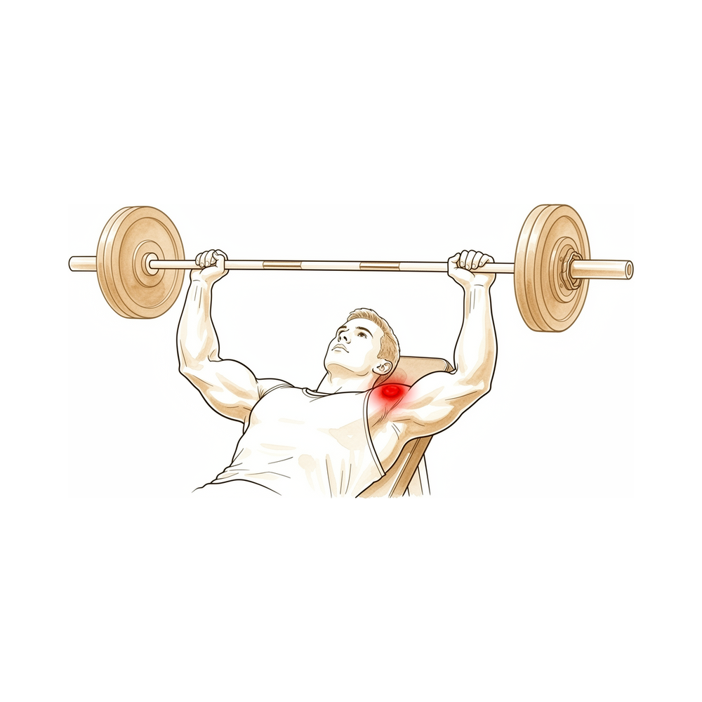
آپ کو کیا محسوس ہوتا ہے
درد آپ کی ہنسلی کی ہڈی کے بیرونی سرے پر ہوتا ہے، بالکل وہاں جہاں یہ آپ کے کندھے کے اوپری حصے سے ملتی ہے۔ اس جگہ کو ایکرومیو کلاویکولر جوڑ (acromioclavicular joint) کہتے ہیں۔ بازو استعمال کرتے وقت، خاص طور پر دھکیلنے، اٹھانے یا دبانے (پریس) والی حرکتوں میں، یہ عموماً سب سے زیادہ دکھتا ہے، اور آرام کرنے سے عموماً کم ہو جاتا ہے۔
ویٹ ٹریننگ (وزن اٹھانے کی ورزش) اس کی ایک عام وجہ ہے۔ بینچ پریس اور دوسری بھاری ورزشیں براہ راست اس جوڑ پر بوجھ ڈالتی ہیں، اور درد اکثر کسی ایک واضح چوٹ سے شروع ہونے کے بجائے ہفتوں کی ورزش کے دوران آہستہ آہستہ بڑھتا ہے۔ کچھ لوگ اسے کندھے کی چوٹ کے بعد بھی محسوس کرتے ہیں، جب باقی کندھا ٹھیک ہو جانے کے کافی عرصہ بعد تک درد باقی رہتا ہے۔
یہ دکھن رات کو یا جم میں ورزش کے بعد بڑھ سکتی ہے۔ اپنے جسم کے آر پار ہاتھ بڑھانا، کرسی سے ہاتھوں کے زور پر اٹھنا، اسی طرف خریداری کا سامان اٹھانا یا گھاس کاٹنے کی مشین دھکیلنا، یہ سب اسے بڑھا سکتے ہیں۔ چونکہ یہ درد کندھے کے بالکل اوپری حصے پر ہوتا ہے، اس لیے شروع میں اسے آسانی سے پٹھے کا کھنچاؤ سمجھ لیا جاتا ہے۔
یہ مسئلہ کندھے کے دوسرے مسائل سے بہت ملتا جلتا ہو سکتا ہے، اس لیے یہ جاننے کے لیے کہ اصل میں کیا ہو رہا ہے، اسکین کیے جاتے ہیں۔ جوڑ کا ایک خاص زاویے سے لیا گیا ایکسرے، جو موازنے کے لیے دونوں طرف کا لیا جاتا ہے، اور ایم آر آئی اسکین ہنسلی کی ہڈی کے سرے پر تبدیلیاں دکھا سکتے ہیں۔
ان علامات پر نظر رکھیں اور ان کے ظاہر ہونے پر قدم اٹھائیں:
اگر آپ کا بازو گرم، سرخ، سوجا ہوا اور دردناک ہو جائے، خاص طور پر بخار کے ساتھ، تو اسی دن ہسپتال کی ایمرجنسی جائیں۔ اگر چوٹ کے بعد اچانک آپ کے بازو میں احساس یا حرکت ختم ہو جائے تو بھی اسی دن ہسپتال کی ایمرجنسی جائیں۔
اگر درد کم نہ ہو رہا ہو، ہفتوں کے دوران بڑھ رہا ہو، آپ کو رات کو جگا دیتا ہو، یا آپ کو کام کرنے یا بازو استعمال کرنے سے روکتا ہو، تو اپنے فیملی ڈاکٹر (GP) سے ملیں یا ماہر سے معائنہ کروانے کا کہیں۔ اگر آپ کلینک سے رابطہ نہ کر سکیں، مثلاً کلینک کے اوقات کے بعد یا ہفتے کے آخر میں، تو اپنے قریب ترین ہسپتال کی ایمرجنسی جائیں۔
دراصل کیا ہو رہا ہے
ایکرومیو کلاویکولر جوڑ آپ کے کندھے کے اوپر ایک چھوٹا جوڑ ہے، جہاں آپ کی ہنسلی کی ہڈی کا بیرونی سرا ہڈی کے ایک چھجے سے ملتا ہے جسے ایکرومیون (acromion) کہتے ہیں۔ ہنسلی کی ہڈی ایک ٹیک (سہارے) کا کام کرتی ہے جو آپ کے بازو کو سینے سے دور رکھتی ہے، اور یہ چھوٹا جوڑ ان جگہوں میں سے ایک ہے جہاں یہ کندھے کی پچھلی چپٹی ہڈی (shoulder blade) سے جڑتی ہے۔ نیچے کی طرف لیگامنٹوں (ligaments) کی ایک مضبوط پٹی، یعنی وہ مضبوط بندھن جو ایک ہڈی کو دوسری ہڈی سے جوڑتے ہیں، ان دونوں ہڈیوں کو اس وقت سیدھ میں رکھتی ہے جب آپ کچھ اٹھاتے اور دباتے ہیں۔
اس مسئلے میں ہنسلی کی ہڈی کا بیرونی سرا ٹوٹ پھوٹ کا شکار ہونے لگتا ہے۔ ہڈی کے اس طرح گھلنے کے لیے طبی لفظ آسٹیولائسس (osteolysis) ہے۔ عام حالت میں جسم گھسی ہوئی ہڈی کی جگہ نئی ہڈی بناتا رہتا ہے، لیکن یہاں ٹوٹ پھوٹ مرمت سے آگے نکل جاتی ہے، اس لیے ہنسلی کی ہڈی کے سرے کی ہڈی نرم پڑ جاتی ہے اور گھستی جاتی ہے۔ اس کے اوپر والا جوڑ صحیح سلامت رہتا ہے؛ مسئلہ صرف ہنسلی کی ہڈی والی طرف تک محدود رہتا ہے۔
سب سے عام وجہ بار بار پڑنے والا بھاری بوجھ ہے، خاص طور پر بینچ پریس اور ویٹ ٹریننگ۔ ہر بار دبانے سے جوڑ کی دونوں ہڈیاں ایک دوسرے سے ٹکراتی ہیں، کچھ ایسے جیسے زمین کے ایک ہی چھوٹے ٹکڑے پر بار بار پاؤں مارتے رہیں یہاں تک کہ وہ گھس جائے۔ یہی عمل کندھے کی چوٹ کے بعد بھی ہو سکتا ہے، جس میں اصل چوٹ کے بعد کئی ہفتوں سے کئی مہینوں تک درد باقی رہتا ہے۔ کم صورتوں میں، ہلکے درجے کا انفیکشن، یا دھات کی وہ پلیٹ جو جوڑ کا اپنی جگہ سے نکل جانے (dislocation) کے بعد ہنسلی کی ہڈی کو نیچے دبائے رکھنے کے لیے لگائی جاتی ہے، بھی اسے شروع کر سکتی ہے۔
جیسے جیسے ہڈی گھستی ہے، جوڑ میں سوزش اور سوجن آ جاتی ہے، اسی لیے دکھن ہفتوں کی ورزش کے دوران بڑھتی ہے اور رات کو بھڑک اٹھتی ہے۔ گھسا ہوا سرا ایکرومیون کے ساتھ اپنی ہموار نشست بھی کھو دیتا ہے، اس لیے دبانے، اٹھانے اور جسم کے آر پار ہاتھ بڑھانے سے ننگی، کھردری ہڈی پر رگڑ پڑتی ہے۔ یہی وہ درد ہے جو آپ کو کندھے کے بالکل اوپری حصے پر محسوس ہوتا ہے۔
ہم اس کے لیے کیا کر سکتے ہیں
ڈاکٹر کیرن ہِرپرا، جو Mater Private Hospital Rockhampton میں بازو اور ہاتھ کے سرجن (upper-limb surgeon) ہیں، سب سے کم چیر پھاڑ والے ان طریقوں سے آغاز کرتے ہیں جو آپ کے مسئلے کے لیے مناسب ہوں۔ مریضوں کو عموماً ان کے فیملی ڈاکٹر ہمارے کلینک بھیجتے ہیں؛ اگر کسی فزیوتھراپسٹ نے آپ کو ہمیں دکھانے کا مشورہ دیا ہے، تب بھی میڈیکیئر (Medicare) کی رقم واپسی کا حق دار ہونے کے لیے آپ کو اپنے فیملی ڈاکٹر سے ریفرل (referral) لینا ہوگا۔ جب ہم آپ کی تکلیف کی تفصیل، معائنے اور اسکین کے ذریعے تشخیص کی تصدیق کر لیں، تو ہم سب سے آسان علاج سے شروع کر کے ترتیب وار علاج آزماتے ہیں۔
پہلا قدم یہ بدلنا ہے کہ آپ جوڑ پر کس طرح بوجھ ڈالتے ہیں۔ چونکہ بار بار بھاری وزن دبانے سے ہی ہڈی گھستی ہے، اس لیے درد کو بھڑکانے والی ورزشوں کو کم کرنے یا ان کی جگہ دوسری ورزشیں کرنے سے ہڈی کو سنبھلنے کا موقع ملتا ہے۔ جب آپ یہ کر رہے ہوں، فزیوتھراپی کا مقصد سوزش کو کم کرنا اور آپ کے کندھے کو مضبوط اور حرکت کے قابل رکھنا ہوتا ہے۔ جو کھلاڑی سیزن کے بیچ میں ہوں، ان کے لیے یہ طریقہ علاج کے اثر کرنے کے دوران کھیل جاری رکھنا ممکن بناتا ہے۔ اس میں کتنا وقت لگے گا، اس کا انحصار اس پر ہے کہ کیا آپ واقعی اپنی ورزش کا طریقہ بدل سکتے ہیں، اور ہم آپ سے بات کریں گے کہ آپ کے کھیل یا کام کے لیے اس کا کیا مطلب ہے۔
اس کے بعد دوا کا مرحلہ آتا ہے۔ سوزش کم کرنے والی گولیاں، جنہیں کبھی کبھی این ایس اے آئی ڈیز (NSAIDs) کہا جاتا ہے، جوڑ میں سوجن اور دکھن کو کم کرتی ہیں۔ ہم سُن کرنے والی دوا اور کورٹیسون (cortisone)، جو سوزش کم کرنے والی ایک طاقتور دوا ہے، کا مرکب سیدھا جوڑ میں انجکشن کے ذریعے بھی لگا سکتے ہیں۔ یہ انجکشن دو کام کرتا ہے: اس سے تصدیق ہوتی ہے کہ یہی جوڑ آپ کے درد کی وجہ ہے، اور اس کے بعد کچھ عرصے کے لیے یہ درد کم بھی ہو سکتا ہے۔
اگر ان اقدامات سے آپ کو کافی آرام نہ ملا ہو، تو ہم سرجری کے بارے میں بات کر سکتے ہیں۔ اس آپریشن میں آپ کی ہنسلی کی ہڈی کے بیرونی سرے سے ہڈی کی ایک پتلی سی پرت نکال دی جاتی ہے، تاکہ کھلی، گھسی ہوئی سطحیں ایک دوسرے سے مزید نہ رگڑیں۔ صرف چند ملی میٹر ہڈی نکالی جاتی ہے، اور اگر ہڈی کا کوئی ابھار (bone spur) قریبی ٹینڈن (tendon)، یعنی وہ مضبوط ڈوری جو پٹھے کو ہڈی سے جوڑتی ہے، کو دبا رہا ہو تو ہم اسی وقت اسے بھی ٹھیک کر دیتے ہیں۔ ہم آپ کو بتائیں گے کہ آپریشن میں کیا کیا جاتا ہے، صحت یابی کیسی ہوتی ہے، اور یہ کیا بدل سکتا ہے اور کیا نہیں، تاکہ آپ ہمارے ساتھ مل کر فیصلہ کر سکیں کہ آیا یہ آپ کے لیے مناسب ہے۔
کیا توقع رکھیں
زیادہ تر لوگوں میں یہ مسئلہ تب ٹھیک ہو جاتا ہے جب وہ بوجھ رک جائے جس کی وجہ سے یہ ہوا تھا۔ جب بار بار دبانا اور اٹھانا کم ہو جائے تو ہنسلی کی ہڈی کے سرے کی ہڈی خود کو دوبارہ بنا سکتی ہے۔ سچی بات یہ ہے کہ اس میں ایک شرط ہے: علاج تب کام کرتا ہے جب آپ واقعی اپنی ورزش یا کام کا طریقہ بدل سکیں۔ اگر آپ جوڑ پر اسی طرح بوجھ ڈالتے رہیں تو درد عموماً برقرار رہتا ہے یا بار بار لوٹ آتا ہے۔
آپریشن کے بغیر صحت یابی کوئی جلدی کا حل نہیں ہے۔ دکھن ہفتوں میں بڑھتی ہے، اور اسے کم ہونے میں عموماً کئی ہفتوں سے کئی مہینوں تک بوجھ کم رکھنا پڑتا ہے۔ کچھ لوگوں کو اس کے بجائے کندھے کی چوٹ کے بعد درد محسوس ہوتا ہے، اور اس صورت میں باقی کندھا ٹھیک ہو جانے کے کافی عرصہ بعد تک یہ باقی رہ سکتا ہے۔ اگر مناسب عرصے تک آرام اور ورزش میں تبدیلی کے بعد بھی درد باقی ہو، تو اسے برداشت کر کے ورزش جاری رکھنے کے بجائے اس کا معائنہ کروانا بہتر ہے۔
اگر آپریشن کے بغیر علاج سے آپ کو کافی آرام نہ ملے تو سرجری ایک راستہ ہے۔ اس آپریشن میں آپ کی ہنسلی کی ہڈی کے بیرونی سرے سے ہڈی کی ایک پتلی سی پرت نکال دی جاتی ہے تاکہ کھلی سطحیں آپس میں رگڑنا بند کر دیں۔ جن لوگوں کا یہ آپریشن ہوتا ہے وہ بتاتے ہیں کہ ان کا درد کم ہو گیا اور کندھے کی حرکت بہتر ہو گئی، اور وہ عموماً نتیجے سے مطمئن ہوتے ہیں۔ دوربین کے ذریعے آپریشن (keyhole) کے بعد کھلے آپریشن کی نسبت عموماً کم دیرپا درد رہتا ہے، اور لوگ اس کے ساتھ اپنی سرگرمیوں پر جلدی واپس آ جاتے ہیں۔
سرجری اس بات کی ضمانت نہیں کہ کندھا بالکل درد سے پاک ہو جائے گا۔ تھوڑے سے لوگوں کو بعد میں دوبارہ آپریشن کی ضرورت پڑتی ہے، 4 سے 10 فیصد کے درمیان۔ ایسا تب ہو سکتا ہے اگر بہت کم یا بہت زیادہ ہڈی نکالی گئی ہو، اگر کندھے میں کوئی اور مسئلہ تھا جو نظر میں نہیں آیا، یا اگر ٹھیک ہوتے وقت جوڑ میں نئی ہڈی بن جائے۔ کچھ بھی فیصلہ کرنے سے پہلے آپ کے سرجن آپ کے ساتھ ان خطرات پر بات کریں گے۔
آپ جو بھی راستہ اپنائیں، مقصد ایک ہی ہے: سوزش کو کم کرنا، ہموار حرکت بحال کرنا، اور آپ کو کندھے کے اوپر اس تیز درد کے بغیر دوبارہ بازو استعمال کرنے کے قابل بنانا۔ اگر آپ کی علامات کم نہ ہو رہی ہوں، ہفتوں کے دوران بڑھ رہی ہوں، آپ کو رات کو جگا رہی ہوں، یا آپ کو کام کرنے یا بازو استعمال کرنے سے روک رہی ہوں، تو اپنے فیملی ڈاکٹر سے ملیں یا ماہر سے معائنہ کروانے کا کہیں۔
ڈاکٹر کو کب دکھائیں
اس مسئلے کو آسانی سے پٹھے کا کھنچاؤ سمجھ لیا جاتا ہے، اس لیے شروع میں یہ اکثر نظر انداز ہو جاتا ہے۔ اگر کندھے کی چوٹ کے بعد آپ کی ہنسلی کی ہڈی کے بیرونی سرے پر درد باقی رہے، اور فریکچر (ہڈی کا ٹوٹنا) یا جوڑ کا ڈھیلا پن خارج از امکان قرار دیا جا چکا ہو، تو اپنے فیملی ڈاکٹر سے ملیں۔ اگر ورزش کے ساتھ دکھن بڑھتی رہے، یا آرام اور بوجھ میں تبدیلی کے باوجود کم نہ ہو رہی ہو، تو ماہر سے معائنہ کروانے کا کہیں۔ اسکین تشخیص کی تصدیق کر سکتے ہیں: ایک خاص زاویے سے لیا گیا ایکسرے جو موازنے کے لیے دونوں کندھوں کا لیا جاتا ہے، اور ایم آر آئی جو ہنسلی کی ہڈی کے سرے کے اندر گہرائی میں سوجن دکھاتا ہے۔ صحیح تشخیص اہم ہے، کیونکہ یہی درد کندھے کے دوسرے مسائل سے بھی ہو سکتا ہے، اور غلط وجہ کو نشانہ بنانے والا علاج فائدہ نہیں دے گا۔ اگر آپ کی علامات کم نہ ہو رہی ہوں، ہفتوں کے دوران بڑھ رہی ہوں، آپ کو رات کو جگا رہی ہوں، یا آپ کو کام کرنے یا بازو استعمال کرنے سے روک رہی ہوں، تو اپنے فیملی ڈاکٹر سے ملیں یا ماہر سے معائنہ کروانے کا کہیں۔ اگر آپ کلینک سے رابطہ نہ کر سکیں، مثلاً کلینک کے اوقات کے بعد یا ہفتے کے آخر میں، تو اپنے قریب ترین ہسپتال کی ایمرجنسی جائیں۔
مزید تفصیل
Advanced reading: the deeper science (optional)
یہ حصہ اس سے زیادہ تفصیل میں جاتا ہے جتنی آپ کو اپنے علاج کے فیصلوں کے لیے ضرورت ہے۔ ہنسلی کی ہڈی کے بیرونی سرے کا گھلنا اس اضافی مطالعے کے لائق ہے کیونکہ اس کی وجہ تقریباً ہمیشہ پہچانی جا سکتی ہے، زیادہ تر لوگ آپریشن کے بغیر ٹھیک ہو جاتے ہیں، اور مشکل حصہ یہ ہے کہ کیا وہ بوجھ جس کی وجہ سے یہ ہوا، واقعی بدلا جا سکتا ہے۔
بوجھ عموماً پہچانا جا سکتا ہے، اور عموماً یہ بینچ پریس ہوتا ہے
سال 2026 کے ایک اسکوپنگ جائزے (scoping review) نے 8 تحقیقات کو یکجا کیا جن میں 483 مریض شامل تھے اور پایا کہ درد کو چھیڑنے والی سب سے عام واحد سرگرمی بینچ پریس تھی، 49.1 فیصد کے ساتھ، اور عمومی ویٹ ٹریننگ مزید 24.4 فیصد تھی [1]۔ 69.9 فیصد مریضوں میں ہنسلی کی ہڈی کے بیرونی سرے یا ایکرومیو کلاویکولر جوڑ پر درد ہی وہ علامت تھی جس کے ساتھ وہ آئے [1]۔ جس تحقیقی سلسلے نے پہلی بار اس مسئلے کی تعریف کی تھی، اس میں بھی یہی انداز دیکھا گیا: 46 مرد جن میں بغیر کسی اچانک چوٹ کے یہ مسئلہ تشخیص ہوا، ان میں سے 45 اپنی ورزش کے حصے کے طور پر وزن اٹھاتے تھے [2]۔
زیادہ تر سرجری کے بغیر ٹھیک ہو جاتے ہیں، لیکن صرف تب جب بوجھ واقعی بدلے
یکجا جائزے میں زیادہ تر مریض آپریشن کے بغیر علاج سے ٹھیک ہو گئے، اور بغیر آپریشن علاج ناکام ہونے کے بعد 16 فیصد کو سرجری کروانی پڑی [1]۔ ایک چھوٹا گروپ، 4.4 فیصد (3 مریض)، اس لیے سرجری تک پہنچا کہ بیماری ہڈی کی ساخت میں بڑھ گئی تھی یا وہ کھیل یا پیشے کی وجوہات کی بنا پر اپنا بوجھ بدلنے پر تیار نہیں تھے [1]۔ یہ آخری گروپ اس مسئلے کی سچی تصویر ہے: آپریشن کے بغیر علاج تب کام کرتا ہے جب درد کو چھیڑنے والی سرگرمی واقعی رک جائے، اور مشکل شاذ و نادر ہی تشخیص کرنے میں ہوتی ہے۔
جب ہڈی کا سرا نکالا جاتا ہے، تو تھوڑی سی ہڈی نکالی جاتی ہے اور صحت یابی جلدی ہوتی ہے
ویٹ لفٹروں کے ایک تحقیقی سلسلے میں، اوسطاً 4.5 ملی میٹر ہڈی کو دوربین کے ذریعے محدود طور پر نکالنے کے بعد کھیل پر واپسی اوسطاً 3.2 دن میں اور آپریشن سے پہلے والے ویٹ ٹریننگ پروگرام پر واپسی 9.1 دن میں ہو گئی، اور تمام مریض بعد میں بھی ورزش کرتے رہے اور اپنا وزن بڑھاتے رہے [3]۔ 59 مریضوں کے ایک جدید تحقیقی سلسلے میں، جن کی کم از کم دو سال تک پیروی کی گئی، 24 ماہ تک درد 10 میں سے 8.20 سے گھٹ کر 1.36 رہ گیا اور SPADI معذوری اسکور 62.65 سے گھٹ کر 6.13 رہ گیا، جبکہ کھیل پر واپسی 1.72 ماہ میں اور کام پر واپسی 3.02 ماہ میں ہوئی [4]۔ اصل تحقیقی سلسلے میں 21 مریضوں میں ہڈی کا سرا نکالا گیا اور جن 19 کی پیروی کی گئی ان سب کی علامات ختم ہو گئیں [2]۔
امیجنگ اس کی تصدیق کرتی ہے؛ صرف طبی علامات سے نہیں ہوتی
اس جائزے کا اپنا نتیجہ یہ ہے کہ اس مسئلے کی طبی نشانیاں اور علامات کندھے کی دوسری بیماریوں سے الگ پہچانی نہیں جا سکتی تھیں [1]۔ ایم آر آئی سب سے زیادہ استعمال ہونے والا ٹیسٹ تھا، جو ہنسلی کی ہڈی کے سرے پر ہڈی کے گودے میں سوجن (marrow oedema) اور کارٹلیج کے نیچے کی ہڈی میں فریکچر (subchondral fracture) دکھاتا ہے، اور مصنفین واضح طور پر کہتے ہیں کہ اسے معائنے کی جگہ نہیں بلکہ معائنے کے ساتھ ملا کر سمجھنا چاہیے [1]۔ عملی طور پر یہی وجہ ہے کہ آپ کی تکلیف کی تفصیل، یعنی آپ کیا اٹھاتے ہیں، کتنی بار اور کتنا بھاری، اتنی ہی اہمیت رکھتی ہے جتنی اسکین۔
حوالہ جات
[1] Wilkinson M, Groch N, Freestone C, et al. Risk factors and management of atraumatic distal clavicular osteolysis: a scoping review. Shoulder Elbow. 2026. https://doi.org/10.1177/17585732261479715 [2] Cahill BR. Osteolysis of the distal part of the clavicle in male athletes. J Bone Joint Surg Am. 1982;64(7):1053-58. https://doi.org/10.2106/00004623-198264070-00015 [3] Auge WK, Fischer RA. Arthroscopic distal clavicle resection for isolated atraumatic osteolysis in weight lifters. Am J Sports Med. 1998;26(2):189-92. https://doi.org/10.1177/03635465980260020701 [4] Leon JV, Hermans D, Venkatesha V, et al. Patient outcomes following arthroscopic distal clavicle excision: a prospective case series. JSES Int. 2023;7(6):2400-05. https://doi.org/10.1016/j.jseint.2023.07.014
Evidence & references
This is the clinical evidence summary written for health professionals. It is technical, and it lists the research this page was built from. You do not need to read it to understand your treatment or to make a decision about it.
Overview
Clinical Presentation and Diagnosis
- Clinical signs and symptoms for atraumatic distal clavicular osteolysis are not distinguishable from other shoulder pathologies [1].
- Post-traumatic osteolysis of the distal clavicle should be considered if there is persistent pain after a shoulder injury and bony lesions or instabilities have been excluded [3].
- Osteolysis of the distal end of the clavicle is a rare condition that can occur during judo [5].
- Special care must be taken to properly identify the acromioclavicular joint and rule out pre-existing distal clavicle stress fracture or osteolysis before performing the arthroscopic Mumford procedure [22].
Etiology and Pathogenesis
- Propionibacterium acnes–mediated distal clavicular osteolysis is a documented etiology [2].
- Heterotopic bone formation is a more common cause of failure of acromioplasty or distal claviculectomy than is acknowledged by the literature [13].
- Subacromial osteolysis can occur following hook plate fixation for acromioclavicular dislocation [4].
- The risk of subacromial osteolysis following hook plate fixation is minimized by removing the implant within 5.5 months and preventing severe osteolysis by removal no more than 11.9 months after surgery [15].
- Maintaining the acromion-hook angle at 10 degrees or less is recommended to minimize the risk of subacromial osteolysis following hook plate fixation [15].
Surgical Management
- The combination of distal clavicle resection and antibiotics halted osteolysis in a patient with Propionibacterium acnes–mediated disease, who remained symptom-free at 10 months after surgery [2].
- Arthroscopic distal clavicle excision results in statistically and clinically significant improvements in range of motion and patient-reported outcome measures in carefully selected patients with isolated acromioclavicular joint pathology [7].
- Arthroscopic and open distal clavicle excisions both provide significant pain reduction at 1 year with no significant difference in outcome measures between groups, except for VAS pain score improvement [9].
- Patients undergoing arthroscopic distal clavicle excision for acromioclavicular joint pathology can expect a faster return to activities while obtaining similar long-term outcomes compared with the open procedure [11].
- Limited distal clavicle excision of patients with acromioclavicular joint osteoarthritis resistant to conservative treatment reduced pain and improved shoulder function at midterm follow-up [14].
- Open and arthroscopic distal clavicle excision are both effective surgeries to treat recalcitrant acromioclavicular joint pain, providing similarly good to excellent results regarding patient satisfaction and shoulder function at intermediate-term follow-up [32].
- Less residual pain was found using the arthroscopic technique compared to the open technique for distal clavicle excision [32].
- Both the direct superior approach and the indirect subacromial approach to arthroscopic distal clavicle resection result in successful clinical outcome with clinically insignificant difference at final follow-up [24].
- A well-performed distal clavicle excision will likely perform better than a poorly performed one, regardless of whether an open or arthroscopic approach is chosen [21].
- Portal placement remains paramount in both facilitating surgery and avoiding injury to adjacent extra-articular structures regardless of the technique chosen for distal clavicle resection [19].
- Intraoperative use of ultrasound and cannulated dilators allows surgeons to perform distal clavicle excisions in a more efficient, reproducible, and safer manner [33].
- The supraspinatus fossa portal technique provides a technical option for distal clavicle resection when the patient is in the lateral decubitus position, allowing convenient removal of the posterior edge of the distal clavicle to prevent impingement [38].
- Coracoclavicular ligament reconstruction is an effective surgical approach for decreasing the incidence of subacromial osteolysis following hook plate fixation [4].
Surgical Technique and Biomechanics
- A 5 mm resection of the distal clavicle guaranteed no bone-to-bone abutment in a cadaver model, whereas excision with 2.5 mm of bone was successful in many specimens [16].
- Surgery for osteolysis of the distal clavicle progressing to osteoarthritic change in the acromioclavicular joint may involve resection of the distal clavicle and capsule, transfer of the acromial attachment, and repair of the supraspinatus tendon [10].
Indications and Contraindications
- Routine distal clavicle resection in the setting of rotator cuff repair does not result in improved outcomes for patients with no difference being observed at 24 months post surgery [23].
- Distal clavicle resection in patients with rotator cuff tears did not result in better clinical outcome scores or shoulder range of motion and was not associated with a lower risk of reoperation [26].
- Routine distal clavicle excision is not absolutely necessary, even in patients with symptomatic acromioclavicular joint osteoarthritis [29].
Anatomy & Pathophysiology
Bony Anatomy
- The clavicle is the only long bone to ossify by intramembranous ossification [39].
- The clavicle serves as the primary stabilizer between the axial skeleton via the sternoclavicular joint and the appendicular skeleton via the acromioclavicular joint [39].
- The clavicle forms a unique S-shaped curve on the axial view [39].
- The distal clavicle is flat in the AP plane [39].
- The primary blood supply to the clavicle is periosteal, with no nutrient blood supply [39].
- The clavicle is subcutaneous, and its muscular envelope includes the platysma, pectoralis major, deltoid, and some of the strap muscles of the neck [39].
- In the transverse plane, the clavicle resembles an italic S [52].
- The greater radius of curvature of the clavicle occurs at its medial curve, which is anteriorly convex [52].
- The smaller lateral curve of the clavicle is posteriorly convex [52].
- The bone is somewhat rounded in its midsection and medially and relatively flat laterally [52].
- The medial end of the clavicle has a 30% incidence of a rhomboid fossa on its inferior surface where the costoclavicular ligaments insert [52].
- The medial end of the clavicle has a 2.5% incidence of actual articular surface facing inferiorly toward the first rib [52].
- The middle portion of the clavicle contains the subclavian groove where the subclavius muscle has a fleshy insertion [52].
- The lateral portion of the clavicle has the coracoclavicular process when present [52].
- The conoid ligament attaches to the clavicle at the conoid tubercle [52].
- The trapezoid ligament attaches at the trapezoid line, which lies in an anteroposterior direction just lateral to the conoid tubercle [52].
- The distance from the lateral edge of the clavicle to the medial edge of the conoid tubercle is approximately 45 mm in male and female specimens [52].
- The distance from the lateral edge of the clavicle to the center of the trapezoid tuberosity is approximately 25 mm in male and female specimens [52].
- The trapezius inserts on the posterosuperior surface of the distal end of the clavicle [52].
- The subclavius muscle has a fleshy insertion on the inferior surface of the middle third of the clavicle [52].
- The deltoid originates on the anterior portion of the inner surface of the lateral curve of the clavicle [52].
- The pectoralis major originates from the anterior portion of the medial two-thirds of the clavicle [52].
- The sternocleidomastoid largely originates on the posterior portion of the middle third of the clavicle [52].
- The sternohyoid originates on the clavicle just medial to the origin of the sternocleidomastoid [52].
- The subclavian vein and artery and brachial plexus lie posterior to the clavicle [52].
- The medial anterior curve of the clavicle is often described as an accommodation for the subclavian vein and artery and brachial plexus [52].
- The clavicle is one of the first bones to ossify, beginning from two primary ossification centers (medial and lateral) by 5 to 6 weeks of gestation [51].
- By 7 to 8 weeks of gestation, the clavicle has already assumed its overall contour and “S” shape [51].
- Most growth (80%) of the clavicle occurs from the medial physis [51].
- The lateral epiphysis of the clavicle forms and fuses at around 18 to 19 years of age [51].
- The medial epiphysis of the clavicle is the last in the body to ossify, at the age of 18 to 20 years [51].
- The medial epiphysis of the clavicle is the last to complete ossification, at the age of 23 to 25 years [51].
Ligaments and Soft Tissue Stabilizers
- The coracoclavicular ligaments consist of the conoid (medial) and trapezoid (lateral) components [39].
- The coracoclavicular ligaments are the primary stabilizers to superior (vertical) translation of the distal clavicle [39].
- The superior shoulder suspensory complex is a bone–soft-tissue ring that provides a stable connection of the glenoid and scapula to the clavicle [39].
- The superior shoulder suspensory complex is composed of four bony landmarks: distal clavicle, acromion, coracoid process, and glenoid neck [39].
- The superior shoulder suspensory complex includes the supporting ligamentous complexes of the AC joint and the CC ligaments [39].
- The acromion has three ossification centers: the meta-acromion (base), the mesoacromion (mid), and the preacromion (tip) [50].
- The coracoid process is the site of ligament attachments for the CC ligaments [50].
- The superior shoulder suspensory complex plays an important role in shoulder stability and biomechanics [50].
- The pectoralis major muscle originates from the clavicular shaft anteroinferiorly [56].
- The sternocleidomastoid originates superiorly on the clavicle [56].
- The pectoralis origin merges with the origin of the anterior deltoid laterally [56].
- The trapezius insertion blends superiorly with the deltoid origin at the lateral margin of the clavicle [56].
- The subclavius muscle inserts on the undersurface of the clavicle [56].
- The platysma usually envelopes the anterior and superior aspects of the clavicle and runs in the subcutaneous tissues [56].
- The supraclavicular nerves originate from cervical roots C3 and C4 [58].
- The supraclavicular nerves exit from a common trunk behind the posterior border of the sternocleidomastoid muscle [58].
- There are typically three major branches of the supraclavicular nerves (anterior, middle, and posterior) that cross the clavicle superficially from medial to lateral [58].
- The subclavian vein runs directly below the subclavius muscle and above the first rib [58].
- The subclavian artery and the brachial plexus lie more posteriorly than the subclavian vein, separated from the vein and clavicle by the scalenus anterior muscle medially [58].
- The brachial plexus is closest to the clavicle in its midportion [58].
- In the middle third of the clavicle, the subclavian artery is a mean of 17 mm from the clavicle [58].
- In the middle third of the clavicle, the subclavian vein is a mean of 13 mm from the clavicle [58].
- In the middle third of the clavicle, the subclavian vessels are located at an approximate angle of 60 degrees to the horizontal [58].
- Laterally, the subclavian artery is a mean of 63 mm from the clavicle [58].
- Laterally, the subclavian vein is a mean of 76 mm from the clavicle [58].
Pathophysiology
- Clinical signs and symptoms for atraumatic distal clavicular osteolysis were not distinguishable from other shoulder pathologies [1].
- Post-traumatic osteolysis of the distal clavicle occurs from several weeks to several months after injury to the acromioclavicular joint [17].
- In post-traumatic osteolysis, radiographs show no luxation or fracture initially [17].
- Shoulder pain in post-traumatic osteolysis develops gradually with swelling of the acromioclavicular joint and limitation of shoulder motion [17].
- The process of osteolysis in post-traumatic distal clavicle osteolysis occurs only in the distal clavicle, and there is no osteolytic lesion in the acromion [17].
- Post-traumatic osteolysis of the distal clavicle was first described by Werder in 1950 [17].
- More than 100 cases of post-traumatic osteolysis have been reported [17].
- The pathogenesis of post-traumatic osteolysis remains unclear [17].
- Osteoclastic resorption has been hypothesized as a pathogenesis for post-traumatic osteolysis [17].
- Nervous system dysfunction has been hypothesized as a pathogenesis for post-traumatic osteolysis [17].
- Avascular necrosis has been hypothesized as a pathogenesis for post-traumatic osteolysis [17].
- Microfracture has been hypothesized as a pathogenesis for post-traumatic osteolysis [17].
- Synovial invasion has been hypothesized as a pathogenesis for post-traumatic osteolysis [17].
- Non-traumatic osteolysis of the acromial end of the clavicle should be considered in cases of pain in the shoulder in athletes [6].
- It is important to consider the possibility of post-traumatic osteolysis of the distal clavicle if there is persistent pain after a shoulder injury, and bony lesions or instabilities have been excluded [3].
- The combination of distal clavicle resection and antibiotics halted the osteolysis in a case of Propionibacterium acnes–mediated distal clavicular osteolysis [2].
- The patient with Propionibacterium acnes–mediated distal clavicular osteolysis remained symptom free at 10 months after surgery [2].
- For all patients with atraumatic osteolysis of the distal clavicle, return to sports activities was dependent on changes in the way of training [8].
- Distal clavicle excision is a widely accepted surgical treatment for symptomatic acromioclavicular joint pathology, including osteoarthritis, post-traumatic degeneration, and osteolysis [47].
- Mechanisms of failure following distal clavicle excision include inadequate resection [47].
- Mechanisms of failure following distal clavicle excision include excessive bone removal resulting in instability [47].
- Mechanisms of failure following distal clavicle excision include unrecognized concomitant pathology [47].
- Mechanisms of failure following distal clavicle excision include iatrogenic disruption of the coracoclavicular ligaments [47].
- The reported incidence of reoperation following distal clavicle excision ranges from 4% to 10% [47].
- A 5-mm distal clavicle resection guaranteed no abutment but decreased joint stiffness [35].
- If the resection was 2.5 mm or less, abutment was likely to occur between the acromion and the clavicle, particularly inferiorly and posteriorly [53].
- Joint stiffness diminished with the amount of bone resected [53].
- Resection of more than 7.6 mm of the distal clavicle in men can violate the superior AC ligament [53].
- Resection of more than 5.2 mm of the distal clavicle in women can violate the superior AC ligament [53].
- Resection of less than 11.0 mm should never violate any portion of the trapezoid ligament in 98% of men or women [53].
- Resection of less than 24.0 mm should never violate any of the conoid ligament [53].
- Patients with a resection of more than 1.0 cm have more pain than patients with less than 1 cm of resection [53].
- The anterior-posterior load to clinical failure of the AC joint after 5 mm of resection from the distal clavicle and medial acromion is significantly greater than 1 cm of the resected distal clavicle alone [77].
- Intact acromioclavicular ligaments protect the coracoclavicular reconstruction by decreasing the in situ graft force [75].
- Inaccurate resection from lack of depth perception and inadequate visualization has been reported to lead to failure and revision surgery [28].
Classification
- Distal clavicular osteolysis presents as isolated pain at the distal clavicle and acromioclavicular joint that tends to worsen with activity [27].
- There are two forms of distal clavicular osteolysis: posttraumatic distal clavicular osteolysis caused by a direct traumatic injury, and atraumatic distal clavicular osteolysis attributable to repetitive stress [27].
- In adults, the most common risk factor for stress-induced distal clavicular osteolysis is weightlifting [27].
- Post-traumatic osteolysis of the distal clavicle is an uncommon condition that occurs more often than is generally recognized, with many cases potentially overlooked or misinterpreted [17].
- In post-traumatic osteolysis, radiographs initially show no luxation or fracture, followed by gradual development of shoulder pain, swelling of the acromioclavicular joint, and limitation of shoulder motion [17].
- The process of post-traumatic osteolysis occurs only in the distal clavicle, with no osteolytic lesion in the acromion [17].
- Hypothesized pathogenesis for post-traumatic osteolysis includes osteoclastic resorption, nervous system dysfunction, avascular necrosis, microfracture, and synovial invasion [17].
- Propionibacterium acnes–mediated distal clavicular osteolysis is a recognized etiology for the condition [2].
- Aseptic foreign-body reaction to Gore-Tex grafts used for clavicle-to-coracoid stabilization can result in clavicle osteolysis [20].
- Prolonged implant retention and higher-grade fracture types significantly increase the risk of subacromial osteolysis following hook plate fixation [78].
Clinical Presentation
Symptoms and Signs
- Patients with distal clavicular osteolysis present with isolated pain at the distal clavicle and acromioclavicular joint that tends to worsen with activity [27, 65].
- Post-traumatic osteolysis of the distal clavicle is characterized by shoulder pain that develops gradually with swelling of the acromioclavicular joint and limitation of shoulder motion [17].
- In post-traumatic osteolysis, radiographs initially show no luxation or fracture [17].
- Persistent pain after a shoulder injury, after bony lesions or instabilities have been excluded, should prompt consideration of post-traumatic osteolysis of the distal clavicle [3].
- Non-traumatic osteolysis of the acromial end of the clavicle should be considered in cases of shoulder pain in athletes [6].
- Osteolysis of the distal end of the clavicle can occur during judo [5].
- A 58-year-old male carpenter presented with osteolysis of the distal clavicle progressing to osteoarthritic change in the acromioclavicular joint [10].
Imaging Findings
- Radiographic findings for distal clavicular osteolysis may range from mild osteopenia to overt osteolysis [27, 65].
- The bilateral Zanca view is helpful for diagnosing distal clavicular osteolysis as it profiles the acromioclavicular joint and allows for comparison with the contralateral side [27, 65].
- MRI characteristics of distal clavicular osteolysis include distal clavicular bone marrow edema that is out of proportion to the edema at the acromion [27, 65].
- MRI characteristics of distal clavicular osteolysis include subchondral cystic change [27, 65].
- In advanced cases of distal clavicular osteolysis, periostitis may be observed at the distal clavicle on MRI [27, 65].
- The process of osteolysis in post-traumatic cases occurs only in the distal clavicle, with no osteolytic lesion in the acromion [17].
Risk Factors and Etiology
- Posttraumatic distal clavicular osteolysis is caused by a direct traumatic injury [27, 65].
- Atraumatic distal clavicular osteolysis is attributable to repetitive stress [27, 65].
- Post-traumatic osteolysis of the distal clavicle is an uncommon condition that occurs more often than generally recognized, with many cases potentially overlooked or misinterpreted [17].
- More than 100 cases of post-traumatic osteolysis have been reported since Werder first described the condition in 1950 [17].
- Propionibacterium acnes infection has been identified as a cause of distal clavicular osteolysis [2].
- Aseptic foreign-body reaction to Gore-Tex grafts can cause clavicle osteolysis [20].
- Bone osteolysis can occur following acromioclavicular joint reconstruction using synthetic ligament (Surgilig™) [31].
Investigations
Clinical Presentation and Diagnosis
- In post-traumatic osteolysis, radiographs typically show no luxation or fracture initially [17].
- The process of osteolysis in post-traumatic distal clavicle injury occurs only in the distal clavicle, with no osteolytic lesion in the acromion [17].
- A high index of suspicion is needed to diagnose complications such as osteolysis early before irretrievable bone loss [31].
Imaging and Anatomical Landmarks
- Special care must be taken to properly identify the AC joint and rule out pre-existing distal clavicle stress fracture or osteolysis before performing the arthroscopic Mumford procedure [22].
- The cross-sectional A-frame morphology of the superior cortex of the distal clavicle provides a reproducible landmark that is eliminated approximately 1.0 cm medial to the distal, lateral end of the clavicle [49].
- The A-frame morphology of the distal clavicle can be used intraoperatively to determine when adequate resection has been completed [49].
Treatment
Non-Operative
- Activity modification, NSAIDs, and physical therapy are first-line treatments for distal clavicular osteolysis [27].
- Intra-articular injection of lidocaine and corticosteroids into the acromioclavicular joint is both diagnostic and therapeutic for distal clavicular osteolysis [27].
- Nonoperative treatment with injections, medication, and physical therapy is a good option for in-season athletes with distal clavicular osteolysis [27].
- The combination of distal clavicle resection and antibiotics halted osteolysis in a case of Propionibacterium acnes–mediated distal clavicular osteolysis [2].
Operative
- Distal clavicle resection is the definitive operative treatment for distal clavicular osteolysis failing nonoperative management [27].
- Patients undergoing arthroscopic distal clavicle excision through the direct approach can expect a faster return to activities while obtaining similar long-term outcomes compared with the open procedure [11].
- Open and arthroscopic distal clavicle excision provide similarly good to excellent results regarding patient satisfaction and shoulder function at intermediate-term follow-up, though less residual pain was found using the arthroscopic technique [32].
- A 5 mm resection of the distal clavicle guaranteed no bone-to-bone abutment in a cadaver model [16].
- A 5-mm distal clavicle resection guaranteed no abutment but decreased joint stiffness in a cadaveric model [35].
- Anterior acromioplasty is needed in cases with subacromial impingement during distal clavicle resection [18].
- SLAP lesion repair after initial distal clavicle resection successfully treated 15 patients, indicating that associated or underlying superior labral detachment may cause similar symptoms leading to misdiagnosis [25].
- The use of intraoperative ultrasound and cannulated dilators allows surgeons to perform distal clavicle excisions in a more efficient, reproducible, and safer manner [33].
- The introduction of the supraspinatus fossa portal technique provides a special technical option for distal clavicle resection when the patient is in the lateral decubitus position, allowing convenient removal of the posterior edge of the distal clavicle to prevent impingement [38].
- Coracoclavicular ligament reconstruction is an effective surgical approach for decreasing the incidence of subacromial osteolysis following hook plate fixation for acromioclavicular dislocation [4].
- Removing the implant within 5.5 months minimizes osteolysis risk and no more than 11.9 months prevents severe osteolysis, while maintaining the acromion-hook angle at 10 degrees or less [15].
- Foreign-body reaction and osteolysis have been reported with the use of Gore-Tex graft to stabilize the clavicle to the coracoid [20].
Complications
Diagnostic and Pathologic Complications
- Post-traumatic osteolysis of the distal clavicle is an uncommon condition that occurs more often than generally recognized, with many cases overlooked or misinterpreted [17].
- In post-traumatic osteolysis, radiographs initially show no luxation or fracture, and shoulder pain develops gradually with swelling of the acromioclavicular joint and limitation of shoulder motion [17].
- Heterotopic bone formation may be a more common cause of failure of acromioplasty or distal clavicectomy than acknowledged in the literature [30].
- Aseptic foreign-body reaction and clavicle osteolysis can occur following acromioclavicular reconstruction using Gore-Tex graft [20].
- A high index of suspicion is needed to diagnose complications such as osteolysis early before irretrievable bone loss occurs [31].
- Distal clavicle fracture is a complication of arthroscopic distal clavicle resection [22].
- Pre-existing distal clavicle stress fracture or osteolysis must be ruled out before performing the arthroscopic Mumford procedure [22].
Operative Complications and Failure Mechanisms
- Multiple mechanisms of failure following distal clavicle excision include inadequate resection, excessive bone removal resulting in instability, unrecognized concomitant pathology, and iatrogenic disruption of the coracoclavicular ligaments [47].
- A 5 mm resection of the distal clavicle guaranteed no bone-to-bone abutment in a cadaver model, whereas 2.5 mm resection was successful in many specimens [16].
- SLAP lesions can cause continued symptoms and failure of distal clavicle resection due to misdiagnosis of associated or underlying superior labral detachment [25].
- Resection of the lateral end of the clavicle following osteolysis is a described surgical management [6].
- Surgery for osteolysis of the distal clavicle progressing to osteoarthritic change can involve resection of the distal clavicle and capsule, transfer of the acromial attachment, and repair of the supraspinatus tendon [10].
Outcomes and Prognosis
- A subset of patients experience persistent pain, residual instability, or functional limitation following primary distal clavicle excision that ultimately requires additional surgical intervention [47].
- Outcomes following revision surgery for persistent pain and/or instability after distal clavicle excision remain poorly characterized and are primarily composed of small cohort studies [47].
- Return to sports activities for patients with atraumatic osteolysis of the distal clavicle was dependent on changes in the way of training [8].
- In carefully selected patients with isolated acromioclavicular joint pathology, arthroscopic distal clavicle excision results in statistically and clinically significant improvements in range of motion and patient-reported outcome measures [7].
Recovery
Post-operative Outcomes and Return to Activity
- Patients undergoing arthroscopic distal clavicle excision, specifically through the direct approach, can expect a faster return to activities while obtaining similar long-term outcomes compared with the open procedure [11].
Surgical Technique and Biomechanics
Complications and Failure Modes
- The successful treatment of 15 patients by SLAP lesion repair after initial distal clavicle resection indicates that there may be misdiagnosis due to associated or underlying superior labral detachment causing similar symptoms [25].
- A high index of suspicion is needed to diagnose complications such as bone osteolysis following acromioclavicular joint reconstruction using synthetic ligament early before irretrievable bone loss [31].
- The combination of distal clavicle resection and antibiotics halted osteolysis in a case of Propionibacterium acnes–mediated distal clavicular osteolysis, with the patient remaining symptom free at 10 months after surgery [2].
Key Evidence
- [L4] Clinical signs and symptoms for atraumatic distal clavicular osteolysis (ADCO) were not distinguishable from other shoulder pathologies. [1] (10.1177/17585732261479715)
- [Case_report] The combination of distal clavicle resection and antibiotics halted the osteolysis, and the patient has remained symptom free at 10 months after surgery. [2] (10.1016/j.jse.2015.03.004)
- [L4] It is important to consider the possibility of post-traumatic osteolysis of the distal clavicle if there is persistent pain after a shoulder injury, and bony lesions or instabilities have been excluded. [3] (10.1007/bf00573456)
- [L1] The current analysis suggests coracoclavicular ligament reconstruction as an effective surgical approach for decreasing the incidence of subacromial osteolysis. [4] (10.1016/j.jse.2024.03.018)
- [L5] Osteolysis of the distal end of the clavicle is a rare condition and it can occur during judo. [5] (10.1016/s1058-2746(96)80543-x)
- [L4] Non-traumatic osteolysis of the acromial end of the clavicle should be borne in mind in cases of pain in the shoulder in athletes. [6] (10.1016/0020-1383(87)90010-6)
- [L4] In carefully selected patients with isolated ACJ pathology, arthroscopic distal clavicle excision results in statistically and clinically significant improvements in range of motion and patient-reported outcome measures. [7] (10.1016/j.jseint.2023.07.014)
- [L4] For all patients, return to sports activities was dependent on changes in the way of training. [8] (10.1016/s1058-2746(96)80377-6)
- [L1] Arthroscopic and open distal clavicle excisions both provide significant pain reduction at 1 year with no significant difference in outcome measures between groups, except for VAS pain score improvement. [9] (10.1016/j.jse.2006.10.006)
- [L5] A 58-year-old male carpenter with osteolysis of the distal clavicle progressing to osteoarthritic change in the acromioclavicular joint underwent surgery involving resection of the distal clavicle and capsule, transfer of the acromial attachment, and repair of the supraspinatus tendon. [10] (10.1016/s1058-2746(95)80313-0)
- [L3] Among patients undergoing distal clavicle excision for acromioclavicular joint pathology, those having an arthroscopic procedure, specifically through the direct approach, can expect a faster return to activities while obtaining similar long-term outcomes compared with the open procedure. [11] (10.1016/j.arthro.2009.12.007)
- [L4] This retrospective series suggests that heterotopic bone formation is a more common cause of failure of acromioplasty or distal claviculectomy than is acknowledged by the literature. [13] (10.1016/s1058-2746(05)80050-3)
- [L4] Limited distal clavicle excision of patients with AC joint osteoarthritis resistant to conservative treatment reduced pain and improved shoulder function at midterm follow-up. [14] (10.1016/j.otsr.2016.01.008)
- [L3] The authors recommend removing the implant within 5.5 months to minimize osteolysis risk and no more than 11.9 months to prevent severe osteolysis, while maintaining the acromion-hook angle at 10 degrees or less. [15] (10.1016/j.jse.2024.09.027)
- [Abstract] Although distal clavicle excision with 2.5 mm of bone was successful in many specimens, a 5 mm resection guaranteed no bone-to-bone abutment. [16] (10.1016/j.jse.2007.02.105)
- [L5] [17] (10.1067/mse.2002.122229)
- [L4] Additionally, the anterior acromioplasty is needed in the cases with subacromial impingement. [18] (10.1016/s1058-2746(96)80520-9)
- [Case_report] Regardless of the technique chosen for distal clavicle resection, portal placement remains paramount in both facilitating surgery and avoiding injury to adjacent extra-articular structures. [19] (10.1016/j.jse.2010.08.032)
- [L5] The case presented reports foreign-body reaction and osteolysis with the use of Gore-Tex graft to stabilize the clavicle to the coracoid. [20] (10.1016/j.jse.2004.01.027)
- [L5] A well-performed distal clavicle excision will likely perform better than a poorly performed one, regardless of whether an open or arthroscopic approach is chosen. [21] (10.1016/j.arthro.2018.03.004)
- [L4] Special care must be taken to properly identify the AC joint and rule out pre-existing distal clavicle stress fracture or osteolysis before performing the arthroscopic Mumford procedure. [22] (10.1016/j.arthro.2009.02.008)
- [L2] Routine distal clavicle resection in the setting of rotator cuff repair does not result in improved outcomes for patients with no difference being observed at 24 months post surgery. [23] (10.1177/1758573217741124)
- [L2] Both the direct superior approach and the indirect subacromial approach to the arthroscopic distal clavicle resection result in successful clinical outcome with clinically insignificant difference at final follow-up. [24] (10.1177/0363546506294855)
- [L4] The successful treatment of 15 patients by SLAP lesion repair after initial distal clavicle resection indicates that there may be misdiagnosis due to associated or underlying superior labral detachment causing similar symptoms. [25] (10.1016/s1058-2746(96)80319-3)
- [L1] Distal clavicle resection in patients with rotator cuff tears did not result in better clinical outcome scores or shoulder ROM and was not associated with a lower risk of reoperation. [26] (10.1097/corr.0000000000000424)
- [Paper] [28] (10.1016/j.eats.2020.10.010)
- [L2] Routine distal clavicle excision is not absolutely necessary, even in patients with symptomatic ACJ osteoarthritis. [29] (10.1007/s00167-020-06098-y)
- [L4] Heterotopic bone formation may be a more common cause of failure of acromioplasty or distal clavicectomy than acknowledged in the literature. [30] (10.1016/s1058-2746(96)80126-1)
- [L4] A high index of suspicion is needed to diagnose such complications early before irretrievable bone loss to osteolysis. [31] (10.1111/sae.12035)
- [L3] Open and arthroscopic distal clavicle excision are both effective surgeries to treat recalcitrant acromioclavicular joint pain, providing similarly good to excellent results regarding patient satisfaction and shoulder function at intermediate-term follow-up, though less residual pain was found using the arthroscopic technique. [32] (10.1177/0363546511419633)
- [L5] The technique will allow surgeons to perform distal clavicle excisions in a more efficient, reproducible and safer manner. [33] (10.1016/j.eats.2024.103331)
- [L5] A 5-mm distal clavicle resection guaranteed no abutment but decreased joint stiffness. [35] (10.1016/j.arthro.2007.07.004)
- [Paper] The introduction of this technique provides a special technical option for distal clavicle resection when the patient is in the lateral decubitus position, allowing convenient removal of the posterior edge of the distal clavicle to prevent impingement. [38] (10.1016/j.eats.2020.08.040)
- [L4] [47] (10.1016/j.jse.2026.08.016)
- [L5] The cross-sectional A-frame morphology of the superior cortex of the distal clavicle provides a reproducible landmark that is eliminated approximately 1.0 cm medial to the distal, lateral end of the clavicle, which can be used intraoperatively to determine when adequate resection has been completed. [49] (10.1016/j.jse.2021.10.013)
- [L5] Intact acromioclavicular ligaments protect the coracoclavicular reconstruction by decreasing the in situ graft force. [75] (10.1177/0363546510374447)
- [L5] This cadaveric study demonstrates that the anterior-posterior load to clinical failure of the AC joint after 5 mm of resection from the distal clavicle and medial acromion is significantly greater than 1 cm of the resected distal clavicle alone. [77] (10.1177/0363546512469873)
- [L3] Prolonged implant retention and higher-grade fracture types significantly increase the risk of subacromial osteolysis (SAO). [78] (10.1186/s12891-026-09516-3)
References
[1] Risk factors and management of atraumatic distal clavicular osteolysis: A scoping review. Shoulder & Elbow. 2026. DOI: 10.1177/17585732261479715
[2] Propionibacterium acnes–mediated distal clavicular osteolysis: a case report. Journal of Shoulder and Elbow Surgery. 2015. DOI: 10.1016/j.jse.2015.03.004
[3] Post-traumatic osteolysis of the distal clavicle. Archives of Orthopaedic and Trauma Surgery. 1996. DOI: 10.1007/bf00573456
[4] Subacromial osteolysis following hook plate fixation for acromioclavicular dislocation: a systematic review and meta-analysis. Journal of Shoulder and Elbow Surgery. 2024. DOI: 10.1016/j.jse.2024.03.018
[5] Post-traumatic osteolysis of the distal end of the clavicle in judo athlete: Case report. Journal of Shoulder and Elbow Surgery. 1996. DOI: 10.1016/s1058-2746(96)80543-x
[6] Resection of the lateral end of the clavicle following osteolysis, with emphasis on non-traumatic osteolysis of the acromial end of the clavicle in athletes. Injury. 1987. DOI: 10.1016/0020-1383(87)90010-6
[7] Patient outcomes following arthroscopic distal clavicle excision: a prospective case series. JSES International. 2023. DOI: 10.1016/j.jseint.2023.07.014
[8] Atraumatic osteolysis of the distal clavicle. Journal of Shoulder and Elbow Surgery. 1996. DOI: 10.1016/s1058-2746(96)80377-6
[9] Arthroscopic versus open distal clavicle excision: Comparative results at six months and one year from a randomized, prospective clinical trial. Journal of Shoulder and Elbow Surgery. 2007. DOI: 10.1016/j.jse.2006.10.006
[10] Osteolysis of the distal end of the clavicle: A case report. Journal of Shoulder and Elbow Surgery. 1995. DOI: 10.1016/s1058-2746(95)80313-0
[11] Open Versus Arthroscopic Distal Clavicle Resection. Arthroscopy. 2010. DOI: 10.1016/j.arthro.2009.12.007
[13] Heterotopic ossification after acromioplasty and distal clavicle resection. Journal of Shoulder and Elbow Surgery. 1995. DOI: 10.1016/s1058-2746(05)80050-3
[14] Limited distal clavicle excision of acromioclavicular joint osteoarthritis. Orthopaedics & Traumatology: Surgery & Research. 2016. DOI: 10.1016/j.otsr.2016.01.008
[15] Predictive value of implant removal time and acromion-hook angle on subacromial osteolysis following hook plate fixation for acromioclavicular dislocation. Journal of Shoulder and Elbow Surgery. 2025. DOI: 10.1016/j.jse.2024.09.027
[16] Arthroscopic Distal Clavicle Resection: A Biomechanical Analysis In A Cadaver Model. Journal of Shoulder and Elbow Surgery. 2007. DOI: 10.1016/j.jse.2007.02.105
[17] A case of post-traumatic osteolysis of the distal clavicle: Histologic lesion of the acromion. Journal of Shoulder and Elbow Surgery. 2002. DOI: 10.1067/mse.2002.122229
[18] The usefulness of distal clavicle resection for acromioclavicular joint disease. Journal of Shoulder and Elbow Surgery. 1996. DOI: 10.1016/s1058-2746(96)80520-9
[19] Acromioclavicular dislocation after arthroscopic distal clavicle resection: a case report. Journal of Shoulder and Elbow Surgery. 2011. DOI: 10.1016/j.jse.2010.08.032
[20] Failure of acromioclavicular reconstruction using Gore-Tex graft due to aseptic foreign-body reaction and clavicle osteolysis: A case report. Journal of Shoulder and Elbow Surgery. 2004. DOI: 10.1016/j.jse.2004.01.027
[21] Editorial Commentary: The “Mumford” & Sons: For Distal Clavicle Excisions, What Are Our Young Surgeons Doing, and How Well Are They Doing It?. Arthroscopy. 2018. DOI: 10.1016/j.arthro.2018.03.004
[22] Distal Clavicle Fracture as a Complication of Arthroscopic Distal Clavicle Resection. Arthroscopy. 2009. DOI: 10.1016/j.arthro.2009.02.008
[23] Is routine distal clavicle resection necessary in rotator cuff repair surgery? A systematic review and meta-analysis. Shoulder & Elbow. 2017. DOI: 10.1177/1758573217741124
[24] Arthroscopic Distal Clavicle Resection in Athletes. The American Journal of Sports Medicine. 2007. DOI: 10.1177/0363546506294855
[25] The slap lesion as a cause of continued symptoms and failure of distal clavicle resection. Journal of Shoulder and Elbow Surgery. 1996. DOI: 10.1016/s1058-2746(96)80319-3
[26] Does Distal Clavicle Resection Decrease Pain or Improve Shoulder Function in Patients With Acromioclavicular Joint Arthritis and Rotator Cuff Tears? A Meta-analysis. Clinical Orthopaedics & Related Research. 2018. DOI: 10.1097/corr.0000000000000424
[27] Arthroscopic Treatment of Acromioclavicular Joint Osteolysis, Arthritis, and Instability. 2016.
[28] Distal Clavicle Excision for Acromioclavicular Joint Osteoarthritis Using a Fluoroscopic Kirschner Wire Guide. Arthroscopy Techniques. 2021. DOI: 10.1016/j.eats.2020.10.010
[29] Acromioclavicular joint arthritis is not an indication for routine distal clavicle excision in arthroscopic rotator cuff repair. Knee Surgery, Sports Traumatology, Arthroscopy. 2020. DOI: 10.1007/s00167-020-06098-y
[30] Heterotopic ossification after acromioplasty and distal clavicle resection. Journal of Shoulder and Elbow Surgery. 1996. DOI: 10.1016/s1058-2746(96)80126-1
[31] Bone Osteolysis Following Acromioclavicular Joint Reconstruction Using Synthetic Ligament (Surgilig™). Shoulder & Elbow. 2014. DOI: 10.1111/sae.12035
[32] Arthroscopic Versus Open Distal Clavicle Excision. The American Journal of Sports Medicine. 2011. DOI: 10.1177/0363546511419633
[33] Intraoperative Use of Ultrasound and Cannulated Dilators to Safely Identify and Access the Acromioclavicular Joint for Distal Clavicle Excision: A Technique Guide. Arthroscopy Techniques. 2024. DOI: 10.1016/j.eats.2024.103331
[35] Arthroscopic Distal Clavicle Resection: A Biomechanical Analysis of Resection Length and Joint Compliance in a Cadaveric Model. Arthroscopy. 2007. DOI: 10.1016/j.arthro.2007.07.004
[38] Arthroscopic Distal Clavicle Resection Through the Supraspinatus Fossa Portal. Arthroscopy Techniques. 2020. DOI: 10.1016/j.eats.2020.08.040
[39] Aaos Comprehensive Orthopaedic Review 3. Fractures of the Clavicle, Scapula, and Glenoid > I. Clavicular Fractures.
[47] Clinical Outcomes and Predictors of Dissatisfaction Following Revision Surgery After Distal Clavicle Excision. Journal of Shoulder and Elbow Surgery. 2026. DOI: 10.1016/j.jse.2026.08.016
[49] Distal clavicle “A-frame” morphology: a reliable intraoperative guide for arthroscopic distal clavicle excision. Journal of Shoulder and Elbow Surgery. 2022. DOI: 10.1016/j.jse.2021.10.013
[50] Aaos Comprehensive Orthopaedic Review 3. Fractures of the Clavicle, Scapula, and Glenoid > II. Scapular and Glenoid Fractures.
[51] Rockwood And Matsen S The Shoulder. Fractures, Dislocations, and Acquired Problems of the Shoulder in Children > FRACTURES OF THE CLAVICLE.
[52] Rockwood And Matsen S The Shoulder. Developmental Anatomy of the Shoulder and Anatomy of the Glenohumeral Joint > Clavicle.
[53] Rockwood And Matsen S The Shoulder. Shoulder and Elbow Specialty Clinic Workers’ Survey > Neurovascular Structures > EXCISION OF THE DISTAL CLAVICLE.
[56] Rockwood And Green S Fractures In Adults. 29: Principles of Nonunion and Bone Defect Treatment > Muscular Anatomy of the Clavicle.
[58] Rockwood And Green S Fractures In Adults. 29: Principles of Nonunion and Bone Defect Treatment > Neurovascular Anatomy of the Clavicle.
[65] Rockwood And Matsen S The Shoulder. Arthroscopic Treatment of Acromioclavicular Joint Osteolysis, Arthritis, and Instability > Distal Clavicle Osteolysis > Indications.
[75] The Effect of Distal Clavicle Excision on in Situ Graft Forces in Coracoclavicular Ligament Reconstruction. The American Journal of Sports Medicine. 2010. DOI: 10.1177/0363546510374447
[77] The Biomechanical Stability of Distal Clavicle Excision Versus Symmetric Acromioclavicular Joint Resection. The American Journal of Sports Medicine. 2012. DOI: 10.1177/0363546512469873
[78] What is the optimal implant removal timing following hook plate fixation in distal clavicle fractures: a retrospective analysis from subacromial osteolysis cases. BMC Musculoskeletal Disorders. 2026. DOI: 10.1186/s12891-026-09516-3