Patients › Hand
ٹینڈن کے غلاف کا جائنٹ سیل ٹیومر (انگلی یا انگوٹھے پر گانٹھ)
A giant cell tumour of tendon sheath is a common, benign (non-cancerous) lump on a finger or thumb. What causes it, how it is diagnosed and removed, and how often it comes back.
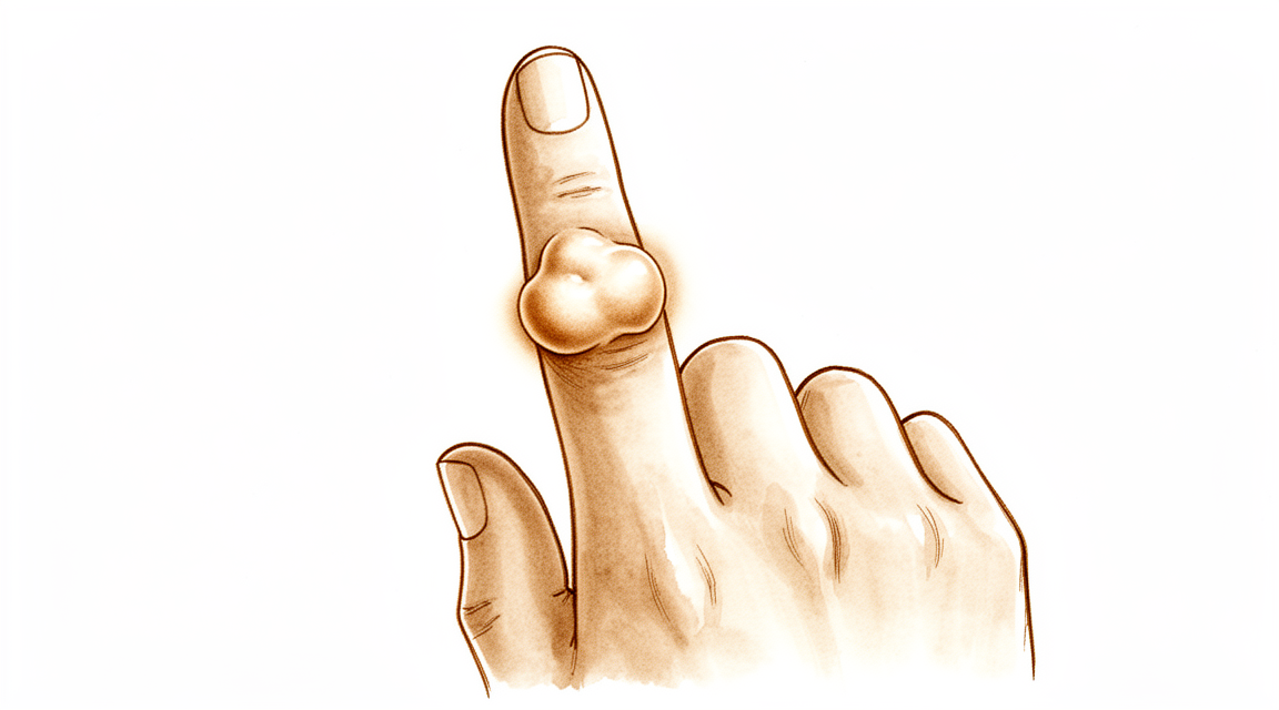
آپ کو کیا محسوس ہوتا ہے
زیادہ تر لوگوں کو کسی انگلی یا انگوٹھے پر ایک گانٹھ نظر آتی ہے جس میں درد نہیں ہوتا۔ یہ عام طور پر سخت ہوتی ہے، اور آہستہ آہستہ بڑھتی ہے۔ کچھ گانٹھیں بالکل کسی جوڑ کے ساتھ ہوتی ہیں، اکثر اس جوڑ کے ساتھ جو انگلی کے سرے کے سب سے قریب ہے، اور یہ انگلی کے اوپر، پہلو میں یا ہتھیلی والی طرف ظاہر ہو سکتی ہیں۔ سب سے عام جگہ شہادت کی انگلی ہے۔
تقریباً ہر کیس میں خود گانٹھ میں درد نہیں ہوتا۔ جب تکلیف ہو تو عموماً اس وجہ سے ہوتی ہے کہ بڑھتی ہوئی گانٹھ آس پاس کے حصوں پر دباؤ ڈالتی ہے۔ اگر گانٹھ کسی عصب پر دباؤ ڈالے تو تھوڑے سے لوگوں کو انگلی میں جھنجھناہٹ یا سُن پن محسوس ہوتا ہے۔ کچھ لوگوں کو لگتا ہے کہ حرکت کرتے وقت انگلی اٹکتی ہے یا اس میں کلک ہوتا ہے، کچھ کچھ ٹریگر فنگر (trigger finger، انگلی کا اٹک جانا) کی طرح، جس میں انگلی ایک لمحے کے لیے اٹکتی ہے اور پھر جھٹکے سے آزاد ہو جاتی ہے۔ اگر گانٹھ کسی جوڑ کے قریب ہو تو اس انگلی کو پوری طرح موڑنا مشکل ہو سکتا ہے کیونکہ گانٹھ راستے میں آ جاتی ہے۔
روزمرہ کے وہ کام جن میں وہ انگلی استعمال ہوتی ہے، مشکل اور بے ڈھنگے ہو سکتے ہیں۔ اگر گانٹھ عین اس جگہ ہو جہاں آپ کی گرفت پڑتی ہے تو چھوٹے سکے اٹھانا، بٹن لگانا، قلم پکڑنا یا مرتبان کا ڈھکن پکڑنا اناڑی پن سے ہوتا محسوس ہو سکتا ہے۔ گانٹھ دستانوں یا جیبوں میں بھی اٹک سکتی ہے۔
اکثر گانٹھ اس سے بہت پہلے سے موجود ہوتی ہے کہ وہ آپ کو اتنا پریشان کرے کہ آپ اس کے بارے میں پوچھیں۔ بہت سے لوگ مہینوں یا برسوں انتظار کرتے ہیں اور اسے آہستہ آہستہ بدلتے دیکھتے رہتے ہیں۔
اس مسئلے کے بارے میں چند باتیں جاننا مفید ہے۔ یہ بے ضرر (benign) ہے، یعنی یہ جسم کے دوسرے حصوں میں نہیں پھیلتا۔ یہ مردوں کی نسبت عورتوں میں تھوڑا زیادہ عام ہے، اور عام طور پر 32 سے 51 سال کی عمر کے درمیان ظاہر ہوتا ہے۔ زیادہ تر لوگوں کو ایک ہی گانٹھ ہوتی ہے، اگرچہ کچھ لوگوں کو مختلف جگہوں پر ایک سے زیادہ گانٹھیں ہوتی ہیں۔
اس بات پر نظر رکھیں کہ کہیں انگلی یا ہاتھ گرم، سرخ، سوجا ہوا اور دردناک نہ ہو جائے، خاص طور پر بخار کے ساتھ۔ اگر ایسا ہو تو اسی دن ہسپتال کی ایمرجنسی جائیں۔ اگر گانٹھ بڑھتی رہے، اس میں درد ہونے لگے، یا انگلی ٹھیک سے کام نہ کر رہی ہو تو اپنے فیملی ڈاکٹر (GP) کو دکھائیں یا کسی ماہر ڈاکٹر (specialist) سے معائنے کی درخواست کریں۔
دراصل کیا ہو رہا ہے
آپ کے ٹینڈن (tendon) وہ مضبوط ڈوریاں ہیں جو پٹھے کو ہڈی سے جوڑتی ہیں اور آپ کی انگلیوں کو مڑنے دیتی ہیں۔ ہر ٹینڈن ایک پتلی آستین کے اندر چلتا ہے جسے ٹینڈن کا غلاف (tendon sheath) کہتے ہیں، اور یہ کچھ کچھ رسی کے گرد لگے گسکٹ (gasket) کی طرح کام کرتا ہے، یعنی ڈوری کو چکنا رکھتا ہے تاکہ انگلی کے حرکت کرتے وقت یہ پھسل سکے۔
اس مسئلے میں اس آستین کے خلیے ضرورت سے زیادہ بڑھنے لگتے ہیں۔ اس کا نتیجہ نرم ٹشو کی ایک گانٹھ ہوتی ہے جو ٹینڈن کے اوپر یا اس کے ساتھ ہوتی ہے، اکثر کسی جوڑ کے قریب۔ یہ بے ضرر ہے، یعنی یہ پھیلتی نہیں، اور آہستہ آہستہ بڑھتی ہے۔ زیادہ تر گانٹھیں چھوٹی ہوتی ہیں، اوسطاً تقریباً 1.35 سینٹی میٹر، اگرچہ کچھ اس سے بڑی ہو جاتی ہیں۔
گانٹھ چند چیزوں کے ایک ساتھ جمع ہونے سے بنتی ہے: سوزش والے خلیے، جائنٹ سیل (giant cells)، جو دراصل ایسے خلیے ہیں جن میں کئی مرکزے ہوتے ہیں، اور ایک گہرے رنگ کا مادہ جسے ہیموسائیڈرن (haemosiderin) کہتے ہیں، یعنی وہی چیز جو پرانے نیل کو رنگ دیتی ہے۔ اسی رنگ دار مادے کی وجہ سے اگر سرجری کے دوران یہ گانٹھیں دیکھی جائیں تو بھوری سی نظر آ سکتی ہیں۔
اوپر والے حصے میں آپ نے جو علامات محسوس کیں وہ اس لیے ہوتی ہیں کہ گانٹھ راستے میں آ جاتی ہے۔ بڑھنے کے ساتھ یہ ٹینڈن، جوڑ یا کسی عصب پر دباؤ ڈال سکتی ہے، جس سے اٹکنے، اکڑاؤ اور کبھی کبھار کی جھنجھناہٹ کی وضاحت ہوتی ہے۔ زیادہ تر اوقات اس میں بالکل درد نہیں ہوتا، اور ہر 100 میں سے صرف تقریباً 16 لوگ درد کی شکایت کرتے ہیں۔
چند باتیں جاننا مفید ہے۔ زیادہ تر لوگوں کو ایک ہی گانٹھ ہوتی ہے، لیکن ہر 100 میں سے تقریباً 21 لوگوں کو دو یا زیادہ الگ الگ گانٹھیں ہوتی ہیں۔ گانٹھ عام طور پر کسی جوڑ کے ساتھ ہوتی ہے، اور انگلی کے تقریباً ایک تہائی کیسوں میں یہ اس جوڑ پر ہوتی ہے جو انگلی کے سرے کے سب سے قریب ہے۔ بہت شاذ و نادر، گانٹھ دب کر خود ہڈی کے اندر گھس سکتی ہے، جو ایکسرے پر نظر آتا ہے۔
اسی مسئلے کی ایک زیادہ نایاب، زیادہ پھیلی ہوئی (diffuse) صورت بھی ہے جو ایک صاف ستھری گانٹھ بنانے کے بجائے ٹینڈن کے ساتھ ساتھ پتلی شاخوں کی صورت میں پھیلتی ہے۔ یہ صورت انگلی میں بہت کم پائی جاتی ہے اور اس کا رویہ مختلف ہوتا ہے، اسی لیے کسی بھی منصوبہ بندی سے پہلے آپ کے سرجن یہ واضح طور پر جاننا چاہیں گے کہ یہ بالکل کس قسم کی گانٹھ ہے۔
ہم اس کے لیے کیا کر سکتے ہیں
ڈاکٹر کیرن ہِرپرا، جو Mater Private Hospital Rockhampton میں بازو اور ہاتھ کے سرجن (upper-limb surgeon) ہیں، سب سے کم چیر پھاڑ والے ان طریقوں سے آغاز کرتے ہیں جو آپ کے مسئلے کے لیے مناسب ہوں۔ مریض عام طور پر اپنے فیملی ڈاکٹر (GP) کے ذریعے ہمارے کلینک بھیجے جاتے ہیں؛ اگر کسی فزیوتھراپسٹ نے آپ کو ہم سے ملنے کا مشورہ دیا ہے، تب بھی میڈیکیئر (Medicare) کی رقم کی واپسی کا حق دار ہونے کے لیے آپ کو اپنے فیملی ڈاکٹر سے ریفرل (referral) لینا ہوگا۔ کلینک میں ہم آپ سے مسئلے کی تفصیل پوچھتے ہیں، آپ کے ہاتھ کا معائنہ کرتے ہیں، اور جہاں ضرورت ہو وہاں تصویری ٹیسٹ (imaging) کا انتظام کرتے ہیں تاکہ ٹھیک ٹھیک معلوم ہو سکے کہ یہ کس قسم کی گانٹھ ہے۔
چونکہ یہ گانٹھ بے ضرر ہے اور آہستہ آہستہ بڑھتی ہے، اس لیے کچھ نہ کرنا بھی ایک حقیقی انتخاب ہے۔ ایک چھوٹی، بغیر درد کی گانٹھ کے لیے بس اس پر نظر رکھنا صحیح انتخاب ہو سکتا ہے۔ اگر گانٹھ رکاوٹ بن رہی ہو تو ہینڈ تھراپی (ہاتھ کی بحالی کا علاج) اکڑاؤ اور اٹکنے میں مدد دے سکتی ہے۔ تھراپی کا مقصد انگلی کو حرکت میں رکھنا اور روزمرہ کے کاموں کو آسان بنانا ہے۔ سرجری کے بارے میں سوچنے سے پہلے ہم عام طور پر اسے پوری طرح آزمانے کا مشورہ دیتے ہیں۔
اس مسئلے کی کوئی ایسی دوا نہیں جو اسے سکیڑ دے، اس لیے درد کش گولیاں اور سوزش کم کرنے والی دوائیں صرف اس تکلیف کو کم کرتی ہیں جو گانٹھ کے آس پاس کے ٹشو پر دباؤ ڈالنے سے ہوتی ہے۔ یہ گانٹھ کو چھوٹا نہیں کرتیں۔
جب گانٹھ بڑی ہو، دردناک ہو، یا انگلی کو ٹھیک سے کام کرنے سے روک رہی ہو تو عام علاج سرجری ہے۔ مقصد پوری گانٹھ کو ٹینڈن کی آستین پر اس کے جڑنے کی جگہ سمیت نکالنا ہے، کیونکہ جڑنے کی اس جگہ کو پیچھے چھوڑ دینا ہی گانٹھ کو دوبارہ بننے کا موقع دیتا ہے۔ ہم آپریشن سے پہلے احتیاط سے منصوبہ بندی کرتے ہیں، آپریشن کے دوران عدسے سے بڑا کر کے دیکھتے ہیں (magnification)، اور گانٹھ کو مکمل طور پر نکالتے ہیں۔ اس مسئلے میں سب سے بڑا خطرہ گانٹھ کا دوبارہ بن جانا ہے، اور ایسا عام طور پر نکالنے کے 36 ماہ کے اندر ہوتا ہے۔ 605 لوگوں کے ایک گروپ میں، جن کی انگلی کی گانٹھ نکالی گئی، 14.8 فیصد میں یہ دوبارہ بن گئی۔ اسے دوسرے طریقے سے یوں کہا جا سکتا ہے: زیادہ تر لوگوں میں، یعنی ہر 100 میں سے تقریباً 85 میں، یہ دوبارہ نہیں بنتی۔ اگر یہ دوبارہ بن جائے تو دوبارہ بڑھنے سے روکنے کے اضافی اقدامات کے ساتھ اسے دوبارہ نکالنا پھر بھی مسئلہ حل کر سکتا ہے۔
کیا توقع رکھیں
اس گانٹھ کا مستقبل ڈرامائی نہیں بلکہ یکساں رہتا ہے۔ یہ بے ضرر ہے، اس لیے آپ کے جسم میں کہیں اور نہیں پھیلتی۔ اگر اسے ویسے ہی چھوڑ دیا جائے تو ایک چھوٹی، بغیر درد کی گانٹھ عموماً آہستہ آہستہ بڑھتی رہتی ہے، یا بس ویسی کی ویسی رہتی ہے۔ یہ شاذ و نادر ہی خود سے ختم ہوتی ہے، اور یہ کسی خطرناک چیز میں نہیں بدلتی۔
وقت کے ساتھ یہ مسئلہ بنیادی طور پر یہ کرتا ہے کہ نکالنے کے بعد دوبارہ بن جاتا ہے۔ اس گانٹھ کا بنیادی خطرہ دوبارہ بن جانا ہے، اور ایسا عام طور پر نکالنے کے 36 ماہ کے اندر ہوتا ہے۔ کچھ گانٹھوں کے دوبارہ بننے کا امکان دوسروں سے زیادہ ہوتا ہے: وہ جو دو یا زیادہ الگ الگ ٹکڑوں پر مشتمل ہوں جو آپس میں جڑے ہوئے نہ ہوں، اور وہ جو خود ٹینڈن میں یا جوڑ کی اندرونی جھلی میں بڑھ گئی ہوں۔ جب پوری گانٹھ کو اس کے جڑنے کی جگہ سمیت نکال دیا جائے تو یہ خطرہ بہت کم ہو جاتا ہے۔
علاج کے بعد ہاتھ کا کام عموماً اچھا رہتا ہے۔ جن لوگوں کی انگلی سے ایسی گانٹھ نکالی گئی ہو، وہ بازو اور ہاتھ کے کام کو جانچنے والے ایک معیاری پیمانے پر اوسطاً معمول کے کام کا 92 فیصد اسکور کرتے ہیں۔ رپورٹ کیے گئے کیسوں میں لوگوں کی انگلی کی پوری حرکت برقرار رہی، اور دو سال کے معائنے تک ایکسرے پر انگلی کی ہڈی کی شکل معمول پر آ گئی۔
اگر گانٹھ زیادہ نایاب، زیادہ پھیلنے والی قسم کی ہو جو ٹینڈن کے گرد شاخوں کی صورت میں لپٹ جاتی ہے، تو اسے مکمل طور پر نکالنا زیادہ مشکل ہوتا ہے اور اس کے دوبارہ بننے کا امکان زیادہ ہوتا ہے۔ ایسے کیسوں میں اس جگہ پر شعاعوں سے کیا جانے والا علاج (radiation therapy) آپ کے ہاتھ کو کام کرتا رکھتے ہوئے گانٹھ پر قابو پانے میں مدد دے سکتا ہے۔
اگر آپ ایک چھوٹی، بغیر درد کی گانٹھ پر بس نظر رکھنے کا انتخاب کریں تو حقیقت پسندانہ تصویر یہ ہے کہ گانٹھ آہستہ آہستہ بڑھے گی یا ویسی ہی رہے گی، اور ہو سکتا ہے وہ رفتہ رفتہ انگلی کو موڑنا یا اسے چھوٹے کاموں کے لیے استعمال کرنا زیادہ مشکل بنا دے۔ اگر اس میں درد ہونے لگے، یہ بڑھتی رہے، یا انگلی ٹھیک سے کام کرنا چھوڑ دے، تو یہی وہ مرحلہ ہے جہاں علاج عموماً فائدہ مند ثابت ہوتا ہے۔
ڈاکٹر کو کب دکھائیں
اس طرح کی زیادہ تر گانٹھیں فوری نوعیت کی نہیں ہوتیں، اور علاج میں کبھی جلدی کی ضرورت نہیں ہوتی۔ اگر گانٹھ ٹھیک نہ ہو رہی ہو، ہفتوں میں بڑھ رہی ہو، اس میں درد ہونے لگے، یا یہ آپ کو انگلی یا ہاتھ معمول کے مطابق استعمال کرنے سے روکے تو اپنے فیملی ڈاکٹر کو دکھائیں۔ اگر حرکت کرتے وقت انگلی اٹکتی ہو یا اس میں کلک ہوتا ہو، اگر آپ کو جھنجھناہٹ یا سُن پن محسوس ہو، یا اگر گانٹھ کے راستے میں آنے کی وجہ سے انگلی کو پوری طرح موڑنا مشکل ہو گیا ہو تو کسی ماہر ڈاکٹر سے معائنے کی درخواست کریں۔ یہ وہ باتیں ہیں جن کا معائنہ دیر کے بجائے جلد کروا لینا بہتر ہے، کیونکہ جو گانٹھ ٹینڈن یا جوڑ کی اندرونی جھلی میں بڑھ گئی ہو اسے مکمل طور پر نکالنا زیادہ مشکل ہوتا ہے اور اس کے دوبارہ بننے کا امکان زیادہ ہوتا ہے۔
علامات کا ایک مجموعہ ایسا ہے جس کے لیے اسی دن علاج ضروری ہے۔ اگر آپ کی انگلی یا ہاتھ گرم، سرخ، سوجا ہوا اور دردناک ہو جائے، خاص طور پر بخار کے ساتھ، تو ہسپتال کی ایمرجنسی جائیں۔ اس کے لیے پہلے فیملی ڈاکٹر کے ریفرل کی ضرورت نہیں۔ اگر دفتری اوقات کے بعد یا ہفتے کے آخری دنوں (ویک اینڈ) میں کلینک سے رابطہ نہ ہو سکے تو اپنے قریب ترین ہسپتال کی ایمرجنسی جائیں۔
مزید تفصیل
Advanced reading: the deeper science (optional)
یہ حصہ اس سے آگے جاتا ہے جتنا آپ کو اپنے علاج کے فیصلوں کے لیے جاننے کی ضرورت ہے۔ ٹینڈن کے غلاف کے جائنٹ سیل ٹیومر کے بارے میں مزید پڑھنا اس لیے فائدہ مند ہے کہ اس کا بنیادی مسئلہ دوبارہ بن جانا ہے، اور شواہد بتاتے ہیں کہ دوبارہ بننے کا تعلق اسے نکالنے کے طریقے سے زیادہ ہر ٹیومر کی اپنی حیاتیاتی فطرت سے ہے۔
دوبارہ بن جانا ٹیومر کی خاصیت ہے، صرف سرجری کی نہیں
گانٹھ کے واپس آنے کی فطری وضاحت یہ لگتی ہے کہ اس کا کچھ حصہ پیچھے رہ گیا تھا۔ انگلیوں کے 605 کیسوں کے ایک منظم جائزے (systematic review) کا نتیجہ کچھ اور تھا: دوبارہ بننے میں ٹیومر کی اندرونی حیاتیاتی فطرت اس کی جگہ یا آس پاس کے ٹشو میں گھسنے کی صلاحیت سے زیادہ بنیادی کردار ادا کرتی نظر آتی ہے، اور مصنفین نے بڑی آئندہ کی تحقیقات (prospective studies) کا مطالبہ کیا تاکہ پہچانا جا سکے کہ کون سے ٹیومر دوبارہ بننے کا رجحان رکھتے ہیں [1]۔
آپریشن سے پہلے یہ بات جان لینا واقعی مفید ہے۔ اچھی طرح کیے گئے آپریشن کے بعد گانٹھ کا دوبارہ بننا اس ٹیومر کا ایک تسلیم شدہ رویہ ہے، اس بات کا ثبوت نہیں کہ کچھ غلط ہوا۔
علاج اختیاری ہے، اور کچھ نہ کرنا بھی ایک حقیقی انتخاب ہے
جب آپریشن کی منصوبہ بندی ہو رہی ہو تو یہ بات نظر سے اوجھل ہو جانا آسان ہے۔ پہلے درجے کے علاج کا اصول مکمل طور پر نکال دینا ہے، لیکن علاج کبھی فوری نوعیت کا نہیں ہوتا، اور اس کی ضرورت کو علامات، بڑھوتری، جگہ اور آپ کے اپنے حالات کے مقابلے میں تولنا چاہیے [4]۔
ایک چھوٹی، بغیر درد کی، آہستہ بڑھنے والی گلٹی کے لیے اس پر نظر رکھنا ایک جائز انتخاب ہے۔ ٹیومر بے ضرر ہے اور پھیلتا نہیں، اس لیے آپریشن کی دلیل ہاتھ کے کام، سائز اور تکلیف کے بارے میں ہے — خطرے کے بارے میں نہیں۔
لیکن سرجری کے دو عوامل واقعی اہم ہیں
حیاتیاتی فطرت پوری کہانی نہیں ہے۔ محدود قسم کے ٹینوسائنوویئل جائنٹ سیل ٹیومر (tenosynovial giant cell tumour) والے 941 مریضوں میں، نکالنے کے بعد دوبارہ بننے سے جڑے عوامل ٹیومر کا بڑا سائز اور ابتدائی علاج آرتھروسکوپی (دوربین سے جوڑ کے اندر دیکھ کر علاج) سے کرنا تھے۔ پیچیدگیوں کی نسبتاً کم شرح اور جوڑ کے کام کے اچھے نتائج کو دیکھتے ہوئے، مصنفین مشورہ دیتے ہیں کہ زیادہ خطرے والے کیسوں میں دوبارہ بننے کو کم کرنے کے لیے جہاں ممکن ہو کھلا آپریشن کر کے مکمل طور پر نکالا جائے [2]۔
اس کے برعکس یہ بات ہے کہ 1,448 مریضوں کے ایک جائزے میں محدود قسم کے لیے چار جوڑوں میں دوربین کے ذریعے گانٹھ نکالنا مؤثر دکھایا گیا ہے، جبکہ پھیلی ہوئی (diffuse) قسم میں دوربین کے ذریعے جوڑ کی اندرونی جھلی نکالنے (arthroscopic synovectomy) کی افادیت صرف گھٹنے میں دکھائی گئی ہے [3]۔
ان دونوں کو ملا کر دیکھیں تو: ایک چھوٹی، محدود، واضح حد والی گانٹھ کے لیے دونوں میں سے کوئی بھی طریقہ کام کر سکتا ہے۔ جیسے جیسے سائز بڑھتا ہے، اور پھیلی ہوئی قسم کے لیے، مکمل کھلے آپریشن کی حمایت زیادہ ہے۔ اس کی وجہ میکانکی ہے، یہ ٹیومر ٹینڈن، عصب اور جوڑ کے گرد پتوں جیسی شاخوں کی صورت میں پھیلتا ہے، اور جو حصے سب سے آسانی سے رہ جاتے ہیں وہ وہ ہیں جو ایسے حصوں کے پیچھے چھپے ہوتے ہیں جنہیں اٹھا کر براہِ راست دیکھنا ضروری ہوتا ہے۔
نام کیوں اہم ہے
یہ مسئلہ اب ٹینوسائنوویئل جائنٹ سیل ٹیومر کے تحت آتا ہے، جس میں ہاتھ کی محدود قسم اور جوڑ کے اندر کی پھیلی ہوئی قسم دونوں شامل ہیں، جسے پہلے پگمنٹڈ ولونوڈیولر سائنوویٹس (pigmented villonodular synovitis) کہا جاتا تھا [4]۔ یہ ایک ہی بیماری ہے جو مختلف جگہوں پر اور بڑھنے کے مختلف انداز میں ہوتی ہے۔
اگر آپ اس موضوع پر پڑھیں تو یہ جاننا مفید ہے، کیونکہ تلاش کرنے پر گھٹنوں اور کولہوں کے بارے میں ایسا مواد سامنے آئے گا جو آپ ہی کے مسئلے کو اس کی پھیلی ہوئی صورت میں بیان کر رہا ہے، اور پھیلی ہوئی بیماری کے لیے بتائی جانے والی دوبارہ بننے کی شرحیں انگلی کی محدود گانٹھ کی شرحوں سے کافی زیادہ ہیں۔ گھٹنے کے اعداد و شمار کو انگلی کی گانٹھ پر لاگو کرنا خطرے کو بڑھا چڑھا کر دکھاتا ہے۔
ریڈیو تھراپی کی جگہ کہاں ہے
پھیلی ہوئی بیماری جو دوبارہ بن گئی ہو یا پوری طرح نکالی نہ جا سکتی ہو، اس کے لیے بعض اوقات اضافی ریڈیو تھراپی (adjuvant radiotherapy) پر غور کیا جاتا ہے۔ ایک میٹا اینالیسس (meta-analysis) میں پایا گیا کہ کھلے آپریشن سے جوڑ کی اندرونی جھلی نکالنا (open synovectomy)، یا اس کے ساتھ آپریشن کے آس پاس کے وقت میں ریڈیو تھراپی، پھیلی ہوئی پگمنٹڈ ولونوڈیولر سائنوویٹس میں دوبارہ بننے کی کم شرح سے جڑی تھی، اگرچہ اس کی تصدیق کے لیے بڑی، طویل مدتی آئندہ کی تحقیقات کا مطالبہ کیا گیا [5]۔
انگلی کے محدود ٹیومر کے لیے، جو ہاتھ کے کیسوں کی بھاری اکثریت ہے، یہ سوال پیدا نہیں ہوتا۔ اس کا تعلق پھیلی ہوئی، بار بار ہونے والی، جوڑ میں موجود قسم سے ہے، اور اس کا ذکر یہاں صرف اس لیے ہے کہ اس مسئلے کے نام پر تلاش کرنے سے یہ سامنے آئے گا۔
یہ کیا نہیں ہے
نام کے باوجود یہ ایک بے ضرر ٹیومر ہے۔ یہ جسم میں کہیں اور نہیں پھیلتا۔ یہاں لفظ "ٹیومر" اس سے زیادہ وزن رکھتا ہے جتنا اسے رکھنا چاہیے، اور دوبارہ بننے کے بارے میں فکر بار بار کی مقامی سرجری، اکڑاؤ اور اعصاب کے قریب ہونے کے بارے میں ہے، کینسر کے بارے میں نہیں۔
حوالہ جات
[1] Fotiadis E, Papadopoulos A, Svarnas T, Akritopoulos P, Sachinis NP, Chalidis BE. Giant cell tumour of tendon sheath of the digits. A systematic review. Hand (N Y). 2011;6(3):244-9. https://doi.org/10.1007/s11552-011-9341-9
[2] Mastboom M, Staals E, Verspoor F, Rueten-Budde A, Stacchiotti S, Palmerini E, et al. Surgical treatment of localized-type tenosynovial giant cell tumors of large joints: a study based on a multicenter-pooled database of 31 international sarcoma centers. J Bone Joint Surg Am. 2019;101(14):1309-18. https://doi.org/10.2106/JBJS.18.01147
[3] Noailles T, Brulefert K, Briand S, Longis P, Andrieu K, Chalopin A, et al. Giant cell tumor of tendon sheath: open surgery or arthroscopic synovectomy? A systematic review of the literature. Orthop Traumatol Surg Res. 2017;103(5):809-14. https://doi.org/10.1016/j.otsr.2017.03.016
[4] Gouin F, Noailles T. Localized and diffuse forms of tenosynovial giant cell tumor (formerly giant cell tumor of the tendon sheath and pigmented villonodular synovitis). Orthop Traumatol Surg Res. 2017;103(1):S91-S97. https://doi.org/10.1016/j.otsr.2016.11.002
[5] Mollon B, Lee A, Busse JW, Griffin AM, Ferguson PC, Wunder JS, et al. The effect of surgical synovectomy and radiotherapy on the rate of recurrence of pigmented villonodular synovitis of the knee. Bone Joint J. 2015;97-B(4):550-7. https://doi.org/10.1302/0301-620X.97B4.34907
Evidence & references
This is the clinical evidence summary written for health professionals. It is technical, and it lists the research this page was built from. You do not need to read it to understand your treatment or to make a decision about it.
Overview
- The diagnosis and treatment of suspected hand neoplasms should proceed in an orderly fashion similar to accepted methods applied to tumors in other locations of the extremities [1].
- Giant cell tumors of the tendon sheaths in the hand are benign lesions where recurrence is the primary risk [3, 4].
- Recurrence of giant cell tumors of the tendon sheaths typically occurs within 36 months of excision [3].
- The fundamental consideration in the management of hand tumours is to reconcile adequate tumour clearance with the maintenance of function and appearance [20].
- Complete surgical resection remains the treatment of choice for most patients with tenosynovial giant cell tumors [15].
- Diffuse tenosynovial giant cell tumor disease presents challenges due to high recurrence rates [15].
- Curettage, cryosurgery, and cementation performed by experienced surgeons appears to be a safe, effective, and reliable method for treating selected giant cell tumors of the hand [5].
- In cases of infiltrative giant cell tumor of the tendon sheath, radiation therapy may provide local tumor control with preservation of hand function [12].
- En bloc resection of the tumour and reconstruction with a fibular graft where necessary should be considered as the treatment of choice in giant cell tumour of the hand [10].
- En bloc resection of the tumour and reconstruction with an autograft should be considered in the treatment of recurrent giant cell tumour of the hand [8].
- En bloc resection and matched nonvascularized toe phalangeal transfer for Campanacci Grade 2 or 3 giant cell tumor of the phalanges resulted in a functional tumor-free digit with a low complication rate and no recurrences [35].
- Well-designed studies combining the recurrence rates of several hand surgery centers implementing a standardized treatment are needed to better demonstrate the associated risk factors for recurrence [28].
Anatomy & Pathophysiology
Clinical Presentation and Demographics
- Giant cell tumour of tendon sheath (GCTTS) is the second most common benign proliferative tumour in the upper extremities after ganglion cysts [102].
- GCTTS are usually slow-growing, painless, benign soft tissue tumours [102].
- GCTTS are most commonly found in the fingers and among women in their fourth and fifth decades [102].
- The male-to-female ratio for GCTTS of the digits is 1:1.47 [23].
- The mean age range for GCTTS of the digits is 32 to 51 years [23].
- Pain was reported in 15.7% of GCTTS cases and sensory disturbances in 4.57% of cases [23].
- A definite history of trauma was recorded in 5% of GCTTS lesions [23].
- The most frequent tumour location for GCTTS of the digits is the index finger (29.7%) [23].
- In a series of 64 cases, the most frequent location for GCTTS was the long finger (23.5%), followed by the thumb, index finger, and hand (20.3% each) [49].
- Patients with GCTTS arising in the fingers complained of a painless mass in almost every instance [24].
- The duration of symptoms for finger GCTTS ranged from two weeks to fifteen years, with an average duration of thirty-eight months [24].
- GCTTS are usually firm, lobulated, and non-tender masses [24].
- As the tumour grows, patients may present with swelling, pain, and limitation of movement [102].
Anatomical Distribution and Morphology
- Fifty-three xanthomas (GCTTS) arose adjacent to joints in the fingers, whereas twenty-nine bore no relation to joints [24].
- Thirty-one of ninety-one finger GCTTS occurred at the distal joints [24].
- Of the thirty-one distal joint GCTTS, eighteen were on the dorsal aspect and thirteen were distributed evenly on the radial, ulnar, and volar aspects [24].
- Type I GCTTS (single lesions) were more frequently detected (78.7%) than Type II tumours (two or more distinct tumours that were not joined together) (21.3%) [23].
- Type 1 GCTTS includes a nodular or multinodular lesion surrounded by a capsule, while Type 2 describes tumours with no connective tissue membrane and satellite, diffuse, or multicentric nodules [102].
- The average size of GCTTS was 1.35 cm, with a range of 0.3 cm to 5 cm [49].
- Bone erosion was found in 3 patients (4.7%) in a series of 64 GCTTS cases [49].
- Tendon involvement with flexors/extensors ratio of 4:3 was found in 7 cases (10.9%) in a series of 64 GCTTS cases [49].
- Involvement of the neurovascular bundle was presented in 7 patients (10.9%) in a series of 64 GCTTS cases [49].
- A case report describes the first patient with three separate GCTTS developing from both flexor and extensor tendons in a single digit [7].
Histology and Pathogenesis
- Microscopically, all GCTTS contained multinucleated giant cells, histiocytes, and haemosiderin deposits [49].
- GCTTS originates from the synovial membrane, tendon sheath, or synovial bursa [102].
- Inflammation resulting from reactive or regenerative hyperplasia is the generally accepted theory of pathogenesis for GCTTS [102].
- Genetic factors have been observed in previous studies regarding GCTTS pathogenesis [102].
- Fibroma of tendon sheath is histologically distinct from giant cell tumor of tendon sheath, a lesion with which it is commonly confused [16].
Diagnostic Imaging
- Radiography can be helpful in evaluating cortical destruction but is not helpful in the definitive diagnosis of GCTTS [102].
- Magnetic resonance imaging (MRI) is the most useful examination for the diagnosis and treatment planning of GCTTS [102].
- Al-Qattan classified GCTTS with MRI into two types according to the risk of recurrence [102].
Classification
- Giant cell tumors of the synovial sheaths in the hand are benign lesions in which recurrence is the primary risk [4].
- Recurrence for giant cell tumors of the tendon sheaths typically occurs within 36 months of excision [3].
- The Al-Qattan classification divides giant cell tumours of tendon sheath into two main types based on whether the entire tumour is surrounded by one pseudocapsule as assessed by the surgeon during surgery [90].
- Type I tumours are defined as those where the entire tumour is surrounded by one pseudocapsule [90].
- Type II tumours are defined as those where the entire tumour is not surrounded by one pseudocapsule [90].
- Type I tumours are sub-classified according to the thickness of the capsule, lobulation of the tumour, the presence of satellite lesions, and the diffuse or multicenteric nature of the tumour [90].
- Type II tumours are sub-classified according to the thickness of the capsule, lobulation of the tumour, the presence of satellite lesions, and the diffuse or multicenteric nature of the tumour [90].
- In a prospective study of 43 consecutive cases, none of the type I tumours (n=30) recurred [90].
- In a prospective study of 43 consecutive cases, recurrence occurred in five out of 13 type II tumours [90].
- Second recurrences were seen with type II B and C tumours, but not type II A tumours [90].
- Type I tumours (single lesions) were more frequently detected (78.7%) than type II tumours (two or more distinct tumours that were not joined together) (21.3%) in a systematic review of 605 patients [23].
- Type II tumours were associated with higher recurrence rates compared to Type I tumours in a systematic review of 605 patients [23].
- Direct involvement of the extensor tendons, flexor tendons, or joint capsule puts patients in a high-risk category with respect to recurrence [43].
- Preoperative diagnosis and meticulous surgical technique were found to be the only predictive factor of recurrence in a series of 14 cases [34].
- The overall recurrence rate in a systematic review of 605 patients was 14.8% [23].
Clinical Presentation
- Patients with fibrous xanthomas arising in the fingers complained of a painless mass in almost every instance [24].
- The duration of symptoms for finger tumors ranged from two weeks to fifteen years, with an average duration of thirty-eight months [24].
- The tumors were usually firm, lobulated, and non-tender [24].
- Fifty-three xanthomas arose adjacent to joints in the fingers, whereas twenty-nine bore no relation to joints [24].
- Thirty-one of the ninety-one tumors in the fingers occurred at the distal joints [24].
- Eighteen of the thirty-one distal joint tumors were on the dorsal aspect of the joint, and thirteen were distributed about evenly on the radial, ulnar, and volar aspects of the joint [24].
- The most frequent tumour location for giant cell tumour of tendon sheath of the digits was the index finger (29.7%) [23].
- Pain was reported in 15.7% of cases and sensory disturbances in 4.57% of cases [23].
- A definite history of trauma was recorded in 5% of lesions [23].
- Type I tumours (single lesions) were more frequently detected (78.7%) than type II tumours (two or more distinct tumours that were not joined together) (21.3%) [23].
- The male-to-female ratio for giant cell tumour of tendon sheath of the digits was 1:1.47 [23].
- The mean age for giant cell tumour of tendon sheath of the digits ranged from 32 to 51 years [23].
- A tendon tumour can present as a trigger finger [18].
- Trigger wrist caused by a giant cell tumour of tendon sheath has been reported [26].
- The trigger phenomenon in trigger wrist caused by a giant cell tumour of tendon sheath probably occurred when the tumour entered and left the distal carpal tunnel [26].
- Fibroma of tendon sheath is a benign soft tissue tumor that has a predilection for the hand [16].
- Fibroma of tendon sheath may be more common than has been previously recognized [16].
- The differential diagnosis of a soft tissue tumor in the digits and hand is usually limited to a ganglion, an inclusion cyst, or a giant cell tumor of tendon sheath (xanthofibroma) [16].
- Fibroma of tendon sheath should be included in the differential diagnosis of a soft tissue tumor in the digits and hand [16].
- A case of fibroma of tendon sheath presented as a 6-year history of a slowly enlarging palmar mass of the left index finger [16].
- Examination of a fibroma of tendon sheath case demonstrated a soft, compressible, lobulated, nontender mass extending from the second web space to the tip of the finger [16].
- Flexion of the digit was limited by the bulge in a case of fibroma of tendon sheath [16].
- The unusual location of a fibroma of the tendon sheath and atypical nature of the triggering phenomenon can lead to it being reported as a rare cause of median nerve compression at the wrist [50].
- Giant cell tumor of tendon sheath and pigmented villonodular synovitis are benign, locally invasive tumors involving synovium, tendon sheaths, and bursae [47].
- Patients with giant cell tumor of tendon sheath or pigmented villonodular synovitis may present with a discrete mass or with joint swelling, pain, or locking or catching [47].
- The knee and digits of the hand are the most common locations of lesions for giant cell tumor of tendon sheath and pigmented villonodular synovitis [47].
- Multifocal lesions of giant cell tumor of tendon sheath and pigmented villonodular synovitis are rare [47].
- The authors report the uniqueness of a case of multicentric giant cell tumor in the upper extremity [9].
- Radiological changes in the form of bony indentation was seen in only 2 cases of giant cell tumor of tendon sheath in a series of 12 patients [34].
- The most common presentation for giant cell tumor of tendon sheath in a series of 12 patients was with a mass over the hand, with a predilection to the thumb [34].
- A 65-year-old man presented with a slowly enlarging mass on the dorsal aspect of the left thumb consistent with a recurrent giant cell tumor of the tendon sheath [33].
Investigations
- The differential diagnosis of a soft tissue tumor in the digits and hand is usually limited to a ganglion, an inclusion cyst, or a giant cell tumor of tendon sheath [16].
- Fibroma of tendon sheath should be included in the differential diagnosis as it may be more common than has been previously recognized [16].
- Fibro-osseous pseudotumor of the digit should be considered in the differential diagnosis of rapidly enlarging digital lesions [64].
- Although MRI findings and location might help in the diagnosis of tenosynovial giant cell tumors, careful assessment is mandatory, especially in unusual locations [129].
Treatment
General Principles
Surgical Excision
- Complete surgical resection remains the treatment of choice for most patients with tenosynovial giant cell tumors, though diffuse disease presents challenges due to high recurrence rates [15].
- En bloc resection of metacarpal giant cell tumors with reconstruction by iliac graft appears to be an effective treatment [14].
- En bloc resection and matched nonvascularized toe phalangeal transfer resulted in a functional tumor-free digit with a low complication rate and no recurrences for Campanacci Grade 2 or 3 giant cell tumors of the phalanges [35].
- Total surgical excision ensuring removal of the attachment site (flexor sheath/palmar fascia) resulted in no local recurrences at a mean follow-up of 3.2 years for fibroma of tendon sheath [11].
- Local excision of the tumour appears to be a satisfactory method of treatment for Nora’s lesion [109].
- Preoperative planning aided by a tissue diagnosis with fine needle aspiration cytology, wide surgical exposure, and meticulous dissection with help of magnification are imperative for a successful outcome in giant cell tumor of tendon sheath [34].
- In all cases of giant cell tumor of tendon sheath, magnifying loupe or operating microscope was used during surgical excision [49].
- In cases of infiltrative giant cell tumor of tendon sheath, radiation therapy may provide local tumor control with preservation of hand function [12].
Recurrence and Adjuvant Therapy
- Giant cell tumors of the tendon sheaths in the hand are benign lesions where recurrence is the primary risk, typically occurring within 36 months of excision [3].
- In a study of 64 cases of giant cell tumor of tendon sheath, the recurrence rate was 4.7% (n=3) [49].
- In a systematic review of 605 patients with giant cell tumor of tendon sheath of the digits, 14.8% of patients had tumour recurrence [23].
- Repeated curettage with adjuvants eventually resulted in the cure for all patients and is therefore a reasonable treatment for both primary and recurrent giant cell tumor of the small bones of the hands and feet [105].
- Intralesional excision with cautery and methylmethacrylate provides a reliable method of treatment of giant cell tumors with good long-term functional results [71].
- Intralesional excision with local adjuvant therapy is recommended for the treatment of giant cell tumor of bone because it results in a good functional outcome compared to extralesional excision [98].
- Intralesional excision remains a viable, and likely the standard, mode of treatment for most giant cell tumors of the distal radius unless there is extensive bone loss [29].
- Based on data obtained from the number of studies available, intralesional excision appears to be more appropriate for the treatment of local lesions (eg, Grades 1 and 2) than Grade 3 giant cell tumors of the distal radius [52].
- Well-designed studies combining the recurrence rates of several hand surgery centers implementing a standardized treatment are needed to better demonstrate the associated risk factors for recurrence in giant cell tumor of tendon sheath [28].
Reconstruction and Resection Options
- Both curettage and resection/amputation are acceptable treatment options for the rare condition of giant cell tumour of bone in the hand, with a need to individualize treatment decisions based on the site and extent of disease to minimize treatment morbidity while maximizing disease control [6].
- Aggressive and malignant bone tumors of the second through fifth metacarpals generally require en bloc bone excision [17].
- Ray excision is usually best for lesions of the second and fifth metacarpals [17].
- If bone graft has been used for reconstruction, carpometacarpal arthrodesis and MCP ligament reconstruction or silicone arthroplasty may be practical [17].
- Whatever level is chosen for reconstruction, it is essential that an adequate, safe margin of normal tissue first be excised en bloc with the tumor [17].
- Aggressive tumors of the second through fifth metacarpals that have invaded soft tissue or sustained pathologic fractures often require excision of not only the involved ray but also the contiguous ray or rays radial and ulnar to the involved digit [17].
- If a malignant tumor has broken into the midpalm and extends across the metacarpals, removal of all digital rays may be needed to gain an adequate soft tissue margin [17].
- Retention of a sensate and relatively mobile thumb may be considerably more esthetic and functionally satisfactory than a forearm- or wrist-level amputation [17].
- If a tumor extends proximally from the metacarpal level, a more proximal level of hand, wrist, or forearm amputation is required for safe tumor management [17].
- Malignant soft tissue tumors in the palm or carpal tunnel often require at least partial hand amputation [17].
- If treated by only local or limited excision, the chance of recurrence is extremely high particularly in the setting of a positive margin for malignant soft tissue tumors in the palm or carpal tunnel [17].
- Below-elbow amputation is necessary to treat larger tumors of the palm or carpal tunnel [17].
- Wide en bloc excision of soft tissue sarcomas with negative margins is required to achieve local control of the lesion [17].
- At a minimum, aggressive soft tissue tumors, such as bone tumors, require ray resection or removal of multiple rays [17].
- Central palmar lesions more likely require sacrifice of three rays; those on the border are more likely than those in the center to be salvageable by removing just two rays [17].
- In the presence of proximal, broader, and larger lesions, all four digits or the entire hand may have to be sacrificed to save the patient [17].
- The use of a massive biocompatible bipolar unconstrained prosthesis is a viable treatment option for distal radius reconstruction after en-bloc resection of a giant cell tumour, offering rapid functional improvement without donor-site morbidity [68].
- Reconstruction after wide excision by nonvascularized fibular graft is a viable alternative for giant cell tumors of the lower end of radius though it is a challenging procedure and may be accompanied by major complications [111].
- En bloc resection is recommended for aggressive giant cell tumours (GCT) of the bone involving the distal end of the radius to minimize the risk of recurrence [91].
- The distal ulna may be widely resected with or without stabilization of the residual ulnar stump, yielding satisfactory local disease control and functional outcome [48].
- This is a simple and effective modality of reconstruction after resection of distal radial tumors using centralization of the ulna [30].
Complications
- Recurrence is the primary risk associated with giant cell tumors of the tendon sheaths in the hand [3].
- The overall recurrence rate for giant cell tumour of tendon sheath of the digits is 14.8% [23].
- Type II tumours (two or more distinct tumours that were not joined together) are associated with a higher risk of recurrence compared to Type I tumours [23].
- Direct involvement of the extensor tendons, flexor tendons, or joint capsule places patients in a high-risk category for recurrence [43].
- Surgical treatment of pigmented villonodular synovitis led to good functional results with an average Enneking score of 92% of normal limb function [32].
- A tendon tumour arising from the superficialis tendon can present as a trigger finger [18].
- A giant cell tumour of the tendon sheath can cause a trigger wrist phenomenon when the tumour enters and leaves the distal carpal tunnel [26].
- A fibroma of the tendon sheath in an unusual location can cause a triggering phenomenon and median nerve compression at the wrist [50].
- Total surgical excision ensuring removal of the attachment site (flexor sheath/palmar fascia) for fibroma of tendon sheath resulted in no local recurrences at a mean follow-up of 3.2 years [11].
- Patients with giant cell tumor of bone in the hand who are at higher risk of recurrence should be clinically followed more closely [41].
- Both curettage and resection/amputation are acceptable treatment options for giant cell tumour of bone in the hand, with a need to individualize treatment decisions based on the site and extent of disease to minimize treatment morbidity while maximizing disease control [6].
- Below-elbow amputation is necessary to treat larger malignant soft tissue tumors in the palm or carpal tunnel [17].
- Patients who received preoperative radiotherapy for hand tumors had a decrease in functional outcomes and grip strength that averaged 66% compared with the contralateral hand [55].
- None of the patients who underwent single-ray amputation for tumors of the hand, including giant cell tumors of bone, had local recurrences [55].
- A 29-year-old patient with recurrent giant-cell tumor of the digit was treated by phalangeal excision and toe phalanx transplant [54].
Recovery
- Recurrence of giant cell tumors of the tendon sheaths in the hand typically occurs within 36 months of excision [3].
- Total surgical excision ensuring removal of the attachment site (flexor sheath/palmar fascia) resulted in no local recurrences at a mean follow-up of 3.2 years [11].
- In cases of infiltrative GCTTS, radiation therapy may provide local tumor control with preservation of hand function [12].
- At 2-year final follow-up examination, no recurrence was noted, finger range of motion was full, and the x-ray film contour of the phalanx had returned to normal [13].
- Surgical treatment led to good functional results with an average Enneking score of 92% of normal limb function [32].
- En bloc resection and matched nonvascularized toe phalangeal transfer resulted in a functional tumor-free digit with a low complication rate and no recurrences [35].
Key Evidence
- [L5] The diagnosis and treatment of suspected hand neoplasms should proceed in an orderly fashion similar to accepted methods applied to tumors in other locations of the extremities. [1] (10.1016/s0363-5023(83)80277-9)
- [L4] Giant cell tumors of the tendon sheaths in the hand are benign lesions where recurrence is the primary risk, typically occurring within 36 months of excision. [3] (10.1016/j.otsr.2013.03.008)
- [L4] Giant cell tumors of the synovial sheaths in the hand are benign lesions in which recurrence is the primary risk. [4] (10.1016/j.jhsa.2013.08.051)
- [L4] Curettage, cryosurgery, and cementation performed by experienced surgeons appears to be a safe, effective, and reliable method for treating selected giant cell tumors of the hand. [5] (10.1053/jhsu.2001.22525)
- [L4] Both curettage and resection/amputation are acceptable treatment options for the rare condition of giant cell tumour of bone in the hand, with a need to individualize treatment decisions based on the site and extent of disease to minimize treatment morbidity while maximizing disease control. [6] (10.1177/17531934211007820)
- [L5] We believe this case report describes the first patient with three separate GCTTS developing from both flexor and extensor tendons in a single digit. [7] (10.1142/s2424835518720189)
- [L4] En bloc resection of the tumour and reconstruction with an autograft should be considered in the treatment of recurrent giant cell tumour of the hand. [8] (10.1016/s0266-7681(98)80197-6)
- [L5] The authors report the uniqueness of this case of multicentric giant cell tumor in the upper extremity. [9] (10.1016/j.main.2011.11.006)
- [L4] En bloc resection of the tumour and reconstruction with a fibular graft where necessary should be considered as the treatment of choice in giant cell tumour of the hand. [10] (10.1016/s0266-7681(96)80161-6)
- [L4] Total surgical excision ensuring removal of the attachment site (flexor sheath/palmar fascia) resulted in no local recurrences at a mean follow-up of 3.2 years. [11] (10.1177/1753193412469146)
- [L4] In cases of infiltrative GCTTS, radiation therapy may provide local tumor control with preservation of hand function. [12] (10.1016/j.jhsa.2012.01.011)
- [L5] At 2-year final follow-up examination, no recurrence was noted, finger range of motion was full, and the x-ray film contour of the phalanx had returned to normal. [13] (10.1016/s0363-5023(05)80151-0)
- [L5] En bloc resection of metacarpal giant cell tumors with reconstruction by iliac graft appears to be an effective treatment. [14] (10.1016/s1297-3203(03)00042-8)
- [L5] Complete surgical resection remains the treatment of choice for most patients with tenosynovial giant cell tumors, though diffuse disease presents challenges due to high recurrence rates. [15] (10.5435/jaaos-d-24-01255)
- [L5] [16] (10.1016/s0363-5023(84)80031-3)
- [L5] At operation a tumour arising from the superficials tendon was found and removed. [18] (10.1016/0266-7681(86)90283-4)
- [L5] The fundamental consideration in the management of hand tumours is to reconcile adequate tumour clearance with the maintenance of function and appearance. [20] (10.1016/0266-7681(91)90159-l)
- [L1] [23] (10.1007/s11552-011-9341-9)
- [L4] [24] (10.2106/00004623-196951010-00005)
- [L5] [26] (10.1016/s0266-7681(85)80038-3)
- [L3] Well-designed studies combining the recurrence rates of several hand surgery centers implementing a standardized treatment are needed to better demonstrate the associated risk factors for recurrence. [28] (10.1186/s12891-019-2866-8)
- [L3] Intralesional excision remains a viable, and likely the standard, mode of treatment for most giant cell tumors of the distal radius unless there is extensive bone loss. [29] (10.1007/s11999-014-4054-3)
- [L4] This is a simple and effective modality of reconstruction after resection of distal radial tumors. [30] (10.1016/j.jhsa.2022.05.011)
- [L4] Surgical treatment led to good functional results with an average Enneking score of 92% of normal limb function. [32] (10.1097/01.blo.0000224051.01873.fb)
- [L4] [33] (10.1016/j.jhsa.2012.11.001)
- [L4] [34] (10.1007/s12593-010-0020-9)
- [L4] En bloc resection and matched nonvascularized toe phalangeal transfer resulted in a functional tumor-free digit with a low complication rate and no recurrences. [35] (10.1016/j.jhsa.2024.06.013)
- [L4] Our observations suggest there are subsets of patients with giant cell tumor of bone who are at higher risk of recurrence and should be clinically followed more closely. [41] (10.1007/s11999-011-2172-8)
- [L3] Direct involvement of the extensor tendons, flexor tendons, or joint capsule puts patients in a high-risk category with respect to recurrence. [43] (10.1016/j.jhsa.2009.12.004)
- [Case_report] [47] (10.1016/j.jhsa.2014.11.010)
- [L3] The distal ulna may be widely resected with or without stabilization of the residual ulnar stump, yielding satisfactory local disease control and functional outcome. [48] (10.1177/1558944717743598)
- [L4] [49] (10.11138/gchir/2013.34.5.149)
- [L5] The unusual location of the fibroma of the tendon sheath (FGT) and the atypical nature of the triggering phenomenon led to the reporting of this observation as a rare cause of median nerve compression at the wrist. [50] (10.1016/j.main.2006.03.001)
- [L3] Based on data obtained from the number of studies available, intralesional excision appears to be more appropriate for the treatment of local lesions (eg, Grades 1 and 2) than Grade 3 GCTs of the distal radius. [52] (10.1007/s11999-012-2464-7)
- [L5] [54] (10.1016/s0363-5023(79)80134-3)
- [Paper] Fibro-osseous pseudotumor of the digit should be considered in the differential diagnosis of rapidly enlarging digital lesions. [64] (10.1177/15589447261481209)
- [Case_report] The use of a massive biocompatible bipolar unconstrained prosthesis is a viable treatment option for distal radius reconstruction after en-bloc resection of a giant cell tumour, offering rapid functional improvement without donor-site morbidity. [68] (10.1016/j.otsr.2013.04.001)
- [L4] Intralesional excision with cautery and methylmethacrylate provides a reliable method of treatment of giant cell tumors with good long-term functional results. [71] (10.1097/01.blo.0000128280.59965.e3)
- [L3] [90] (10.1054/jhsb.2000.0522)
- [L4] [91] (10.1177/17531934211068622)
- [L3] Intralesional excision with local adjuvant therapy is recommended for the treatment of giant cell tumor of bone because it results in a good functional outcome compared to extralesional excision. [98] (10.1007/s004020100317)
- [L4] [102] (10.1177/17531934231222401)
- [L3] Repeated curettage with adjuvants eventually resulted in the cure for all patients and is therefore a reasonable treatment for both primary and recurrent GCT of the small bones of the hands and feet. [105] (10.1302/0301-620x.95b6.30876)
- [L5] Local excision of the tumour appears to be a satisfactory method of treatment. [109] (10.1016/s0266-7681(97)80269-0)
- [L4] Reconstruction after wide excision by nonvascularized fibular graft is a viable alternative for giant cell tumors of the lower end of radius though it is a challenging procedure and may be accompanied by major complications. [111] (10.1007/s00402-010-1059-6)
- [L4] Although MRI findings and location might help in the diagnosis of a T-GCT, careful assessment is mandatory, especially in unusual locations. [129] (10.1186/s12891-016-1050-7)
References
[1] Tumors in the hand. The Journal of Hand Surgery. 1983. DOI: 10.1016/s0363-5023(83)80277-9
[3] Giant cell tumors of the tendon sheaths in the hand: Review of 96 patients with an average follow-up of 12 years. Orthopaedics & Traumatology: Surgery & Research. 2013. DOI: 10.1016/j.otsr.2013.03.008
[4] Giant Cell Tumors of the Tendon Sheaths in the Hand: Review of 96 Patients With an Average Follow-Up of 12 Years. The Journal of Hand Surgery. 2013. DOI: 10.1016/j.jhsa.2013.08.051
[5] Giant cell tumor of the hand: Superior results with curettage, cryosurgery, and cementation. The Journal of Hand Surgery. 2001. DOI: 10.1053/jhsu.2001.22525
[6] Giant cell tumour of hand bones: outcomes of treatment. Journal of Hand Surgery (European Volume). 2021. DOI: 10.1177/17531934211007820
[7] Multiple Giant Cell Tumor of Tendon Sheath Involving Both Flexor and Extensor Tendons in a Single Digit: A Case Report and Review of the Literatures. The Journal of Hand Surgery (Asian-Pacific Volume). 2018. DOI: 10.1142/s2424835518720189
[8] Recurrent Metacarpal Giant Cell Tumour Treated by En Bloc Resection and Metatarsal Transfer. Journal of Hand Surgery. 1998. DOI: 10.1016/s0266-7681(98)80197-6
[9] Tumeurs à cellules géantes à localisations multiples du membre supérieur. Chirurgie de la Main. 2012. DOI: 10.1016/j.main.2011.11.006
[10] Giant Cell Tumours of the Hand. Journal of Hand Surgery. 1996. DOI: 10.1016/s0266-7681(96)80161-6
[11] Fibroma of tendon sheath of the hand: a series of 20 patients with 23 tumours. Journal of Hand Surgery (European Volume). 2012. DOI: 10.1177/1753193412469146
[12] Radiation Therapy for Infiltrative Giant Cell Tumor of the Tendon Sheath. The Journal of Hand Surgery. 2012. DOI: 10.1016/j.jhsa.2012.01.011
[13] Vascular leiomyoma of the finger causing bone erosion. The Journal of Hand Surgery. 1995. DOI: 10.1016/s0363-5023(05)80151-0
[14] Tumeur à cellules géantes du troisième métacarpien : à propos d’un cas. Chirurgie de la Main. 2003. DOI: 10.1016/s1297-3203(03)00042-8
[15] Tenosynovial Giant Cell Tumor and Pigmented Villonodular Synovitis. Journal of the American Academy of Orthopaedic Surgeons. 2025. DOI: 10.5435/jaaos-d-24-01255
[16] Fibroma of tendon sheath. The Journal of Hand Surgery. 1984. DOI: 10.1016/s0363-5023(84)80031-3
[17] Green S Operative Hand Surgery. BOX 59.1 Ganglions of the Hand and Wrist > Finger Metacarpals.
[18] A tendon tumour presenting as a trigger finger. The Journal of Hand Surgery: Journal of the British Society for Surgery of the Hand. 1986. DOI: 10.1016/0266-7681(86)90283-4
[20] Soft Tissue Tumours of the Hand. Journal of Hand Surgery. 1991. DOI: 10.1016/0266-7681(91)90159-l
[23] Giant Cell Tumour of Tendon Sheath of the Digits. A Systematic Review. HAND. 2011. DOI: 10.1007/s11552-011-9341-9
[24] Fibrous Xanthoma of Synovium (Giant-Cell Tumor of Tendon Sheath, Pigmented Nodular Synovitis). The Journal of Bone & Joint Surgery. 1969. DOI: 10.2106/00004623-196951010-00005
[26] Trigger Wrist Caused by a Giant Cell Tumour of Tendon Sheath. Journal of Hand Surgery. 1985. DOI: 10.1016/s0266-7681(85)80038-3
[28] Giant cell tumor of tendon sheath in the hand: analysis of risk factors for recurrence in 50 cases. BMC Musculoskeletal Disorders. 2019. DOI: 10.1186/s12891-019-2866-8
[29] Is Intralesional Treatment of Giant Cell Tumor of the Distal Radius Comparable to Resection With Respect to Local Control and Functional Outcome?. Clinical Orthopaedics & Related Research. 2015. DOI: 10.1007/s11999-014-4054-3
[30] Functional Outcomes of Centralization of the Ulna as a Method of Reconstruction Following Resection of Campanacci Grade 3 Giant Cell Tumor of the Distal Radius. The Journal of Hand Surgery. 2024. DOI: 10.1016/j.jhsa.2022.05.011
[32] What Affects the Recurrence and Clinical Outcome of Pigmented Villonodular Synovitis?. Clinical Orthopaedics and Related Research. 2006. DOI: 10.1097/01.blo.0000224051.01873.fb
[33] Giant Cell Tumor of the Tendon Sheath. The Journal of Hand Surgery. 2013. DOI: 10.1016/j.jhsa.2012.11.001
[34] Giant Cell Tumor of Tendon Sheath: Case Series and Review of Literature. Journal of Hand and Microsurgery. 2010. DOI: 10.1007/s12593-010-0020-9
[35] Campanacci Grade 2 or 3 Giant Cell Tumor of the Phalanges: En Bloc Resection and Matched Nonvascularized Toe Phalangeal Transfer. The Journal of Hand Surgery. 2025. DOI: 10.1016/j.jhsa.2024.06.013
[41] Giant Cell Tumor of Bone: Are We Stratifying Results Appropriately?. Clinical Orthopaedics & Related Research. 2012. DOI: 10.1007/s11999-011-2172-8
[43] Recurrence of Giant Cell Tumors in the Hand: A Prospective Study. The Journal of Hand Surgery. 2010. DOI: 10.1016/j.jhsa.2009.12.004
[47] Recurrent Pigmented Villonodular Synovitis and Multifocal Giant Cell Tumor of the Tendon Sheath: Case Report. The Journal of Hand Surgery. 2015. DOI: 10.1016/j.jhsa.2014.11.010
[48] Extensor Carpi Ulnaris Tenodesis Versus No Stabilization After Wide Resection of Distal Ulna Giant Cell Tumors. HAND. 2017. DOI: 10.1177/1558944717743598
[49] Giant cell tumor of tendon sheath: study of 64 cases and review of literature. Giornale di Chirurgia - Journal of Surgery. 2013. DOI: 10.11138/gchir/2013.34.5.149
[50] Fibrome de la gaine tendineuse occasionnant un « poignet à ressaut » et un syndrome du canal carpien. Chirurgie de la Main. 2006. DOI: 10.1016/j.main.2006.03.001
[52] Which Treatment is the Best for Giant Cell Tumors of the Distal Radius? A Meta-analysis. Clinical Orthopaedics & Related Research. 2012. DOI: 10.1007/s11999-012-2464-7
[54] Treatment of recurrent giant-cell tumor of the digit by phalangeal excision and toe phalanx transplant: A case report. The Journal of Hand Surgery. 1979. DOI: 10.1016/s0363-5023(79)80134-3
[55] Green S Operative Hand Surgery. Index Finger Ray Amputation.
[64] Recurrent Fibro-Osseous Pseudotumour of the Digit Mimicking Infection: A Narrative Review and Case Report. HAND. 2026. DOI: 10.1177/15589447261481209
[68] Massive wrist prosthesis for giant cell tumour of the distal radius: A case report with a 3-year follow-up. Orthopaedics & Traumatology: Surgery & Research. 2013. DOI: 10.1016/j.otsr.2013.04.001
[71] Results of Giant Cell Tumor of Bone Treated With Intralesional Excision. Clinical Orthopaedics & Related Research. 2004. DOI: 10.1097/01.blo.0000128280.59965.e3
[90] Giant Cell Tumours of Tendon Sheath: Classification and Recurrence Rate. Journal of Hand Surgery. 2001. DOI: 10.1054/jhsb.2000.0522
[91] En bloc resection and vascularized ulnar pedicle graft reconstruction with plate fixation for giant cell tumour of the distal radius. Journal of Hand Surgery (European Volume). 2022. DOI: 10.1177/17531934211068622
[98] Oncologic and functional results after treatment of giant cell tumors of bone. Archives of Orthopaedic and Trauma Surgery. 2001. DOI: 10.1007/s004020100317
[102] The effect of surgical factors on recurrence of tendon sheath giant cell tumours. Journal of Hand Surgery (European Volume). 2024. DOI: 10.1177/17531934231222401
[105] Giant cell tumours of the small bones of the hands and feet. The Bone & Joint Journal. 2013. DOI: 10.1302/0301-620x.95b6.30876
[109] Nora’s Lesion. Journal of Hand Surgery. 1997. DOI: 10.1016/s0266-7681(97)80269-0
[111] Autogenous non-vascularized fibula for treatment of giant cell tumor of distal end radius. Archives of Orthopaedic and Trauma Surgery. 2010. DOI: 10.1007/s00402-010-1059-6
[129] Tenosynovial giant cell tumors in unusual locations detected by positron emission tomography imaging confused with malignant tumors: report of two cases. BMC Musculoskeletal Disorders. 2016. DOI: 10.1186/s12891-016-1050-7