Patients › Shoulder
بازو کی اوپری ہڈی کے سرے کے تازہ فریکچر میں کندھے کے جوڑ کی تبدیلی
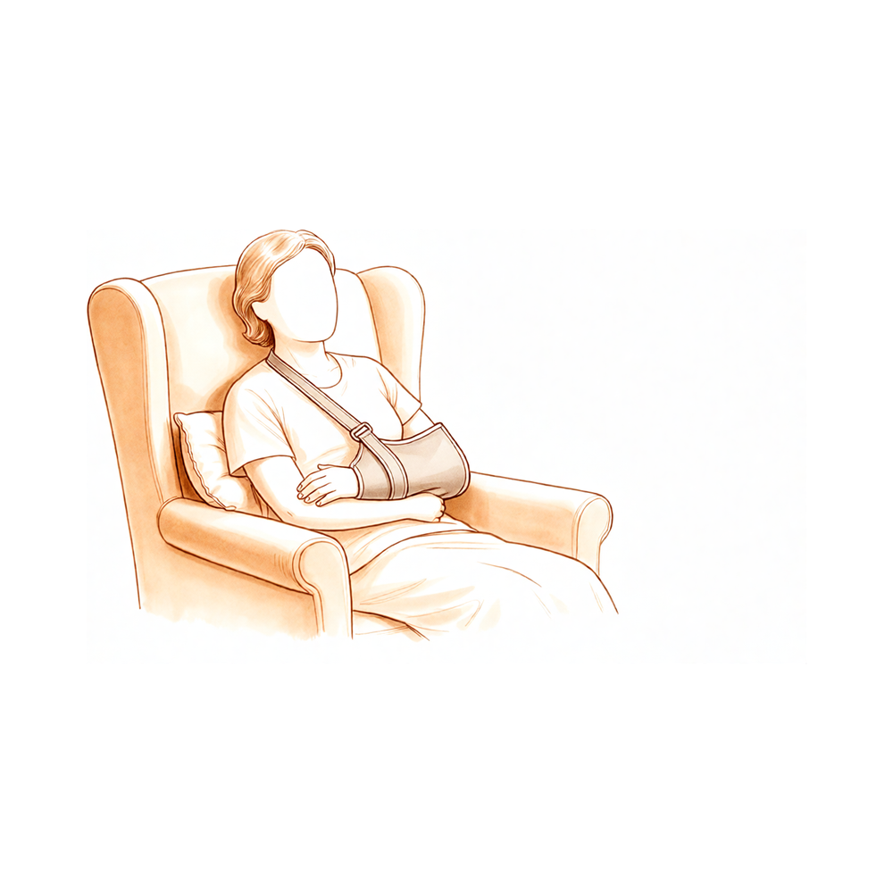
یہ آپریشن کیوں تجویز کیا گیا ہے
ڈاکٹر کیرن ہِرپرا، جو Mater Private Hospital Rockhampton میں بازو اور ہاتھ کے سرجن (upper-limb surgeon) ہیں، علاج کو آپ کی مخصوص چوٹ کے مطابق طے کرتے ہیں۔ ہم ان کم سے کم چیر پھاڑ والے طریقوں سے آغاز کرتے ہیں جو آپ کی حالت کے لیے موزوں ہوں۔ کندھے کے بہت سے فریکچر (ہڈی کا ٹوٹنا) میں اس کا مطلب ہے کہ پہلے آرام، سلنگ (sling)، یعنی گلے سے لٹکی وہ پٹی جو بازو کو سہارا دیتی ہے، اور فزیوتھراپی۔ اس قسم کے زیادہ تر فریکچر سرجری کے بغیر جڑ جاتے ہیں، اور بغیر آپریشن کے علاج سے 90 فیصد سے زیادہ اچھی طرح جڑ جاتے ہیں۔ لیکن کچھ فریکچر میں ہڈی بری طرح اپنی جگہ سے ہٹ جاتی ہے، یعنی ٹوٹے ہوئے ٹکڑے ایک دوسرے سے دور ہو گئے ہوتے ہیں۔ ایسی صورتوں میں، خاص طور پر بڑی عمر کے مریضوں میں، فوری طور پر سرجری کا مشورہ دیا جا سکتا ہے۔
یہ آپریشن کندھے کے جوڑ کی تبدیلی ہے جو خاص طور پر فریکچر کے لیے بنائی گئی ہے۔ آپ کے بازو کی ہڈی کا گول اوپری سرا نکال دیا جاتا ہے اور اس کی جگہ ایک مصنوعی جوڑ لگا دیا جاتا ہے۔ یہ عام طور پر ان بڑی عمر کے مریضوں کو پیش کیا جاتا ہے جن کے پیچیدہ فریکچر میں ٹوٹے ہوئے ٹکڑوں کو قابلِ اعتماد طریقے سے آپس میں جوڑ کر نہیں رکھا جا سکتا۔ انتظار کرنا اور پہلے بغیر آپریشن کے علاج آزمانا کندھے کے کام کاج کو زیادہ خراب اور پیچیدگیوں کو زیادہ کر سکتا ہے، اس لیے ہم چوٹ کے کچھ ہی عرصے بعد آپریشن کا مشورہ دے سکتے ہیں۔ اصل مقصد درد میں قابلِ اعتماد آرام ہے۔ بازو کو کتنا حرکت دے سکتے ہیں اور کتنا استعمال کر سکتے ہیں، اس میں بہتری ایک اضافی مقصد ہے، اگرچہ اس کے نتائج ہر شخص میں زیادہ مختلف ہوتے ہیں۔ ہم ان سب باتوں پر آپ سے بات کریں گے اور آپ کے ساتھ مل کر فیصلہ کریں گے۔
آپریشن سے پہلے
سرجری سے پہلے آپ کو کچھ اسکین کروانے ہوں گے تاکہ ہم آپریشن کی منصوبہ بندی کر سکیں۔ سب سے پہلے آپ کے کندھے کے ایکسرے ہوتے ہیں۔ بعض اوقات ایم آر آئی (ایسا اسکین جو ٹینڈن جیسی نرم بافتوں کو دکھاتا ہے) یا الٹراساؤنڈ بھی کروایا جاتا ہے۔ ہم آپ کو بالکل بتائیں گے کہ آپ کو کون سے اسکین کروانے ہیں۔ آپریشن کے دن، اپنے آپریشن کے وقت سے سات گھنٹے پہلے کھانا پینا بند کر دیں۔ ہم چھ کے بجائے سات گھنٹے اس لیے کہتے ہیں تاکہ اگر آپریشن کی فہرست وقت سے پہلے آگے بڑھ جائے تو ہم آپ کو پہلے بلا سکیں۔ آپ کے سرجن آپ کو بتائیں گے کہ اپنی معمول کی ادویات میں سے کون سی بند کرنی ہیں اور کب۔ جو کچھ بھی آپ لیتے ہیں اس کی لکھی ہوئی فہرست ساتھ لائیں۔ آپریشن کے بعد آپ کو گاڑی میں گھر لے جانے کے لیے کسی کا انتظام کریں، اور ڈھیلے، آرام دہ کپڑے پہنیں۔ اگر آپ کو دوسری طبی بیماریاں ہیں تو ہو سکتا ہے آپ کو خون کے ٹیسٹ یا بے ہوشی کے ڈاکٹر (anaesthetist) سے معائنے کی ضرورت ہو۔
آپریشن کے دن
آپ ہسپتال کے سرجیکل داخلے کے شعبے میں پہنچیں گے، جہاں آپ کا اندراج ہوگا اور آپ کو آپریشن تھیٹر کے لیے تیار کیا جائے گا۔ بے ہوشی کے ڈاکٹر آپریشن سے پہلے آپ سے ملیں گے اور دونوں حصوں کے بارے میں آپ کو سمجھائیں گے۔ یہ آپریشن جنرل اینستھیزیا (مکمل بے ہوشی) کے ساتھ اعصابی بلاک (nerve block)، یعنی ایک انجکشن جو جسم کے ایک حصے کو سُن کر دیتا ہے، ملا کر کیا جاتا ہے۔ پھر آپ کو آپریشن تھیٹر میں لے جایا جاتا ہے، جہاں آپریشن کیا جاتا ہے۔ اس کے بعد آپ ریکوری روم میں جاگیں گے، جہاں بے ہوشی کا اثر ختم ہونے تک نرسیں آپ کی نگرانی کرتی ہیں۔ جب آپ کی حالت مستحکم ہو جائے گی تو آپ وارڈ میں جائیں گے۔
آپریشن میں کیا کیا جاتا ہے
یہ کندھے کے جوڑ کی تبدیلی ہے جو خاص طور پر فریکچر کے لیے بنائی گئی ہے۔ آپ کے سرجن آپریشن والی جگہ پر ایک چیرا لگاتے ہیں اور اسی کے ذریعے کندھے کے ٹوٹے ہوئے جوڑ تک پہنچتے ہیں۔ آپ کے بازو کی ہڈی کا گول اوپری سرا، جو ٹوٹا ہوا حصہ ہے، نکال دیا جاتا ہے۔ اس کی جگہ دھات اور پلاسٹک سے بنا ایک مصنوعی جوڑ لگایا جاتا ہے، کچھ ایسے جیسے ٹوٹی ہوئی چھڑی کے سرے پر ایک نئی ہموار گیند لگا دی جائے۔
پھر سرجن نئے جوڑ کے اردگرد کے حصوں کو دوبارہ بناتے ہیں۔ آپ کے بازو کی ہڈی کے اوپری حصے کے قریب ہڈی کے ابھار (tuberosity)، جہاں آپ کے کندھے کے ٹینڈن (tendon)، یعنی وہ مضبوط ڈوری جو پٹھے کو ہڈی سے جوڑتی ہے، جڑے ہوتے ہیں، واپس اپنی جگہ پر لائے جاتے ہیں اور مضبوط ٹانکوں سے امپلانٹ (جسم میں لگایا گیا مصنوعی پرزہ) کے ساتھ باندھ دیے جاتے ہیں۔ ان ٹکڑوں کا اپنی جگہ پر واپس آنا اور جڑ جانا اہم ہے، کیونکہ انہی سے وہ ٹینڈن جڑے ہوتے ہیں جو آپ کے بازو کو حرکت دیتے ہیں۔ سرجن جوڑ کے اردگرد کی نرم بافتوں کا توازن بھی درست کرتے ہیں اور نئے پرزوں کو اس طرح لگاتے ہیں کہ بازو اپنی قدرتی لمبائی اور زاویے پر رہے۔
جب سب کچھ اپنی جگہ پر آ جائے تو زخم بند کر دیا جاتا ہے۔ بند زخم پر پہلے ایک باریک، خود چپکنے والی جالی رکھی جاتی ہے جو جلد کے کناروں کو آپس میں ملائے رکھتی ہے۔ پھر جالی کے اوپر جلد پر لگانے والا مائع گوند لگایا جاتا ہے، جو سوکھ کر پورے حصے کو بند کر دیتا ہے۔ یہ تقریباً ایک سے دو ہفتے تک لگا رہتا ہے اور پھر خود ہی اٹھ کر اتر جاتا ہے، اس لیے کچھ بھی نکالنے کی ضرورت نہیں ہوتی۔
آپریشن کے بعد
جب آپ جاگیں گے تو آپ ریکوری روم میں ہوں گے، اور بے ہوشی کا اثر ختم ہونے تک نرسیں آپ پر گہری نظر رکھیں گی۔ اس آپریشن کے بعد زیادہ تر مریض ایک یا دو راتیں ہسپتال میں رہتے ہیں۔ شروع میں آپ کے کندھے میں درد ہوگا، اور ہم آپ کو آرام میں رکھنے کے لیے دوا دیں گے۔ آرام کے لیے آپ کا بازو ایک سادہ سلنگ میں رہے گا، جو ورزشوں اور نہانے دھونے کے لیے اتار دی جاتی ہے۔ ہم پٹی تقریباً 10 دن تک لگی رہنے دیتے ہیں؛ براہ کرم اس سے پہلے اسے نہ اتاریں، جب تک ہم آپ کو ایسا کرنے کو نہ کہیں۔ جب ہم آپ کو دیکھیں گے تو پٹی بدل دیں گے یا اتار دیں گے۔ سرجری کے کچھ ہی دیر بعد ایک نرس آپ کو اٹھنے اور چلنے پھرنے میں مدد دے گی۔ براہ کرم گھر جانے کے بعد پہلے 24 گھنٹے کے لیے کسی کو اپنے ساتھ رہنے کا انتظام کریں۔
صحت یابی
سرجری کے بعد پہلے چند دنوں میں آپ کے کندھے میں درد اور سوجن ہوگی۔ یہ معمول کی بات ہے اور آہستہ آہستہ کم ہو جاتی ہے۔ برف کی پٹیاں (ice packs)، آرام اور آپ کی درد کش دوا تکلیف کم کرنے میں مدد دیتی ہیں۔ آپریشن تھیٹر میں لگایا گیا اعصابی بلاک پہلے دن کے دوران ختم ہو جاتا ہے، اس لیے درد بڑھنے سے پہلے ہی اپنی دوا لے لیں۔
آرام کے لیے آپ کا بازو ایک سادہ سلنگ میں رہتا ہے۔ یہ ورزشوں اور نہانے دھونے کے لیے اتار دی جاتی ہے۔ آپ کے فزیوتھراپسٹ شروع سے ہی ہلکی حرکات میں آپ کی رہنمائی کریں گے، اور یہ ورزشیں آپ کے کیے ہوئے کسی بھی دوسرے کام سے زیادہ اہم ہیں۔ جب پیش رفت سست محسوس ہو تب بھی انہیں جاری رکھیں۔ آپ پہلے دن سے گھر میں چل پھر سکتے ہیں، لیکن شروع میں آپ کو کھانے، کپڑے پہننے اور خریداری میں مدد کی ضرورت ہوگی۔ ابتدائی ہفتوں میں کرسی پر ٹیک لگا کر یا اضافی تکیوں کے سہارے سونا اکثر زیادہ آرام دہ ہوتا ہے۔
پیش رفت کے مراحل تاریخوں سے نہیں بلکہ واقعات سے آتے ہیں۔ جب سوجن کم ہو جاتی ہے تو روزمرہ کے کام آسان ہو جاتے ہیں۔ جیسے جیسے آپ کی حرکت واپس آتی ہے، آپ ہاتھ زیادہ دور تک لے جا سکیں گے اور تھوڑا زیادہ وزن اٹھا سکیں گے۔ جب آپ کے سرجن آپ کو گاڑی چلانے کی اجازت دے دیں، جو عام طور پر چھ ہفتے والے معائنے پر ہوتا ہے، تو آپ دوبارہ گاڑی چلا سکتے ہیں؛ دیکھیں بازو اور ہاتھ کی سرجری کے بعد گاڑی چلانا۔ مکمل صحت یابی میں وقت لگتا ہے، اور ورزشوں پر مسلسل محنت آپ کو اچھے نتیجے کا بہترین موقع دیتی ہے۔
ہر شخص اپنی رفتار سے ٹھیک ہوتا ہے۔ آپ کی صحت یابی کا دورانیہ مختلف ہو سکتا ہے، اور آپ کے سرجن اور فزیوتھراپسٹ ہر مرحلے پر آپ کی رہنمائی کریں گے۔
کیا مسائل پیدا ہو سکتے ہیں
زیادہ تر مریض ٹھیک رہتے ہیں، لیکن کبھی کبھار مسائل پیدا ہو سکتے ہیں۔ آپ کے سرجن اور ٹیم آپ کی قریب سے نگرانی کرتے ہیں تاکہ کسی بھی مسئلے کو جلد پہچانا جا سکے۔
انفیکشن سب سے سنگین مسئلہ ہے جس پر نظر رکھنی چاہیے۔ یہ گہرے، دھڑکتے ہوئے درد کی صورت میں ظاہر ہو سکتا ہے جو عام درد کش دواؤں سے کم نہ ہو، زخم سے باہر کی طرف پھیلتی ہوئی سرخی، یا بخار کی صورت میں۔ اگر آپ کو ان میں سے کوئی بھی علامت نظر آئے تو فوراً ہمیں بتائیں۔ گہرے انفیکشن کا جلد علاج ضروری ہے، اس لیے اپنی اگلی ملاقات کا انتظار نہ کریں۔
نیا جوڑ بعض اوقات اپنی جگہ سے نکل سکتا ہے۔ آپ کو اچانک ایک جھٹکے کی آواز یا احساس ہوگا، جس کے بعد درد ہوگا اور بازو معمول کے مطابق حرکت نہیں کرے گا۔ اگر ایسا ہو تو فوری طور پر کلینک سے رابطہ کریں یا ہسپتال کی ایمرجنسی جائیں۔
مصنوعی پرزے وقت کے ساتھ ڈھیلے ہو سکتے ہیں۔ یہ عام طور پر اچانک نہیں بلکہ آہستہ آہستہ ہوتا ہے۔ آپ محسوس کر سکتے ہیں کہ کچھ عرصہ بہتر رہنے کے بعد درد واپس آ رہا ہے، اور ساتھ ہی کندھے میں کلک یا رگڑ کا احساس ہو رہا ہے۔ اپنے اگلے معائنے میں اس کا ذکر کریں، جہاں اسکین کے ذریعے جوڑ کو جانچا جا سکتا ہے۔
امپلانٹ کے اردگرد کی ہڈی ٹوٹ سکتی ہے۔ ایسا گرنے یا چوٹ لگنے کے بعد ہو سکتا ہے، اور یہ اچانک درد اور سوجن کے ساتھ ایک نئی چوٹ جیسا محسوس ہوتا ہے۔ اگر ایسا ہو تو ہسپتال کی ایمرجنسی جائیں۔
کندھے کے اردگرد کسی عصب کو چوٹ لگنا اس آپریشن کا ایک معلوم خطرہ ہے۔ اس سے بازو میں سُن پن، جھنجھناہٹ یا کمزوری ہو سکتی ہے۔ عصب کی زیادہ تر جلن (irritation) خود ہی ٹھیک ہو جاتی ہے، لیکن اگر ایسا نہ ہو تو معائنے پر اپنے سرجن کو بتائیں۔
سرجری کے بعد اکڑاؤ اور مسلسل درد پیدا ہو سکتا ہے۔ کندھا جکڑا ہوا محسوس ہوتا ہے اور ورزشوں کے باوجود حرکت محدود رہتی ہے۔ اپنے معائنے پر اس کا ذکر کریں تاکہ آپ کی فزیوتھراپی کے منصوبے میں تبدیلی کی جا سکے۔
امپلانٹ کے اردگرد ہڈی کے ٹکڑے بعض اوقات جڑ نہیں پاتے، یا ہٹی ہوئی حالت میں جڑ جاتے ہیں۔ اس سے کندھا توقع سے زیادہ کمزور یا کم مستحکم رہ سکتا ہے۔ آپ کے سرجن فالو اپ اسکینوں پر اسے دیکھ لیں گے اور آپ سے علاج کے اختیارات پر بات کریں گے۔
کچھ مریضوں کو بعد میں، بعض اوقات کئی سال بعد، ایک اور آپریشن کی ضرورت پڑتی ہے۔ اس کی وجوہات میں امپلانٹ کا ڈھیلا ہو جانا، جوڑ کا اپنی جگہ سے نکل جانا، انفیکشن یا جوڑ کی سطحوں کا گھس جانا شامل ہیں۔ دوبارہ آپریشن (revision) پہلے آپریشن سے زیادہ پیچیدہ ہوتا ہے اور صحت یابی سست ہو سکتی ہے۔
اگر آپ تفصیل جاننا چاہتے ہیں تو اس صفحے پر پیچیدگیوں کا جدول ان کی عام شرحیں بتاتا ہے۔
ہمیں کب فون کریں
زیادہ تر مسائل کی ابتدائی علامات ہوتی ہیں، اور ہم چاہتے ہیں کہ آپ انتظار کرنے کے بجائے ہم سے رابطہ کریں۔ اگر آپ کو بخار ہو، اگر آپ کے زخم کے اردگرد کی جلد زیادہ سرخ ہو جائے یا اس سے رطوبت رسنے لگے، یا اگر آپ کا درد کم ہونے کے بجائے بڑھتا ہی جائے تو ہمیں فون کریں۔ اگر اچانک آپ اپنے بازو کو محسوس نہ کر سکیں یا اسے حرکت نہ دے سکیں، اگر آپ کی پنڈلی کے پچھلے حصے میں سوجن یا درد ہو، یا اگر آپ کا سانس پھولنے لگے تو ہسپتال کی ایمرجنسی جائیں۔ یہ خون کے لوتھڑے کی علامت ہو سکتی ہیں۔ گرنے یا چوٹ لگنے کے بعد اچانک شدید درد کی صورت میں بھی فوری طبی امداد کی ضرورت ہے۔
اس مسئلے کے بارے میں مزید کہاں پڑھیں
یہ صفحہ خود آپریشن کے بارے میں ہے۔ جس مسئلے کا یہ علاج کرتا ہے، بشمول اس کے کہ شواہد کیا بتاتے ہیں کہ سرجری کب فائدہ دیتی ہے اور کب نہیں، اس کی مزید تفصیل بازو کی اوپری ہڈی کے سرے کا فریکچر والے صفحے پر ہے۔
Evidence & references
This is the clinical evidence summary written for health professionals. It is technical, and it lists the research this page was built from. You do not need to read it to understand your treatment or to make a decision about it.
Overview
- Patients undergoing arthroplasty for acute proximal humerus fractures may achieve satisfactory long-term pain relief [1].
- The result for overall shoulder motion after arthroplasty for acute proximal humerus fractures is less predictable [1].
- Anatomic tuberosity healing improves objective and subjective outcomes in elderly patients who have undergone reverse shoulder arthroplasty for acute proximal humeral fractures [2].
- Elderly patients who require admission after sustaining a proximal humeral fracture are frail and subject to a greater-than-average risk of mortality for their age [3].
- Patients who undergo initial periods of nonoperative management have worse functional outcomes than those who undergo acute reverse total shoulder arthroplasty for proximal humeral fractures [4].
- Patients who undergo initial periods of nonoperative management have higher complication rates than those who undergo acute reverse total shoulder arthroplasty for proximal humeral fractures [4].
- Clinical results at 1-year follow-up confirmed the advantage of applying a new intramedullary support nail and plate system to 3- or 4-part proximal humeral fractures in older patients [5].
- Patients with acute proximal humeral fractures who undergo reverse shoulder arthroplasty appear to achieve superior 5-year functional outcomes compared with patients who undergo hemiarthroplasty [7].
- The study by [11] is the largest long-term follow-up study of acute proximal humeral fractures treated with hemiarthroplasty [11].
- In most studies of proximal humeral fractures, only 1 or 2 patients experiencing an alternative outcome or lost to follow-up would change the conclusions for the dichotomous outcome studied [14].
- The available literature suggests that reverse shoulder arthroplasty performed to address complex proximal humeral fractures might result in more favorable clinical outcomes than hemiarthroplasty performed for the same indication [32].
- The increased in-hospital risk for major adverse events and surgical complications may moderate the enthusiasm associated with reverse total shoulder arthroplasty for proximal humeral fractures in patients 65 years and older [33].
- Reverse total shoulder arthroplasty performed for acute 3- and 4-part proximal humeral fractures yields overall worse clinical outcomes compared with reverse total shoulder arthroplasty performed for elective indications [111].
- Reverse total shoulder arthroplasty performed for acute 3- and 4-part proximal humeral fractures yields worse active range of motion compared with reverse total shoulder arthroplasty performed for elective indications [111].
- No clear benefits were observed in treating patients 65 years or older with four-part fractures of the proximal humerus with either hemiarthroplasty or nonoperative treatment [114].
Anatomy & Pathophysiology
Bony Anatomy
- The proximal humerus comprises four main parts: the humeral head, greater tuberosity (GT), lesser tuberosity (LT), and humeral shaft [37].
- The articular head is spherical with a diameter of 37 to 57 mm [37].
- The most superior portion of the articular surface of the humeral head averages 8 mm above the greater tuberosity [37].
- Humeral version averages 29.8 degrees, with a range of 10 to 55 degrees [37].
- The humeral head is inclined approximately 130 degrees with respect to the humeral shaft [37].
- The anatomic neck is located at the junction of the articular surface and the tuberosities [37].
- The surgical neck represents an indistinct region, or metadiaphyseal junction, below the tuberosities but above the humeral shaft [37].
- The greater tuberosity is located in a posterior-superior position relative to the humeral shaft and serves as the attachment site for the supraspinatus, infraspinatus, and teres minor tendons [37].
- The lesser tuberosity is located on the anterior aspect of the proximal humerus and serves as the attachment site for the subscapularis tendon [37].
- The bicipital groove lies between the greater and lesser tuberosities and serves as a pathway for the long head of the biceps tendon [37].
- The distal aspect of the bicipital groove is internally rotated with respect to the proximal portion [37].
- The glenoid is a convex structure of shallow depth shaped like an inverted pear [37].
- The acromion, coracoacromial ligament, and coracoid process form the coracoacromial arch, a rigid bony-ligamentous structure that imparts stability to the shoulder girdle [37].
- The humeral head averages 19° of retroversion and 41° of inclination (neck-shaft angle) [39].
- The neck-shaft angle measures an average of 135 degrees, and the humeral head is retroverted an average of 30 degrees [38].
- The proximal humerus has three centers of ossification: the humeral head (4 to 6 months), the greater tuberosity (1 to 3 years), and the lesser tuberosity (3 to 5 years) [39].
- The ossification centers of the proximal humerus fuse to the shaft at age 17 to 20 years [39].
- The control volume is an important anatomic and functional area of the proximal humerus [73].
Vascular Supply
- The proximal humerus receives its blood supply from the anterior and posterior humeral circumflex branches from the third division of the axillary artery [37].
- The posterior humeral circumflex artery travels with the axillary nerve, enters the quadrilateral space posteriorly, and anastomoses with a branch of the anterior circumflex to supply the posterior cuff [37].
- The anterior humeral circumflex artery arises from the axillary artery at the inferior border of the subscapularis [37].
- The anterior humeral circumflex artery provides vascular inflow to the humeral head by way of its terminal anterolateral branch, known as the artery of Laing or arcuate artery [37].
- The ascending branch of the anterior humeral circumflex artery courses parallel to the lateral aspect of the long head biceps tendon and enters the humeral head at the interface of the bicipital groove and greater tuberosity [37].
- Injury to the arcuate artery may result in osteonecrosis of the humeral head [37].
- Additional extraosseous collateral branches can permit humeral head perfusion despite complete ligation of the arcuate artery [37].
- The anterolateral ascending branch of the anterior humeral circumflex artery provides the primary blood supply to the humeral head [39].
- The terminal intraosseous portion of the anterior humeral circumflex artery enters at the proximal aspect of the intertubercular groove as the arcuate artery [39].
- Fractures of the anatomic neck have a poor prognosis because of complete disruption of the blood supply to the head [38].
- Surgical neck fractures are common, and with these, the blood supply to the head is preserved [38].
Soft Tissue & Neurovascular Anatomy
- The rotator cuff consists of four muscles: the subscapularis, supraspinatus, infraspinatus, and teres minor [38].
- The teres major is not a rotator cuff muscle [38].
- The cuff muscles serve as depressors of the humeral head to allow the deltoid to efficiently abduct the humerus [38].
- The infraspinatus and teres minor are external rotators, while the subscapularis is an internal rotator of the humerus [38].
- The deltoid and pectoralis major muscles, along with the rotator cuff, cause predictable displacement of fractures around the proximal humerus [38].
- The subscapularis inserts on the lesser tuberosity and causes medial displacement [37].
- The supraspinatus and infraspinatus insert on the greater tuberosity and cause superior and posterior displacement [37].
- The pectoralis major inserts on the humeral shaft and displaces it medially [37].
- The axillary nerve is a terminal branch coming off the posterior cord of the brachial plexus just proximal to the coracoid process [42].
- The axillary nerve passes beneath the conjoined tendon anterior to the subscapularis 3 to 5 mm medial to the musculotendinous junction [42].
- The axillary nerve is adjacent to the inferior capsule before entering the quadrilateral space posteriorly [42].
- The axillary nerve splits into the anterior and posterior branches within the quadrangular space [42].
- The anterior and middle deltoid muscle receives sole innervation from the anterior branch of the axillary nerve [42].
- Posterior deltoid muscle innervation varies, with supply only from the anterior branch in 2.3% of cases, from the posterior branch in 8.5%, and from both branches in 89.1% [42].
- The posterior branch of the axillary nerve branches to supply the teres minor muscle and then terminates as the superior lateral brachial cutaneous nerve [42].
- In the anterior deltopectoral approach, the axillary nerve can be palpated by sweeping a finger inferiorly across the subscapularis muscle tendon interface [42].
- In the anterolateral deltoid splitting approach, the axillary nerve crosses approximately 5 cm inferior to the anterolateral acromial corner [42].
- In the posterior deltoid splitting approach, the axillary nerve is approximately 7 cm from the posterior acromial corner [42].
- Vertical abduction has the greatest effect on axillary nerve position, while horizontal glenohumeral forward flexion and humeral rotation have little effect [70].
- An axillary nerve injury from proximal humeral fracture or fracture-dislocation would result in paralysis of the deltoid muscle and anesthesia over the “badge” region at the lateral proximal arm [38].
Bursae & Joints
- The subacromial bursa and subscapular bursa are two bursae in the shoulder region with clinical importance [40].
- The subscapular bursa lies between the subscapularis tendon and the neck of the scapula [40].
- The subscapular bursa communicates with the joint cavity between the superior and middle glenohumeral ligaments [40].
- The subscapular bursa protects the tendon of the subscapularis at the point where it passes under the base of the coracoid process and over the neck of the scapula [40].
- The subscapular bursa is linked to the coracoid process by a suspensory ligament [40].
- In 28% of specimens dissected by Colas and colleagues, the subscapular bursae merged with the subcoracoid bursae, forming a unique wide bursa in this region [40].
- The subscapular bursa often houses loose bodies in the shoulder [40].
- The subscapular bursa is a region in which synovitis of the shoulder may be most intense, where small fringes, or villi, can project into the joint cavity [40].
- A soft tissue sheath consistently covers the long head of the biceps tendon to the level of the proximal margin of the pectoralis major tendon and contributes to the roof of the bicipital tunnel [40].
- The fibro-osseous bicipital tunnel consists of three distinct anatomic zones [40].
- Zone 1 of the bicipital tunnel represents the traditional bony bicipital groove beginning at the articular margin and ending at the distal margin of the subscapularis tendon [40].
- Zone 2 of the bicipital tunnel extends from the distal margin of the subscapularis tendon to the proximal margin of the pectoralis major tendon and represents a “no man’s land” because it is not viewable from arthroscopy above or from subpectoral exposure below [40].
- Zone 3 of the bicipital tunnel is distal to the proximal margin of the pectoralis major tendon and represents the subpectoral region [40].
- The rotator interval is defined medially by the base of the coracoid, superiorly by the supraspinatus tendon, and inferiorly by the subscapularis tendon [39].
- The rotator interval contains the coracohumeral ligament, the superior glenohumeral ligament, and the intra-articular portion of the long head of the biceps tendon [39].
- Laxity of the rotator interval results in inferior laxity (the sulcus sign), and contracture of the interval is seen with adhesive capsulitis [39].
- The coracohumeral ligament restricts external rotation in adduction, and it is a static restraint to inferior and posterior translation in adduction and external rotation [39].
- The superior glenohumeral ligament is a primary static restraint against anterior translation with the arm at the side [39].
- With the coracohumeral ligament, the superior glenohumeral ligament forms a pulley that provides restraint against medial subluxation of the long head of the biceps tendon [39].
- The middle glenohumeral ligament is a primary static restraint against anterior translation with the arm in external rotation and 45° of abduction [39].
- The anterior band of the inferior glenohumeral ligament is a primary static restraint against anterior-inferior dislocation of the glenohumeral joint in 90° of abduction and external rotation (position of apprehension) [39].
- The posterior band of the inferior glenohumeral ligament is a primary static restraint against posterior-inferior translation in internal rotation and adduction [39].
- The superior transverse scapular ligament arises from the medial base of the coracoid overlying the suprascapular notch [39].
- The suprascapular artery runs superior to the superior transverse scapular ligament; the nerve runs deep to the ligament [39].
- Entrapment of the suprascapular nerve at the superior transverse scapular ligament causes denervation of both the supraspinatus and the infraspinatus [39].
- The spinoglenoid ligament overlies the suprascapular nerve at the spinoglenoid notch [39].
- Entrapment, traction, or compression of the suprascapular nerve at the spinoglenoid notch causes denervation of the infraspinatus [39].
Pathophysiology & Biomechanics
- Stability and function of the glenohumeral joint is provided by the interaction of the glenohumeral joint that promote a near global range of motion and purposeful function [37].
- External loads transferred to the shoulder girdle are initially offset by joint surface anatomy, joint volume, atmospheric pressure, and joint fluid cohesion and adhesion [37].
- Moderate and large loads are counterbalanced by the deltoid and rotator cuff and by the capsulolabral and bone structures, respectively [37].
- Proximal humerus fractures alter complex interactions of the shoulder girdle, resulting in pain, decreased range of motion and stiffness, and disability [37].
- Displaced proximal humerus fractures can impede normal movement of the rotator cuff, subacromial bursa, and subdeltoid bursa, causing impingement and disruption of normal glenohumeral motion [37].
- In displaced and nondisplaced proximal humerus fractures, the subdeltoid and subacromial bursae can become thickened and fibrotic, forming adhesions that can limit normal glenohumeral motion [37].
- Early range of motion exercises after a fracture have been hypothesized to decrease the formation of such adhesions [37].
- Adhesions in the humeroscapular motion interface can limit shoulder mobility with examples such as scarring after proximal humerus fracture trauma [42].
- The humeroscapular motion interface lies between the inner structures of the proximal humerus, rotator cuff, coracohumeral ligament, and biceps tendon sheath and the superficial layer of the acromion, deltoid, coracoacromial ligament, coracoid process, and the conjoined tendon [42].
- Smooth, unrestricted motion at the humeroscapular motion interface is vital to shoulder mobility [42].
- Varus and antecurvatum proximal humerus deformities as small as 15 degrees were associated with statistically significant alterations in glenohumeral joint mechanics [65].
- With minimal and moderate amounts of glenohumeral abduction, glenohumeral joint forces are significantly displaced superiorly [50].
- Greater tuberosity healing does not seem to impact reverse shoulder arthroplasty biomechanics during abduction or forward flexion; however, it does affect biomechanics during external rotation [48].
- The double plate strategy can increase the stability of the medial column of the proximal humerus, and enhance the overall biomechanical property of the repaired proximal humerus [67].
- The two-screw fixation construct augmented with a cerclage wire has higher mechanical performance than the conventional two-screw configuration for the fixation of humeral greater tuberosity fractures [74].
- Despite the suture button construct having superior interface contact immediately after fixation, the double-row construct offered better contact performance at all abduction angles with applied force [64].
Classification
Reliability and Reproducibility
- Classifications of proximal humeral fractures using the Neer system based on CT scans and plain radiographs are not very reliable or reproducible due to difficulty in determining which segments are fractured [103].
- The Neer and AO classifications of proximal humeral fractures have limited intraobserver and interobserver reliability [104].
- Neer indicated that his classification was meant to be applied after operative exposure and believed that radiographs alone would be unreliable [104].
- The addition of two-dimensional (2-D) computed tomography (CT) scans did not improve the interobserver reproducibility of either the Neer or AO classification systems [104].
- Interobserver agreement on the Neer classification was slight, with a kappa measure ranging from 0.069 to 0.14 [104].
- Interobserver agreement on treatment based on fracture classification was fair, with a kappa measure ranging from 0.28 to 0.33 [104].
- The lack of a reliable classification confounds efforts to compare the outcomes of treatment methods among different clinical trials and reports [104].
Specific Classification Systems
- The revised Neer classification covers 98% of all proximal humeral fractures and is appropriate for clinical practice [101].
- The HGLS classification is a reliable method of describing fractures of the proximal humerus compared with the Neer and AO systems [98].
- A new classification system with emphasis on the qualitative aspects of proximal humeral fractures showed high reliability when based on a standardized imaging protocol including computed tomography scans [89].
Clinical Presentation
- Patients presenting with a traumatic shoulder injury and normal radiographs may have an occult greater tuberosity fracture identified by the anterior bruise sign, which is a highly sensitive and specific clinical aid [29].
- Fractures of the proximal humerus follow characteristic patterns [24].
- In patients with proximal humeral fractures, a majority underwent non-operative treatment [8].
- Nonoperative treatment of proximal humeral fractures produces considerable variation in shoulder-specific and general health outcomes at 1 year [25].
- A substantial proportion of patients treated nonoperatively for proximal humeral fractures have poor perceived functional outcomes at 1 year [25].
- Compared with the general population, patients sustaining a proximal humeral fracture have a significantly higher risk of mortality up to one year after the injury [16].
- Mortality at 1 year for fragility proximal humerus fractures is universally high regardless of risk factors [31].
- As the majority of unplanned hospital readmissions after surgical treatment of proximal humerus fractures were associated with medical diagnoses, patient medical comorbidities must be considered before surgical treatment and during the postoperative care phase [21].
- In a cohort of 46 patients undergoing shoulder arthroplasty for posttraumatic avascular necrosis of the humeral head after proximal humerus fracture, the average age was 64 years (range, 49–82 years) [6].
- In a cohort of 46 patients undergoing shoulder arthroplasty for posttraumatic avascular necrosis of the humeral head after proximal humerus fracture, the initial injury occurred as a result of a simple fall in 58% of patients [6].
- In a cohort of 46 patients undergoing shoulder arthroplasty for posttraumatic avascular necrosis of the humeral head after proximal humerus fracture, 66% were four-part fractures and 13% were three-part fractures [6].
- In a study of 37 patients undergoing surgical fixation of the proximal humerus, the mean age at the time of surgery was 66.0 ± 9.3 years (range, 32–83 years) [69].
- In a study of 37 patients undergoing surgical fixation of the proximal humerus, fractures were classified as two-part in 29%, three-part in 58%, and four-part in 13% of cases [69].
- In a randomized study of patients aged 70 years or older with acute proximal humeral fractures, indications for shoulder arthroplasty included complex fractures not amenable to reconstruction, including displaced 4-part fractures, fracture-dislocations with 3-part fractures, and head-splitting fractures with more than 40% articular surface involvement [72].
- In a randomized study of patients aged 70 years or older with acute proximal humeral fractures, preoperative confirmation of fracture type was made by computed tomography [72].
- In a randomized study of patients aged 70 years or older with acute proximal humeral fractures, exclusion criteria included contraindications to surgery, prior surgery in the shoulder, associated ipsilateral upper limb fracture, and neurologic disorder [72].
- In a study of 463 proximal humeral fractures, prevention of local complications, in particular those leading to severe varus deviation, appears essential to improve shoulder function [30].
- In a study of 463 proximal humeral fractures, factors associated with poor results include being a woman, four-part fracture dislocation, and absence of metaphyseal head extension [99].
- In a study of 463 proximal humeral fractures, surgical treatment outcomes are unpredictable [99].
- In a study of 463 proximal humeral fractures, surgical treatment remains far from straightforward [99].
- In a study of 463 proximal humeral fractures, being a woman is a factor associated with poor results [99].
- In a study of 463 proximal humeral fractures, four-part fracture dislocation is a factor associated with poor results [99].
- In a study of 463 proximal humeral fractures, absence of metaphyseal head extension is a factor associated with poor results [99].
- In a study of 463 proximal humeral fractures, surgical treatment of proximal humerus fractures remains far from straightforward [99].
- In a study of 463 proximal humeral fractures, unpredictable outcomes are observed [99].
- In a study of 463 proximal humeral fractures, factors associated with poor results include being a woman [99].
- In a study of 463 proximal humeral fractures, factors associated with poor results include four-part fracture dislocation [99].
- In a study of 463 proximal humeral fractures, factors associated with poor results include absence of metaphyseal head extension [99].
Investigations
Imaging Modalities and Protocols
- AP and lateral plain radiographs are often the only required studies needed for assessing acute shoulder trauma, including fractures or dislocations [47].
- CT imaging is frequently used to evaluate fractures of the shoulder [47].
- MRI is the modality of choice for evaluating the rotator cuff, biceps, and subacromial/subdeltoid bursa [47].
- T1-weighted MRI can reveal Hill-Sachs lesions and is often used with magnetic resonance arthrograms to provide a more detailed picture of the joint surfaces [47].
- T2-weighted MRI provides better visualization of full thickness rotator cuff tears [47].
- Arthrography involves injection of contrast agent in conjunction with either an MRI or CT scan, enhancing imaging of the joint to enable better identification of normal structures and pathology involving the joint surfaces [47].
- MR arthrography is considered the benchmark for evaluation for labral tears and rarely is indicated for evaluation of rotator cuff pathology [47].
- When MRI or MR arthrography is contraindicated, CT arthrography is indicated [47].
- Ultrasonography is a low-cost alternative to MRI and arthrography for evaluating both skeletal and soft-tissue structures of the shoulder [47].
- Ultrasonography can provide immediate, real-time visualization of the rotator cuff, biceps tendon, and calcific deposits [47].
- Ultrasonography can be used to measure the subacromial space and detect atrophy of rotator cuff muscles [47].
- Ultrasonography can evaluate impingement in various positions and motions due to real-time imaging [47].
- Ultrasonography is highly operator dependent and is not as useful for evaluating labral tears or rotator cuff tears that are very small or larger than 3 cm [47].
- The purpose of imaging of the shoulder is to help establish the diagnosis, determine the severity of the pathoanatomy, assist in surgical planning, and enable the surgeon to illustrate the condition of the shoulder to the patient [19].
- Unless a specific research protocol is in place, the temptation to “overimage” should be resisted, obtaining only the scans or reconstructions that are necessary for the care of the patient [19].
- Standardized plain films are almost always sufficient to garner the information needed, and there is information that can be gathered from properly taken plain films that cannot be obtained from CT scans [19].
- Proper radiographic technique is as important as proper surgical technique to achieve the desired outcome [19].
- The first key view is the anteroposterior (AP) in the plane of the scapula taken so that the x-ray beam passes through the glenohumeral joint [19].
- The AP view shows the superoinferior position of the humeral head relative to the glenoid, the presence of osteophytes on the humeral head and glenoid, narrowing of the joint space, the degree of medial displacement of the humerus in relation to the lateral acromial line, the quality of the humeral and glenoid bone, the presence of loose bodies, and whether there is humeral head collapse or deformity [19].
- The second key view is the axillary view taken with the arm in the functional position of elevation in the plane of the scapula and oriented so that both the spinoglenoid notch and the scapular neck are visible [19].
- The axillary view shows a different perspective of the humeral anatomy, the amount of glenoid bone, the shape of the glenoid, its version in relation to the plane of the scapula, and the relationship of the humeral head to the glenoid fossa [19].
- The axillary view is referred to as the “truth view” because it demonstrates the glenohumeral relationships in the functional position of elevation [19].
- CT scans have the disadvantage of being taken with the arm in the adducted position [19].
- Many of the “axillary views” sent for consultation are taken without standardization, making it impossible to determine the important features of the glenohumeral joint [19].
- When taken properly, the standardized anteroposterior and axillary views indicate the thickness of the cartilage space between the humerus and the glenoid, relative positions of the humeral head and the glenoid, presence of osteophytes, degree of osteopenia, and extent of bony deformity and erosion [19].
- Joint space narrowing is most evident on the truth view as opposed to images made with the arm at the side [19].
- The axillary truth view can show posterior subluxation or “functional decentering” that is not evident in images taken with the arm at the side [19].
- The degree of posterior subluxation can be measured as the position of the center of the humeral head in relation to the plane of the scapula, the position of the center of the humeral head in relation to the glenoid face, or the point of contact of the humeral articular surface on the glenoid articular surface [19].
- The point of contact of the humeral articular surface on the glenoid articular surface reflects the degree of centering of the net humeral joint reaction force on the glenoid [19].
- Malcentering of the joint reaction force leads to posterior instability, posterior glenoid wear, and “rocking horse” loosening of prosthetic glenoid components [19].
- Three-dimensional reconstructions can reveal fine details of the shoulder anatomy, but this additional information rarely changes the planning or conduct of the arthroplasty [19].
- A robust approach to imaging the shoulder needs to recognize that the shoulder is a three-dimensional structure that cannot be represented by a single planar view [45].
- Critical relationships—such as the degree of centering of the humeral head—change with the position of the arm [45].
- Shoulder pathology may be found in a large number of different bones and soft tissues [45].
- Overlying and superimposed structures as well as metallic implants may complicate imaging the structures of interest [45].
- Surgeons need to develop a judicious approach that yields the information necessary to treat the patient while avoiding the tendency to "over-image" [45].
Fracture-Specific Imaging Findings
- There is relevant variability in displacement measurements between shoulder radiographs and CT scans in the coronal plane, with nearly 30% of cases suggesting surgical treatment on radiographs being reclassified for conservative treatment based on CT findings [132].
- The routine use of 3D-printed models may not be beneficial for classifying proximal humeral fracture patterns beyond the information gained from currently available imaging modalities, and their use as the sole determinant for recommending surgical intervention should be avoided at this time [134].
- CNNs proficiently rule out proximal humerus fractures on plain radiographs [136].
- In patients presenting with a traumatic shoulder injury with normal radiographs, the anterior bruise sign (ABS) is a highly sensitive and specific clinical aid to identify patients with an occult greater tuberosity fracture [29].
- Undisplaced greater tuberosity fractures can be managed non-operatively with good results, but patients with persistent post-traumatic shoulder pain and limitation of function warrant MRI investigation to identify occult fractures [124].
- Missed posterior dislocation of the shoulder after intramedullary fixation of proximal humeral fractures is an extremely rare injury that can be missed due to inadequate initial and postoperative x-ray images and incorrect interpretation [139].
Treatment
Arthroplasty Outcomes and Indications
- Patients undergoing arthroplasty for acute proximal humerus fractures may achieve satisfactory long-term pain relief, but overall shoulder motion results are less predictable [1].
- Primary shoulder hemiarthroplasty for proximal humeral fracture is associated with satisfactory prosthetic survival at an average of 6.3 years [17].
- The available literature suggests that reverse shoulder arthroplasty for complex proximal humeral fractures might result in more favorable clinical outcomes than hemiarthroplasty for the same indication [32].
- Reverse shoulder arthroplasty is advocated for complex proximal humerus fractures in elderly patients because it provides more consistent and predictable results compared with hemiarthroplasty or plate osteosynthesis [118].
- Treatment with reverse shoulder arthroplasty provides superior functional outcomes compared with conservative treatment for patients presenting with an acute proximal humeral fracture [96].
- Patients who undergo initial periods of nonoperative management have worse functional outcomes and higher complication rates than those who undergo acute reverse total shoulder arthroplasty for proximal humeral fractures [4].
- With narrow indications, use of a specific fracture stem, and adequate tuberosity management, successful radiographic and functional results are presented after a mean follow-up of 4.8 years following hemiarthroplasty for primary nonreconstructable humeral head fractures [109].
- A reverse shoulder prosthesis using a dedicated stem is a viable solution to treat complex proximal humerus fractures with reliable restoration of elevation [117].
- Reverse shoulder arthroplasty is a powerful tool for managing proximal humerus fracture sequelae when joint-preserving options are not optimal, provided there is careful management of the tuberosities and understanding of associated pearls and pitfalls [28].
Tuberosity Management
- In elderly patients who have undergone a reverse shoulder arthroplasty for acute proximal humeral fractures, anatomic tuberosity healing improves objective and subjective outcomes [2].
- Shoulder rotational ability is improved by systematically repairing the tuberosities around the implant, provided their consolidation is anatomic [13].
Surgical Technique and Approach
- The goals of prosthetic reconstruction for displaced three- and four-part fractures include replacement of the humeral-head articular fragment, stable fixation of the prosthetic component, restoration of humeral length and version, stable and anatomic reduction of the tuberosities to achieve bone union, and functional restoration of the rotator cuff mechanism [107].
- The surgical approach for reverse shoulder arthroplasty has been either the standard deltopectoral approach or the anterosuperior approach [110].
- The deltopectoral approach allows adequate access to fracture fragments for suture fixation and provides excellent glenoid exposure after fracture fragment mobilization, though exposure and reduction of the greater tuberosity can be challenging [110].
- The anterosuperior approach offers greater tuberosity access but requires deltoid detachment, carries a potential for dehiscence, and has limited extensibility which is relevant during humeral shaft exposure with comminution [110].
- Optimizing proximal humerus fracture fixation involves preoperative planning, understanding indications and implant design, obtaining effective exposure and proper intraoperative imaging, using various reduction modalities, ensuring proper implant placement, and using supplemental fixation methods when helpful [108].
Non-Operative Management
- A majority of patients with proximal humeral fractures underwent non-operative treatment [8].
- Nonsurgical management of proximal humerus fractures decreased during the study period [78].
- Nonoperative treatment of proximal humeral fractures produces considerable variation in shoulder-specific and general health outcomes at 1 year, and a substantial proportion of patients have poor perceived functional outcomes [25].
- Short and long periods of immobilization yield similar results for nonoperatively treated proximal humeral fractures, independent of the fracture pattern [85].
- Nonsurgical treatment should have a more prominent role in the treatment of proximal humeral fractures [90].
Internal Fixation
- Clinical results at 1-year follow-up confirmed the advantage of applying an intramedullary support nail and plate system to 3- or 4-part proximal humeral fractures in older patients [5].
- A less-invasive locking plate fixation method is a feasible treatment option in proximal humeral fractures with acceptable complications and considerable improvement during the first six months, although a lengthy recovery time is required [86].
- This trial found no significant difference in clinical outcomes at 2 years between surgery and non-operative treatment in patients 60 years of age or older with displaced 2-part fractures of the proximal humerus [87].
- Shoulder function was restored to preinjury levels for most patients, and osteoporosis may not be regarded as a contraindication for open reduction and internal fixation of unilateral displaced 3- or 4-part fractures at 12-month follow-up [27].
- Internal fixation of three-part and four-part proximal humerus fracture dislocations resulted in poor shoulder function and complications in a high number of patients, although fracture union was achieved in most patients [120].
- Proximal humerus fractures in the elderly can be reliably fixed with a "hybrid" locked-plating technique, with no early or late reoperations reported in a series of 35 patients older than 75 years [106].
- Percutaneous treatment of selected proximal humeral fractures results in predictable union and good clinical results with a low rate of complications [116].
- The Humerus Block technique is a very good and predictable, minimally invasive, percutaneous operative technique for treatment of various types of proximal humeral fractures that results in very good pain relief, mobility, and pull force in the arm with a low incidence of avascular necrosis [35].
Complications and Mortality
- At 1-year postinjury, the mortality rate for older patients after proximal humerus fracture has been reported to be as high as 10% [26].
- Most studies on periprosthetic postoperative humeral fractures have reported occurrence rates between 0.5% and 3%, but the field has changed dramatically with the use of reverse shoulder arthroplasty and new implant designs [34].
- The most important risk factors for postoperative opioid dependence following proximal humerus fractures are preoperative dependence and fracture complexity [113].
- This systematic review highlights significant heterogeneity in the terminology and definitions used to describe complications following non-surgical management of proximal humeral fractures, calling for standardized definitions to improve evidence synthesis [10].
Context and Consensus
- Consensus when managing proximal humerus fractures is limited to specific scenarios, whereas lack of consensus still exists in others [9].
- The introduction and widespread adoption of reverse total shoulder arthroplasty in the management of three- and four-part proximal humeral fractures represents a paradigm shift with respect to operative treatment options [15].
- This is the largest long-term follow-up study of acute proximal humeral fractures treated with hemiarthroplasty [11].
Complications
Mortality and Systemic Risk
- Elderly patients requiring admission after sustaining a proximal humeral fracture are frail and subject to a greater-than-average risk of mortality for their age [3].
- Postoperative mortality after surgical fixation of proximal humeral fractures was 3.5%, which was approximately sixfold higher than the ninety-day mortality rate observed after nonoperative treatment [53].
- As the majority of unplanned hospital readmissions were associated with medical diagnoses, it is important to consider patient medical comorbidities before surgical treatment of proximal humerus fractures and during the postoperative care phase [21].
General Complication Rates and Risk Factors
- The rate of complications after operative treatment of proximal humeral fractures is high [143].
- Patient age of sixty-five years or older, male sex, residence in an area with an income in the lowest two quintiles, and the presence of preexisting comorbidities were associated with elevated risks of short-term complications following surgical fixation [53].
- There is significant heterogeneity in the terminology and definitions used to describe complications following non-surgical management of proximal humeral fractures [10].
Arthroplasty-Specific Complications
- The most common postoperative complications reported in a systematic review of shoulder arthroplasty were postoperative stiffness/pain (n = 47), anterior dislocation or shoulder instability (n = 16), and progressive arthritis requiring revision (n = 7) [22].
- Periprosthetic fracture occurred in 3 patients and infection in 1 patient in a systematic review of shoulder arthroplasty complications [22].
- In-hospital complications are more likely to occur after reverse shoulder arthroplasty than after locked plating for proximal humeral fractures [33].
- The increased in-hospital risk for major adverse events and surgical complications may moderate the enthusiasm associated with RTSA for proximal humeral fractures in patients 65 years and older [33].
- The most common reasons for reoperation after primary reverse shoulder arthroplasty were humeral component fracture, humeral loosening, dislocation, infection, and glenoid failure, each occurring at a rate under 1% [130].
- Humeral loosening is rare in reverse shoulder arthroplasty, occurring in 0.7% of primary and 2.8% of revision cases [135].
- The most common complications after reverse shoulder arthroplasty for fracture sequelae were humeral loosening (9 cases) and instability (4 cases), all of which occurred in patients with type 3 and 4 sequelae [81].
- Revision surgery for failed arthroplasty of proximal humerus fracture is complex with a high likelihood of inferior outcomes compared with primary arthroplasty [80].
- Revision shoulder arthroplasty remains challenging with a high rate of complications [126].
- In a series of 157 revisions from failed hemiarthroplasty to reverse total arthroplasty, 11 patients (7%) underwent repeated revision surgeries secondary to glenoid component loosening, instability, humeral component disassembly, or humeral stem loosening [83].
- Nerve injury following reverse shoulder arthroplasty is a known risk factor with wide ranging incidences reported [82].
Fixation-Specific Complications
- Acute deep infection after ORIF of proximal humeral fractures is a devastating complication with high complication rates, poor functional outcomes, and a notably high nonunion rate [127].
- Prevention of local complications, in particular those leading to severe varus deviation, appears essential to improve shoulder function after a proximal humeral fracture [30].
- Overall, there was a moderate rate of complications but low rate of revision following intramedullary nailing of humerus fractures [141].
Nonunion and Functional Outcomes
- The prevalence of nonunion after proximal humeral fracture is higher than previously reported, with most patients having a very low risk but a smaller subgroup at much higher risk [129].
- Patients who undergo initial periods of nonoperative management have worse functional outcomes and higher complication rates than those who undergo acute rTSA for proximal humeral fractures [4].
- Patients undergoing arthroplasty as treatment of an acute fracture of the proximal humerus may achieve satisfactory long-term pain relief; however, the result for overall shoulder motion is less predictable [1].
Recovery
Operative Rehabilitation
- Early active motion rehabilitation for postoperative treatment after locking plate fixation of proximal humerus fractures was not inferior to a restrictive treatment protocol after a follow-up period of 24 months [88].
- Trauma surgeons have a more aggressive approach to rehabilitation following operative proximal humerus fracture repair compared to shoulder surgeons regarding time to weightbearing status and passive range of motion [92].
Non-Operative Rehabilitation
- Patients with proximal humeral fractures treated with early range of motion exercises do well, largely returning to baseline functional status by 1 year [56].
- Immediate physiotherapy after a minimally displaced proximal humeral fracture results in faster recovery, with maximal functional benefit being achieved at 1 year [79].
- Patients with minimally displaced (<3 mm) or non-displaced fractures of the proximal humerus can be reassured that a favorable outcome is anticipated with a staged rehabilitation protocol, although full recovery might take an average of 8 months [71].
Functional Outcomes
- Patients with acute proximal humeral fractures who undergo RSA appear to achieve superior 5-year functional outcomes compared with patients who undergo hemiarthroplasty [7].
- Shoulder function was restored to preinjury levels for most patients, and osteoporosis may not be regarded as a contraindication for this treatment [27].
Complications and Mortality
- Sixty-six complications were reported by a total of 4 studies, with the most common postoperative complications being postoperative stiffness/pain (n = 47), anterior dislocation or shoulder instability (n = 16), and progressive arthritis requiring revision (n = 7) [22].
- Periprosthetic fracture (n = 3) and infection (n = 1) were reported as postoperative complications in the systematic review of return to work after shoulder arthroplasty [22].
- In total, 14 patients were reported to have returned to the operating room for revision in the systematic review of return to work after shoulder arthroplasty [22].
Measurement and Study Quality
- The SFInX is a feasible outcome measure which clinicians can use to reliably measure and detect clinically important changes in the construct of 'shoulder function', the ability to perform activities in which the shoulder is involved, in people recovering from a proximal humeral fracture [122].
Key Evidence
- [L3] Patients undergoing arthroplasty as treatment of an acute fracture of the proximal humerus may achieve satisfactory long-term pain relief; however, the result for overall shoulder motion is less predictable. [1] (10.1016/j.jse.2007.06.025)
- [L3] In elderly patients who have undergone a reverse shoulder arthroplasty for acute proximal humeral fractures, anatomic tuberosity healing improves objective and subjective outcomes. [2] (10.1016/j.jse.2018.05.030)
- [L3] Elderly patients who require admission after sustaining a proximal humeral fracture are frail and subject to a greater-than-average risk of mortality for their age. [3] (10.1016/j.jse.2019.05.030)
- [L3] Patients who undergo initial periods of nonoperative management have worse functional outcomes and higher complication rates than those who undergo acute rTSA for proximal humeral fractures. [4] (10.1016/j.jse.2021.06.020)
- [L3] Clinical results at 1-year follow-up confirmed the advantage of applying it to 3- or 4-part proximal humeral fractures in older patients. [5] (10.1186/s12891-022-05998-z)
- [L4] [6] (10.1097/blo.0b013e318159cb7a)
- [L3] Patients with acute proximal humeral fractures who undergo RSA appear to achieve superior 5-year functional outcomes compared with patients who undergo hemiarthroplasty. [7] (10.1016/j.jse.2012.03.006)
- [L3] A majority of patients with proximal humeral fractures underwent non-operative treatment. [8] (10.1186/s12891-019-2812-9)
- [L5] Consensus when managing proximal humerus fractures is limited to specific scenarios, whereas lack of consensus still exists in others. [9] (10.1016/j.jse.2024.12.005)
- [L1] This systematic review highlights significant heterogeneity in the terminology and definitions used to describe complications following non-surgical management of proximal humeral fractures, calling for standardized definitions to improve evidence synthesis. [10] (10.1186/s12891-019-2459-6)
- [L3] This is the largest long-term follow-up study of acute proximal humeral fractures treated with hemiarthroplasty. [11] (10.1302/0301-620x.103b6.bjj-2020-1753.r1)
- [L3] Shoulder rotational ability is improved by systematically repairing the tuberosities around the implant, provided their consolidation is anatomic. [13] (10.1016/j.jse.2012.03.011)
- [L2] In most studies of proximal humeral fractures, only 1 or 2 patients experiencing an alternative outcome or lost to follow-up would change the conclusions for the dichotomous outcome studied. [14] (10.1016/j.jse.2022.01.141)
- [L5] [15] (10.1016/j.jhsa.2025.07.009)
- [L3] Compared with the general population, patients sustaining a proximal humeral fracture have a significantly higher risk of mortality up to one year after the injury. [16] (10.1302/0301-620x.102b11.bjj-2020-0627.r1)
- [L2] Primary shoulder hemiarthroplasty for proximal humeral fracture is associated with satisfactory prosthetic survival at an average of 6.3 years. [17] (10.2106/jbjs.l.01115)
- [L3] As the majority of unplanned hospital readmissions were associated with medical diagnoses, it is important to consider patient medical comorbidities before surgical treatment of proximal humerus fractures and during the postoperative care phase. [21] (10.1007/s11999-014-3613-y)
- [L1] [22] (10.1016/j.jse.2018.12.011)
- [L4] Fractures of the proximal humerus follow characteristic patterns. [24] (10.1016/j.jse.2017.05.014)
- [L1] Nonoperative treatment of proximal humeral fractures produces considerable variation in shoulder-specific and general health outcomes at 1 year, and a substantial proportion of patients have poor perceived functional outcomes. [25] (10.2106/jbjs.20.02018)
- [L5] [26] (10.5435/jaaos-d-24-01073)
- [L1] Shoulder function was restored to preinjury levels for most patients, and osteoporosis may not be regarded as a contraindication for this treatment. [27] (10.1016/j.jse.2022.07.008)
- [L5] Reverse shoulder arthroplasty is a powerful tool for managing proximal humerus fracture sequelae when joint-preserving options are not optimal, provided there is careful management of the tuberosities and understanding of associated pearls and pitfalls. [28] (10.5435/jaaos-d-23-00740)
- [L2] In patients presenting with a traumatic shoulder injury with normal radiographs, the anterior bruise sign (ABS) is a highly sensitive and specific clinical aid to identify patients with an occult greater tuberosity fracture. [29] (10.1016/j.jse.2023.07.044)
- [L2] Prevention of local complications, in particular those leading to severe varus deviation, appears essential to improve shoulder function after a proximal humeral fracture. [30] (10.1016/j.jse.2011.06.009)
- [L3] Mortality at 1 year for fragility proximal humerus fractures is universally high regardless of risk factors. [31] (10.1016/j.jse.2022.03.006)
- [L1] The available literature suggests that reverse shoulder arthroplasty performed to address complex proximal humeral fractures might result in more favorable clinical outcomes than hemiarthroplasty performed for the same indication. [32] (10.1016/j.jse.2015.08.030)
- [L3] The increased in-hospital risk for major adverse events and surgical complications may moderate the enthusiasm associated with RTSA for proximal humeral fractures in patients 65 years and older. [33] (10.1097/corr.0000000000001776)
- [L4] Most studies on periprosthetic postoperative humeral fractures have reported occurrence rates between 0.5% and 3%, but the field has changed dramatically with the use of reverse shoulder arthroplasty and new implant designs. [34] (10.5435/jaaos-d-21-01001)
- [L4] The Humerus Block technique is a very good and predictable, minimally invasive, percutaneous operative technique for treatment of various types of proximal humeral fractures that results in very good pain relief, mobility, and pull force in the arm with a low incidence of avascular necrosis. [35] (10.1016/j.jse.2011.07.029)
- [L5] Greater tuberosity healing does not seem to impact reverse shoulder arthroplasty biomechanics during abduction or forward flexion; however, it does affect biomechanics during external rotation. [48] (10.1016/j.jse.2019.07.022)
- [L5] With minimal and moderate amounts of glenohumeral abduction, glenohumeral joint forces are significantly displaced superiorly. [50] (10.1016/j.jse.2007.06.017)
- [L4] [53] (10.2106/jbjs.m.01039)
- [L3] Patients with proximal humeral fractures treated with early range of motion exercises do well, largely returning to baseline functional status by 1 year. [56] (10.1016/j.jse.2007.07.016)
- [L5] Findings suggest that despite the SB construct having superior interface contact immediately after fixation, the DR construct offered better contact performance at all abduction angles with applied force. [64] (10.1186/s12891-019-2412-8)
- [L5] Varus and antecurvatum proximal humerus deformities as small as 15 degrees were associated with statistically significant alterations in glenohumeral joint mechanics. [65] (10.5435/jaaos-d-20-00555)
- [L5] The double plate strategy can increase the stability of the medial column of the proximal humerus, and enhance the overall biomechanical property of the repaired proximal humerus. [67] (10.1186/s12891-024-08216-0)
- [L3] [69] (10.1007/s11999-010-1760-3)
- [L5] Vertical abduction has the greatest effect on axillary nerve position, while horizontal glenohumeral forward flexion and humeral rotation have little effect. [70] (10.1016/j.jse.2008.12.001)
- [L4] Patients with minimally displaced (<3 mm) or non-displaced fractures of the proximal humerus can be reassured that a favorable outcome is anticipated with a staged rehabilitation protocol, although full recovery might take an average of 8 months. [71] (10.1016/j.jse.2013.01.033)
- [L1] [72] (10.1016/j.jse.2014.06.035)
- [L5] The control volume is an important anatomic and functional area of the proximal humerus. [73] (10.1016/j.jse.2017.12.004)
- [L5] The current biomechanical cadaveric study demonstrated that the two-screw fixation construct augmented with a cerclage wire has higher mechanical performance than the conventional two-screw configuration for the fixation of humeral GT fractures. [74] (10.1186/s12891-021-04215-7)
- [L4] Nonsurgical management of proximal humerus fractures decreased during the study period. [78] (10.1016/j.jhsa.2020.03.022)
- [L1] Immediate physiotherapy after a minimally displaced proximal humeral fracture results in faster recovery, with maximal functional benefit being achieved at 1 year. [79] (10.1016/j.jse.2006.06.003)
- [L5] Revision surgery for failed arthroplasty of proximal humerus fracture is complex with a high likelihood of inferior outcomes compared with primary arthroplasty. [80] (10.5435/jaaos-d-17-00051)
- [L4] [81] (10.1016/j.jse.2020.03.010)
- [L4] [82] (10.1016/j.jse.2022.10.022)
- [L4] [83] (10.1016/j.jse.2017.06.038)
- [L2] Short and long periods of immobilization yield similar results for nonoperatively treated proximal humeral fractures, independent of the fracture pattern. [85] (10.2106/jbjs.20.02137)
- [L3] Although the less-invasive surgical procedure is a feasible treatment option in proximal humeral fractures with acceptable complications and considerable improvement during the first six months, a lengthy recovery time is required. [86] (10.1186/s12891-015-0618-y)
- [L1] This trial found no significant difference in clinical outcomes at 2 years between surgery and non-operative treatment in patients 60 years of age or older with displaced 2-part fractures of the proximal humerus. [87] (10.1371/journal.pmed.1002855)
- [L2] Early active motion rehabilitation for postoperative treatment after locking plate fixation of proximal humerus fractures was not inferior to a restrictive treatment protocol after a follow-up period of 24 months. [88] (10.1016/j.jse.2025.01.042)
- [L3] The new classification system with emphasis on the qualitative aspects of proximal humeral fractures showed high reliability when based on a standardized imaging protocol including computed tomography scans. [89] (10.1016/j.jse.2015.08.006)
- [L3] Nonsurgical treatment should have a more prominent role in the treatment of proximal humeral fractures. [90] (10.1016/j.jse.2011.01.025)
- [L4] Trauma surgeons have a more aggressive approach to rehabilitation following operative proximal humerus fracture repair compared to shoulder surgeons regarding time to weightbearing status and passive range of motion. [92] (10.1016/j.jse.2021.12.045)
- [L1] Treatment with reverse shoulder arthroplasty provides superior functional outcomes compared with conservative treatment for patients presenting with an acute proximal humeral fracture. [96] (10.1016/j.jse.2024.02.023)
- [L3] The HGLS classification is a reliable method of describing fractures of the proximal humerus compared with the Neer and AO systems. [98] (10.1016/j.jse.2012.09.018)
- [L5] Surgical treatment of proximal humerus fractures remains far from straightforward, with unpredictable outcomes where factors associated with poor results include being a woman, four-part fracture dislocation, and absence of metaphyseal head extension. [99] (10.1097/corr.0000000000002242)
- [L4] The revised Neer classification covers 98% of all proximal humeral fractures and is appropriate for clinical practice. [101] (10.1016/j.jse.2009.01.018)
- [L4] Classifications of proximal humeral fractures using the Neer system based on CT scans and plain radiographs are not very reliable or reproducible due to difficulty in determining which segments are fractured. [103] (10.2106/00004623-199609000-00012)
- [L3] [104] (10.2106/jbjs.l.00586)
- [L4] [106] (10.1007/s11999-011-1894-y)
- [L5] [107] (10.5435/00124635-200011000-00005)
- [L5] [108] (10.5435/jaaos-d-22-01211)
- [L4] With narrow indications, use of a specific fracture stem and adequate tuberosity management, successful radiographic and functional results are presented after a mean follow-up of 4.8 years after hemiarthroplasty for primary nonreconstructable humeral head fractures. [109] (10.1016/j.jse.2023.02.118)
- [L5] [110] (10.1016/j.jse.2013.10.003)
- [L1] RTSA performed for acute 3- and 4-part proximal humeral fractures yields overall worse clinical outcomes and active ROM compared with RTSA performed for elective indications. [111] (10.1016/j.jse.2021.07.014)
- [L3] The most important risk factors for postoperative opioid dependence following proximal humerus fractures are preoperative dependence and fracture complexity. [113] (10.1186/s13018-019-1233-7)
- [L1] We observed no clear benefits in treating patients 65 years or older with four-part fractures of the proximal humerus with either hemiarthroplasty or nonoperative treatment. [114] (10.1007/s11999-012-2531-0)
- [L4] [116] (10.1016/j.jse.2006.09.006)
- [L4] Reverse shoulder prosthesis using a dedicated stem is a very viable solution to treat complex proximal humerus fractures with reliable restoration of elevation. [117] (10.1186/s13018-015-0261-1)
- [L5] Reverse shoulder arthroplasty is advocated for complex proximal humerus fractures in elderly patients because it provides more consistent and predictable results compared with hemiarthroplasty or plate osteosynthesis. [118] (10.5435/jaaos-d-24-00890)
- [L3] Internal fixation of three-part and four-part proximal humerus fracture dislocations resulted in poor shoulder function and complications in a high number of patients, although fracture union was achieved in most patients. [120] (10.1097/corr.0000000000002190)
- [L3] The SFInX is a feasible outcome measure which clinicians can use to reliably measure and detect clinically important changes in the construct of 'shoulder function', the ability to perform activities in which the shoulder is involved, in people recovering from a proximal humeral fracture. [122] (10.1186/s12891-016-1138-0)
- [L4] Undisplaced greater tuberosity fractures can be managed non-operatively with good results, but patients with persistent post-traumatic shoulder pain and limitation of function warrant MRI investigation to identify occult fractures. [124] (10.1186/s12891-018-2225-1)
- [L4] Nevertheless, revision shoulder arthroplasty remains challenging with a high rate of complications. [126] (10.1016/j.jse.2013.07.041)
- [L4] Acute deep infection after ORIF of proximal humeral fractures is a devastating complication with high complication rates, poor functional outcomes, and a notably high nonunion rate. [127] (10.1016/j.jse.2006.09.021)
- [L3] The prevalence of nonunion after proximal humeral fracture is higher than previously reported, with most patients having a very low risk but a smaller subgroup at much higher risk. [129] (10.2106/jbjs.20.01139)
- [L4] The most common reasons for reoperation were humeral component fracture for 1 particular implant, humeral loosening, dislocation, infection, and glenoid failure, each occurring at a rate under 1%. [130] (10.1016/j.jse.2019.01.026)
- [L3] There is relevant variability in displacement measurements between shoulder radiographs and CT scans in the coronal plane, with nearly 30% of cases suggesting surgical treatment on radiographs being reclassified for conservative treatment based on CT findings. [132] (10.1016/j.jse.2016.05.016)
- [L5] The routine use of 3D-printed models may not be beneficial for classifying proximal humeral fracture patterns beyond the information gained from currently available imaging modalities, and their use as the sole determinant for recommending surgical intervention should be avoided at this time. [134] (10.1097/corr.0000000000002017)
- [L4] Humeral loosening is rare in RSA, occurring in 0.7% of primary and 2.8% of revision cases. [135] (10.1016/j.jse.2023.02.006)
- [L3] CNNs proficiently rule out proximal humerus fractures on plain radiographs. [136] (10.1302/0301-620x.106b11.bjj-2024-0264.r1)
- [L4] Missed posterior dislocation of the shoulder after intramedullary fixation of proximal humeral fractures is an extremely rare injury that can be missed due to inadequate initial and postoperative x-ray images and incorrect interpretation. [139] (10.1016/j.jse.2008.10.020)
- [L4] Overall, there was a moderate rate of complications but low rate of revision following IMN of humerus fractures. [141] (10.1016/j.jse.2024.07.049)
- [L4] The rate of complications after operative treatment of proximal humeral fractures is high. [143] (10.1016/j.jse.2006.05.008)
References
[1] Shoulder hemiarthroplasty for acute fractures of the proximal humerus: A minimum five-year follow-up. Journal of Shoulder and Elbow Surgery. 2008. DOI: 10.1016/j.jse.2007.06.025
[2] How the greater tuberosity affects clinical outcomes after reverse shoulder arthroplasty for proximal humeral fractures. Journal of Shoulder and Elbow Surgery. 2018. DOI: 10.1016/j.jse.2018.05.030
[3] Mortality after inpatient stay for proximal humeral fractures. Journal of Shoulder and Elbow Surgery. 2020. DOI: 10.1016/j.jse.2019.05.030
[4] Minimum 2-year outcomes of reverse total shoulder arthroplasty for fracture: how does acute arthroplasty compare with salvage?. Journal of Shoulder and Elbow Surgery. 2022. DOI: 10.1016/j.jse.2021.06.020
[5] Technique and clinical results of a new intramedullary support nail and plate system for fixation of 3- or 4- part proximal humeral fractures in older adults. BMC Musculoskeletal Disorders. 2022. DOI: 10.1186/s12891-022-05998-z
[6] Shoulder Arthroplasty for Traumatic Avascular Necrosis. Clinical Orthopaedics & Related Research. 2007. DOI: 10.1097/blo.0b013e318159cb7a
[7] Functional outcomes of reverse shoulder arthroplasty compared with hemiarthroplasty for acute proximal humeral fractures. Journal of Shoulder and Elbow Surgery. 2013. DOI: 10.1016/j.jse.2012.03.006
[8] Readmissions, revisions, and mortality after treatment for proximal humeral fractures in three large states. BMC Musculoskeletal Disorders. 2019. DOI: 10.1186/s12891-019-2812-9
[9] Consensus statement on the treatment of proximal humerus fractures: a Delphi approach by the Neer Circle of the American Shoulder and Elbow Surgeons. Journal of Shoulder and Elbow Surgery. 2025. DOI: 10.1016/j.jse.2024.12.005
[10] Complications after non-surgical management of proximal humeral fractures: a systematic review of terms and definitions. BMC Musculoskeletal Disorders. 2019. DOI: 10.1186/s12891-019-2459-6
[11] Ten-year follow-up of stemmed hemiarthroplasty for acute proximal humeral fractures. The Bone & Joint Journal. 2021. DOI: 10.1302/0301-620x.103b6.bjj-2020-1753.r1
[13] Improvement in shoulder rotation in complex shoulder fractures treated by reverse shoulder arthroplasty. Journal of Shoulder and Elbow Surgery. 2013. DOI: 10.1016/j.jse.2012.03.011
[14] Fragility of randomized controlled trials on treatment of proximal humeral fracture. Journal of Shoulder and Elbow Surgery. 2022. DOI: 10.1016/j.jse.2022.01.141
[15] Controversies in the Management of Proximal Humerus Fractures. The Journal of Hand Surgery. 2026. DOI: 10.1016/j.jhsa.2025.07.009
[16] Mortality after a proximal humeral fracture. The Bone & Joint Journal. 2020. DOI: 10.1302/0301-620x.102b11.bjj-2020-0627.r1
[17] Comparison of Hemiarthroplasty and Reverse Arthroplasty for Treatment of Proximal Humeral Fractures. Journal of Bone and Joint Surgery. 2013. DOI: 10.2106/jbjs.l.01115
[19] Rockwood And Matsen S The Shoulder. Arthroscopic Management of Prearthritic and Arthritic Conditions of the Shoulder and the Postarthroplasty Shoulder > Radiographic Evaluation.
[21] Hospital Readmissions After Surgical Treatment of Proximal Humerus Fractures: Is Arthroplasty Safer Than Open Reduction Internal Fixation?. Clinical Orthopaedics & Related Research. 2014. DOI: 10.1007/s11999-014-3613-y
[22] Return to work after shoulder arthroplasty: a systematic review and meta-analysis. Journal of Shoulder and Elbow Surgery. 2019. DOI: 10.1016/j.jse.2018.12.011
[24] Fracture line morphology of complex proximal humeral fractures. Journal of Shoulder and Elbow Surgery. 2017. DOI: 10.1016/j.jse.2017.05.014
[25] Functional Outcome After Nonoperative Treatment of a Proximal Humeral Fracture in Adults. Journal of Bone and Joint Surgery. 2021. DOI: 10.2106/jbjs.20.02018
[26] Contemporary Management of Proximal Humeral Fractures. Journal of the American Academy of Orthopaedic Surgeons. 2025. DOI: 10.5435/jaaos-d-24-01073
[27] Osteoporosis does not affect bone mineral density change in the proximal humerus or the functional outcome after open reduction and internal fixation of unilateral displaced 3- or 4-part fractures at 12-month follow-up. Journal of Shoulder and Elbow Surgery. 2023. DOI: 10.1016/j.jse.2022.07.008
[28] Reverse Shoulder Arthroplasty to Treat Proximal Humerus Fracture Sequelae: A Review. Journal of the American Academy of Orthopaedic Surgeons. 2024. DOI: 10.5435/jaaos-d-23-00740
[29] A new clinical sign to detect radiologically occult greater tuberosity fractures. Journal of Shoulder and Elbow Surgery. 2024. DOI: 10.1016/j.jse.2023.07.044
[30] Path analysis of factors for functional outcome at one year in 463 proximal humeral fractures. Journal of Shoulder and Elbow Surgery. 2011. DOI: 10.1016/j.jse.2011.06.009
[31] Morbidity and mortality of fragility proximal humerus fractures: a retrospective cohort study of patients presenting to a level one trauma center. Journal of Shoulder and Elbow Surgery. 2022. DOI: 10.1016/j.jse.2022.03.006
[32] Hemiarthroplasty versus reverse shoulder arthroplasty for treatment of proximal humeral fractures: a meta-analysis. Journal of Shoulder and Elbow Surgery. 2016. DOI: 10.1016/j.jse.2015.08.030
[33] In-hospital Complications Are More Likely to Occur After Reverse Shoulder Arthroplasty Than After Locked Plating for Proximal Humeral Fractures. Clinical Orthopaedics & Related Research. 2021. DOI: 10.1097/corr.0000000000001776
[34] Periprosthetic Postoperative Humeral Fractures After Shoulder Arthroplasty. Journal of the American Academy of Orthopaedic Surgeons. 2022. DOI: 10.5435/jaaos-d-21-01001
[35] Functional and radiographic medium-term outcome evaluation of the Humerus Block, a minimally invasive operative technique for proximal humeral fractures. Journal of Shoulder and Elbow Surgery. 2012. DOI: 10.1016/j.jse.2011.07.029
[37] Rockwood And Matsen S The Shoulder. Shoulder and Elbow Specialty Clinic Workers’ Survey > ANATOMY.
[38] A Lange Medical Book Current Diagnosis Treatment In Orthopedics Fifth Edition. 2Musculoskeletal Trauma Surgery > SHOULDER AND ARM INJURIES.
[39] Aaos Comprehensive Orthopaedic Review 3. Anatomy of the Shoulder, Arm, and Elbow > I. Shoulder.
[40] Rockwood And Matsen S The Shoulder. Developmental Anatomy of the Shoulder and Anatomy of the Glenohumeral Joint > Bursae.
[42] Rockwood And Matsen S The Shoulder. Developmental Anatomy of the Shoulder and Anatomy of the Glenohumeral Joint > EDITOR COMMENTARY.
[45] Rockwood And Matsen S The Shoulder. Developmental Anatomy of the Shoulder and Anatomy of the Glenohumeral Joint > SENIOR EDITOR COMMENTARY.
[47] Orthopaedic Knowledge Update 13 Ebook Without Multimedia. Shoulder Anatomy and Biomechanics, Clinical Evaluation, Imaging > Clinical Evaluation > Imaging.
[48] The role of greater tuberosity healing in reverse shoulder arthroplasty: a finite element analysis. Journal of Shoulder and Elbow Surgery. 2020. DOI: 10.1016/j.jse.2019.07.022
[50] Neer Award 2006: Biomechanical assessment of inferior tuberosity placement during hemiarthroplasty for four-part proximal humeral fractures. Journal of Shoulder and Elbow Surgery. 2008. DOI: 10.1016/j.jse.2007.06.017
[53] Factors Predicting Complication and Reoperation Rates Following Surgical Fixation of Proximal Humeral Fractures. Journal of Bone and Joint Surgery. 2014. DOI: 10.2106/jbjs.m.01039
[56] Functional outcome following one-part proximal humeral fractures: A prospective study. Journal of Shoulder and Elbow Surgery. 2008. DOI: 10.1016/j.jse.2007.07.016
[64] Different suture anchor fixation techniques affect contact properties in humeral greater tuberosity fracture: a biomechanical study. BMC Musculoskeletal Disorders. 2019. DOI: 10.1186/s12891-019-2412-8
[65] Altered Glenohumeral Biomechanics in Proximal Humeral Fracture Malunion. Journal of the American Academy of Orthopaedic Surgeons. 2020. DOI: 10.5435/jaaos-d-20-00555
[67] Biomechanical study of two different fixation methods for the treatment of Neer III proximal humerus fractures. BMC Musculoskeletal Disorders. 2024. DOI: 10.1186/s12891-024-08216-0
[69] Nerve Monitoring During Proximal Humeral Fracture Fixation: What Have We Learned?. Clinical Orthopaedics & Related Research. 2011. DOI: 10.1007/s11999-010-1760-3
[70] Effects of shoulder position on axillary nerve positions during the split lateral deltoid approach. Journal of Shoulder and Elbow Surgery. 2009. DOI: 10.1016/j.jse.2008.12.001
[71] Minimally displaced fractures of the greater tuberosity: outcome of non-operative treatment. Journal of Shoulder and Elbow Surgery. 2013. DOI: 10.1016/j.jse.2013.01.033
[72] Reverse shoulder arthroplasty versus hemiarthroplasty for acute proximal humeral fractures. A blinded, randomized, controlled, prospective study. Journal of Shoulder and Elbow Surgery. 2014. DOI: 10.1016/j.jse.2014.06.035
[73] A morphovolumetric study of head malposition in proximal humeral fractures based on 3-dimensional computed tomography scans: the control volume theory. Journal of Shoulder and Elbow Surgery. 2018. DOI: 10.1016/j.jse.2017.12.004
[74] Augmented cerclage wire improves the fixation strength of a two-screw construct for humerus split type greater tuberosity fracture: a biomechanical study. BMC Musculoskeletal Disorders. 2021. DOI: 10.1186/s12891-021-04215-7
[78] Cost-Minimization Analysis and Treatment Trends of Surgical and Nonsurgical Treatment of Proximal Humerus Fractures. The Journal of Hand Surgery. 2020. DOI: 10.1016/j.jhsa.2020.03.022
[79] Rehabilitation of two-part fractures of the neck of the humerus (two-year follow-up). Journal of Shoulder and Elbow Surgery. 2007. DOI: 10.1016/j.jse.2006.06.003
[80] Management of the Failed Arthroplasty for Proximal Humerus Fracture. Journal of the American Academy of Orthopaedic Surgeons. 2019. DOI: 10.5435/jaaos-d-17-00051
[81] SECEC Grammont Award 2017: the prejudicial effect of greater tuberosity osteotomy or excision in reverse shoulder arthroplasty for fracture sequelae. Journal of Shoulder and Elbow Surgery. 2020. DOI: 10.1016/j.jse.2020.03.010
[82] How common is nerve injury after reverse shoulder arthroplasty? A systematic review. Journal of Shoulder and Elbow Surgery. 2023. DOI: 10.1016/j.jse.2022.10.022
[83] Revision of failed shoulder hemiarthroplasty to reverse total arthroplasty: analysis of 157 revision implants. Journal of Shoulder and Elbow Surgery. 2018. DOI: 10.1016/j.jse.2017.06.038
[85] One Versus 3-Week Immobilization Period for Nonoperatively Treated Proximal Humeral Fractures. Journal of Bone and Joint Surgery. 2021. DOI: 10.2106/jbjs.20.02137
[86] Mid-term results of a less-invasive locking plate fixation method for proximal humeral fractures: a prospective observational study. BMC Musculoskeletal Disorders. 2015. DOI: 10.1186/s12891-015-0618-y
[87] Operative versus non-operative treatment for 2-part proximal humerus fracture: A multicenter randomized controlled trial. PLOS Medicine. 2019. DOI: 10.1371/journal.pmed.1002855
[88] Postoperative treatment of proximal humerus fractures with an early active motion protocol: a prospective randomized controlled trail. Journal of Shoulder and Elbow Surgery. 2025. DOI: 10.1016/j.jse.2025.01.042
[89] Classification of proximal humeral fractures based on a pathomorphologic analysis. Journal of Shoulder and Elbow Surgery. 2016. DOI: 10.1016/j.jse.2015.08.006
[90] Locking plate versus nonsurgical treatment for proximal humeral fractures: better midterm outcome with nonsurgical treatment. Journal of Shoulder and Elbow Surgery. 2011. DOI: 10.1016/j.jse.2011.01.025
[92] Understanding postoperative rehabilitation preferences in operatively managed proximal humerus fractures: do trauma and shoulder surgeons differ?. Journal of Shoulder and Elbow Surgery. 2022. DOI: 10.1016/j.jse.2021.12.045
[96] Reverse shoulder arthroplasty or nothing for patients with displaced proximal humeral fractures: a randomized controlled trial. Journal of Shoulder and Elbow Surgery. 2024. DOI: 10.1016/j.jse.2024.02.023
[98] A comprehensive classification of proximal humeral fractures: HGLS system. Journal of Shoulder and Elbow Surgery. 2013. DOI: 10.1016/j.jse.2012.09.018
[99] CORR Insights®: What Factors Are Associated With Poor Shoulder Function and Serious Complications After Internal Fixation of Three-part and Four-part Proximal Humerus Fracture-dislocations?. Clinical Orthopaedics & Related Research. 2022. DOI: 10.1097/corr.0000000000002242
[101] Four-segment classification of proximal humeral fractures revisited: A multicenter study on 509 cases. Journal of Shoulder and Elbow Surgery. 2009. DOI: 10.1016/j.jse.2009.01.018
[103] Evaluation of the Neer System of Classification of Proximal Humeral Fractures with Computerized Tomographic Scans and Plain Radiographs. The Journal of Bone & Joint Surgery*. 1996. DOI: 10.2106/00004623-199609000-00012
[104] Interobserver Reliability of Classification and Characterization of Proximal Humeral Fractures. Journal of Bone and Joint Surgery. 2013. DOI: 10.2106/jbjs.l.00586
[106] Proximal Humerus Fractures in the Elderly Can Be Reliably Fixed With a “Hybrid” Locked-plating Technique. Clinical Orthopaedics & Related Research. 2011. DOI: 10.1007/s11999-011-1894-y
[107] Displaced Three- and Four-Part Proximal Humerus Fractures: Evaluation and Management. Journal of the American Academy of Orthopaedic Surgeons. 2000. DOI: 10.5435/00124635-200011000-00005
[108] Technical Tips for Reduction and Stable Fixation of Proximal Humerus Fractures. Journal of the American Academy of Orthopaedic Surgeons. 2023. DOI: 10.5435/jaaos-d-22-01211
[109] Is there still a place for anatomic hemiarthroplasty in patients with high functional demands in primary, nonreconstructable proximal humeral fractures? A clinical and radiographic assessment. Journal of Shoulder and Elbow Surgery. 2023. DOI: 10.1016/j.jse.2023.02.118
[110] Reverse shoulder arthroplasty for proximal humeral fractures: update on indications, technique, and results. Journal of Shoulder and Elbow Surgery. 2014. DOI: 10.1016/j.jse.2013.10.003
[111] Clinical outcomes of reverse total shoulder arthroplasty for elective indications versus acute 3- and 4-part proximal humeral fractures: a systematic review and meta-analysis. Journal of Shoulder and Elbow Surgery. 2022. DOI: 10.1016/j.jse.2021.07.014
[113] Can patient and fracture factors predict opioid dependence following upper extremity fractures?: a retrospective review. Journal of Orthopaedic Surgery and Research. 2019. DOI: 10.1186/s13018-019-1233-7
[114] Hemiarthroplasty for Humeral Four-part Fractures for Patients 65 Years and Older: A Randomized Controlled Trial. Clinical Orthopaedics & Related Research. 2012. DOI: 10.1007/s11999-012-2531-0
[116] Outcomes after percutaneous reduction and fixation of proximal humeral fractures. Journal of Shoulder and Elbow Surgery. 2007. DOI: 10.1016/j.jse.2006.09.006
[117] Reverse shoulder arthroplasty for proximal humerus fracture using a dedicated stem: radiological outcomes at a minimum 2 years of follow-up—case series. Journal of Orthopaedic Surgery and Research. 2015. DOI: 10.1186/s13018-015-0261-1
[118] Tuberosity Management in Reverse Shoulder Arthroplasty for Proximal Humerus Fractures. Journal of the American Academy of Orthopaedic Surgeons. 2025. DOI: 10.5435/jaaos-d-24-00890
[120] What Factors Are Associated With Poor Shoulder Function and Serious Complications After Internal Fixation of Three-part and Four-part Proximal Humerus Fracture-dislocations?. Clinical Orthopaedics & Related Research. 2022. DOI: 10.1097/corr.0000000000002190
[122] The Shoulder Function Index (SFInX): evaluation of its measurement properties in people recovering from a proximal humeral fracture. BMC Musculoskeletal Disorders. 2016. DOI: 10.1186/s12891-016-1138-0
[124] Missed fractures of the greater tuberosity. BMC Musculoskeletal Disorders. 2018. DOI: 10.1186/s12891-018-2225-1
[126] Increased-offset reverse shoulder arthroplasty for the treatment of failed post-traumatic humeral head replacement. Journal of Shoulder and Elbow Surgery. 2014. DOI: 10.1016/j.jse.2013.07.041
[127] Acute deep infection after surgical fixation of proximal humeral fractures. Journal of Shoulder and Elbow Surgery. 2007. DOI: 10.1016/j.jse.2006.09.021
[129] Prediction of Nonunion After Nonoperative Treatment of a Proximal Humeral Fracture. Journal of Bone and Joint Surgery. 2021. DOI: 10.2106/jbjs.20.01139
[130] Primary reverse shoulder arthroplasty using contemporary implants is associated with very low reoperation rates. Journal of Shoulder and Elbow Surgery. 2019. DOI: 10.1016/j.jse.2019.01.026
[132] Coronal displacement in proximal humeral fractures: correlation between shoulder radiographic and computed tomography scan measurements. Journal of Shoulder and Elbow Surgery. 2017. DOI: 10.1016/j.jse.2016.05.016
[134] CORR Insights®: 3D-printed Handheld Models Do Not Improve Recognition of Specific Characteristics and Patterns of Three-part and Four-part Proximal Humerus Fractures. Clinical Orthopaedics & Related Research. 2021. DOI: 10.1097/corr.0000000000002017
[135] Humeral loosening in reverse shoulder arthroplasty: an analysis of 2,342 cases. Journal of Shoulder and Elbow Surgery. 2023. DOI: 10.1016/j.jse.2023.02.006
[136] Detection, classification, and characterization of proximal humerus fractures on plain radiographs. The Bone & Joint Journal. 2024. DOI: 10.1302/0301-620x.106b11.bjj-2024-0264.r1
[139] Missed posterior dislocation of the shoulder after intramedullary fixation of humeral fractures: A report of three cases. Journal of Shoulder and Elbow Surgery. 2009. DOI: 10.1016/j.jse.2008.10.020
[141] Complications following intramedullary nailing of proximal humerus and humeral shaft fractures: a systematic review. Journal of Shoulder and Elbow Surgery. 2025. DOI: 10.1016/j.jse.2024.07.049
[143] Early complications of operatively treated proximal humeral fractures. Journal of Shoulder and Elbow Surgery. 2007. DOI: 10.1016/j.jse.2006.05.008