Patients › Wrist
ڈی کویروین ریلیز
De Quervain’s release surgery widens the tendon sheath to relieve pain from thumb tendon inflammation.
یہ آپریشن کیوں تجویز کیا گیا ہے
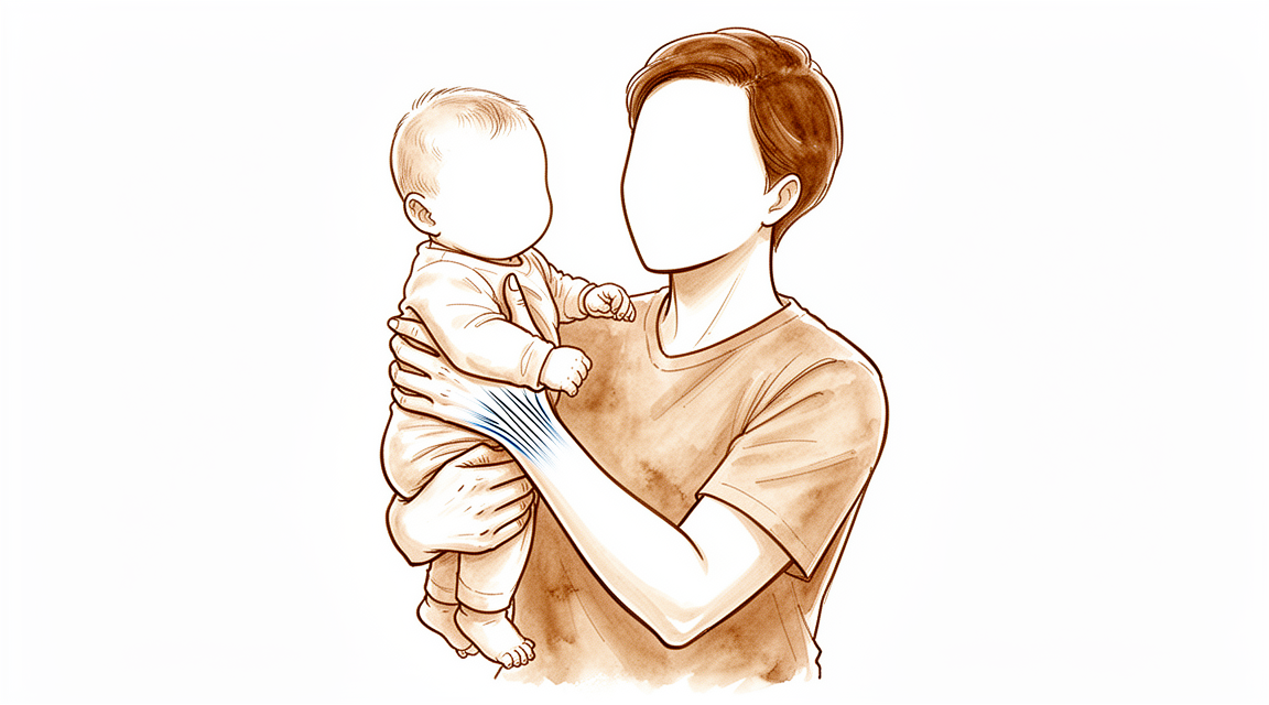
ڈاکٹر کیرن ہِرپرا، جو Mater Private Hospital Rockhampton میں بازو اور ہاتھ کے سرجن (upper-limb surgeon) ہیں، سب سے پہلے وہ طریقے آزماتے ہیں جن میں کم سے کم چیر پھاڑ ہو اور جو آپ کی حالت کے مطابق ہوں۔ ڈی کویروین ریلیز (De Quervain's release) ایک آپریشن ہے جو کلائی پر انگوٹھے کے دو ٹینڈنوں (tendon، یعنی وہ مضبوط ڈوریاں جو پٹھے کو ہڈی سے جوڑتی ہیں) کے گرد ٹشو کی سرنگ کو کھول دیتا ہے، تاکہ ٹینڈن اٹکے بغیر پھسل سکیں۔ ہم عموماً یہ آپریشن اس وقت پیش کرتے ہیں جب دوسرے علاجوں سے آپ کو کافی آرام نہ ملا ہو۔
مریض عموماً اپنے فیملی ڈاکٹر (GP) کے ریفرل (referral) پر ہمارے کلینک آتے ہیں؛ اگر کسی فزیوتھراپسٹ نے آپ کو ہمیں دکھانے کا مشورہ دیا ہے تو بھی میڈیکیئر (Medicare) کی رقم کی واپسی کا حق دار ہونے کے لیے آپ کو اپنے فیملی ڈاکٹر سے ریفرل لینا ہو گا۔ آپ کی ملاقات پر ہم آپ کی بیماری کی تفصیل لیتے ہیں، آپ کی کلائی کا معائنہ کرتے ہیں، اور اگر آپ کے درد کی وجہ کی تصدیق کے لیے ضرورت ہو تو اسکین کرواتے ہیں۔ اس بیماری میں عموماً پہلے آپریشن کے بغیر علاج کیا جاتا ہے۔ اس کا مطلب ہے کورٹیسون کا انجکشن، جو ابتدائی علاج کے طور پر ترجیحی طریقہ ہے، اور اکثر اس کے ساتھ اسپلنٹ اور ہینڈ تھراپی (ہاتھ کی بحالی کے ماہر سے علاج)۔ کورٹیسون کے ایک ہی انجکشن سے 82 فیصد مریضوں کی علامات میں آرام آیا، اور آدھے سے زیادہ کم از کم 12 ماہ تک علامات سے پاک رہے۔ دوبارہ انجکشن بھی اچھا کام کر سکتے ہیں۔ سرجری پر اس وقت غور کیا جاتا ہے جب انجکشن اور اسپلنٹ سے آپ کا درد ٹھیک نہ ہوا ہو، یا جب آپ کی علامات اتنی شدید ہوں کہ روزمرہ زندگی متاثر ہو۔ آپریشن کا مقصد درد سے آرام دینا اور آپ کو دوبارہ اپنا انگوٹھا اور کلائی معمول کے مطابق استعمال کرنے کے قابل بنانا ہے۔
آپریشن سے پہلے
آپ کے سرجن آپ کی ملاقات پر تشخیص کی تصدیق کریں گے، عموماً کلائی کے ہلکے معائنے سے اور، اگر ضرورت ہو تو، آپریشن کی منصوبہ بندی کے لیے ایکسرے، الٹراساؤنڈ یا ایم آر آئی اسکین جیسی تصویروں سے۔ زیادہ تر لوگوں کو سرجری کے دن سے پہلے اس کے علاوہ کسی چیز کی ضرورت نہیں ہوتی۔ اگر آپ کو دوسری بیماریاں ہیں تو آپ کو خون کے ٹیسٹ یا بے ہوشی کے ڈاکٹر (anaesthetist) سے معائنے کی ضرورت پڑ سکتی ہے۔ آپریشن والے دن سات گھنٹے پہلے کھانا پینا بند کر دیں؛ ہم یہ اس لیے کہتے ہیں تاکہ اگر آپریشن کی فہرست وقت سے پہلے چل رہی ہو تو آپ کا آپریشن پہلے کیا جا سکے۔ آپ کے سرجن آپ کو بتائیں گے کہ آپ اپنی معمول کی دواؤں میں سے کون سی نہ لیں۔ جو کچھ بھی آپ لیتے ہیں اس کی لکھی ہوئی فہرست ساتھ لائیں۔ اس بات کا انتظام کریں کہ کوئی آپ کو بعد میں گاڑی میں گھر لے جائے، اور ڈھیلے، آرام دہ کپڑے پہنیں جن کی آستین آسانی سے کلائی سے اوپر چڑھ جائے۔
آپریشن کے دن
آپ ہسپتال کے سرجیکل داخلے کے شعبے میں پہنچتے ہیں، جہاں آپ کا اندراج کیا جاتا ہے اور آپ کو آپریشن تھیٹر کے لیے تیار کیا جاتا ہے۔ پھر آپ بے ہوشی کے ڈاکٹر سے ملتے ہیں۔ یہ آپریشن لوکل اینستھیزیا (ایک انجکشن جو صرف آپریشن والی جگہ کو سُن کرتا ہے، اور آپ جاگ رہے ہوتے ہیں) یا جنرل اینستھیزیا (مکمل بے ہوشی) میں کیا جا سکتا ہے۔ زیادہ تر لوگ لوکل اینستھیزیا کا انتخاب کرتے ہیں: صحت یابی زیادہ تیز ہوتی ہے اور آپ جلد ہی گھر جا سکتے ہیں۔ اگر آپ بے ہوش ہونا پسند کریں تو یہ بھی ایک معقول انتخاب ہے؛ اس بارے میں اپنے سرجن اور بے ہوشی کے ڈاکٹر سے بات کریں۔
پھر آپ کو آپریشن تھیٹر میں لے جایا جاتا ہے، جہاں آپریشن کیا جاتا ہے۔ بعد میں آپ ریکوری روم میں ہوش میں آتے ہیں، جہاں اینستھیزیا کا اثر ختم ہونے تک نرسیں آپ کی نگرانی کرتی ہیں۔ جب آپ کی حالت مستحکم ہو جائے تو آپ یا تو وارڈ میں چلے جاتے ہیں یا گھر، یہ اس پر منحصر ہے کہ کون سا طریقہ کیا گیا اور آپ کیسے صحت یاب ہو رہے ہیں۔
آپریشن میں کیا کیا جاتا ہے
اس آپریشن کو پہلے ڈورسل کمپارٹمنٹ کی ریلیز (release of the first dorsal compartment) کہتے ہیں۔ یہ آپ کی کلائی کی انگوٹھے والی طرف ٹشو کی وہ سرنگ ہے جو اپنے اندر سے گزرنے والے دو ٹینڈنوں کو دباتی ہے۔ آپ کے سرجن اس سرنگ کے اوپر، کلائی کی انگوٹھے والی طرف، ایک چھوٹا چیرا لگاتے ہیں اور سرنگ کو کھول دیتے ہیں تاکہ ٹینڈن آزادی سے پھسل سکیں۔
بعض اوقات دونوں میں سے ایک ٹینڈن سرنگ کے اندر اپنی الگ جگہ میں ہوتا ہے، جسے ٹشو کی ایک دیوار الگ کرتی ہے۔ اگر آپ کے سرجن کو آپریشن کے دوران ایسا ملے تو وہ اس جگہ کو بھی کھول دیتے ہیں، تاکہ دونوں ٹینڈن آزاد ہو جائیں۔
جب ٹینڈن اٹکے بغیر پھسلنے لگیں تو آپ کے سرجن چیرے کو ٹانکوں سے بند کرتے ہیں اور اس پر زخم کی پٹی لگا دیتے ہیں۔ آپ یہ پٹی تقریباً 10 دن تک لگائے رکھتے ہیں، جیسا کہ اس کے بعد والے حصے میں بتایا گیا ہے۔
پورا آپریشن آپ کی کلائی کے ایک چھوٹے سے حصے پر کیا جانے والا ایک مختصر اور مرکوز طریقہ ہے۔ آپ اسی دن زخم کی دیکھ بھال کی ہدایات کے ساتھ گھر چلے جائیں گے۔
آپریشن کے بعد
آپ ریکوری روم میں ہوش میں آتے ہیں، جہاں اینستھیزیا کا اثر ختم ہونے تک نرسیں آپ پر نظر رکھتی ہیں۔ یہ عموماً ڈے کیس ہوتا ہے، اس لیے آپ اسی دن گھر جانے کی توقع رکھ سکتے ہیں، اگرچہ کبھی کبھار مریض رات بھر رکتے ہیں۔ پہلے 24 گھنٹے کسی کو آپ کے ساتھ رہنا چاہیے۔ آپ کی کلائی پر ٹانکوں کے اوپر زخم کی پٹی ہو گی، اور ہم یہ پٹی تقریباً 10 دن تک لگی رہنے دیتے ہیں؛ براہ کرم اس سے پہلے اسے نہ اتاریں جب تک ہم آپ کو نہ کہیں۔ جب ہم آپ کو دیکھتے ہیں تو ہم اسے بدلتے یا اتارتے ہیں۔ آپ اپنی انگلیاں، انگوٹھا اور کلائی اتنا ہلا سکتے ہیں جتنا آرام سے ہو سکے، اور زیادہ تر لوگ فوراً ہی ہلکے کاموں کے لیے اپنا ہاتھ استعمال کرنے لگتے ہیں۔ بہت سے لوگوں کو پہلے ایک دو دن میں ہی آرام محسوس ہونے لگتا ہے۔
صحت یابی
بہت سے لوگ پہلے ایک دو دن میں ہی آرام محسوس کرتے ہیں، جب تنگ سرنگ کھل جاتی ہے اور ٹینڈن پھسلنے لگتے ہیں۔ شروع کے دنوں میں زخم کے اردگرد کچھ درد اور سوجن معمول کی بات ہے۔ ہاتھ کو تکیے پر اونچا رکھ کر آرام کرنے سے اس میں کمی آتی ہے، اور اسی طرح انگلیوں، انگوٹھے اور کلائی کو اتنا ہلکا ہلانے سے بھی جتنا آرام سے ہو سکے۔
آپ فوراً ہی ہلکے کاموں کے لیے اپنا ہاتھ استعمال کر سکتے ہیں۔ زخم کی پٹی کو صاف اور خشک رکھیں، اور اسے اس وقت تک لگا رہنے دیں جب تک ہم اسے نہ دیکھ لیں۔ سرجری کے بعد ہینڈ تھراپی Extend Rehabilitation میں Ruby Doolan کے ساتھ ہوتی ہے۔ آپ کے ہینڈ تھراپسٹ سادہ ورزشوں میں آپ کی رہنمائی کریں گے تاکہ ٹینڈن ہمواری سے پھسلتے رہیں، اور اگر ضرورت ہو تو آپ کے لیے اسپلنٹ بنائیں گے۔
جیسے جیسے سوجن کم ہوتی ہے، روزمرہ کی سرگرمیاں آسان ہو جاتی ہیں۔ آپ دیکھیں گے کہ آپ کی گرفت اور انگوٹھے کی حرکت آہستہ آہستہ واپس آ رہی ہے۔ جب آپ اپنے ہاتھ کو بچائے بغیر اسٹیئرنگ کو پکڑ اور گھما سکیں، اور زخم آرام دہ ہو، تو عموماً آپ دوبارہ گاڑی چلا سکتے ہیں؛ ہمارا صفحہ بازو اور ہاتھ کی سرجری کے بعد گاڑی چلانا دیکھیں۔ کام اور دوسری سرگرمیاں آپ کے آرام اور طاقت کے مطابق دوبارہ شروع کی جا سکتی ہیں۔
صحت یابی ہر شخص میں مختلف ہوتی ہے۔ آپ کا وقت مختلف ہو سکتا ہے، اور آپ کے سرجن اور ہینڈ تھراپسٹ اس سارے عرصے میں آپ کی رہنمائی کریں گے۔
کیا مسائل پیدا ہو سکتے ہیں
زیادہ تر مریض ٹھیک رہتے ہیں، لیکن کبھی کبھار مسائل پیدا ہو سکتے ہیں۔ آپ کے سرجن اور ٹیم آپ کی قریب سے نگرانی کرتے ہیں تاکہ کوئی بھی مسئلہ جلد پکڑا جا سکے۔
بعض اوقات تنگ سرنگ پوری طرح نہیں کھلتی، یا پھسلتے وقت کوئی ٹینڈن اپنی نالی سے باہر نکل جاتا ہے۔ آپ کو آپریشن سے پہلے جیسا اٹکنے یا چٹکی کاٹنے جیسا احساس ہو سکتا ہے، یا انگوٹھا ہلاتے وقت اس کی جڑ پر جھٹکے سے کھسکنے کا احساس ہو سکتا ہے۔ اگر ایسا ہو تو اگلے معائنے پر اس کا ذکر کریں۔ اگر ٹینڈن بار بار کھسکتے رہیں تو مزید سرجری سے سرنگ کو دوبارہ بنایا جا سکتا ہے اور ٹینڈنوں کو اپنی جگہ پر رکھا جا سکتا ہے۔
وہ اعصاب جو آپ کے ہاتھ کی پشت اور انگوٹھے کو احساس دیتے ہیں، سرنگ کے قریب سے گزرتے ہیں۔ اگر سرجری کے دوران ان میں سے کسی میں جلن (irritation) پیدا ہو جائے تو آپ کو اپنے انگوٹھے یا ہاتھ کی پشت میں جھنجھناہٹ، سُن پن یا بجلی کے جھٹکے جیسا تیز احساس ہو سکتا ہے۔ ان میں سے زیادہ تر احساسات ٹھیک ہو جاتے ہیں، لیکن اگر ٹھیک نہ ہوں، یا بہتر ہونے کے بجائے بدتر ہونے لگیں، تو اپنے سرجن کو بتائیں۔
صحت یابی میں آپ کی امید سے زیادہ وقت بھی لگ سکتا ہے۔ کچھ لوگوں میں درد اور اکڑاؤ دنوں کے بجائے ہفتوں تک رہتا ہے، اور یہ پریشان کن ہو سکتا ہے۔ اگر مناسب وقت گزرنے کے بعد بھی آپ کی کلائی میں درد ہو، یا جس آرام کی آپ کو توقع تھی وہ نہ ملا ہو، تو معائنے کی ملاقات پر اس کا ذکر کریں تاکہ ہم دیکھ سکیں کہ کیا ہو رہا ہے۔
کبھی کبھار سرجری سے پہلے لگایا گیا کورٹیسون کا انجکشن توقع کے مطابق کام نہیں کرتا، اور درد جاری رہتا ہے۔ اگر آپ کے ساتھ ایسا ہوا ہے تو اس کا مطلب یہ نہیں کہ آپ کی کلائی میں کچھ غلط ہو گیا ہے۔ اس کا مطلب صرف یہ ہے کہ اگلا قدم، یعنی سرجری، آپ کے لیے درست ہے۔
اگر آپ تفصیل جاننا چاہیں تو اس صفحے پر پیچیدگیوں کی جدول میں عام شرحیں دی گئی ہیں۔
ہمیں کب فون کریں
زیادہ تر لوگ بغیر کسی مسئلے کے صحت یاب ہو جاتے ہیں، لیکن کچھ علامات پر فوری توجہ کی ضرورت ہوتی ہے۔ اگر آپ کو بخار ہو، اگر زخم زیادہ سرخ ہو جائے یا اس سے رطوبت رسنے لگے، یا اگر آپ کا درد اچانک بہت بڑھ جائے تو ہمیں فون کریں۔ اگر آپ کی پنڈلی میں سوجن یا درد ہو، یا سانس پھول رہی ہو تو ہسپتال کی ایمرجنسی جائیں۔ اگر آپ کا ہاتھ یا انگوٹھا سُن ہو جائے اور سُن ہی رہے، یا آپ اپنی انگلیاں یا انگوٹھا نہ ہلا سکیں تو ہمیں فوراً فون کریں۔ جب شک ہو تو ہمیں فون کریں۔
اس مسئلے کے بارے میں مزید کہاں پڑھیں
یہ صفحہ خود آپریشن کے بارے میں ہے۔ جس بیماری کا یہ علاج کرتا ہے، بشمول اس کے کہ شواہد کیا بتاتے ہیں کہ سرجری کب مدد کرتی ہے اور کب نہیں، اس کی مزید تفصیل ڈی کویروین ٹینوسائنووائٹس کے صفحے پر ہے۔
Evidence & references
This is the clinical evidence summary written for health professionals. It is technical, and it lists the research this page was built from. You do not need to read it to understand your treatment or to make a decision about it.
Anatomy & Pathophysiology
First Extensor Compartment Anatomy
- A common septum is found between the abductor pollicis longus (APL) and extensor pollicis brevis (EPB) tendons in 80% of patients requiring surgical release for de Quervain tenosynovitis [2].
- The first dorsal compartment of the wrist contains the APL and EPB tendons [7].
- The fibrous and osseous components of the first extensor compartment of the wrist have been characterized in cadaveric studies [1].
Wrist Bony Anatomy
- The wrist includes the distal radioulnar, radiocarpal, and ulnocarpal joints, as well as the eight carpal bones and their proximal and distal articulations [3].
- The proximal row of carpal bones consists of the scaphoid, lunate, triquetrum, and pisiform [3].
- The distal row of carpal bones consists of the trapezium, trapezoid, capitate, and hamate [3].
- The distal radius has three articular components: the scaphoid fossa, the lunate fossa, and the sigmoid notch [7].
- The concave elliptical distal radius is oriented in the sagittal plane with an average of 11 degrees of volar tilt [7].
- In the frontal plane, the average radial inclination of the distal radius is 23 degrees [7].
- Radial length is measured from the tip of the radial styloid to the ulnar articular surface and averages 13 mm [7].
- The distal ulnar convexity articulates at the lesser sigmoid notch of the distal radius [3].
- There is about a 20-degree inclination of the distal ulna at its articulation with the radius [3].
Wrist Ligaments and Soft Tissue
- Extrinsic carpal ligaments connect the radius or the ulna to the carpus [8].
- In general, volar wrist ligaments are stronger than dorsal ligaments [8].
- The triangular fibrocartilage complex (TFCC) attaches to the ulnar margin of the lunate fossa of the radius and includes the ulnar collateral ligament, dorsal and volar radioulnar ligaments, articular disc, meniscal homologue, extensor carpi ulnaris sheath, and ulnolunate and ulnotriquetral ligaments [3].
- The space of Poirier is a relatively thin area on the palmar side of the carpus situated between the radiolunotriquetral ligament and the radioscapocapitate ligament, overlying the palmar surface of the lunate [3].
- The space of Poirier is a weak area vulnerable to instability where the distal carpal row separates from the lunate during a perilunate dislocation [8].
- The dorsal radiocarpal ligament has a trapezoidal shape and passes from the dorsal rim of the distal radius to the lunate and the triquetrum [8].
- The scapholunate interosseous ligament is C-shaped, consisting of dorsal, palmar, and interosseous portions, with the dorsal portion being the strongest and thickest [8].
- The lunotriquetral interosseous ligament is C-shaped, with the volar portion being the thickest and strongest [8].
Wrist Biomechanics and Kinematics
- The radius bears 80% of the axial load transmitted through the radiocarpal joint, while the ulna bears 20% in neutral ulnar variance [8].
- Approximately 62° of wrist extension occurs through the radiocarpal joint [8].
- Approximately 62% of wrist flexion occurs through the midcarpal joint [8].
- The midcarpal joint is mostly responsible for 20° of radial deviation and 40° of ulnar deviation [8].
- The midcarpal joint is responsible for the “dart thrower’s motion,” which involves moving from radial extension into ulnar flexion positioning of the wrist [8].
- During wrist flexion from neutral, the proximal row translates dorsally [8].
- During wrist extension from neutral, the proximal row translates palmarly [8].
- The extensor carpi ulnaris (ECU) ulnarly deviates the wrist when the forearm is in pronation and extends the wrist when the forearm is supinated [2].
- The ECU provides both static and dynamic support to the wrist [2].
- The volar ECU sheath is adjacent to the TFCC, and ECU injury may coexist with concomitant TFCC injury [2].
Pathophysiology and Associated Conditions
- Tenosynovitis of the ECU is a common cause of ulnar-sided wrist pain [2].
- Painful dorsal wrist swelling may be the presenting symptom in rheumatoid arthritis, and tenosynovial swelling may contribute to de Quervain disease [11].
- In rheumatoid arthritis, synovitis can spread and cause massive swelling in the shape of an hourglass, with the middle constricted by the extensor retinaculum [11].
- The human wrist is described as the most complex articular system in the human body, with each carpal bone capable of moving in different degrees or directions dependent on hand position, motion, and force generation [4].
Physical Examination
- Patients with ECU tenosynovitis present with tenderness over the ECU tendon [2].
- Patients with ECU tenosynovitis present with swelling [2].
- Patients with ECU tenosynovitis present with pain with resisted wrist extension [2].
- Patients with ECU tenosynovitis do not have instability [2].
- The ECU synergy test helps distinguish a TFCC injury from ECU tenosynovitis [2].
- In the ECU synergy test, the patient rests the elbow of the supinated forearm on a table with the digits extended [2].
- In the ECU synergy test, the examiner grasps the patient’s thumb and long finger and asks the patient to deviate the thumb radially against resistance [2].
- In the ECU synergy test, the examiner’s other hand gently palpates the ECU and flexor carpi ulnaris [2].
- The presence of pain during the ECU synergy test suggests pathology in the ECU rather than intra-articular pathology [2].
Imaging
- Ultrasonography can be used to distinguish tenosynovitis from ECU subsheath injury in patients whose examination is equivocal [2].
- MRI can be used to distinguish tenosynovitis from ECU subsheath injury in patients whose examination is equivocal [2].
Investigations
Magnetic Resonance Imaging
- MRI has been an important diagnostic tool for hand and wrist imaging since its first report in 1986 [9].
- The primary advantages of MRI compared with CT and radiography are improved tissue characterization, especially of soft tissues such as ligamentous structures and synovium, and the lack of ionizing radiation [9].
- MRI is the modality of choice for imaging radiographically occult fractures of the hand and wrist [9].
- Early MRI was limited by low magnetic field strength (0.15 T) and limited image options and processing [9].
- Modern MRI is generally performed at 1.5T or 3T with a wide variety of imaging options and powerful image processing [9].
- 3T MRI is much preferred for hand and wrist imaging, especially for imaging small fields of view [9].
- Dedicated extremity magnets have been marketed, but their image quality is poor compared with conventional MR imaging [9].
- 7T MRI has recently become approved for clinical use and has the potential to become a powerful tool for hand and wrist imaging as applications are developed [9].
- MRI with contrast enhancement is most commonly used to determine whether soft-tissue lesions are solid or cystic or to better visualize erosions and synovial burden in rheumatologic imaging [9].
- Dynamic contrast enhancement has been used with inconsistent results to assess for the presence of avascular necrosis in the lunate or scaphoid after injury [9].
- MR angiography of the hand and wrist can be helpful in situations such as the diagnosis of hypothenar hammer syndrome [9].
- MR arthrography can be performed for evaluation of triangular fibrocartilage and intercarpal ligament tears, but this is generally unnecessary with the increasing availability of high-field MRI [9].
- The use of MRI for hand and wrist imaging was first reported in 1986 [9].
Radiography and Computed Tomography
- Hybrid SPECT/CT has been used for the diagnosis of radiographically occult fractures of the wrist [5].
- Cone-beam CT has been used in the diagnosis of scaphoid fractures [5].
- CT and MRI have a diagnostic impact on wrist injuries in young adults [5].
- Distal radioulnar joint stress radiography has been used for detecting radioulnar ligament injury [5].
- Cine MRI has been used as a new approach to the diagnosis of scapholunate dissociation [5].
- Radiographic stress views have been compared for scapholunate dynamic instability in a cadaver model [5].
- Cone-beam computed tomography arthrography is an innovative modality for the evaluation of wrist ligament and cartilage injuries [5].
- Simple plain radiographic signs and measures have been evaluated for the accuracy of diagnosing acute scapholunate ligament injuries of the wrist [10].
- Radiographic clues have been used for determining carpal instability and treatment protocols for scaphoid fractures [10].
References
[1] Campbell S Operative Orthopaedics 4 Volume Set. ANATOMIC RECONSTRUCTION OF THE DISTAL RADIOULNAR LIGAMENTS > ANATOMY AND BIOMECHANICS.
[2] Aaos Comprehensive Orthopaedic Review 3. Tendinopathy of the Hand and Wrist* > VII. Extensor Carpi Ulnaris Tenosynovitis.
[3] Campbell S Operative Orthopaedics 4 Volume Set. NERVE INJURIES AT THE LEVEL OF THE HAND AND WRIST > ANATOMY.
[4] Green S Operative Hand Surgery. INTRODUCTION.
[5] Campbell S Operative Orthopaedics 4 Volume Set. ANATOMIC RECONSTRUCTION OF THE DISTAL RADIOULNAR LIGAMENTS > RADIOGRAPHIC TECHNIQUES.
[7] A Lange Medical Book Current Diagnosis Treatment In Orthopedics Fifth Edition. 2Musculoskeletal Trauma Surgery > FRACTURES AND DISLOCATIONS OF THE DISTAL AND MID-FOREARM.
[8] Aaos Comprehensive Orthopaedic Review 3. Carpal Instability* > II. Anatomy and Biomechanics (See Chapter 92).
[9] Orthopaedic Knowledge Update 13 Ebook Without Multimedia. Anatomy, Evaluation, Clinical Examination, and Imaging > Imaging: Advances in Imaging of the Hand and Upper Extremity > Magnetic Resonance Imaging.
[10] Campbell S Operative Orthopaedics 4 Volume Set. ANATOMIC RECONSTRUCTION OF THE DISTAL RADIOULNAR LIGAMENTS > DIAGNOSIS AND EVALUATION.
[11] Campbell S Operative Orthopaedics 4 Volume Set. THUMB CARPODNETACARPAL ARTHRODESIS WITH KIRSCHNER WIRE OR BLADE-PLATE FIXATION > SYNOVITIS OF THE WRIST.