Patients › Hand
ہاتھ کے انفیکشن
Paronychia, deep-space and flexor sheath infections — recognition, antibiotics, drainage.
آپ کو کیا محسوس ہوتا ہے
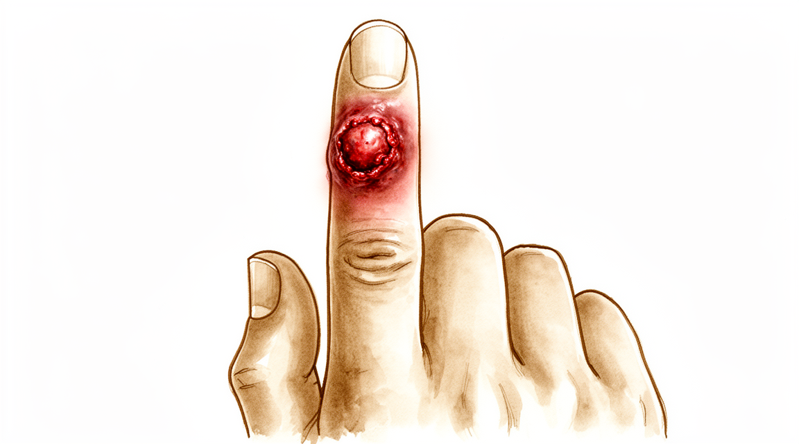
ہاتھ کے انفیکشن میں عام طور پر ایک ہی جگہ پر درد ہوتا ہے۔ اس جگہ کی جلد سرخ، گرم اور سوجی ہوئی ہو سکتی ہے۔ انگلی ہلانے یا کوئی چیز پکڑنے پر درد اکثر بڑھ جاتا ہے۔ ہاتھ کو آرام دینے سے درد کچھ کم ہو جاتا ہے، لیکن یہ تکلیف خود بخود ختم نہیں ہوتی۔
روزمرہ کے کام مشکل ہو جاتے ہیں۔ بٹن بند کرنا، چابی گھمانا، چائے کا کپ پکڑنا یا ٹائپ کرنا، ان سب میں چبھن ہو سکتی ہے۔ اگر انفیکشن انگلی کے ٹینڈن (tendon)، یعنی وہ مضبوط ڈوری جو پٹھے کو ہڈی سے جوڑتی ہے، کے غلاف میں ہو (یعنی وہ سرنگ جس میں انگلی موڑنے والا ٹینڈن پھسلتا ہے)، تو پوری انگلی سوج سکتی ہے اور ہلکی سی مڑی ہوئی حالت میں رہ سکتی ہے۔ اسے سیدھا کرنے میں درد ہوتا ہے، اور یہ درد اس وقت بھی ہوتا ہے جب کوئی دوسرا شخص اسے آہستہ سے سیدھا کرنے کی کوشش کرے۔ اگر کسی جوڑ میں انفیکشن ہو، تو وہ جوڑ سوجا ہوا اور گرم ہوتا ہے، اسے چھونے پر درد ہوتا ہے، اور اسے کسی بھی سمت میں ہلانا کافی تکلیف دہ ہوتا ہے۔
کچھ انفیکشن جلد پر سرخ لکیروں کی صورت میں پھیلتے ہیں جنہیں چھونے پر درد ہوتا ہے، اور آپ کی بغل کی گلٹیوں میں درد ہو سکتا ہے۔ سوجن کی وجہ سے ہاتھ کی پشت پھولی ہوئی نظر آ سکتی ہے، اور یہ بتانا مشکل ہو سکتا ہے کہ سیال کہاں جمع ہوا ہے۔ رات کا وقت تکلیف دہ ہو سکتا ہے کیونکہ جب ہاتھ ساکن رہتا ہے تو سوجن بڑھ جاتی ہے۔
زیادہ تر انفیکشن ہفتوں کے بجائے چند گھنٹوں یا دنوں میں ظاہر ہوتے ہیں۔ اگر سرخی، سوجن اور درد کم ہونے کے بجائے بڑھ رہے ہوں، تو یہ عام موچ کے بجائے انفیکشن کی نشانی ہے۔ کچھ دوسری بیماریاں تقریباً ایک جیسی نظر آتی ہیں، جیسے گاؤٹ (gout)، کیڑے کا کاٹنا، لکڑی کی پھانس (splinter) پر جسم کا ردِعمل یا آرتھرائٹس (جوڑ کی خرابی اور سوزش) کا بھڑک اٹھنا، اس لیے شروع میں وجہ ہمیشہ واضح نہیں ہوتی۔
اگر آپ کو ذیابیطس (شوگر)، گردے کی بیماری، یا کوئی ایسی بیماری یا دوا ہے جو آپ کے مدافعتی نظام کو کمزور کرتی ہے، تو اپنے سرجن کو ضرور بتائیں۔ ان سے یہ بدل جاتا ہے کہ انفیکشن کیسا برتاؤ کرتا ہے اور اس کا علاج کیسے کرنا ہوتا ہے۔ یہی بات کسی ایسے انفیکشن پر بھی لاگو ہوتی ہے جو اینٹی بائیوٹک دواؤں کے بعد یا زخم کی صفائی کے بعد ٹھیک نہ ہوا ہو، کیونکہ کچھ آہستہ بڑھنے والے انفیکشن کی شناخت کے لیے خاص ٹیسٹ درکار ہوتے ہیں۔
بروقت علاج سے ہاتھ کے نرم بافتوں (soft tissue) کے زیادہ تر انفیکشن مکمل طور پر ٹھیک ہو جاتے ہیں۔ انتظار کرنے کا خطرہ یہ ہے کہ اس کے بعد اکڑاؤ، انگلی کا مستقل طور پر مڑا رہ جانا، یا شدید صورتوں میں انگلی سے محروم ہونا ہو سکتا ہے۔
دراصل کیا ہو رہا ہے
آپ کا ہاتھ ایک بھری ہوئی جگہ ہے۔ ستائیس ہڈیاں ایک دوسرے کے بہت قریب جلد کے نیچے موجود ہیں، اور یہ جلد ہاتھ کی پشت پر پتلی اور ہتھیلی میں مضبوطی سے جمی ہوتی ہے۔ ٹینڈن، یعنی وہ ڈوریاں جو آپ کی انگلیوں کو کھینچتی ہیں، تنگ سرنگوں میں سے پھسلتے ہیں۔ ان میں سے ایک سرنگ، جسے فلیکسر شیتھ (flexor sheath) یعنی ٹینڈن کا غلاف کہتے ہیں، انگلی کو موڑنے والے ٹینڈن کے گرد لپٹی ہوتی ہے اور اس کے اندر ایک چکنی تہہ ہوتی ہے جو ٹینڈن کو غذا اور چکناہٹ فراہم کرتی ہے۔ جب جراثیم کسی کٹ، لکڑی کی پھانس یا کسی نوکیلی چیز کے چبھنے سے بنے زخم کے ذریعے اندر داخل ہوتے ہیں، تو اس بند جگہ میں ان کے پاس سرنگ کے ساتھ ساتھ پھیلنے کے سوا کوئی راستہ نہیں ہوتا۔
اصل مسئلہ سوجن ہے۔ ایسی سرنگ کے اندر سوجن کے لیے کوئی جگہ نہیں ہوتی، اس لیے دباؤ بڑھ جاتا ہے۔ اگر دباؤ بڑھتا رہے تو یہ خود ٹینڈن کو خون کی فراہمی بند کر سکتا ہے۔ اس کے بعد ٹینڈن زخم بھرنے سے بننے والے سخت ریشے (scar tissue) کے ذریعے اپنے گرد کی سرنگ سے چپک سکتا ہے، جس سے انفیکشن ختم ہو جانے کے بعد بھی انگلی اکڑی رہ جاتی ہے۔ یہی وجہ ہے کہ ٹینڈن کے غلاف کے انفیکشن کا علاج چند دن انتظار کرنے کے بجائے جلدی کرنا ضروری ہے۔
دوسری جگہوں کے اپنے خطرات ہیں۔ آپ کی انگلی کے سرے پر موجود چربی کی گدی مضبوط ریشوں کے ذریعے چھوٹے چھوٹے بند خانوں میں بٹی ہوتی ہے، اس لیے وہاں کا انفیکشن، یعنی انگلی کے پور کا پھوڑا (felon)، تیزی سے دباؤ پیدا کرتا ہے اور ہڈی یا قریبی جوڑ تک پھیل سکتا ہے۔ آپ کے ناخن کے ساتھ والی جلد میں بھی ایک آہستہ، اندر ہی اندر سلگنے والا انفیکشن (chronic paronychia، ناخن کے کنارے کا دیرینہ انفیکشن) رہ سکتا ہے، جو اکثر خمیر (yeast) کی وجہ سے ہوتا ہے، اور اس میں نمی ناخن کی جڑ کی جلد (cuticle) کے نیچے گھس جاتی ہے اور اس میں سوزش برقرار رکھتی ہے۔ اور کچھ لوگوں میں انگوٹھے اور چھوٹی انگلی کے ٹینڈنوں کی سرنگیں آپس میں جڑی ہوتی ہیں، اس لیے انفیکشن ہتھیلی کی ایک طرف سے دوسری طرف تک پہنچ سکتا ہے۔
ہاتھ کے زیادہ تر انفیکشن ان جراثیم (bacteria) سے ہوتے ہیں جو عام طور پر جلد پر رہتے ہیں اور جلد میں کسی کٹ یا شگاف کے ذریعے اندر داخل ہو جاتے ہیں۔ کچھ انفیکشن انسان یا جانور کے کاٹنے سے ہوتے ہیں۔ ذیابیطس، اسٹیرائیڈ دوائیں یا کمزور مدافعتی نظام انفیکشن کے لیے جڑ پکڑنا اور ٹینڈن، ہڈی یا جوڑ جیسے گہرے حصوں تک پہنچنا آسان بنا دیتے ہیں۔ اسی لیے آپ کا سرجن آپ کی صحت کی سابقہ تاریخ کے بارے میں پوچھے گا، اور اسی لیے جلد علاج اہم ہے: بروقت علاج سے نرم بافتوں کے زیادہ تر انفیکشن مکمل طور پر ٹھیک ہو جاتے ہیں، جبکہ تاخیر سے دیرپا اکڑاؤ کا خطرہ ہوتا ہے۔
ہم اس کے لیے کیا کر سکتے ہیں
ڈاکٹر کیرن ہِرپرا، جو Mater Private Hospital Rockhampton میں بازو اور ہاتھ کے سرجن (upper-limb surgeon) ہیں، ہاتھ کے انفیکشن کا علاج تیزی سے قدم اٹھا کر کرتے ہیں، کیونکہ تاخیر ہی دیرپا نقصان کا سبب بنتی ہے۔ مریضوں کو عام طور پر ان کا فیملی ڈاکٹر (GP) ہمارے کلینک میں بھیجتا ہے؛ اگر کسی فزیوتھراپسٹ نے آپ کو ہمارے پاس آنے کا مشورہ دیا ہے، تب بھی میڈیکیئر (Medicare) کی رقم کی واپسی (rebate) کا اہل ہونے کے لیے آپ کو اپنے فیملی ڈاکٹر سے ریفرل (referral) لینا ہوگا۔ اس ملاقات میں ہم آپ کی بیماری کی تفصیل پوچھتے ہیں، آپ کے ہاتھ کا معائنہ کرتے ہیں اور ضروری ٹیسٹ کرواتے ہیں تاکہ پتا چل سکے کہ کون سا جراثیم بڑھ رہا ہے اور انفیکشن کتنا گہرا جا چکا ہے۔
بہت سے ابتدائی انفیکشن میں پہلا قدم آپریشن کے بغیر علاج ہوتا ہے۔ اگر ٹینڈن کے غلاف کا انفیکشن علامات شروع ہونے کے 48 گھنٹوں کے اندر پکڑ لیا جائے، تو اینٹی بائیوٹک دوائیں اور اسپلنٹ اسے پھیلنے سے روک سکتے ہیں۔ اس کے بعد آپ کی قریبی نگرانی کی جائے گی، اور اگر حالت جلد بہتر نہ ہو تو ہم انتظار کرنے کے بجائے آپریشن کی طرف جائیں گے۔ کسی بھی علاج کے بعد ہاتھ کو ایک محفوظ حالت میں اسپلنٹ میں رکھنا اور پھر انگلیوں کو جلد ہلانا شروع کرنا اکڑاؤ سے بچانے میں مدد دیتا ہے۔ یہاں بحالی (rehabilitation) بہت اہم ہے: جس ہاتھ میں انفیکشن ہو اور اسے ہلایا نہ جائے وہ اکڑ جائے گا، اس لیے تھراپی علاج کے منصوبے کا حصہ ہے، کوئی اختیاری اضافہ نہیں۔
اینٹی بائیوٹک دوائیں علاج کا دوسرا طبی حصہ ہیں۔ پہلا نسخہ اس طرح چنا جاتا ہے کہ وہ ان جراثیم پر اثر کرے جو آپ کے انفیکشن کا سب سے زیادہ ممکنہ سبب ہیں، اور جب لیبارٹری کے نتائج سے ٹھیک ٹھیک پتا چل جائے کہ کون سا جراثیم ہے، تو ہم اس کے لیے موزوں اینٹی بائیوٹک پر منتقل ہو جاتے ہیں۔ کچھ انفیکشن میں گولیوں کے بجائے ڈرپ کے ذریعے اینٹی بائیوٹک دینی پڑتی ہے، اور سنگین انفیکشن میں علاج کا کورس 4 سے 6 ہفتے تک چل سکتا ہے۔ اگر آپ کا مدافعتی نظام کسی بیماری یا اسٹیرائیڈ جیسی دواؤں کی وجہ سے کمزور ہو، تو ہم زیادہ تیزی سے اور زیادہ بھرپور طریقے سے علاج کرتے ہیں، کیونکہ ایسے انفیکشن زیادہ جلدی سنگین ہو سکتے ہیں۔ انسان یا جانور کے کاٹنے کی صورت میں عام طور پر احتیاطی اینٹی بائیوٹک دوائیں بھی دینی پڑتی ہیں۔
آپریشن پر اس وقت غور کیا جاتا ہے جب نکالنے کے لیے پیپ موجود ہو یا انفیکشن ٹینڈن، ہڈی یا جوڑ تک پہنچ چکا ہو۔ پھوڑے (abscess) یا جلد کے نیچے جمع مواد کو کھولنا پڑتا ہے تاکہ دباؤ نکل سکے، اور شروع میں زخم کے اندر اکثر پٹی بھر دی جاتی ہے تاکہ مواد نکلتا رہے۔ انگلی کے پور کا پھوڑا (felon) یا ناخن کے ساتھ والا انفیکشن (paronychia) ایک چھوٹے چیرے کے ذریعے نکالا جاتا ہے، اور بعض اوقات ناخن کا کچھ حصہ بھی ہٹایا جاتا ہے۔ ٹینڈن کے غلاف کے انفیکشن میں اس سرنگ کو دھونا پڑتا ہے، اور ہڈی کے انفیکشن (osteomyelitis) میں مردہ بافتوں کو صاف کرنا اور ساتھ لمبے عرصے تک اینٹی بائیوٹک دینا ضروری ہوتا ہے۔ ایک اہم استثنا: اگر آپ کی انگلی کا کوئی چھالا وائرل انفیکشن (herpetic whitlow) نکلے، تو اس کا علاج آپریشن سے نہیں کیا جاتا، اور اسے کاٹ کر کھولنے سے حالت مزید بگڑ سکتی ہے۔
کیا توقع رکھیں
ہاتھ کے زیادہ تر انفیکشن جو جلد پکڑ لیے جائیں، علاج سے مکمل طور پر ٹھیک ہو جاتے ہیں۔ بروقت علاج سے نرم بافتوں کے زیادہ تر انفیکشن پوری طرح ختم ہو جاتے ہیں، اور ہاتھ معمول پر آ جاتا ہے۔ آپ کا معائنہ جتنی جلدی ہو، اکڑاؤ یا دیرپا نقصان سے بچنے کا امکان اتنا ہی بہتر ہوتا ہے۔
صحت یابی میں کتنا وقت لگتا ہے، یہ اس بات پر منحصر ہے کہ انفیکشن کتنا گہرا گیا تھا۔ جلد یا ناخن کا انفیکشن مواد نکالنے یا اینٹی بائیوٹک کے چند دنوں کے اندر ٹھیک ہو سکتا ہے۔ ٹینڈن کے غلاف، ہڈی یا کسی جوڑ کے انفیکشن میں زیادہ وقت لگتا ہے، اور کچھ میں کئی ہفتوں تک اینٹی بائیوٹک کی ضرورت ہوتی ہے۔ انفیکشن ختم ہو جانے کے بعد بھی انگلی کچھ عرصے تک اکڑی رہ سکتی ہے۔ جب آپ کا سرجن کہے کہ ایسا کرنا محفوظ ہے، تو انگلیوں کو جلد ہلانا شروع کرنا اس اکڑاؤ کو مستقل بننے سے روکنے میں مدد دیتا ہے۔
اگر علاج میں تاخیر ہو جائے تو نتیجے کے بارے میں اتنے یقین سے کچھ نہیں کہا جا سکتا۔ ٹینڈن کے غلاف کا جو انفیکشن کئی دنوں سے موجود ہو، وہ زخم کے سخت ریشے کے ذریعے ٹینڈن کو اس کی سرنگ سے چپکا سکتا ہے، جس سے انفیکشن ختم ہونے کے بعد بھی انگلی اکڑی رہ جاتی ہے۔ مکمل اور بروقت علاج کے باوجود بھی شدید انفیکشن ہاتھ کی کارکردگی کے دیرپا نقصان کا، یا بعض صورتوں میں انگلی سے محرومی کا سبب بن سکتے ہیں۔ یہی وجہ ہے کہ ان انفیکشن کو چند دن تک دیکھتے رہنے کے بجائے ہنگامی مسئلہ سمجھ کر ان کا علاج کیا جاتا ہے۔
کچھ انفیکشن مختلف طرح سے برتاؤ کرتے ہیں۔ ذیابیطس یا کمزور مدافعتی نظام والے لوگوں میں انفیکشن تیزی سے پھیل کر گہرے حصوں تک پہنچ سکتے ہیں، اس لیے انہیں زیادہ بھرپور علاج کی اور بعض اوقات ایک سے زیادہ آپریشن کی ضرورت ہوتی ہے۔ آہستہ بڑھنے والے انفیکشن، جیسے مائیکوبیکٹیریا (mycobacteria، جراثیم کا ایک خاندان جو ٹی بی پیدا کرنے والے جراثیم سے تعلق رکھتا ہے) سے ہونے والے انفیکشن، کو ختم کرنے کے لیے آپریشن کے ساتھ ساتھ لمبے عرصے تک اینٹی بائیوٹک کی ضرورت ہوتی ہے، اور صحت یابی دنوں کے بجائے مہینوں پر پھیل جاتی ہے۔
علاج کے بعد عام طور پر آپ کو دوبارہ دیکھا جائے گا تاکہ یہ جانچا جا سکے کہ انفیکشن ختم ہو رہا ہے اور آپ کا ہاتھ ٹھیک سے حرکت کر رہا ہے۔ یہ ملاقاتیں ضرور رکھیں چاہے آپ بہتر محسوس کر رہے ہوں، کیونکہ کچھ مسائل سوجن کم ہونے کے بعد ہی سامنے آتے ہیں۔ اگر انفیکشن ٹھیک ہوتا دکھائی دینے کے بعد درد، سرخی یا سوجن واپس آ جائے، تو انتظار کرنے کے بجائے اپنے سرجن سے رابطہ کریں۔
ڈاکٹر کو کب دکھائیں
ہاتھ کے انفیکشن میں وقت بہت اہم ہوتا ہے۔ تاخیر سے ہونے والا نقصان تیزی سے ہوتا ہے، اس لیے یہ دیکھنے کا انتظار نہ کریں کہ حالت خود ٹھیک ہوتی ہے یا نہیں۔
اگر آپ کے ہاتھ یا بازو پر اوپر کی طرف جاتی ہوئی سرخ لکیریں ہوں، اگر پوری انگلی سوجی ہوئی اور مڑی ہوئی ہو اور اتنی دردناک ہو کہ سیدھی نہ ہو سکے، یا اگر جلد تیزی سے سیاہ پڑ رہی ہو، اس پر چھالے بن رہے ہوں یا وہ گل کر پھٹ رہی ہو، تو ہسپتال کی ایمرجنسی جائیں۔ یہی بات اس صورت میں بھی لاگو ہوتی ہے اگر ہاتھ کی علامات کے ساتھ آپ کو بخار محسوس ہو اور طبیعت خراب ہو، یا اگر ہاتھ پر چوٹ لگی ہو اور اب وہ بہت زیادہ سوجا ہوا اور تنا ہوا ہو۔ یہ علامات کسی پھیلتے ہوئے یا گہرے انفیکشن کی نشانی ہو سکتی ہیں جس کا اسی دن معائنہ اور علاج ضروری ہے۔
اگر درد شدید ہو اور بڑھ رہا ہو، اگر انگلی کا کوئی جوڑ گرم، سوجا ہوا اور اتنا دردناک ہو کہ ہل نہ سکے، یا اگر آپ کو جلد کے نیچے یا ناخن کے ساتھ پیپ جمع ہوتی نظر آئے، تو فوری معائنے کی درخواست کریں، اگر ممکن ہو تو اسی دن۔ جو انفیکشن ایک یا دو دن اینٹی بائیوٹک لینے کے بعد بہتر نہ ہوا ہو، اس کا بھی دوبارہ معائنہ ضروری ہے، نسخہ ختم ہونے کا انتظار نہ کریں۔
ہاتھ کے کسی بھی انفیکشن کے لیے، چاہے وہ چھوٹا ہی کیوں نہ ہو، بلا تاخیر اپنے فیملی ڈاکٹر کو دکھائیں۔ یہ بات اور بھی زیادہ اہم ہے اگر آپ کو ذیابیطس، گردے کی بیماری، یا کوئی ایسی بیماری یا دوا ہے جو آپ کے مدافعتی نظام کو کمزور کرتی ہے، کیونکہ ایسی صورتوں میں انفیکشن تیزی سے پھیل کر زیادہ گہرائی تک پہنچ سکتے ہیں۔ اپنے فیملی ڈاکٹر کو حال ہی میں کسی جانور یا انسان کے کاٹنے، کسی نوکیلی چیز کے چبھنے سے لگے زخم، یا اپنے کام اور سفر کی ایسی تفصیل کے بارے میں بتائیں جو کسی غیر معمولی جراثیم کی طرف اشارہ کر سکتی ہو۔
اگر کوئی انفیکشن بار بار واپس آتا رہے یا علاج کے باوجود کبھی پوری طرح ٹھیک نہ ہو، تو کسی ماہر ڈاکٹر (specialist) سے معائنے کی درخواست کریں، کیونکہ کچھ آہستہ بڑھنے والے انفیکشن کی شناخت کے لیے خاص ٹیسٹ درکار ہوتے ہیں۔
مزید تفصیل
Advanced reading: the deeper science (optional)
یہ حصہ اس سے زیادہ تفصیل میں جاتا ہے جتنی آپ کو اپنے علاج کے فیصلوں کے لیے ضرورت ہے۔ ہاتھ کے انفیکشن کے بارے میں یہ اضافی مطالعہ اس لیے مفید ہے کہ زیادہ تر لوگوں کو جو اینٹی بائیوٹک سب سے پہلے دی جاتی ہیں، وہ اس خاص صورتحال میں غلط ہوتی ہیں، اور اس لیے کہ ہاتھ کا ایک انفیکشن سرجری کی ایمرجنسی ہے جسے اکثر معمولی انفیکشن سمجھ لیا جاتا ہے۔
سب سے عام جراثیم سب سے عام نسخوں کے خلاف مزاحمت رکھتا ہے
شہری علاقوں میں ہاتھ کے 815 انفیکشن پر کی گئی ایک دس سالہ طویل مدتی تحقیق سے معلوم ہوا کہ اگرچہ ایم آر ایس اے (MRSA، اینٹی بائیوٹک کے خلاف مزاحمت رکھنے والا ایک جراثیم) کی سالانہ شرح مجموعی طور پر کم ہوئی ہے، لیکن یہ اب بھی سب سے عام جراثیم ہے، جبکہ کلنڈامائسن (clindamycin) اور لیووفلوکساسن (levofloxacin) کے خلاف مزاحمت مسلسل بڑھتی رہی۔ مصنفین کا نتیجہ بالکل واضح ہے: ہاتھ کے انفیکشن میں لیبارٹری کے نتیجے سے پہلے اندازے پر دی جانے والی اینٹی بائیوٹک میں پینسلین، بیٹا لیکٹم (beta-lactam) دواؤں، کلنڈامائسن اور لیووفلوکساسن سے پرہیز کیا جائے [1]۔
جن دواؤں سے پرہیز کرنا ہے ان کی یہ فہرست زیادہ تر لوگوں کی توقع سے لمبی ہے، اور اس میں وہ بہت سی دوائیں شامل ہیں جو جسم کے دوسرے حصوں میں جلد کے انفیکشن کے لیے معمول کے طور پر دی جاتی ہیں۔ عملی نتیجہ یہ ہے کہ اگر ہاتھ کا انفیکشن پہلی دی گئی اینٹی بائیوٹک سے ٹھیک نہ ہو تو یہ کوئی غیر معمولی بات نہیں بلکہ ایک عام اور متوقع صورتحال ہے، اور یہ دوا جاری رکھنے کے بجائے دوبارہ معائنہ کروانے کی وجہ ہے۔
جراثیم کی مزاحمت کے انداز ہر علاقے میں مختلف ہوتے ہیں، اس لیے یہ ایک اصول ہے، کوئی نسخہ نہیں، لیکن اس سے یہ سمجھ آتا ہے کہ ہاتھ کے انفیکشن کا علاج اکثر ان کے سائز کے لحاظ سے توقع سے زیادہ تیزی سے سخت کیوں کیا جاتا ہے۔
ٹینڈن کے غلاف کا انفیکشن: وہ تفصیلات جو نتیجہ بدل دیتی ہیں
پیپ والی فلیکسر ٹینوسائنووائٹس (pyogenic flexor tenosynovitis)، یعنی اس بند سرنگ کے اندر انفیکشن جس میں انگلی موڑنے والا ٹینڈن چلتا ہے، وہ انفیکشن ہے جو ہاتھ کے دوسرے انفیکشن سے مختلف برتاؤ کرتا ہے۔ یہ غلاف ایک تنگ اور بند جگہ ہے جس میں خون کی فراہمی کم ہوتی ہے، اس لیے اس کے اندر دباؤ میں جمع پیپ چند دنوں میں ٹینڈن کی پھسلنے والی سطح کو تباہ کر سکتی ہے۔
مجموعی طور پر 763 مریضوں میں دو چیزوں سے حرکت کی حد بہتر ہوئی: علاج کے ایک حصے کے طور پر اینٹی بائیوٹک کا استعمال، اور سرنگ کو کھول کر دھونے کے بجائے باریک نلکی (catheter) کے ذریعے دھلائی۔ شواہد نے جلد علاج اور پورے جسم میں اثر کرنے والی (systemic) اینٹی بائیوٹک کے استعمال، دونوں کی تائید کی [2]۔
یاد رکھیں کہ جو نتیجہ ناپا گیا وہ حرکت کی حد تھی، انفیکشن کا خاتمہ نہیں۔ انفیکشن ٹھیک ہو سکتا ہے جبکہ انگلی اکڑی رہ جائے، اور یہاں تاخیر کی اصل قیمت یہی ہے۔ چار کلاسیکی علامات، یعنی پوری انگلی کا یکساں طور پر "ساسیج" کی طرح سوج جانا، انگلی کا ہلکا سا مڑا رہنا، ٹینڈن کے پورے غلاف کے ساتھ ساتھ چھونے پر درد، اور کسی دوسرے کے سیدھا کرنے پر شدید درد، کو پہچاننا مفید ہے، کیونکہ ان کا ایک ساتھ ہونا گولیوں کے کورس کے بجائے اسی دن معائنے کا تقاضا کرتا ہے۔
وہ انفیکشن جو واقعی ایمرجنسی ہے
ہاتھ سے شروع ہونے والا نیکروٹائزنگ فیشائٹس (necrotising fasciitis، گوشت اور بافتوں کو تیزی سے تباہ کرنے والا انفیکشن) نایاب ہے اور تیزی سے تباہی مچاتا ہے۔ 161 کیسز کے منظم جائزے (systematic review) کا نتیجہ یہ ہے کہ جلد تشخیص انتہائی اہمیت رکھتی ہے، اور یہ کہ جلد اور فیصلہ کن سرجری کا فیصلہ کرنے میں زیادہ ہچکچاہٹ نہ کی جائے (low threshold)، خاص طور پر جہاں خطرے کے عوامل موجود ہوں [3]۔
"زیادہ ہچکچاہٹ نہ کرنا" (low threshold) جان بوجھ کر چنے گئے الفاظ ہیں۔ یہ ایسی تشخیص ہے جس میں یقین ہونے کا انتظار کرنے کی قیمت اعضا کے ضائع ہونے کی صورت میں ادا کرنی پڑتی ہے۔ جو خصوصیات اسے جلد کے عام انفیکشن (cellulitis) سے الگ کرتی ہیں وہ یہ ہیں: ظاہری حالت کے مقابلے میں کہیں زیادہ درد، دنوں کے بجائے گھنٹوں میں تیزی سے بگڑنا، اور پورے جسم کی بیماری، یعنی بخار، ذہنی الجھن اور شدید بیمار محسوس کرنا، اور یہ سب اس کے ساتھ جو شاید سرخی کا ایک معمولی سا حصہ نظر آئے۔
چھوٹا زخم کیوں گمراہ کن ہو سکتا ہے
دو طریقے ایسے انفیکشن پیدا کرتے ہیں جو نظر آنے والے زخم سے کہیں زیادہ گہرے ہوتے ہیں۔ کسی کے منہ پر مکا مارنے سے انگلی کے جوڑ (knuckle) پر دانت لگنے کا زخم منہ کے جراثیم کو ایک ہی حرکت میں جلد، انگلی سیدھی کرنے والے ٹینڈن (extensor tendon) اور جوڑ کی جھلی (capsule) کے پار پہنچا دیتا ہے، اور پھر جلد اس آلودگی کے اوپر بند ہو جاتی ہے۔ ہتھیلی میں کوئی نوکیلی چیز چبھنے سے جراثیم ٹینڈن کے غلاف میں ایسے سوراخ کے ذریعے پہنچ سکتے ہیں جو ایک دن کے اندر بند ہو جاتا ہے۔
دونوں صورتوں میں نظر آنے والا زخم مسئلے کی گہرائی کا اچھا اندازہ نہیں دیتا، اور درد کا انداز، خاص طور پر زخم کی جگہ پر درد کے بجائے انگلی ہلانے پر ہونے والا درد، جلد کی ظاہری حالت سے زیادہ معلومات دیتا ہے۔
حوالہ جات
[1] Kistler JM, Thoder JJ, Ilyas AM. MRSA incidence and antibiotic trends in urban hand infections: a 10-year longitudinal study. Hand (N Y). 2018;14(4):449-54. https://doi.org/10.1177/1558944717750921
[2] Giladi AM, Malay S, Chung KC. A systematic review of the management of acute pyogenic flexor tenosynovitis. J Hand Surg Eur Vol. 2015;40(7):720-8. https://doi.org/10.1177/1753193415570248
[3] Christopoulos G, Khoury A, Johnson M, Sergentanis TN. Necrotizing fasciitis originating in the hand: a systematic review and meta-analysis. Hand (N Y). 2022;19(4):568-74. https://doi.org/10.1177/15589447221141486
Evidence & references
This is the clinical evidence summary written for health professionals. It is technical, and it lists the research this page was built from. You do not need to read it to understand your treatment or to make a decision about it.
Overview
- Treatment of acute hand infections requires a combination of surgical intervention (incision and drainage) and appropriate antibiotic therapy tailored to the organism and infection severity [1].
- Fungal infections of the hand are most commonly cutaneous infections involving the skin and nails [2].
- Cutaneous fungal infections of the hand can be treated with topical or local therapy [2].
- Mycobacterium tuberculosis must be maintained in the differential diagnosis for subacute presentations of flexor tenosynovitis or carpal tunnel syndrome of the hand [3].
- Mycobacterium tuberculosis must be considered in the differential diagnosis of subacute flexor tenosynovitis or carpal tunnel syndrome even in nonendemic areas and in the absence of past tuberculosis infection or exposure [3].
- Stiffness and contracture are the most common sequela of pyogenic flexor tenosynovitis [4].
- Stiffness and contracture resulting from pyogenic flexor tenosynovitis benefit from early motion and hand therapy [4].
- Tuberculous tenosynovitis can be overlooked as a cause of chronic tenosynovitis [5].
- Tuberculous tenosynovitis is particularly easily overlooked in immunocompetent young people lacking risk factors [5].
- The majority of patients with fungal tenosynovitis of the hand and upper extremity were successfully treated with surgical debridement and antimicrobial therapy [6].
- Fungal tenosynovitis of the hand and upper extremity has a recurrence rate of 30% [6].
- A recurrence rate of 30% in fungal tenosynovitis highlights the need for close post-treatment follow-up [6].
- Management of fungal osteomyelitis and fungal septic arthritis is challenging, especially in immunocompromised patients [7].
- Outcomes for fungal osteomyelitis and fungal septic arthritis have historically been poor due to a lack of evidence-based treatment guidelines [7].
- Invasive infections may require a combination of serial surgical debridement and a multidrug antibiotic regimen [8].
Anatomy & Pathophysiology
General Principles & Diagnosis
- Chronic hand infections can be caused by viruses, bacteria, mycobacteria, fungi, Prototheca, protozoa, parasites, and insects [14].
- Hand infections may be superficial (skin or nails), subcutaneous, or deep (nerves, tendons/tenosynovium, joints/synovium, bone, and rarely muscles) [14].
- Chronic lesions of the hand have a nonspecific presentation, and early biopsy and cultures facilitate diagnosis [14].
- Infection must be included in the differential diagnosis for any chronic lesion of the hand, particularly in immunocompromised patients [14].
- An infection that does not respond to antibiotics, incision, drainage, or debridement is suspect for a chronic or atypical etiology [14].
- The adage "culture a tumor and biopsy an infection" is recommended when an unusual lesion is encountered to avoid diagnostic delay [14].
- Nontuberculous mycobacterial (NTM) infections of the hand are now more common than Mycobacterium tuberculosis infections [14].
- Tenosynovial infections are far more common than joint and bone infections in mycobacterial hand disease [14].
- Hansen disease is the most common chronic infection affecting the hand in developing countries and infects peripheral nerves [14].
- High suspicion for Hansen disease is warranted when peripheral neuropathy of the ulnar nerve, with or without nerve enlargement, is seen in an immigrant [14].
- Consultation with an infectious disease specialist, microbiology personnel, and a pathologist improves diagnostic accuracy for chronic hand infections where organisms are scarce or slow-growing [14].
Bacterial Pathophysiology
- Paronychia contains polymicrobial aerobic and anaerobic flora, with mixed aerobic/anaerobic flora isolated in half of the patients [9].
- Human bite injuries to the hand involve 42 different bacterial species identified in normal human mouth flora [48].
- The mechanism for introducing anaerobic bacteria into the joint during a clenched-fist injury involves the lacerated tendon gliding proximally when the finger is extended, creating a closed, anaerobic environment [48].
- Actinomycosis is caused by endogenous human flora, most commonly Actinomyces israelii, which is a normal inhabitant of the oral cavity [32].
- Actinomycosis spreads contiguously in a slow but progressive manner, ignoring tissue planes [32].
- The identification of "yellow sulfur granules" from a draining sinus or pus is diagnostic for actinomycosis [32].
- Actinomycosis cultures are positive in only 25% of cases, often because specimens were not sent in anaerobic media [32].
- Mycetoma presents with a clinical triad of tumification, draining sinuses, and granules in the discharging pus [33].
- Mycetoma grains are 0.2 to 5.0 mm in diameter and may be black, white, yellow, pink, or red depending on the causal organism [33].
- Mycetoma infection begins in the skin and subcutaneous tissues and tends to follow fascial planes as it spreads proximally and mediolaterally [33].
- With increasing depth, mycetoma progressively infects and destroys all connective tissues and eventually bone [33].
Fungal Pathophysiology
- Cutaneous fungal infections are caused by fungi that infect and metabolize keratin, preventing invasion beneath the skin [29].
- Candida albicans and dermatophytes (Trichophyton, Microsporum, and Epidermophyton) cause the majority of chronic cutaneous and nail infections [29].
- Chronic cutaneous C. albicans infection occurs in the moist palms and webs of a "clenched fist" in patients with spasticity [29].
- C. albicans involvement in chronic paronychia is characterized by mycelium in the outer epidermis with no involvement of the dermis [10].
- Fungal tenosynovitis has a recurrence rate of 30% despite treatment with surgical debridement and antimicrobial therapy [6].
- Management of fungal osteomyelitis and fungal septic arthritis is challenging, especially in immunocompromised patients, with historically poor outcomes due to a lack of evidence-based treatment guidelines [7].
Mycobacterial Pathophysiology
- Mycobacterium tuberculosis must be maintained in the differential diagnosis for subacute flexor tenosynovitis or carpal tunnel syndrome, even in nonendemic areas and without past exposure [3].
- Tuberculous tenosynovitis can be overlooked as a cause of chronic tenosynovitis, particularly in immunocompetent young people lacking risk factors [5].
- Extrapulmonary TB accounts for almost 20% of all TB cases in the general population and 72% of all cases in U.S. AIDS patients [30].
- Skeletal TB represents 1% of all TB and 10% of all extrapulmonary TB in the United States [30].
- Of skeletal TB cases, 10% occur in the upper extremity, with 4% specifically in the hand and fingers and an additional 3% in the hand accompanied by multifocal sites elsewhere [30].
- Tuberculous osteomyelitis of the hand typically occurs in the phalanges (tuberculous dactylitis) and metacarpals [30].
Anatomical Structures & Compartments
- The hand consists of 19 bones, 17 articulations, and 19 muscles situated entirely within the hand, plus approximately the same number of tendons activated by forearm muscles [11].
- The skeleton of the hand and wrist consists of 27 bones, of which 19 are long bones [19].
- The thumb ray is the shortest, composed of a metacarpal and two phalanges, and is more mobile and proximal than the other rays [19].
- The trapezium is angled out in front of the carpal plane, allowing the first metacarpal to make an angle of about 45 degrees with the second metacarpal in the sagittal plane [19].
- The transverse axis of the palm is oblique, forming an acute angle of approximately 75 degrees with the longitudinal axis [19].
- The extrinsic extensors run through six different fibroosseous retinacular compartments at the wrist level [18].
- The first extensor compartment contains the abductor pollicis longus and the extensor pollicis brevis [18].
- The second extensor compartment contains the extensor carpi radialis longus and the extensor carpi radialis brevis [18].
- The third extensor compartment contains the extensor pollicis longus, which turns abruptly radialward about Lister tubercle [18].
- The fourth extensor compartment contains the extensor indicis proprius lying deep to the four tendons of the extensor digitorum communis [18].
- The fifth extensor compartment contains the extensor digiti quinti [18].
- The sixth extensor compartment contains the extensor carpi ulnaris [18].
- The sagittal bands stabilize the digital extensor tendons over the midline of the metacarpophalangeal joint and transmit proximal extrinsic extensor tension to the proximal phalanx [18].
- Rupture or attenuation of the sagittal band fibers allows the extrinsic extensor tendon to sublux to the ulnar side of the metacarpal head, causing ulnar deviation of the finger [18].
- The flexor digitorum profundus inserts on the proximal volar aspect of the distal phalanx and flexes the distal, proximal interphalangeal, and metacarpophalangeal joints [18].
- The flexor digitorum superficialis inserts via radial and ulnar slips into the proximal metaphysis of the middle phalanx and primarily flexes the proximal interphalangeal joint [18].
- The tenosynovial sheath of the flexor pollicis longus is continuous with the radial bursa, and the sheath to the little finger is continuous with the ulnar digital bursa [28].
- In some patients, the radial and ulnar bursae communicate, allowing a horseshoe abscess to spread between the thumb and little finger if infection occurs in either flexor tendon sheath [28].
- The fibroosseous tunnel of the digital flexor sheath consists of annular pulleys for mechanical stability and cruciate pulleys for flexibility [28].
- The A2 and A4 annular pulleys are the most essential in maintaining the mechanical advantage of the flexor tendons [28].
- The tenosynovium lining the fibroosseous tunnel supplies nutrition and lubrication to the poorly vascularized flexor tendons [28].
- Within the flexor tendon sheath, tendon vascularity is supplied via the vincula system, specifically the vinculum longus and brevis [28].
- There are seven interosseous muscles in the hand: four dorsal and three volar [24].
- The dorsal interossei are abductors, while the volar interossei are adductors [24].
- The middle finger has two dorsal interossei (abductors) and no volar interossei (adductors) because the central axis of the hand lies within it [24].
- The deep head of each dorsal interosseous muscle forms a lateral band that flexes and weakly abducts the proximal phalanx while extending the middle and distal phalanges [24].
- The volar interossei form the ulnar lateral band of the index finger and the radial lateral band of the ring and little fingers [24].
- The abductor digiti quinti and flexor digiti quinti brevis are structurally and functionally similar to the superficial and deep heads of the dorsal interossei, respectively [24].
- The opponens digiti quinti arises from the pisohamate ligament and the hook of the hamate and inserts onto the ulnar side of the diaphysis of the fifth metacarpal [24].
- The flexor retinaculum maintains and restrains the tendons of the extrinsic flexors within the carpal canal, keeping them close to the axis of flexion–extension of the wrist [26].
- The metacarpal arch is adaptable due to the mobility of the peripheral metacarpals, which can deepen the palmar concavity as they approach each other [26].
- The index metacarpal is the most firmly fixed, while the fifth metacarpal is semi-independent with a range of flexion–extension of approximately 20 degrees [26].
- The metacarpophalangeal joints are the keystones of the longitudinal arches and are stabilized by collateral ligaments and the thick volar articular capsule (volar plate) [26].
- The volar plates are interconnected by the transverse interglenoid ligament, which ties together the anterior glenoid ligaments of the metacarpophalangeal articulations [26].
- The palmar skin is subdivided into two zones by the oppositional crease of the thumb: a radial mobile portion and an ulnar/distal fixed portion [23].
- The central triangular part of the palm has fixed, poorly vascularized skin covering almost directly the superficial palmar aponeurosis [23].
- Incisions made along the sides of the diamond-shaped cutaneous contact zones in flexed digits present a minimal chance of retraction [23].
- The dorsal slope of the web spaces has supple, non-adherent skin, while the palmar surface is flat and densely adherent to the commissural skeleton [23].
- The "princeps pollicis" artery is the terminal branch of the radial artery that crosses the first intermetacarpal space and runs along the ulnar side of the first metacarpal [27].
- Only 15% of anatomical dissections of the thumb palmar arteries fall into the classical "typical" category [27].
- In the second segment of the thumb, the ulnar collateral artery is often easier to dissect and larger than the radial collateral artery [27].
- The dorsal arteries of the thumb originate from palmar arteries (princeps, commissural, or anastomoses) at the level of the first metacarpal and head distally along the sides of the distal phalanges [27].
Classification
Flexor Tenosynovitis
- Kanavel identified four cardinal signs of suppurative flexor tenosynovitis: tenderness over the involved sheath, rigid positioning of the finger in flexion, pain on attempts to hyperextend the fingers, and swelling of the involved part [16].
- Of the four cardinal signs of suppurative flexor tenosynovitis, tenderness over the flexor sheath is considered the most significant [16].
- The Michon classification categorizes flexor tenosynovitis into three intraoperative stages based on characteristic findings [16].
- Michon stage I flexor tenosynovitis is characterized by increased fluid in the sheath, primarily serous exudate [16].
- Michon stage II flexor tenosynovitis is characterized by cloudy or purulent fluid and granulomatous synovium [16].
- Michon stage III flexor tenosynovitis is characterized by septic necrosis of the tendon, pulleys, or tendon sheath [16].
- The Michon classification has not been validated [16].
- Direct inoculation is the most common cause of infectious flexor tenosynovitis [16].
- Adjacent spread from a local infection or hematogenous spread are possible causes of infectious flexor tenosynovitis [16].
- While the flexor sheath is usually involved in infectious tenosynovitis, the radial and ulnar bursae may also be involved [16].
Subcutaneous Fungal Infections
- There are three major subcutaneous fungal infections of the hand: chronic paronychia, sporotrichosis, and phaeomycotic cysts [44].
- Chronic paronychia is usually initially misdiagnosed as a bacterial infection [44].
- A phaeomycotic cyst is a deep dermal or subcutaneous infection resulting from the traumatic implantation of a dematiaceous (pigmented) fungus such as Exophiala or Phialophora [44].
Mycetoma
- Mycetoma of the hand and upper extremity has been graded into five stages [47].
- Mycetoma stage I is the nodular stage, characterized by a small, firm, painless subcutaneous nodule or nodules under the skin [47].
- The duration of mycetoma stage I is 2 to 3 months [47].
- Mycetoma stage II is the sinusoidal stage, where nodules become abscesses and drain granules through sinuses to the surface of the skin or to the surface of an ulcerated nodule [47].
- The duration of mycetoma stage II is 4 to 12 months [47].
- Mycetoma stage III is the skeletal stage, where the infection progresses to osteomyelitis [47].
- Mycetoma stage IV is the skeletal stage, where limb deformity occurs over the course of a year or more [47].
- Mycetoma stage V is the metastatic stage, where lesions of the hand may metastasize to the axilla and the chest wall [47].
- Radiographic signs of mycetoma include thinning of the metacarpal, bone erosions and cavities, sclerosis, and periosteal reaction [47].
- A network of connected sinuses is present in the soft tissues from the bone to the skin in mycetoma [47].
- Constitutional symptoms are absent in mycetoma unless there is superimposed bacterial infection [47].
- Pain is not a predominant symptom in mycetoma [47].
- In 80% of cases, patients postpone seeking medical care for mycetoma until the second or third stage is reached due to lack of pain [47].
Clinical Presentation
General Principles and Diagnostic Approach
- Chronic lesions of the hand, both superficial and deep, have a nonspecific presentation [14].
- Early biopsy and cultures facilitate diagnosis of chronic hand infections [14].
- Infection must be included in the differential diagnosis for any chronic lesion of the hand [14].
- In an immunocompromised patient, infection must always be included in the differential diagnosis [14].
- An infection that does not respond to antibiotics, incision, drainage, or debridement is suspect [14].
- "Culture a tumor and biopsy an infection" is a useful adage when an unusual lesion is encountered [14].
- With a careful history and physical examination, the location of the infection, the extent of spread, and the presence of swelling, lymphangitis, lymphadenitis, and joint involvement can be determined [38].
- Fluctuance can be difficult to identify in the hand [38].
- Radiographs are helpful in revealing bone injury [38].
- Radionuclide scanning may show bone infection [38].
- MRI and ultrasound may localize an abscess [38].
- The specificity of all inflammation markers (WBC, C-reactive protein, ESR) was inadequate for diagnosis of finger infections [38].
- If any fluid or tissue is obtained, it is sent to the laboratory for Gram stain, crystals, culture, and antibiotic sensitivity determinations [38].
- Specific requests are usually made of the laboratory to culture for aerobic and anaerobic bacteria, mycobacteria, and fungi [38].
Specific Infection Types
- Tuberculous tenosynovitis can easily be overlooked as a cause of chronic tenosynovitis, particularly in immunocompetent young people lacking risk factors [5].
- M tuberculosis should be maintained in the differential diagnosis of a subacute presentation of flexor tenosynovitis or carpal tunnel syndrome of the hand, even in nonendemic areas and in the absence of past tuberculosis infection or exposure [3].
- In chronic paronychia lesions studied, C. albicans involvement was present with mycelium in the outer epidermis but no involvement of the dermis [10].
- Finger joint infections usually result from the spread of infection in adjacent structures, direct penetration of the joint, and less commonly, hematogenous spread [34].
- The involved joints in finger joint infections are usually swollen, tender, and warm, and the finger is usually held in slight flexion [34].
- Careful inspection and palpation may reveal a fluctuant joint effusion in finger joint infections [34].
- Active and passive motions are usually quite painful in finger joint infections [34].
- Fluid obtained from a septic joint is usually turbid, opaque, or grossly purulent [34].
- The joint fluid WBC is usually greater than 50,000/mm³ in septic joints [34].
- Lowering the cell count threshold to 17,500 increases the sensitivity of the diagnosis of septic arthritis to 83% [34].
- The polymorphonuclear count is usually greater than 75% in septic joint fluid [34].
- The synovial fluid glucose is 40 mg or less in septic joints [34].
- Blue nails and clubbing are both a sign of HIV infection [21].
- Red fingers syndrome has been reported in patients with HIV and hepatitis C infection [21].
- Fingers of patients with AIDS may be red with painless erythema and periungual telangiectasia [21].
- Nails in patients with AIDS may be blue with painless clubbing [21].
- Diagnosis of AIDS should be considered when there is an unusual hand infection [21].
- Diagnosis of AIDS should be taken into consideration for any patient who needs a repeat drainage or debridement procedure [21].
Differential Diagnosis Considerations
- Conditions that can be confused with hand infections include gout, acute calcium deposition, pseudogout, pyogenic granuloma, insect bites, pyoderma gangrenosum, foreign bodies, factitious lesions, herpetic gangrenes, metastatic lesions, silicone synovitis, granuloma annulare, rheumatoid arthritis, nonspecific tenosynovitis, reactions to intravenous medications (e.g., chemotherapeutic agents), and Sweet syndrome [38].
Investigations
- MRI, CT, and 3-phase bone scans may add to the radiographic diagnosis made on plain radiographs for hand osteomyelitis [42].
- MRI, CT, and 3-phase bone scans have poor sensitivity and specificity for the diagnosis of hand osteomyelitis [42].
- Deep tissue cultures are the most accurate method of diagnosis for hand osteomyelitis [42].
- The presence of an indolent course in hand osteomyelitis suggests a fungal or mycobacterial cause [42].
- Incisional biopsy is indicated for patients with lytic destructive lesions of the bones of the hand [46].
- Biopsy must be strongly considered before the initiation of treatment for presumed deep infection because misdiagnosis is the most common error in diagnosis [46].
- MRI is particularly useful in delineating the extent of bone and soft tissue involvement in Ewing’s sarcoma that may not be apparent on plain radiographs [46].
Treatment
General Principles
- Early suspicion, biopsy, and diagnosis of a chronic infection is the mainstay of all ensuing treatment principles [14].
- Pharmacologic treatment of a chronic hand infection requires close monitoring for serious side effects and drug resistance [14].
- Consultation with an infectious disease specialist, microbiology personnel, and a pathologist improves the accuracy of a diagnosis when organisms are scarce, slow to grow, and require special media and temperature to grow [14].
Fingertip Infections
- Early-stage paronychia can be managed with warm soaks and antistaphylococcal antibiotics [35].
- When paronychia has progressed to abscess formation, drainage, with or without removal of the involved portion of the nail plate from the fold, is required [35].
- Following drainage of paronychia, cultures should be taken whenever possible, and antibiotics as well as warm soapy soaks should be recommended [35].
- Routine oral antibiotics usually are not effective for chronic paronychia [35].
- Nail plate removal and eponychial marsupialization is the recommended treatment for chronic paronychia [35].
- The “Swiss roll” technique has also been described to treat chronic paronychia [35].
- Surgical drainage is the mainstay of treatment for felon [35].
- Drainage of a felon should be accomplished without violating the flexor sheath or DIP joint [35].
- A midaxial incision along the non–pressure-bearing side of the digit or a longitudinal incision over the volar pulp skin is preferred for felon drainage [35].
- The wound from a felon drainage is left open, and warm soapy soaks are initiated to allow drainage [35].
- Surgical drainage or débridement of herpetic whitlow lesions is contraindicated [35].
- When administered early, oral acyclovir may lessen symptom severity in herpetic whitlow [35].
- In children with herpetic whitlow, a 10-day course of a penicillinase-resistant oral antibiotic is required if cultures from blistering dactylitis reveal growth [35].
Cellulitis and Abscesses
- Empiric coverage for CA-MRSA should be provided if local prevalence rates exceed 10% to 15% [15].
- Ultrasonography has a 78.4% positive predictive value of identifying an abscess and a negative predictive value of 90% to rule out an abscess [15].
- Early antibiotic administration has not been shown to greatly reduce bacterial culture growth from hand abscess so long as decompression is performed within 24 hours [15].
- After surgical decompression, débridement, and irrigation of the abscess, packing is often used, at least initially, to allow for continued drainage [15].
- No difference has been shown between different soaks and daily dressing changes in clearing the infection postoperatively [15].
- Infections in immunosuppressed patients are more likely to involve deeper structures such as joints, bone, tendons, and muscle [15].
- Immunosuppressed patients with hand infections should be treated rapidly and aggressively as their potential for increased morbidity is high [15].
Flexor Tenosynovitis
- When early tenosynovitis is suspected, immediate treatment with antibiotics and splinting may abort the spread of infection if the patient’s symptoms have been present for less than 48 hours [16].
- If nonsurgical treatment is selected for early tenosynovitis, patients should be followed closely with a low threshold for hospital admission [16].
- Good results have been reported in patients with pyogenic flexor tenosynovitis treated with surgical drainage, followed by outpatient management with intravenous antibiotics, wound care, and rehabilitation [16].
- If gross pus is obtained from the aspiration of the digital flexor sheath, surgical drainage usually is indicated [16].
- Vancomycin is effective for infections caused by gram-positive bacteria [16].
- Ciprofloxacin is most effective for gram-negative organisms, including Pseudomonas [16].
- The Michon classification recommends minimally invasive drainage and catheter irrigation for stage I or II flexor tenosynovitis [16].
- The Michon classification recommends extensile open debridement and possible amputation for stage III flexor tenosynovitis [16].
- The use of a continuous postoperative irrigation catheter has not been shown to improve outcomes, but rather increases postoperative pain and adds difficulty to postoperative care [16].
- Stiffness and contracture are the most common sequela of pyogenic flexor tenosynovitis and benefit from early motion and hand therapy [4].
- Invasive infections may require a combination of a serial surgical debridement and a multidrug antibiotic regimen [8].
Fungal Infections
- Fungal infections of the hand are most commonly cutaneous infections involving the skin and nails and can be treated with topical or local therapy [2].
- Although the majority of patients with fungal tenosynovitis were successfully treated with surgical debridement and antimicrobial therapy, a recurrence rate of 30% highlights the need for close post-treatment follow-up [6].
- Fluconazole and AmB are the drugs of choice for use in patients with deep candidiasis [20].
- For periprosthetic fungal infection, radical synovectomy, tenosynovectomy, and removal of the implant are recommended in addition to chemotherapy combination of amphotericin B and 5-fluorouracil [20].
Mycobacterial Infections
- The case highlights the importance of maintaining M tuberculosis in the differential diagnosis of a subacute presentation of flexor tenosynovitis or carpal tunnel syndrome of the hand, even in nonendemic areas and in the absence of past tuberculosis infection or exposure [3].
Complications
Flexor Tenosynovitis
- Persistent tenosynovial infection can cause pressures within the flexor sheath to exceed 30 mm Hg, rendering tendons ischemic [16].
- Delay in treatment of flexor tenosynovitis may lead to damage to the flexor tendon, resulting in adhesion, loss of excursion, finger stiffness, and impaired function [16].
- The prognosis for function is poor if a flexor tenosynovitis infection produces pus that must be drained [16].
- If an open technique is used for flexor tenosynovitis drainage, healing and rehabilitation are prolonged and full motion may not be regained [16].
- Patients presenting with ischemic changes in flexor tenosynovitis had amputation rates of 59% [16].
- Patients presenting with subcutaneous purulence in flexor tenosynovitis had amputation rates of 8% [16].
- The use of a continuous postoperative irrigation catheter for flexor tenosynovitis has not been shown to improve outcomes, but rather increases postoperative pain and adds difficulty to postoperative care [16].
Fungal Infections
- Fungal infections following organ transplantation can have an associated mortality as high as 76% [40].
- Histoplasmosis necrotizing myofasciitis in severe burns of the upper extremity can result in fatality [40].
- Cutaneous histoplasmosis in a renal transplant patient with hand and forearm necrotizing myofasciitis required below-elbow amputation [40].
- Recurrent histoplasmosis infection after multiple antifungal medications and debridements may require above-elbow amputation to save the patient's life [40].
General Infection Complications
- Hand involvement is an independent risk factor for hospital admission for patients presenting to the emergency department with cellulitis [15].
- Patients with immunosuppression are at a higher risk for atypical infections [15].
- Patients with immunosuppression have a high potential for increased morbidity from hand infections [15].
- M tuberculosis must be maintained in the differential diagnosis of subacute flexor tenosynovitis or carpal tunnel syndrome, even in nonendemic areas and in the absence of past tuberculosis infection or exposure [3].
Recovery
- Stiffness and contracture following pyogenic flexor tenosynovitis benefit from early motion and hand therapy [4].
- A recurrence rate of 30% was observed in patients treated for fungal tenosynovitis of the hand and upper extremity [6].
- The 30% recurrence rate in fungal tenosynovitis highlights the need for close post-treatment follow-up [6].
- After surgical decompression, débridement, and irrigation of a hand abscess, packing is often used initially to allow for continued drainage [17].
- No difference has been shown between different soaks and daily dressing changes in clearing the infection postoperatively after hand abscess drainage [17].
- In a matched cohort study of upper extremity infections, the most frequent immunosuppressive medication was glucocorticoids [17].
- Infections in immunosuppressed patients were more likely to involve deeper structures such as joints, bone, tendons, and muscle [17].
- In a review of 74 HIV-seropositive patients treated for upper extremity infections, 26 infections (29%) required more than one operation [21].
- In a review of 74 HIV-seropositive patients treated for upper extremity infections, 11 infections (12%) resulted in amputation [21].
- Among 14 AIDS patients with hand infections, almost one-third needed multiple debridements and resulted in amputation of a finger or hand [21].
Key Evidence
- [L5] Treatment requires a combination of surgical intervention (incision and drainage) and appropriate antibiotic therapy tailored to the organism and infection severity. [1] (10.1016/j.jhsa.2014.03.031)
- [L5] Fungal infections of the hand are most commonly cutaneous infections involving the skin and nails and can be treated with topical or local therapy. [2] (10.1016/j.hcl.2020.03.009)
- [L5] The case highlights the importance of maintaining M tuberculosis in the differential diagnosis of a subacute presentation of flexor tenosynovitis or carpal tunnel syndrome of the hand, even in nonendemic areas and in the absence of past tuberculosis infection or exposure. [3] (10.5435/jaaosglobal-d-17-00083)
- [L5] Stiffness and contracture are the most common sequela and benefit from early motion and hand therapy. [4] (10.2106/jbjs.rvw.26.00015)
- [L5] Tuberculous tenosynovitis can easily be overlooked as a cause of chronic tenosynovitis, particularly in immunocompetent young people lacking risk factors. [5] (10.1007/s00402-012-1527-2)
- [L4] Although the majority of patients were successfully treated with surgical debridement and antimicrobial therapy, a recurrence rate of 30% highlights the need for close post-treatment follow-up. [6] (10.1016/j.jhsa.2016.11.014)
- [L5] Management of fungal osteomyelitis and fungal septic arthritis is challenging, especially in immunocompromised patients, and historically outcomes have been poor due to a lack of evidence-based treatment guidelines. [7] (10.5435/jaaos-22-06-390)
- [L5] Invasive infections may require a combination of a serial surgical debridement and a multidrug antibiotic regimen. [8] (10.5435/jaaosglobal-d-19-00024)
- [L4] Paronychia contains polymicrobial aerobic and anaerobic flora, with mixed aerobic/anaerobic flora isolated in half of the patients. [9] (10.1016/0266-7681(93)90063-l)
- [L4] Each lesion studied had C. albicans involvement with mycelium in the outer epidermis but no involvement of the dermis. [10] (10.1001/archderm.1962.01590090066015)
References
[1] Acute Hand Infections. The Journal of Hand Surgery. 2014. DOI: 10.1016/j.jhsa.2014.03.031
[2] Fungal Infections of the Hand. Hand Clinics. 2020. DOI: 10.1016/j.hcl.2020.03.009
[3] Flexor Tenosynovitis of the Hand Caused by Mycobacterium tuberculosis. JAAOS: Global Research and Reviews. 2018. DOI: 10.5435/jaaosglobal-d-17-00083
[4] Management of Pyogenic Flexor Tenosynovitis. JBJS Reviews. 2026. DOI: 10.2106/jbjs.rvw.26.00015
[5] Tuberculous extensor tenosynovitis of the hand. Archives of Orthopaedic and Trauma Surgery. 2012. DOI: 10.1007/s00402-012-1527-2
[6] A Rare Diagnosis: Recognizing and Managing Fungal Tenosynovitis of the Hand and Upper Extremity. The Journal of Hand Surgery. 2017. DOI: 10.1016/j.jhsa.2016.11.014
[7] Fungal Osteomyelitis and Septic Arthritis. Journal of the American Academy of Orthopaedic Surgeons. 2014. DOI: 10.5435/jaaos-22-06-390
[8] Finger Flexor Tenosynovitis From Stonefish Envenomation Injury. JAAOS: Global Research and Reviews. 2019. DOI: 10.5435/jaaosglobal-d-19-00024
[9] Paronychia: a Mixed Infection. Journal of Hand Surgery. 1993. DOI: 10.1016/0266-7681(93)90063-l
[10] Chronic Paronychia. Archives of Dermatology. 1962. DOI: 10.1001/archderm.1962.01590090066015
[11] Exam Of The Hand Wrist 2Ed. INTRODUCTION.
[14] Green S Operative Hand Surgery. GENERAL PRINCIPLES.
[15] Orthopaedic Knowledge Update 13 Ebook Without Multimedia. Bone and Soft-Tissue Infections of the Hand and Wrist > Soft-Tissue Infections > Hand Cellulitis and Abscesses.
[16] Campbell S Operative Orthopaedics 4 Volume Set. TUMORS AND TUMOROUS CONDITIONS OF THE HAND > TENOSYNOVITIS.
[17] Exam Of The Hand Wrist 2Ed. REFERENCES.
[18] A Lange Medical Book Current Diagnosis Treatment In Orthopedics Fifth Edition. 9Hand Surgery > Image DISORDERS OF THE MUSCULATURE OF THE HAND.
[19] Exam Of The Hand Wrist 2Ed. 1.1 SKELETON OF THE HAND > The osseous skeleton.
[20] Green S Operative Hand Surgery. Candidiasis.
[21] Green S Operative Hand Surgery. Clinical Manifestations of AIDS Infection in the Hands.
[23] Exam Of The Hand Wrist 2Ed. Functional cutaneous units.
[24] Green S Operative Hand Surgery. Interosseous and Hypothenar Muscles.
[26] Exam Of The Hand Wrist 2Ed. The arches of the hand > The metacarpal arch.
[27] Exam Of The Hand Wrist 2Ed. Techniques of investigation of the arterial supply by J P Melki > Vascularization of the thumb > Palmar aspect.
[28] A Lange Medical Book Current Diagnosis Treatment In Orthopedics Fifth Edition. 9Hand Surgery > FLEXOR TENDON INJURY.
[29] Green S Operative Hand Surgery. Cutaneous Fungal Infections > Etiology and Epidemiology.
[30] Green S Operative Hand Surgery. Mycobacterial Osteomyelitis.
[32] Green S Operative Hand Surgery. CHRONIC BACTERIAL INFECTIONS > Actinomycosis.
[33] Green S Operative Hand Surgery. Clinical Findings.
[34] Campbell S Operative Orthopaedics 4 Volume Set. TUMORS AND TUMOROUS CONDITIONS OF THE HAND > SEPTIC ARTHRITIS.
[35] Aaos Comprehensive Orthopaedic Review 3. Infections of the Hand* > I Fingertip Infections.
[38] Campbell S Operative Orthopaedics 4 Volume Set. TUMORS AND TUMOROUS CONDITIONS OF THE HAND > GENERAL APPROACH TO HAND INFECTIONS.
[40] Green S Operative Hand Surgery. Histoplasmosis.
[42] Orthopaedic Knowledge Update 13 Ebook Without Multimedia. Bone and Soft-Tissue Infections of the Hand and Wrist > Bone Infections.
[44] Green S Operative Hand Surgery. Subcutaneous Fungal Infections.
[46] Green S Operative Hand Surgery. BOX 59.1 Ganglions of the Hand and Wrist > Ewing's Sarcoma.
[47] Green S Operative Hand Surgery. Staging.
[48] Campbell S Operative Orthopaedics 4 Volume Set. TUMORS AND TUMOROUS CONDITIONS OF THE HAND > HUMAN BITE INJURIES.