Patients › Shoulder
ٹینڈن میں کیلشیم جمنے سے سوزش (Calcific Tendinitis)
Calcific tendinitis causes shoulder pain from calcium deposits; treatment ranges from observation to washing out the calcium.
آپ کو کیا محسوس ہوتا ہے
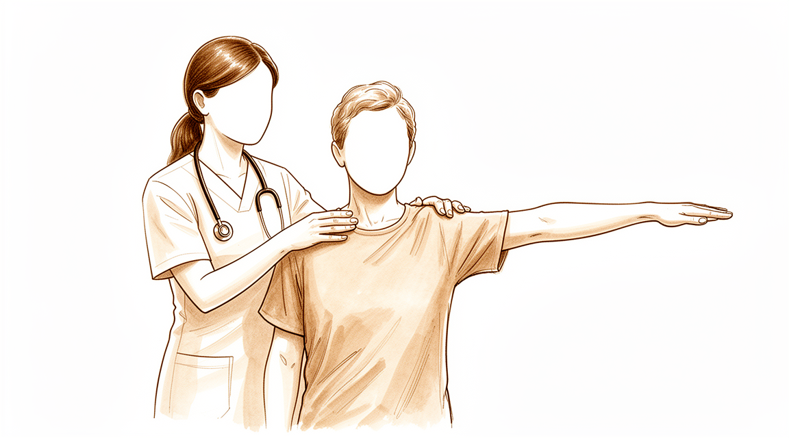
ٹینڈن میں کیلشیم جمنے سے سوزش (calcific tendinitis) کا مطلب ہے کہ آپ کے روٹیٹر کف (rotator cuff)، یعنی ٹینڈنوں کا وہ گروپ جو کندھے کو تھامتا اور حرکت دیتا ہے، کے کسی ایک ٹینڈن (tendon)، یعنی وہ مضبوط ڈوری جو پٹھے کو ہڈی سے جوڑتی ہے، کے اندر تھوڑا سا کیلشیم جمع ہو گیا ہے۔ درد عام طور پر آپ کے کندھے کے پہلو یا اوپر کی طرف، سوپرا اسپائنیٹس ٹینڈن (supraspinatus) کے اوپر محسوس ہوتا ہے، جو سب سے زیادہ متاثر ہونے والا ٹینڈن ہے۔ بازو کو پہلو کی طرف باہر یا سر سے اوپر اٹھانے سے درد عموماً بڑھ جاتا ہے، اور اونچی شیلف تک ہاتھ پہنچانا یا دھلے کپڑے رسی پر لٹکانا مشکل کام بن سکتا ہے۔
درد اکثر رات کو بھڑکتا ہے، خاص طور پر اگر آپ اسی کندھے پر لیٹیں، اور یہ آپ کو جگا سکتا ہے۔ یہ کسی سرگرمی کے بعد یا صبح اٹھتے ہی بھی ہو سکتا ہے۔ کچھ لوگوں کو درد دوروں کی صورت میں ہوتا ہے، جن کے درمیان کچھ پرسکون وقفے آتے ہیں، اور یہ مہینوں تک چل سکتا ہے۔ تکلیف کے اس لمبے عرصے کا مطلب کام سے چھٹی ہو سکتا ہے، اور یہ آہستہ آہستہ آپ کی زندگی کے معیار کو متاثر کر سکتا ہے۔
یہ مسئلہ 30 سے 60 سال کی عمر کی خواتین میں سب سے زیادہ ہوتا ہے، اگرچہ دوسرے لوگوں کو بھی ہوتا ہے۔ ذخیرہ خود عموماً 0.5 سے 1.5 سینٹی میٹر لمبا ہوتا ہے، اور زیادہ تر ذخیرے روئی جیسے دھندلے ہونے کے بجائے ٹھوس ہوتے ہیں۔ تقریباً 9 فیصد لوگوں میں ایک سے زیادہ ٹینڈن متاثر ہوتے ہیں، جس سے درد کندھے کے ارد گرد پھیل سکتا ہے۔
ایک بات جاننا مفید ہے: اسکین پر نظر آنے والا کیلشیم کا ذخیرہ اس امکان کو خارج نہیں کرتا کہ اسی ٹینڈن میں پھٹاؤ بھی ہو۔ اگر آپ کا درد کم نہیں ہو رہا، ہفتوں کے دوران بڑھ رہا ہے، آپ کو رات کو جگا دیتا ہے، یا آپ کو کام کرنے یا بازو استعمال کرنے سے روکتا ہے، تو اپنے فیملی ڈاکٹر (GP) کو دکھائیں یا ماہر ڈاکٹر (specialist) سے معائنے کے لیے کہیں۔
دراصل کیا ہو رہا ہے
آپ کا روٹیٹر کف چار ٹینڈنوں کا گروپ ہے جو آپ کے کندھے کو تھامتے اور حرکت دیتے ہیں۔ یہ ٹینڈن کندھے کے اوپری حصے میں ہڈی کی ایک چھوٹی محراب کے نیچے ایک تنگ جگہ سے پھسل کر گزرتے ہیں، اور ان کے درمیان سیال سے بھری ایک پتلی گدی ہوتی ہے۔ ٹینڈن کو ایک رسی سمجھیں جو ایک تنگ چرخی (pulley) میں سے گزر رہی ہو۔
ٹینڈن میں کیلشیم جمنے سے سوزش میں، اس رسی کا کچھ حصہ بدل جاتا ہے۔ ٹینڈن کے اندر کے خلیے آہستہ آہستہ کارٹلیج (cartilage)، یعنی وہ چکنا ٹشو جو آپ کے جوڑوں میں ہوتا ہے، جیسے خلیوں میں بدل جاتے ہیں، اور اس بدلے ہوئے ٹشو میں کیلشیم جمنا شروع ہو جاتا ہے۔ یہ مرحلہ وار ہوتا ہے: پہلے ٹینڈن بدلتا ہے، پھر کیلشیم بنتا ہے اور وہیں پڑا رہتا ہے، اور آخر میں جسم ذخیرے کو توڑ کر جذب کر لیتا ہے۔ یہ ایک فعال عمل ہے، سادہ گھساؤ نہیں، اور یہ کیلشیم کے اُس جماؤ سے مختلف ہے جو آپ جسم میں کہیں اور کسی بوڑھے، گھسے ہوئے ٹینڈن میں دیکھ سکتے ہیں۔
مرحلہ اس لیے اہم ہے کہ ہر مرحلہ مختلف محسوس ہوتا ہے۔ جب کیلشیم خاموشی سے ٹینڈن میں پڑا ہو، تو ہو سکتا ہے آپ کو تھوڑا درد ہو یا بالکل نہ ہو۔ جب جسم اسے جذب کرنا شروع کرتا ہے، تو درد اکثر اچانک اور شدید ہو جاتا ہے، اور رات کو سب سے زیادہ ہو سکتا ہے۔ ذخیرہ اپنے سائز کی وجہ سے بھی مسئلہ پیدا کر سکتا ہے: جب یہ تنگ جگہ کے اندر پھولتا ہے تو اوپر والی ہڈی کی محراب سے اٹک سکتا ہے، جس سے بازو کی ہموار حرکت رک جاتی ہے۔ اسی لیے بازو کو پہلو کی طرف باہر اٹھانا اکڑا ہوا یا رکا ہوا محسوس ہو سکتا ہے۔
ذخیرہ سب سے زیادہ سوپرا اسپائنیٹس ٹینڈن میں ہوتا ہے، اُس جگہ سے تقریباً 1.5 سے 2 سینٹی میٹر دور جہاں یہ بازو کی ہڈی کے اوپری سرے سے جڑتا ہے۔ اس جگہ سے واضح ہوتا ہے کہ درد وہیں کیوں ہوتا ہے جہاں ہوتا ہے، اور سر سے اوپر ہاتھ پہنچانا سب سے مشکل حرکت کیوں ہے۔
ایک اور بات جاننا مفید ہے: بہت سے لوگوں میں یہ ذخیرے ہوتے ہیں اور انہیں بالکل درد نہیں ہوتا، اور ذخیروں میں سے صرف تقریباً ایک تہائی کبھی علامات پیدا کرتے ہیں۔ چنانچہ آپ کے اسکین پر کیلشیم ایک حقیقی دریافت ہے، لیکن یہ آپ کے درد کی وجہ ہے یا نہیں، یہ اس پر منحصر ہے کہ یہ کس مرحلے میں ہے اور آپ کا جسم اس پر کیسا ردعمل دے رہا ہے۔
ہم اس کے لیے کیا کر سکتے ہیں
ڈاکٹر کیرن ہِرپرا، جو Mater Private Hospital Rockhampton میں بازو اور ہاتھ کے سرجن (upper-limb surgeon) ہیں، آپ کی حالت کے مطابق سب سے کم چیر پھاڑ والے طریقوں سے علاج شروع کرتے ہیں۔ مریض عام طور پر اپنے فیملی ڈاکٹر (GP) کے ریفرل (referral) پر ہمارے کلینک آتے ہیں؛ اگر کسی فزیوتھراپسٹ نے آپ کو ہمارے پاس آنے کا مشورہ دیا ہے، تب بھی میڈیکیئر (Medicare) سے رقم کی واپسی (rebate) کا حق دار ہونے کے لیے آپ کو اپنے فیملی ڈاکٹر سے ریفرل لینا ہوگا۔ آپ کی پہلی ملاقات میں ہم آپ سے آپ کی تکلیف کی تفصیل پوچھتے ہیں، آپ کے کندھے کا معائنہ کرتے ہیں اور جہاں ضرورت ہو وہاں امیجنگ (ایکسرے یا اسکین جیسے ٹیسٹ) کرواتے ہیں۔ اس طرح کے دیرینہ مسئلے میں ہم عام طور پر پہلے آپریشن کے بغیر علاج آزماتے ہیں، اور جب اس سے کافی بہتری نہ ملی ہو تو سرجری پر غور کرتے ہیں۔
پہلا قدم آسان ہے: ان سرگرمیوں کو بدلیں جو درد کو بھڑکاتی ہیں، اور اپنے کندھے کو سر سے اوپر کے کام سے آرام دیں۔ فزیوتھراپی کا مقصد درد کو کم کرنا اور بازو میں ہموار، قابو میں رہنے والی حرکت دوبارہ بنانا ہے۔ اس کے بارے میں رائے قائم کرنے سے پہلے اسے پورا موقع دیں، کیونکہ یہ مسئلہ مہینوں تک چل سکتا ہے۔ اگر آپ کی علامات کم نہیں ہو رہیں، ہفتوں کے دوران بڑھ رہی ہیں، آپ کو رات کو جگا دیتی ہیں، یا آپ کو کام کرنے یا بازو استعمال کرنے سے روکتی ہیں، تو اپنے فیملی ڈاکٹر کو دکھائیں یا ماہر ڈاکٹر سے معائنے کے لیے کہیں۔
سوزش کم کرنے والی دوائیں درد کا ایک عام ابتدائی علاج ہیں۔ اگر یہ کافی نہ ہوں، تو ہم ایسے طریقے پیش کر سکتے ہیں جو براہ راست ذخیرے کو نشانہ بناتے ہیں۔ ایک طریقہ شاک ویو تھراپی (shock wave therapy) ہے، جس میں جسم کے باہر سے آواز کی لہریں کیلشیم پر ڈالی جاتی ہیں تاکہ ذخیرہ ٹوٹ جائے۔ اس میں چیر پھاڑ نہیں ہوتی، یعنی جلد میں کچھ نہیں چبھویا جاتا، اور کیلشیم کو اپنی جگہ سے ہلانے کے لیے یہ ایک طے شدہ طاقت اور لہروں کی طے شدہ تعداد کے ساتھ کیا جاتا ہے۔ دوسرا طریقہ الٹراساؤنڈ کی رہنمائی میں سوئی کے ذریعے علاج (needling) ہے، جس میں الٹراساؤنڈ، یعنی وہ اسکین جو ٹینڈن کی براہ راست تصویریں دکھاتا ہے، کی مدد سے ایک سوئی ذخیرے میں ڈالی جاتی ہے تاکہ کیلشیم کو توڑ کر کھینچ لیا جائے۔ دونوں طریقے درد کم کر سکتے ہیں، کارکردگی بحال کر سکتے ہیں اور کیلشیم کو ختم کر سکتے ہیں۔ اگر علامات برقرار رہیں تو سوئی کے ذریعے علاج دہرایا جا سکتا ہے، بشرطیکہ دو بار کے درمیان کم از کم چھ ہفتے کا وقفہ ہو۔
جب یہ علاج کارگر نہ ہوئے ہوں تو سرجری کی باری آتی ہے۔ اس میں ٹینڈن سے کیلشیم کا ذخیرہ نکال دیا جاتا ہے، اور اگر ٹینڈن خود بھی کافی حد تک متاثر ہو تو اس کی مرمت بھی کی جاتی ہے۔ اگر اوپر دیے گئے طریقوں کو صحیح معنوں میں آزمانے کے بعد بھی آپ کا درد کم نہ ہوا ہو، تو ہم آپ کے ساتھ اس پر بات کریں گے کہ آیا سرجری آپ کے لیے مناسب ہے، اور آپ کے ساتھ مل کر فیصلہ کریں گے۔
کیا توقع رکھیں
ٹینڈن میں کیلشیم جمنے سے سوزش میں، زیادہ تر صورتوں میں، آگے کی توقعات اچھی ہوتی ہیں۔ بہت سے لوگوں میں یہ ذخیرے ہوتے ہیں اور انہیں کبھی پتا نہیں چلتا، اور ان میں سے صرف تقریباً ایک تہائی کبھی درد پیدا کرتے ہیں۔ جب ذخیرہ علامات پیدا کرے، تو طویل مدت میں اس کے مکمل طور پر ختم ہو جانے کا اچھا امکان ہوتا ہے۔ آپ کا جسم خود کیلشیم کو جذب کر سکتا ہے، اور یہ عمل اسی لمحے سے شروع ہو چکا ہوتا ہے جب ذخیرہ بنتا ہے۔
البتہ وہاں تک کا راستہ ہمیشہ جلدی طے نہیں ہوتا۔ علامات مہینوں تک چل سکتی ہیں، اور تکلیف کے اس عرصے کا مطلب کام سے چھٹی اور آپ کی زندگی کے معیار پر واقعی اثر ہو سکتا ہے۔ درد اکثر دوروں کی صورت میں آتا ہے، جن کے درمیان کچھ پرسکون وقفے ہوتے ہیں، اور جب آپ کا جسم ذخیرے کو توڑنا شروع کرتا ہے تو یہ اچانک اور شدید ہو سکتا ہے۔ کچھ ذخیرے چند مہینوں میں ٹھیک ہو جاتے ہیں، کچھ میں زیادہ وقت لگتا ہے، اور ٹھیک ٹھیک یہ بتانے کا کوئی قابلِ اعتماد طریقہ نہیں کہ آپ کا ذخیرہ کیسا برتاؤ کرے گا۔
زیادہ تر لوگ آپریشن کے بغیر ٹھیک رہتے ہیں۔ آپریشن کے بغیر علاج (conservative care)، یعنی ایسا علاج جس میں سرجری شامل نہ ہو، عام طور پر پہلا انتخاب ہوتا ہے، اور اس سے زیادہ تر لوگوں کو نمایاں بہتری ملتی ہے: اس طرح علاج کروانے والے 72 فیصد لوگ اچھے یا بہترین نتیجے کی اطلاع دیتے ہیں، ذخیرے کا سائز یا جگہ چاہے کچھ بھی ہو۔ شاک ویو تھراپی اور الٹراساؤنڈ کی رہنمائی میں سوئی کے ذریعے علاج، دونوں بہت سے لوگوں میں درد کم کرتے اور کیلشیم ختم کرتے ہیں، اور دونوں کا حفاظتی ریکارڈ مضبوط ہے۔ سوئی کے ذریعے علاج عموماً تب سب سے بہتر کام کرتا ہے جب علامات کم عرصے سے ہوں اور ذخیرہ چھوٹا ہو۔ اگر ان میں سے کوئی طریقہ آپ کے خاص ذخیرے کے لیے موزوں نہ ہو، تو ہم آپ کے ساتھ دوسرے راستوں پر بات کریں گے۔
سرجری ان لوگوں کے لیے رکھی جاتی ہے جن کا درد ان علاجوں کو صحیح معنوں میں آزمانے کے بعد بھی کم نہ ہوا ہو۔ اس میں ٹینڈن سے کیلشیم کا ذخیرہ نکال دیا جاتا ہے، اور جن لوگوں کا درد دوسرے علاجوں سے ٹھیک نہیں ہوا، ان میں سے 88.5 فیصد میں یہ اچھا نتیجہ دیتی ہے۔ سرجری کے بعد صحت یابی آہستہ آہستہ ہوتی ہے: پہلے چھ ماہ کے دوران کندھے کی کارکردگی آہستہ آہستہ بہتر ہوتی ہے، اور آرام اور حرکت میں زیادہ تر بہتری یک دم آنے کے بجائے اسی عرصے میں بتدریج آتی ہے۔
اگر آپ کی علامات کم نہیں ہو رہیں، ہفتوں کے دوران بڑھ رہی ہیں، آپ کو رات کو جگا دیتی ہیں، یا آپ کو کام کرنے یا بازو استعمال کرنے سے روکتی ہیں، تو اپنے فیملی ڈاکٹر کو دکھائیں یا ماہر ڈاکٹر سے معائنے کے لیے کہیں۔
ڈاکٹر کو کب دکھائیں
کیلشیم کے زیادہ تر ذخیرے خود ہی ٹھیک ہو جاتے ہیں، اور بہت سے کوئی تکلیف نہیں دیتے۔ لیکن یہ مسئلہ مہینوں تک چل سکتا ہے، اور اس کی وجہ سے آپ کو کام سے چھٹی کرنی پڑ سکتی ہے اور آپ کی زندگی کا معیار متاثر ہو سکتا ہے۔ اگر آپ کے کندھے کا درد کم نہیں ہو رہا، ہفتوں کے دوران بڑھ رہا ہے، آپ کو رات کو جگا دیتا ہے، یا آپ کو کام کرنے یا بازو استعمال کرنے سے روکتا ہے، تو اپنے فیملی ڈاکٹر کو دکھائیں یا ماہر ڈاکٹر سے معائنے کے لیے کہیں۔ اگر بازو کو پہلو کی طرف باہر اٹھاتے وقت درد رکا ہوا یا اٹکا ہوا محسوس ہو تو اس کا بھی ذکر کریں، کیونکہ پھولا ہوا ذخیرہ ٹینڈن کو اپنے اوپر والی ہڈی کی محراب کے نیچے حرکت کرنے سے جسمانی طور پر روک سکتا ہے۔ ایک اور بات جو بتانے کے لائق ہے: آپ کے اسکین پر کیلشیم کا ذخیرہ اس امکان کو خارج نہیں کرتا کہ اسی ٹینڈن میں پھٹاؤ بھی ہو، اس لیے صرف اسکین کی دریافت سے تشخیص طے نہیں ہو جاتی۔ اگر آپ دفتری اوقات کے بعد یا ہفتے کے آخر میں کلینک سے رابطہ نہ کر سکیں، تو اپنے قریب ترین ہسپتال کی ایمرجنسی جائیں۔
مزید تفصیل
Advanced reading: the deeper science (optional)
یہ حصہ اس سے آگے کی بات کرتا ہے جتنی آپ کو اپنے علاج کے فیصلوں کے لیے ضرورت ہے۔ ٹینڈن میں کیلشیم جمنے سے سوزش کے بارے میں مزید پڑھنا اس لیے مفید ہے کہ یہ کندھے کے زیادہ تر مسائل سے مختلف طریقے سے برتاؤ کرتی ہے: یہ ذخیرہ ایسے لوگوں میں بھی عام ہے جنہیں بالکل درد نہیں ہوتا، جسم عام طور پر کسی مدد کے بغیر اسے گھلا دیتا ہے، اور جو علاج کیلشیم کو نکالتا ہے وہ وہ علاج نہیں جو درد کو سب سے جلدی کم کرتا ہے۔
ذخیرہ ایک عام دریافت ہے، خود بخود تشخیص نہیں
آپ کے اسکین کی رپورٹ میں کیلشیم کا ذخیرہ دیکھ کر اسے ہی مسئلے کی وجہ سمجھ لینا آسان ہے، کیونکہ یہ نظر آتا ہے اور درد حقیقی ہے۔ لیکن یہ کتنا عام ہے، اس کے اعداد و شمار معاملے کو پیچیدہ بنا دیتے ہیں۔ 1,219 بالغوں میں، ذخیرے بغیر علامات والے 7.8 فیصد لوگوں میں اور کندھے کی ہڈی کے نیچے والی جگہ کے درد کے سنڈروم (subacromial pain syndrome) والے 42.5 فیصد لوگوں میں موجود تھے [1]۔ 302 کندھوں کی ایک الگ تحقیق میں، عام آبادی میں ذخیرے اکثر پائے گئے اور ان میں سے صرف تقریباً ایک تہائی دردناک تھے [2]۔
چنانچہ ذخیرہ اس امکان کو کافی حد تک بڑھا دیتا ہے کہ درد کی وجہ یہی ہے، لیکن تقریباً ہر تیرہ میں سے ایک بغیر درد والے کندھے میں بھی ذخیرہ ہوتا ہے۔ کسی ذخیرے کو خاموش سے علامات والا بنانے میں جزوی طور پر اس کا سائز اور جگہ کردار ادا کرتے دکھائی دیتے ہیں: واقعی علامات والی بیماری کا سب سے زیادہ امکان 30 سے 60 سال کی ان خواتین میں تھا جنہیں کندھے کی ہڈی کے نیچے والی جگہ میں درد تھا اور جن کا ذخیرہ 1.5 سینٹی میٹر سے لمبا تھا [1]، اور درد کا تعلق سوپرا اسپائنیٹس میں ذخیرے کی جگہ سے اور ایک سے زیادہ ٹینڈنوں کے متاثر ہونے سے تھا [2]۔
یہ عام طور پر گھل جاتا ہے، اور یہی بات ہر چیز پر اثر ڈالتی ہے
قدرتی راستہ یہ ہے کہ ذخیرہ جذب ہو جاتا ہے، اسی لیے اتنے زیادہ علاج کارگر نظر آتے ہیں۔ اس کی سب سے واضح مثال ایک رینڈمائزڈ ٹرائل (randomised trial) سے ملتی ہے جس میں ہر مریض کا سوئی سے علاج اور دھلائی (needling and lavage) کی گئی اور پھر انہیں قرعہ اندازی سے اسٹیرائیڈ یا نمکین پانی (saline) کا انجکشن دیا گیا: بارہ ماہ پر نمکین پانی والے گروپ کے 83 فیصد اور اسٹیرائیڈ والے گروپ کے 74 فیصد مریضوں میں کیلشیم جذب ہو چکا تھا [3]۔
اسے غور سے پڑھیں، کیونکہ اس میں ایک خلافِ توقع نتیجہ ہے۔ اسٹیرائیڈ نے چھ ہفتوں تک درد اور تین ماہ تک کارکردگی بہتر کی، اور کیلشیم کے غائب ہونے یا نہ ہونے پر اس کا کوئی نمایاں اثر نہیں تھا [3]۔ درد سے آرام اور کیلشیم کا صاف ہونا دو الگ عمل ہیں۔ ایک علاج ان میں سے ایک دے سکتا ہے اور دوسرا نہیں، اور اسکین پر نظر آنے والی چیز وہ نہیں جو ہفتہ بہ ہفتہ علامات کو چلا رہی ہوتی ہے۔
اسی لیے آپریشن کے بغیر کے طریقے ایک جیسے نتائج دیتے ہیں
اگر ذخیرہ زیادہ تر خود ہی ختم ہو جاتا ہے، تو علاج دراصل شفا دینے کے بجائے اس درمیانی وقت کو قابلِ برداشت بنانے میں ایک دوسرے سے مقابلہ کر رہے ہوتے ہیں۔ موازنوں سے یہی ظاہر ہوتا ہے۔ 257 مریضوں میں، فزیکل تھراپی، کورٹیکو اسٹیرائیڈ انجکشن اور الٹراساؤنڈ کی رہنمائی میں باربوٹاج (barbotage، سوئی سے کیلشیم کو توڑ کر دھو کر نکالنا) نے سرجری سے بچنے کی ملتی جلتی شرحیں دیں [4]۔ 239 مریضوں میں یہی تین طریقے زیادہ تر کامیاب رہے، اور صرف فزیوتھراپی میں ناکامی کی شرح سب سے زیادہ تھی [5]۔
طبی طریقوں میں سے، زیادہ توانائی والی شاک ویو تھراپی سب سے زیادہ تحقیق شدہ کم چیر پھاڑ والا علاج ہے اور مختصر سے درمیانی مدت میں محفوظ اور مؤثر دکھائی گئی ہے، جبکہ الٹراساؤنڈ کی رہنمائی میں سوئی کے ذریعے علاج کندھے کی ہڈی کے نیچے والی جگہ میں الٹراساؤنڈ کی رہنمائی میں دیے گئے انجکشن سے بہتر ثابت نہیں ہوا [6]۔ 1,258 مریضوں کے اکٹھے تجزیے میں، شاک ویو، سوئی کے ذریعے علاج اور آرتھروسکوپی (دوربین سے جوڑ کے اندر دیکھ کر علاج) سب نے اچھے طبی نتائج دیے [7]، اور 908 مریضوں میں باربوٹاج محفوظ اور زیادہ کامیابی کی شرح والا تھا، لیکن اس کا دوسرے بڑے طریقوں سے براہ راست موازنہ کبھی نہیں کیا گیا [8]۔
سرجری کیا اضافی فائدہ دیتی ہے، ٹھیک ٹھیک الفاظ میں
سرجری کا کوئی فائدہ نہ ہو، ایسا نہیں ہے، اور اس فائدے کی مقدار کو اندازے سے بیان کرنے کے بجائے ٹھیک ٹھیک بتانا بہتر ہے۔ رینڈمائزڈ ٹرائلز کے 2,352 مریضوں کے اکٹھے تجزیے میں، سرجری سے علاج نے آپریشن کے بغیر علاج، خاص طور پر الٹراساؤنڈ کی رہنمائی میں سوئی کے ذریعے علاج، کے مقابلے میں کارکردگی کے اسکور میں زیادہ بہتری اور درد میں ملتی جلتی کمی دی، اور دونوں راستوں سے طبی لحاظ سے نمایاں بہتری حاصل ہوئی [9]۔ سرجری کے مختلف طریقوں کے درمیان کوئی نمایاں فرق نہیں تھا، اور صرف ذخیرہ نکالنے کے نتائج ذخیرہ نکالنے کے ساتھ سب ایکرومیئل ڈیکمپریشن (subacromial decompression، کندھے کی ہڈی کے نیچے کی جگہ کو کشادہ کرنا) کرنے جیسے ہی تھے [10]۔
کارکردگی اور درد کا یہ فرق ہی کام کی بات ہے۔ اگر اصل شکایت درد ہے تو شواہد واضح طور پر آپریشن کے حق میں نہیں ہیں۔ اگر اکڑاؤ اور کارکردگی کی کمی نمایاں ہو اور جاری رہی ہو، تو شواہد آپریشن کے حق میں زیادہ ہیں۔
وہ تعلق جو اکثر نظر انداز ہو جاتا ہے
ٹینڈن میں کیلشیم جمنے سے سوزش سب لوگوں میں یکساں طور پر نہیں ہوتی۔ 102 مریضوں میں، جن لوگوں کو ساتھ ہی ہارمونی غدود کی کوئی بیماری (endocrine disorder) تھی، خاص طور پر تھائرائیڈ کی بیماری اور ذیابیطس (شوگر)، ان میں علامات کم عمر میں شروع ہوئیں، ان کی بیماری نمایاں طور پر زیادہ لمبی چلی، اور انہیں زیادہ اکثر سرجری کی ضرورت پڑی [11]۔
اگر آپ کی بیماری غیر معمولی طور پر لمبی چلی ہو یا جلدی شروع ہوئی ہو تو یہ بات اپنے فیملی ڈاکٹر سے ضرور کریں، اس لیے نہیں کہ ہارمونی بیماری کا علاج کندھے کو ٹھیک کر دیتا ہے، بلکہ اس لیے کہ اس سے یہ بدل جاتا ہے کہ حقیقت پسندانہ وقت کتنا ہو سکتا ہے۔ یہ سن کر کہ کوئی مسئلہ خود بخود ختم ہو جاتا ہے، دو سال کے درد کے ساتھ اس بات کو ماننا مشکل ہوتا ہے؛ ہارمونی بیماری کا تعلق ایک وجہ ہے کہ معمول کی تسلی ہر کیس پر پوری نہیں اترتی۔
حوالہ جات
[1] Louwerens JK, Sierevelt IN, van Hove RP, van den Bekerom MP, van Noort A. Prevalence of calcific deposits within the rotator cuff tendons in adults with and without subacromial pain syndrome: clinical and radiologic analysis of 1219 patients. J Shoulder Elbow Surg. 2015;24(10):1588-93. https://doi.org/10.1016/j.jse.2015.02.024
[2] Sansone V, Consonni O, Maiorano E, Meroni R, Goddi A. Calcific tendinopathy of the rotator cuff: the correlation between pain and imaging features in symptomatic and asymptomatic female shoulders. Skeletal Radiol. 2015;45(1):49-55. https://doi.org/10.1007/s00256-015-2240-3
[3] Darrieutort-Laffite C, Varin S, Coiffier G, Albert J, Planche L, Maugars Y, et al. Are corticosteroid injections needed after needling and lavage of calcific tendinitis? Randomised, double-blind, non-inferiority trial. Ann Rheum Dis. 2019;78(6):837-43. https://doi.org/10.1136/annrheumdis-2018-214971
[4] Gilbert R, Dadoo S, Lin R, Bhardwaj N, McMahon S, Steuer F, et al. Comparison of physical therapy, corticosteroid injections, and ultrasound-guided barbotage for nonoperative management of calcific tendinitis. Orthop J Sports Med. 2026;14(4). https://doi.org/10.1177/23259671261434919
[5] Drummond M, Ayinon C, Lin A, Dunn R. Relative efficacy of three nonsurgical treatments for calcific tendinitis: physical therapy vs steroid injection vs barbotage. Orthop J Sports Med. 2021;9(7_suppl4). https://doi.org/10.1177/2325967121S00210
[6] Louwerens JK, Sierevelt IN, van Noort A, van den Bekerom MP. Evidence for minimally invasive therapies in the management of chronic calcific tendinopathy of the rotator cuff: a systematic review and meta-analysis. J Shoulder Elbow Surg. 2014;23(8):1240-9. https://doi.org/10.1016/j.jse.2014.02.002
[7] Louwerens JK, Veltman ES, van Noort A, van den Bekerom MP. The effectiveness of high-energy extracorporeal shockwave therapy versus ultrasound-guided needling versus arthroscopic surgery in the management of chronic calcific rotator cuff tendinopathy: a systematic review. Arthroscopy. 2015;32(1):165-75. https://doi.org/10.1016/j.arthro.2015.06.049
[8] Gatt DL, Charalambous CP. Ultrasound-guided barbotage for calcific tendonitis of the shoulder: a systematic review including 908 patients. Arthroscopy. 2014;30(9):1166-72. https://doi.org/10.1016/j.arthro.2014.03.013
[9] Angileri HS, Gohal C, Comeau-Gauthier M, Owen MM, Shanmugaraj A, Terry MA, et al. Chronic calcific tendonitis of the rotator cuff: a systematic review and meta-analysis of randomized controlled trials comparing operative and nonoperative interventions. J Shoulder Elbow Surg. 2023;32(8):1746-60. https://doi.org/10.1016/j.jse.2023.03.017
[10] Anam E, Zahran S, Roy A, Daneshvar P, Bicknell RT, Janssen I. Surgical approaches of shoulder calcific tendonitis: a systematic review and meta-analysis. JSES Rev Rep Tech. 2024;4(3):353-8. https://doi.org/10.1016/j.xrrt.2024.03.013
[11] Harvie P, Pollard TC, Carr AJ. Calcific tendinitis: natural history and association with endocrine disorders. J Shoulder Elbow Surg. 2007;16(2):169-73. https://doi.org/10.1016/j.jse.2006.06.007
Evidence & references
This is the clinical evidence summary written for health professionals. It is technical, and it lists the research this page was built from. You do not need to read it to understand your treatment or to make a decision about it.
Overview
Epidemiology and Natural History
- Calcific tendon deposits of the shoulder are a frequent occurrence in the general population [2].
- Only one third of calcific tendon deposits in the shoulder are painful [2].
- Symptomatic calcific tendinitis of the shoulder has a good likelihood to completely resolve in the long-term [3].
Non-Operative Treatment
- The primary choice of treatment for calcific tendinitis is conservative, especially in patients with acute calcific tendinitis [26].
- Ultrasound treatment helps resolve calcifications and is associated with short-term clinical improvement in patients with symptomatic calcific tendinitis of the shoulder [1].
- Conservative treatment for calcific tendinitis of the shoulder showed clinically significant improvement, with 72% of excellent or good results regardless of the location, radiologic type and size, and initial symptoms of calcific deposits [5].
- Extracorporeal shock wave therapy (ESWT) has produced a high rate of success in pain relief and functional restoration with negligible associated complications [11].
- Shock wave therapy is a safe and effective noninvasive treatment for patients with calcific tendinitis of the shoulder, producing a high rate of success in pain relief and functional restoration with negligible complications [19].
- ESWT can be considered as an alternative treatment for chronic calcific tendinitis of the shoulder due to its good tolerance, safety, and clinical radiological response [25].
- Both ultrasound-guided needling and extracorporeal shock wave therapy improved clinical outcomes and eliminated calcium deposits in the treatment of calcific tendinitis [6].
- Utilization of barbotage as a treatment for calcific tendonitis of the shoulder appears to produce notable pain reduction in the short term (specifically at the 2-month follow-up), but begins to lose some efficacy over long-term evaluation [27].
- Patients with calcific tendinitis of the shoulder who have the factors identified for a poor outcome after ESWT should undergo a different procedure [42].
Operative Treatment
- Surgical treatment of calcific tendinitis gives good results in 88.5% of patients resistant to medical treatment [12].
- The aim of arthroscopic treatment in calcific tendinitis is to remove the calcific deposit [15].
- Arthroscopic removal of calcification leads to improved clinical outcomes in patients with chronic calcific tendinitis, but at least 6 months of follow-up is needed for these improvements to become statistically significant [13].
- Patients undergoing arthroscopic treatment of a calcific deposit in the shoulder had satisfactory clinical and radiological outcomes at the final follow-up, with functional scores improving slowly and reaching more than 75 percent at six months after surgery [7].
- Arthroscopic treatment of calcifying tendinitis provides good to excellent clinical results [20].
- Arthroscopic treatment of chronically painful calcific tendinitis of the rotator cuff promises to be successful in more than 90% of the patients if conservative treatment fails [37].
- Arthroscopic removal of calcific deposits of the shoulder shows good clinical results for pain reduction, shoulder function and patient satisfaction [44].
- Endoscopic and open surgery are equally effective in the treatment of chronic calcifying tendinopathy, showing similar clinical and sonographic results [34].
- Preoperative ultrasound-guided marking of calcific deposits is a procedure that statistically significantly improves the clinical results of arthroscopic surgery as seen at 6 weeks and 2 years [33].
- The short-term functional outcome of patients with calcific tendonitis after arthroscopic bursectomy and debridement of the calcific deposit is not influenced if performed in combination with or without a subacromial decompression [40].
- Routine diagnostic glenohumeral exploration does not appear beneficial in arthroscopic treatment of calcific tendinitis due to the low prevalence of intraarticular pathologies which most frequently do not require surgical treatment [23].
Prognostic Factors
- Patients with calcific lesions >1 cm had a 2.8 increased likelihood to undergo operative treatment in the setting of calcific tendinitis of the shoulder [4].
Anatomy & Pathophysiology
Epidemiology & Demographics
- Calcific tendon deposits of the shoulder are a frequent occurrence in the general population, although only one third are painful [2].
- In approximately 80% of patients, calcifying tendinitis occurs in the supraspinatus tendon [115].
- Most patients with calcifying tendinitis are 30-50 years old [115].
- Women are approximately 1.5 times more often affected by calcifying tendinitis than men [115].
- The incidence of calcifying tendinitis in the general population is 2.5-20% [115].
- In about 10% of individuals, calcific deposits are found bilaterally [17].
- In 20% of cases, subjects with rotator cuff calcific tendinopathy are asymptomatic [17].
- Subjects with rotator cuff calcific tendinopathy were often female aged between 30 and 60 [17].
- Demographic, radiographic, and clinical features of calcific tendinitis of the shoulder in the Korean population were not different from those of Western populations [14].
Anatomical Location & Structures
- Calcific deposits are most commonly located at the level of the supraspinatus tendon [17].
- In calcifying tendinitis, deposits are typically located 1.5-2 cm from the tendinous insertion at the greater tuberosity [115].
- The greater tuberosity serves as the attachment site for the supraspinatus, infraspinatus, and teres minor tendons of the rotator cuff [70].
- The rotator cuff consists of four muscles: the subscapularis, supraspinatus, infraspinatus, and teres minor [71].
- The acromion, the coracoacromial ligament, and the coracoid process form the coracoacromial arch [70].
- The rotator cuff, subacromial bursa, and subdeltoid bursa pass underneath the coracoacromial arch [70].
- The subacromial bursa separates the rotator cuff tendons from the coracoacromial arch and allows them to glide [78].
- The infraspinatus and teres minor fuse near their musculotendinous junctions [83].
- The tendons of the infraspinatus and supraspinatus muscles join approximately 15 mm proximal to their insertion [83].
Pathogenesis & Staging
- The pathogenesis of calcific tendinitis remains unclear, and different theories have been proposed [30].
- Calcific tendinitis of the rotator cuff tendons is thought to have a different pathophysiologic mechanism than calcific tendinitis at other sites, such as the Achilles or patellar tendons [30].
- Uhthoff and Loehr described an active, cell-mediated process that is now widely accepted for the pathogenesis of calcific tendinitis [30].
- Uhthoff and Loehr described cartilage metaplasia as a reactionary process in an active biologic environment [30].
- The reactive calcification theory proposed by Uthoff in 1997 consists of a three-stages process: pre-calcific, calcific, and post-calcific stage [17].
- The precalcific stage consists of predominantly fibrocartilaginous metaplasia presumably within less vascular areas of the tendon [30].
- During the pre-calcific stage, tenocytes undergo a metaplastic transformation into chondrocytes within the site where the calcium deposits will subsequently form [17].
- The calcific stage is subdivided into three separate phases: formative, resting, and resorptive [30].
- In the formative phase of the calcific stage, matrix vesicles unite to become calcific deposits that are separated by fibrocartilage [30].
- The resting phase is characterized by the presence of calcium deposits in the rotator cuff [17].
- The resorptive phase is associated with acute, sudden onset of extremely severe pain [49].
- Calcific deposits consist of two different forms of hydroxyapatite: A type and B type [30].
- The composition of an individual calcific deposit can have a mixture of the two types of hydroxyapatite [30].
- Each individual phase of calcific tendinitis has a different composition of hydroxyapatite [30].
- Calcium is deposited in the fibrocartilaginous matrix of the tendon as calcium carbonate apatite [49].
- Endocrine and metabolic disorders were often described as comorbidity in subjects with rotator cuff calcific tendinopathy [17].
- Endocrine disorders probably play an important role in the development of rotator cuff calcific tendinopathy, but the effect of this process is still unknown [17].
- Shorter length of the infraspinatus tendon and specific subacromial bursa anatomy may explain the intramuscular migration of calcium deposits leading to the lesion [10].
Clinical Presentation & Pain Mechanisms
- Subjects with rotator cuff calcific tendinopathy often reported nightly, acute, unilateral and severe pain with spontaneous onset [17].
- Subjects with rotator cuff calcific tendinopathy often reported deficit in active and passive range of motion, mainly in abduction and flexion [17].
- Calcific tendinitis of the supraspinatus does not typically cause loss of external rotation but is frequently associated with mild isolated restriction of abduction [22].
- Patients with calcific tendinitis had less passive abduction, forward flexion, internal rotation, and external rotation than the control group [65].
- The main mechanism of abduction contracture lies in the mechanical blocking between the bulged calcified deposits and the coracoacromial arch [45].
- Abduction contracture is presumably caused by mechanical blocking by the calcified deposits and the coracoacromial arch and by reflex muscle spasm caused by pain [125].
- Patients with calcific tendinitis had significantly greater nerve counts compared with the rotator cuff repair group and the control group [65].
- A greater nerve count positively correlated with a greater degree of neovascularization and also positively correlated with greater frequency of extreme pain [65].
- Neovascularization positively correlated with more frequent pain during sleep and more frequent extreme pain [65].
- The pathophysiology of calcific tendinitis remains largely unknown, with proposed theories including degenerative calcification, repetitive trauma, necrosis of tenocytes and intracellular calcium accumulation, reactive calcification, endochondral ossification, and chondral metaplasia [65].
- Calcific tendinitis of the rotator cuff with tuberosity osteolysis is a distinctive form of calcific tendinitis that should be considered in clinical and surgical practice [28].
- A rotator cuff tear was the only factor affecting the complete recovery of shoulder function in patients treated for calcific tendinitis [113].
Classification
- The supraspinatus is the most frequently affected tendon in rotator cuff calcific tendinopathy, followed by the infraspinatus and subscapularis [38].
- In 9% of patients with rotator cuff calcific deposits, two or more tendons are affected [38].
- The median length of calcific deposits in the rotator cuff is 1.16 cm [38].
- 55.8% of calcific deposits in the rotator cuff have a length between 0.5 and 1.5 cm [38].
- 28.6% of calcific deposits in the rotator cuff have a length greater than 1.5 cm [38].
- Gartner type II morphology is the most common radiographic classification for calcific deposits, present in 47.0% of cases [38].
- Gartner type I morphology is present in 38.4% of calcific deposits [38].
- Gartner type III morphology is present in 14.6% of calcific deposits [38].
- Approximately 76% of calcifications in the shoulder are characterized as dense (type A or B) using the Mole et al. classification scheme [39].
- Approximately 24% of calcifications in the shoulder are characterized as fluffy (type C) using the Mole et al. classification scheme [39].
- Type A calcific deposits are dense, rounded, and sharply delineated [67].
- Type B calcific deposits are multilobular in appearance, radiodense, and sharply outlined [67].
- Type C calcific deposits are more radiolucent and heterogeneous with irregular outlines [67].
- Type D calcific deposits are dystrophic calcific lesions of the tendon insertion [67].
- The pathogenesis of calcific tendinitis involves a three-stage process: precalcific, calcific, and postcalcific [30].
- The calcific stage is subdivided into three phases: formative, resting, and resorptive [30].
- The precalcific stage consists of predominantly fibrocartilaginous metaplasia within less vascular areas of the tendon [30].
- In the formative phase of the calcific stage, matrix vesicles unite to become calcific deposits separated by fibrocartilage [30].
- The composition of an individual calcific deposit can have a mixture of A type and B type hydroxyapatite [30].
- Disease evolution in calcific tendinitis can be classified into four stages: precalcific, calcific, resorptive, and postcalcific [107].
- The resorptive phase of calcific tendinitis is the most painful stage owing to vascular invasion, increased phagocytic cells, and edema from intratendinous pressure [107].
Clinical Presentation
- Subjects complaining of rotator cuff calcific tendinopathy were often female aged between 30 and 60 [17].
- Calcific tendinitis is a poorly understood condition in which symptoms can be protracted, resulting in time off work and impaired quality of life [8].
- The calcium deposits are most commonly located at the level of the supraspinatus tendon [17].
- The supraspinatus was the most frequently affected tendon in 82.7% of patients with calcific deposits, followed by the subscapularis tendon in 8.9% and the infraspinatus in 8.4% [38].
- Two or more tendons were affected in 9% of patients with calcific deposits [38].
- The median length of the calcific deposit was 1.16 cm, with 55.8% ranging from 0.5 to 1.5 cm and 28.6% greater than 1.5 cm [38].
- Approximately 76% of calcifications were characterized as dense (type A or B) and 24% as fluffy (type C) [39].
- Recognition of atypical presentations of calcific tendinitis with bone erosion may prevent unnecessary biopsy and overtreatment [16].
- The presence of a calcium deposit in the rotator cuff of a patient with painful shoulder does not rule out the possibility of a coexisting rotator cuff tear [60].
- Calcific tendinitis in the elbow of a child has not been reported previously [24].
- Atypical calcific tendinitis involving teres minor can affect overhead movement [9].
- Calcific tendinitis of the trapezius insertion is a recognized presentation [18].
- Of twelve histologically verified cases of calcific periarthritis adjacent to joints other than the shoulder, in only one patient was the preoperative clinical diagnosis accurate [59].
Investigations
Plain Radiography
- Standardized plain films are almost always sufficient to garner the information needed for shoulder imaging [47].
- The purpose of imaging the shoulder is to help establish the diagnosis, determine the severity of the pathoanatomy, assist in surgical planning, and enable the surgeon to illustrate the condition of the shoulder to the patient [47].
- At least two X-ray views should be obtained for shoulder imaging: an anteroposterior in the plane of the glenoid and an axillary projection with the arm in abduction [87].
- The axillary view is taken with the arm in the functional position of elevation in the plane of the scapula and oriented so that both the spinoglenoid notch and the scapular neck are visible [47].
- The axillary view demonstrates glenohumeral relationships in the functional position of elevation, which is referred to as the "truth view" [47].
- CT scans may offer a few degrees of increased precision in the measurement of glenoid version, but this precision does not improve the quality of the surgery or the clinical outcome [47].
- The temptation to "overimage" should be resisted, obtaining only the scans or reconstructions that are necessary for the care of the patient [47].
- Calcific tendinitis can be observed on plain radiograph [94].
- Calcific deposits in the fingers may not be seen on standard views and oblique views may be necessary [111].
Ultrasonography
- Ultrasound is a simple and accurate test for identifying rotator cuff tears and calcific tendinitis [87].
- Ultrasound can be useful in guiding injections or barbotage (aspirating calcific deposits in the rotator cuff) [87].
- Ultrasonography is a low-cost alternative to MRI and arthrography for evaluating both skeletal and soft-tissue structures of the shoulder [94].
- Ultrasonography can provide immediate, real-time visualization of the rotator cuff, biceps tendon, and calcific deposits [94].
- Ultrasonography can be used to measure the subacromial space and detect atrophy of rotator cuff muscles [94].
- Ultrasonography can evaluate impingement in various positions and motions due to real-time imaging [94].
- Ultrasonography is highly operator dependent and is not as useful for evaluating labral tears or rotator cuff tears that are very small or larger than 3 cm [94].
- The accuracy of rotator cuff ultrasonography depends on the skill of the scanner operator and an awareness of pitfalls that are encountered [85].
- US elastography is recommended for all patients with calcific tendinosis, except for those with the arc morphological type, before making management decisions [31].
- The surgeon performing ultrasound of the shoulder can diagnose intraosseous calcifying tendinitis [56].
- Calcific tendinitis of the supraspinatus is frequently associated with mild isolated restriction of abduction but does not typically cause loss of external rotation [22].
Magnetic Resonance Imaging
- MRI is the modality of choice for evaluating the rotator cuff, biceps, and subacromial/subdeltoid bursa [94].
- T2-weighted MRI provides better visualization of full thickness rotator cuff tears [94].
- MRI can identify labral tears and rotator cuff tears, although the accuracy for these is enhanced by combining the scan with arthrography [87].
- Shoulder surgeons should be aware of the accuracy limitations of sonographic or MRI evaluation regarding rotator cuff tears as a comorbidity in calcific tendinitis [32].
- The incidence of rotator cuff tears in cases of calcific tendonitis is higher than previously reported in cohorts undergoing MRI [41].
Computed Tomography
- CT imaging is frequently used to evaluate fractures of the shoulder, to assess for bony lesions in recurrent instability cases, or for preoperative templating for shoulder arthritis [94].
- CT is helpful for planning fracture surgery and shoulder joint replacement [87].
Diagnostic Findings and Pathology
- The paper illustrates the dynamic pathological process of calcific tendinopathy, including migration patterns of calcium deposits [29].
Treatment
Non-Operative Management
- Nonoperative treatment consists of physical therapy, therapeutic modalities, and injections [49].
- Anti-inflammatory medications and steroid use are a common first line treatment option [36].
- Extracorporeal shock wave therapy (ESWT) is a safe and effective noninvasive treatment for patients with calcific tendinitis of the shoulder, producing a high rate of success in pain relief and functional restoration with negligible complications [19].
- Treatment of calcific tendinitis of the shoulder with shock waves has produced a high rate of success in pain relief and functional restoration with negligible associated complications [11].
- The effectiveness of ESWT in calcific tendinitis of the shoulder is dose-dependent [43].
- Disintegration of calcific deposits via ESWT requires an energy of at least 0.42 mJ/mm² and 2000 impulses [58].
- Shock wave therapy is a heterogeneous but effective treatment for calcifying tendinitis of the shoulder [53].
- ESWT in calcific tendinitis of the shoulder is very effective [55].
- Both ultrasound-guided needling and extracorporeal shock wave therapy improved clinical outcomes and eliminated calcium deposits in patients with calcific tendinitis [6].
- Ultrasound-guided needling showed a higher decrease in calcium deposits compared to radial shockwave therapy at 6 weeks [104].
- Ultrasound-guided needling showed a significantly higher improvement in the Oxford Score pre- and post-treatment compared to radial shockwave therapy [104].
- After one year, there was no significant difference in NRS and Oxford Score between ultrasound-guided needling and radial shockwave therapy groups [104].
- Patients with calcific tendinitis of the shoulder who have factors identified for a poor outcome after ESWT should undergo a different procedure [42].
- Needle aspiration of calcific deposits (NACD) is safe and effective for calcific tendinitis [35].
- In case of persisting symptoms, patients may be scheduled for another NACD procedure with a minimal term of six weeks between procedures [35].
- Percutaneous needle barbotage of calcium deposits has been demonstrated to provide pain relief and improved symptoms in patients suffering from calcific tendonitis [36].
- Symptoms of calcific tendinitis of the trapezius insertion responded to needle aspiration and injection, with the patient reporting normal shoulder function approximately 1 year after the procedure [18].
Operative Management
- Arthroscopic or open removal of the deposit is occasionally necessary for calcific tendinitis [49].
- In athletes with calcifying tendinitis of the supraspinatus tendon with failed nonoperative treatment, complete arthroscopic removal of calcific deposits and tendon repair without acromioplasty results in significant pain relief and improvement in functional outcomes [102].
- Arthroscopic debridement of calcific tendinitis with intraosseous involvement is a safe and effective treatment method similar to that of pure tendinous involvement [100].
- Complete removal of calcium was achieved in 44 patients (88%) after surgery, with faint specks apparent in 6 (12%) [39].
- Routine exploration of the gleno-humeral joint during arthroscopic removal of rotator cuff calcifications is not advisable as it significantly increases the risk of adhesive capsulitis without identifying a sufficient number of lesions requiring a therapeutic procedure [110].
- The rotator cuff should be repaired if it is significantly involved during treatment for calcific tendinitis [49].
Complications
- Shoulder surgeons should be cautious about rotator cuff tears as a comorbidity in calcific tendinitis and aware of the accuracy limitations of sonographic or MRI evaluation [32].
- The incidence of rotator cuff tears in cases of calcific tendonitis is higher than previously reported in a cohort of patients who underwent MRI [41].
- The incidence and type of intraarticular lesions in calcifying tendinitis are comparable to age-matched shoulders with partial- rather than full-thickness rotator cuff tears [61].
- It is speculated that the main mechanism of abduction contracture lies in the mechanical blocking between the bulged calcified deposits and the coracoacromial arch [45].
- Periarticular calcific deposits are self-limiting and respond to conservative treatment, whereas recurrence and persistent symptoms are not rare in patients with peritendinous calcific deposits in the hand and wrist [21].
Recovery
Natural History and Prognosis
- Symptoms of calcific tendinitis can be protracted, resulting in time off work and impaired quality of life [8].
- The onset of calcium resorption in acute calcific tendinitis occurs most frequently in the summer in Japan [130].
Conservative and Minimally Invasive Management
- Extracorporeal shock wave therapy (ESWT) can be considered as an alternative treatment for chronic calcific tendinitis of the shoulder due to its good tolerance, safety, and clinical radiological response [25].
- Calcific deposits disappeared in the same percentage of patients in both groups of a randomized clinical trial comparing two different energy levels of extracorporeal shock-wave therapy [69].
- Percutaneous needle aspiration and lavage is effective in the short term and in the long term in calcific tendinitis of the shoulder, with results similar to or better than those published for other techniques, and it is only slightly invasive and painful [62].
- There was no significant difference in short- and long-term clinical outcomes between single- and double-needle US-guided percutaneous irrigation of calcific tendinopathy [63].
- A symptom duration of ≤10 months or calcification size of ≤10.82 mm represented the clinical scenarios most likely to show resorption after ESWT [126].
- In SWT studies, Type III calcifications and shorter symptom duration were associated with better outcomes [129].
Operative Management
- Improved Constant scores at 24 months after arthroscopic treatment were inversely related to the number and size of residual calcifications in all patients [67].
Prognostic Factors and Complications
Key Evidence
- [L1] In patients with symptomatic calcific tendinitis of the shoulder, ultrasound treatment helps resolve calcifications and is associated with short-term clinical improvement. [1] (10.1056/nejm199905203402002)
- [L3] Calcific tendon deposits of the shoulder are a frequent occurrence in the general population, although only one third are painful. [2] (10.1007/s00256-015-2240-3)
- [L1] Symptomatic calcific tendinitis of the shoulder has a good likelihood to completely resolve in the long-term. [3] (10.1097/phm.0000000000000939)
- [L3] Patients with calcific lesions >1 cm had a 2.8 increased likelihood to undergo operative treatment in the setting of calcific tendinitis of the shoulder. [4] (10.1016/j.jseint.2021.01.013)
- [L2] Conservative treatment for calcific tendinitis of the shoulder showed clinically significant improvement, with 72% of excellent or good results regardless of the location, radiologic type and size, and initial symptoms of calcific deposits. [5] (10.1016/j.jse.2009.07.008)
- [L2] Both treatment modalities for calcific tendinitis improved clinical outcomes and eliminated calcium deposits. [6] (10.1016/j.jse.2014.06.036)
- [Paper] Patients undergoing arthroscopic treatment of a calcific deposit in the shoulder had satisfactory clinical and radiological outcomes at the final follow-up, with functional scores improving slowly and reaching more than 75 percent at six months after surgery. [7] (10.1016/j.otsr.2020.03.005)
- [L3] Calcific tendinitis is a poorly understood condition in which symptoms can be protracted, resulting in time off work and impaired quality of life. [8] (10.1016/j.jse.2006.06.007)
- [Case_report] [9] (10.1016/j.jisako.2025.101055)
- [L4] Shorter length of the infraspinatus tendon and specific subacromial bursa anatomy may explain the intramuscular migration of calcium deposits leading to the lesion. [10] (10.1016/j.jse.2022.01.092)
- [L2] Treatment of calcific tendinitis of the shoulder with shock waves has produced a high rate of success in pain relief and functional restoration with negligible associated complications. [11] (10.1016/j.jse.2007.03.023)
- [L4] Surgical treatment of calcific tendinitis gives good results in 88.5% of patients resistant to medical treatment. [12] (10.1007/s100670050108)
- [L4] Arthroscopic removal of calcification leads to improved clinical outcomes in patients with chronic calcific tendinitis, but at least 6 months of follow-up is needed for these improvements to become statistically significant. [13] (10.5397/cise.2018.21.2.75)
- [L4] This study reported demographic, radiographic, and clinical features of calcific tendinitis of the shoulder in the Korean population, which were not different from those of Western populations. [14] (10.5397/cise.2020.00010)
- [L4] The aim of arthroscopic treatment in calcific tendinitis is to remove the calcific deposit. [15] (10.1016/s1058-2746(98)90180-x)
- [L5] Recognition of atypical presentations of calcific tendinitis with bone erosion may prevent unnecessary biopsy and overtreatment. [16] (10.1016/j.jse.2009.02.009)
- [L3] [17] (10.1177/17585732241244515)
- [L5] Symptoms responded to needle aspiration and injection, with the patient reporting normal shoulder function approximately 1 year after the procedure. [18] (10.1016/s1058-2746(99)90011-3)
- [L3] Shock wave therapy is a safe and effective noninvasive treatment for patients with calcific tendinitis of the shoulder, producing a high rate of success in pain relief and functional restoration with negligible complications. [19] (10.1177/03635465030310031701)
- [L3] Arthroscopic treatment of calcifying tendinitis provides good to excellent clinical results. [20] (10.1177/03635465211037690)
- [L3] Periarticular calcific deposits are self-limiting and respond to conservative treatment, whereas recurrence and persistent symptoms are not rare in patients with peritendinous calcific deposits in the hand and wrist. [21] (10.1177/1753193413478393)
- [L3] Calcific tendinitis of the supraspinatus does not typically cause loss of external rotation but is frequently associated with mild isolated restriction of abduction. [22] (10.1177/2325967117752907)
- [L3] Routine diagnostic glenohumeral exploration does not appear beneficial in arthroscopic treatment of calcific tendinitis due to the low prevalence of intraarticular pathologies which most frequently do not require surgical treatment. [23] (10.1186/s12891-017-1839-z)
- [L5] Calcific tendinitis in the elbow of a child has not been reported previously. [24] (10.1067/mse.2002.119854)
- [L2] Because of its good tolerance, safety, and clinical radiological response, ESWT can be considered as an alternative treatment for chronic calcific tendinitis of the shoulder. [25] (10.1136/ard.62.3.248)
- [L5] The primary choice of treatment for calcific tendinitis is conservative, especially in patients with acute calcific tendinitis. [26] (10.5397/cise.2020.00318)
- [L4] Utilization of barbotage as a treatment for calcific tendonitis of the shoulder appears to produce notable pain reduction in the short term (specifically at the 2-month follow-up), but begins to lose some efficacy over long-term evaluation. [27] (10.1016/j.jseint.2024.06.005)
- [L2] Imaging and functional data indicate that calcific tendinitis of the rotator cuff with tuberosity osteolysis is a distinctive form of calcific tendinitis that should be considered in clinical and surgical practice. [28] (10.1016/j.jse.2008.09.016)
- [L5] The paper illustrates the dynamic pathological process of calcific tendinopathy, including migration patterns of calcium deposits, and discusses clinical and sonographic assessment to optimize diagnosis, rehabilitation, and interventional management for functional recovery. [29] (10.3390/diagnostics12123097)
- [L5] [30] (10.5435/jaaos-22-11-707)
- [L4] We recommend that US elastography be performed for all patients with calcific tendinosis, except for those with the arc morphological type, before making management decisions. [31] (10.1016/j.jcma.2015.05.006)
- [L3] Shoulder surgeons should be cautious about rotator cuff tears as a comorbidity in calcific tendinitis and aware of the accuracy limitations of sonographic or MRI evaluation. [32] (10.5397/cise.2021.00094)
- [L3] Preoperative ultrasound-guided marking of calcific deposits is a procedure that statistically significantly improves the clinical results of arthroscopic surgery as seen at 6 weeks and 2 years. [33] (10.1016/j.arthro.2006.08.005)
- [L1] Endoscopic and open surgery are equally effective in the treatment of chronic calcifying tendinopathy, showing similar clinical and sonographic results. [34] (10.1097/01.blo.0000063786.32430.22)
- [L4] [35] (10.1016/j.ejrad.2016.01.018)
- [L4] [36] (10.1177/2325967121s00655)
- [L4] This study confirmed that if conservative treatment fails, arthroscopic treatment of chronically painful calcific tendinitis of the rotator cuff promises to be successful in more than 90% of the patients. [37] (10.1016/j.arthro.2006.01.012)
- [L3] [38] (10.1016/j.jse.2015.02.024)
- [L3] [39] (10.1016/j.jse.2010.10.038)
- [L1] This study has demonstrated that the short-term functional outcome of patients with calcific tendonitis after arthroscopic bursectomy and debridement of the calcific deposit is not influenced if performed in combination with or without a subacromial decompression. [40] (10.1016/j.arthro.2015.05.015)
- [L4] The incidence of rotator cuff tears in cases of calcific tendonitis in this cohort of patients who underwent MRI is higher than previously reported. [41] (10.1016/j.arthro.2019.11.127)
- [L3] Patients with calcific tendinitis of the shoulder who have the factors identified for a poor outcome after ESWT should undergo a different procedure. [42] (10.1302/0301-620x.99b12.bjj-2016-1178.r1)
- [L2] The study confirms the effectiveness of ESWT in CT of the shoulder and demonstrates that the results are dose-dependent. [43] (10.1067/mse.2002.126614)
- [L3] Arthroscopic removal of calcific deposits of the shoulder shows good clinical results for pain reduction, shoulder function and patient satisfaction. [44] (10.1007/s00167-008-0507-0)
- [L4] It is speculated that the main mechanism of abduction contracture lies in the mechanical blocking between the bulged calcified deposits and the coracoacromial arch. [45] (10.1016/s1058-2746(96)80285-0)
- [L1] Shock wave therapy is a heterogeneous but effective treatment for calcifying tendinitis of the shoulder. [53] (10.1177/1941738108331197)
- [L1] ESWT in calcific tendinitis of the shoulder is very effective. [55] (10.1007/s00256-004-0849-8)
- [L4] This complex case report illustrates the value of the surgeon performing ultrasound of the shoulder to diagnose an intraosseous calcifying tendinitis. [56] (10.1016/j.xrrt.2023.09.012)
- [L2] [58] (10.1007/s00776-003-0720-0)
- [L4] Of twelve histologically verified cases of calcific periarthritis adjacent to joints other than the shoulder, in only one patient (toe) was the preoperative clinical diagnosis accurate, which signals the need for greater awareness of this entity as a differential diagnostic option. [59] (10.2106/jbjs.k.00874)
- [L4] The presence of a calcium deposit in the rotator cuff of a patient with painful shoulder does not rule out the possibility of a coexisting rotator cuff tear (RCT). [60] (10.1016/s1058-2746(09)80095-5)
- [L3] The incidence and type of intraarticular lesions in calcifying tendinitis are comparable to age-matched shoulders with partial- rather than full-thickness rotator cuff tears. [61] (10.1007/s00402-011-1263-z)
- [L4] Percutaneous needle aspiration and lavage is effective in the short term and in the long term in calcific tendinitis of the shoulder, with results similar to or better than those published for other techniques, and it is only slightly invasive and painful. [62] (10.2214/ajr.07.2254)
- [L1] There was no significant difference in short- and long-term clinical outcomes between single- and double-needle US-guided percutaneous irrigation of calcific tendinopathy. [63] (10.1148/radiol.2017162888)
- [L3] [65] (10.2106/jbjs.o.00417)
- [L4] [67] (10.1016/j.jse.2004.04.001)
- [L2] Calcific deposits disappeared in the same percentage of patients in both groups. [69] (10.2522/ptj.20110252)
- [L3] Arthroscopic debridement of calcific tendinitis with intraosseous involvement is a safe and effective treatment method similar to that of pure tendinous involvement. [100] (10.1007/s00167-022-06870-2)
- [L4] In athletes with calcifying tendinitis of the supraspinatus tendon with failed nonoperative treatment, complete arthroscopic removal of calcific deposits and tendon repair without acromioplasty results in significant pain relief and improvement in functional outcomes. [102] (10.1177/2325967116669310)
- [L2] [104] (10.1016/j.jor.2017.07.011)
- [L1] [107] (10.1148/radiol.11111157)
- [L3] Routine exploration of the gleno-humeral joint during arthroscopic removal of rotator cuff calcifications is not advisable as it significantly increases the risk of adhesive capsulitis without identifying a sufficient number of lesions requiring a therapeutic procedure. [110] (10.1016/j.otsr.2021.102915)
- [L5] The calcification may not be seen on standard views and oblique views may be necessary. [111] (10.1016/0266-7681(85)90036-1)
- [L3] A rotator cuff tear was the only factor affecting the complete recovery of shoulder function. [113] (10.1177/03635465231217733)
- [L2] [115] (10.1177/0269215510396740)
- [L4] The abduction contracture is presumably caused by mechanical blocking by the calcified deposits and the coracoacromial arch and by reflex muscle spasm caused by pain. [125] (10.1016/s1058-2746(97)90074-4)
- [L3] A symptom duration of ≤10 months or calcification size of ≤10.82 mm represented the clinical scenarios most likely to show resorption after ESWT. [126] (10.1177/23259671241231609)
- [L3] In SWT studies, Type III calcifications and shorter symptom duration were associated with better outcomes. [129] (10.1177/17585732251414964)
- [L3] The onset of calcium resorption in acute calcific tendinitis occurs most frequently in the summer in Japan; however, the reasons for seasonal variation remain unclear, and further studies will be needed. [130] (10.1186/s12891-020-03773-6)
References
[1] Ultrasound Therapy for Calcific Tendinitis of the Shoulder. New England Journal of Medicine. 1999. DOI: 10.1056/nejm199905203402002
[2] Calcific tendinopathy of the rotator cuff: the correlation between pain and imaging features in symptomatic and asymptomatic female shoulders. Skeletal Radiology. 2015. DOI: 10.1007/s00256-015-2240-3
[3] Long-Term Course of Shoulders After Ultrasound Therapy for Calcific Tendinitis. American Journal of Physical Medicine & Rehabilitation. 2018. DOI: 10.1097/phm.0000000000000939
[4] Predictive factors for failure of conservative management in the treatment of calcific tendinitis of the shoulder. JSES International. 2021. DOI: 10.1016/j.jseint.2021.01.013
[5] Radiologic course of the calcific deposits in calcific tendinitis of the shoulder: Does the initial radiologic aspect affect the final results?. Journal of Shoulder and Elbow Surgery. 2010. DOI: 10.1016/j.jse.2009.07.008
[6] Which method is more effective in treatment of calcific tendinitis in the shoulder? Prospective randomized comparison between ultrasound-guided needling and extracorporeal shock wave therapy. Journal of Shoulder and Elbow Surgery. 2014. DOI: 10.1016/j.jse.2014.06.036
[7] Recovery pattern after arthroscopic treatment for calcific tendinitis of the shoulder. Orthopaedics & Traumatology: Surgery & Research. 2020. DOI: 10.1016/j.otsr.2020.03.005
[8] Calcific tendinitis: Natural history and association with endocrine disorders. Journal of Shoulder and Elbow Surgery. 2007. DOI: 10.1016/j.jse.2006.06.007
[9] Atypical calcific tendinitis involving teres minor which affects overhead movement: A case report. Journal of ISAKOS. 2026. DOI: 10.1016/j.jisako.2025.101055
[10] Infraspinatus Myotendinous Junction Tears: Analysis Of A Consecutive Series And Their Relationship With Calcifying Tendonitis. Journal of Shoulder and Elbow Surgery. 2022. DOI: 10.1016/j.jse.2022.01.092
[11] Extracorporeal shock wave therapy for calcifying tendinitis of the shoulder. Journal of Shoulder and Elbow Surgery. 2008. DOI: 10.1016/j.jse.2007.03.023
[12] Surgical Management of Calcific Tendinitis of the Shoulder: An Analysis of 26 Cases. Clinical Rheumatology. 1999. DOI: 10.1007/s100670050108
[13] Functional Recovery of the Shoulder after Arthroscopic Treatment for Chronic Calcific Tendinitis. Clinics in Shoulder and Elbow. 2018. DOI: 10.5397/cise.2018.21.2.75
[14] Calcific tendinitis of the shoulder in the Korean population: demographics and its relation with coexisting rotator cuff tear. Clinics in Shoulder and Elbow. 2021. DOI: 10.5397/cise.2020.00010
[15] Arthroscopic treatment of calcific tendinitis of the shoulder. Journal of Shoulder and Elbow Surgery. 1998. DOI: 10.1016/s1058-2746(98)90180-x
[16] Calcific tendinitis of the rotator cuff associated with intraosseous loculation: Two case reports. Journal of Shoulder and Elbow Surgery. 2009. DOI: 10.1016/j.jse.2009.02.009
[17] Clinical features in rotator cuff calcific tendinopathy: A scoping review. Shoulder & Elbow. 2024. DOI: 10.1177/17585732241244515
[18] Calcific tendinitis of the trapezius insertion. Journal of Shoulder and Elbow Surgery. 1999. DOI: 10.1016/s1058-2746(99)90011-3
[19] Shock Wave Therapy for Calcific Tendinitis of the Shoulder. The American Journal of Sports Medicine. 2003. DOI: 10.1177/03635465030310031701
[20] Clinical and Structural Results of Rotator Cuff Repair Compared With Rotator Cuff Debridement in Arthroscopic Treatment of Calcifying Tendinitis of the Shoulder. The American Journal of Sports Medicine. 2021. DOI: 10.1177/03635465211037690
[21] Acute calcium deposits in the hand and wrist; comparison of acute calcium peritendinitis and acute calcium periarthritis. Journal of Hand Surgery (European Volume). 2013. DOI: 10.1177/1753193413478393
[22] Restriction of Passive Glenohumeral Abduction Combined With Normal Passive External Rotation Is a Diagnostic Feature of Calcific Tendinitis. Orthopaedic Journal of Sports Medicine. 2018. DOI: 10.1177/2325967117752907
[23] Examination of concomitant glenohumeral pathologies in patients treated arthroscopically for calcific tendinitis of the shoulder and implications for routine diagnostic joint exploration. BMC Musculoskeletal Disorders. 2017. DOI: 10.1186/s12891-017-1839-z
[24] Calcific tendinitis at the biceps brachii insertion of a child: A case report. Journal of Shoulder and Elbow Surgery. 2002. DOI: 10.1067/mse.2002.119854
[25] Extracorporeal shock wave therapy for chronic calcific tendinitis of the shoulder: single blind study. Annals of the Rheumatic Diseases. 2003. DOI: 10.1136/ard.62.3.248
[26] Diagnosis and treatment of calcific tendinitis of the shoulder. Clinics in Shoulder and Elbow. 2020. DOI: 10.5397/cise.2020.00318
[27] Determining the efficacy of barbotage for pain relief in calcific tendinitis. JSES International. 2024. DOI: 10.1016/j.jseint.2024.06.005
[28] Osteolytic lesion of greater tuberosity in calcific tendinitis of the shoulder. Journal of Shoulder and Elbow Surgery. 2009. DOI: 10.1016/j.jse.2008.09.016
[29] Clinical/Sonographic Assessment and Management of Calcific Tendinopathy of the Shoulder: A Narrative Review. Diagnostics. 2022. DOI: 10.3390/diagnostics12123097
[30] Calcific Tendinitis of the Rotator Cuff. Journal of the American Academy of Orthopaedic Surgeons. 2014. DOI: 10.5435/jaaos-22-11-707
[31] Management of rotator cuff calcific tendinosis guided by ultrasound elastography. Journal of the Chinese Medical Association. 2015. DOI: 10.1016/j.jcma.2015.05.006
[32] Is common the rotator cuff tear in the calcific tendinitis?. Clinics in Shoulder and Elbow. 2021. DOI: 10.5397/cise.2021.00094
[33] Value of Preoperative Ultrasound Marking of Calcium Deposits in Patients Who Require Surgical Treatment of Calcific Tendinitis of the Shoulder. Arthroscopy. 2007. DOI: 10.1016/j.arthro.2006.08.005
[34] Prospective Randomized Surgical Treatments for Calcifying Tendinopathy. Clinical Orthopaedics & Related Research. 2003. DOI: 10.1097/01.blo.0000063786.32430.22
[35] Needle aspiration of calcific deposits (NACD) for calcific tendinitis is safe and effective: Six months follow-up of clinical results and complications in a series of 431 patients. European Journal of Radiology. 2016. DOI: 10.1016/j.ejrad.2016.01.018
[36] Paper 92: Outcomes of Percutaneous Barbotage for Calcific Tendonitis of the Shoulder. Orthopaedic Journal of Sports Medicine. 2022. DOI: 10.1177/2325967121s00655
[37] Arthroscopic Treatment of Chronically Painful Calcifying Tendinitis of the Supraspinatus Tendon. Arthroscopy. 2006. DOI: 10.1016/j.arthro.2006.01.012
[38] Prevalence of calcific deposits within the rotator cuff tendons in adults with and without subacromial pain syndrome: clinical and radiologic analysis of 1219 patients. Journal of Shoulder and Elbow Surgery. 2015. DOI: 10.1016/j.jse.2015.02.024
[39] Calcific tendonitis of the shoulder: is subacromial decompression in combination with removal of the calcific deposit beneficial?. Journal of Shoulder and Elbow Surgery. 2011. DOI: 10.1016/j.jse.2010.10.038
[40] Short‐Term Outcome After Arthroscopic Bursectomy Debridement of Rotator Cuff Calcific Tendonopathy With and Without Subacromial Decompression: A Prospective Randomized Controlled Trial. Arthroscopy. 2015. DOI: 10.1016/j.arthro.2015.05.015
[41] Calcific Tendonitis of the Shoulder: Protector or Predictor of Cuff Pathology? A Magnetic Resonance Imaging–Based Study. Arthroscopy. 2019. DOI: 10.1016/j.arthro.2019.11.127
[42] Prognostic factors for the outcome of extracorporeal shockwave therapy for calcific tendinitis of the shoulder. The Bone & Joint Journal. 2017. DOI: 10.1302/0301-620x.99b12.bjj-2016-1178.r1
[43] Long-term effects of extracorporeal shockwave therapy in chronic calcific tendinitis of the shoulder. Journal of Shoulder and Elbow Surgery. 2002. DOI: 10.1067/mse.2002.126614
[44] Influence of deposit stage and failed ESWT on the surgical results of arthroscopic treatment of calcifying tendonitis of the shoulder. Knee Surgery, Sports Traumatology, Arthroscopy. 2008. DOI: 10.1007/s00167-008-0507-0
[45] Calcific tendinitis of the rotator cuff showing abduction contracture. Journal of Shoulder and Elbow Surgery. 1996. DOI: 10.1016/s1058-2746(96)80285-0
[47] Rockwood And Matsen S The Shoulder. Arthroscopic Management of Prearthritic and Arthritic Conditions of the Shoulder and the Postarthroplasty Shoulder > Radiographic Evaluation.
[49] Miller S Review Of Orthopaedics. ACROMIOCLAVICULAR AND STERNOCLAVICULAR INJURIES > 5. Calcific tendinitis.
[53] Focused Extracorporeal Shock Wave Therapy in Calcifying Tendinitis of the Shoulder: A Meta-Analysis. Sports Health: A Multidisciplinary Approach. 2009. DOI: 10.1177/1941738108331197
[55] Extracorporeal shock wave therapy in calcific tendinitis of the shoulder. Skeletal Radiology. 2004. DOI: 10.1007/s00256-004-0849-8
[56] Intraosseous migration of supraspinatus calcification: benefits of intraoperative ultrasound technique. JSES Reviews, Reports, and Techniques. 2024. DOI: 10.1016/j.xrrt.2023.09.012
[58] Efficacy of extracorporal shock-wave treatment for calcific tendinitis of the shoulder: experimental and clinical results. Journal of Orthopaedic Science. 2003. DOI: 10.1007/s00776-003-0720-0
[59] Calcific Periarthritis: More Than a Shoulder Problem. Journal of Bone and Joint Surgery. 2012. DOI: 10.2106/jbjs.k.00874
[60] Calcific tendinitis and rotator cuff tearing: A clinical and radiographic study. Journal of Shoulder and Elbow Surgery. 1994. DOI: 10.1016/s1058-2746(09)80095-5
[61] Intraarticular lesions in calcifying tendinitis: incidence and association with the acromion index. Archives of Orthopaedic and Trauma Surgery. 2011. DOI: 10.1007/s00402-011-1263-z
[62] Sonographically Guided Percutaneous Needle Lavage in Calcific Tendinitis of the Shoulder: Short- and Long-Term Results. American Journal of Roentgenology. 2007. DOI: 10.2214/ajr.07.2254
[63] Rotator Cuff Calcific Tendinopathy: Randomized Comparison of US-guided Percutaneous Treatments by Using One or Two Needles. Radiology. 2017. DOI: 10.1148/radiol.2017162888
[65] Are the Symptoms of Calcific Tendinitis Due to Neoinnervation and/or Neovascularization?. Journal of Bone and Joint Surgery. 2016. DOI: 10.2106/jbjs.o.00417
[67] Arthroscopic treatment of calcifying tendinitis of the shoulder: Clinical and ultrasonographic follow-up findings at two to five years. Journal of Shoulder and Elbow Surgery. 2004. DOI: 10.1016/j.jse.2004.04.001
[69] Extracorporeal Shock-Wave Therapy for Supraspinatus Calcifying Tendinitis: A Randomized Clinical Trial Comparing Two Different Energy Levels. Physical Therapy. 2012. DOI: 10.2522/ptj.20110252
[70] Rockwood And Matsen S The Shoulder. Shoulder and Elbow Specialty Clinic Workers’ Survey > ANATOMY.
[71] A Lange Medical Book Current Diagnosis Treatment In Orthopedics Fifth Edition. 2Musculoskeletal Trauma Surgery > SHOULDER AND ARM INJURIES.
[78] Apley And Solomon S Concise System Of Orthopaedics And Trauma. DISORDERS OF THE ROTATOR CUFF.
[83] Campbell S Operative Orthopaedics 4 Volume Set. ANTERIOR CRUCIATE LIGAMENT RECONSTRUCTION WITH BONE-PATELLAR TENDON-BONE GRAFT > SHOULDER INJURIES > ANATOMY AND BIOMECHANICS.
[85] Orthopaedic Knowledge Update Sports Medicine 6. Diagnostic Ultrasonography and Ultrasonography-Guided Procedures > Annotated References.
[87] Apley And Solomon S Concise System Of Orthopaedics And Trauma. INVESTIGATION.
[94] Orthopaedic Knowledge Update 13 Ebook Without Multimedia. Shoulder Anatomy and Biomechanics, Clinical Evaluation, Imaging > Clinical Evaluation > Imaging.
[100] Intraosseous calcific tendinitis of the rotator cuff yields similar outcomes to those of intratendinous lesions despite worse preoperative scores. Knee Surgery, Sports Traumatology, Arthroscopy. 2022. DOI: 10.1007/s00167-022-06870-2
[102] Return to Sports After Arthroscopic Treatment of Rotator Cuff Calcifications in Athletes. Orthopaedic Journal of Sports Medicine. 2016. DOI: 10.1177/2325967116669310
[104] Ultrasound guided Needling vs Radial Shockwave Therapy in calcific tendinitis of the shoulder: A prospective randomized trial. Journal of Orthopaedics. 2017. DOI: 10.1016/j.jor.2017.07.011
[107] Rotator Cuff Calcific Tendinitis: Does Warm Saline Solution Improve the Short-term Outcome of Double-Needle US-guided Treatment?. Radiology. 2012. DOI: 10.1148/radiol.11111157
[110] Is routine gleno-humeral exploration a risk factor for adhesive capsulitis after arthroscopic removal of rotator cuff calcifications? A comparative retrospective study in 340 cases. Orthopaedics & Traumatology: Surgery & Research. 2021. DOI: 10.1016/j.otsr.2021.102915
[111] Acute calcification in the fingers. The Journal of Hand Surgery: Journal of the British Society for Surgery of the Hand. 1985. DOI: 10.1016/0266-7681(85)90036-1
[113] Arthroscopic Surgery Versus Nonoperative Treatment for Calcific Tendinitis of the Shoulder: A Retrospective Cohort Study. The American Journal of Sports Medicine. 2024. DOI: 10.1177/03635465231217733
[115] Arm position during extracorporeal shock wave therapy for calcifying tendinitis of the shoulder: a randomized study. Clinical Rehabilitation. 2011. DOI: 10.1177/0269215510396740
[125] Calcific tendinitis of the rotator cuff showing a contracted state of abduction: A report of four cases. Journal of Shoulder and Elbow Surgery. 1997. DOI: 10.1016/s1058-2746(97)90074-4
[126] Treatment Algorithm for the Resorption of Calcific Tendinitis Using Extracorporeal Shockwave Therapy: A Data Mining Study. Orthopaedic Journal of Sports Medicine. 2024. DOI: 10.1177/23259671241231609
[129] Prognostic factors for outcomes following needling and shockwave therapy in rotator cuff calcific tendinitis: A systematic review. Shoulder & Elbow. 2026. DOI: 10.1177/17585732251414964
[130] Seasonal variation in the onset of acute calcific tendinitis of rotator cuff. BMC Musculoskeletal Disorders. 2020. DOI: 10.1186/s12891-020-03773-6