Patients › Rehabilitation
بازو کی ہڈی کے اوپری سرے کے فریکچر کو جوڑنا (ORIF)
Rehabilitation after locking-plate fixation of a proximal humerus fracture, gated on radiographic healing at review.
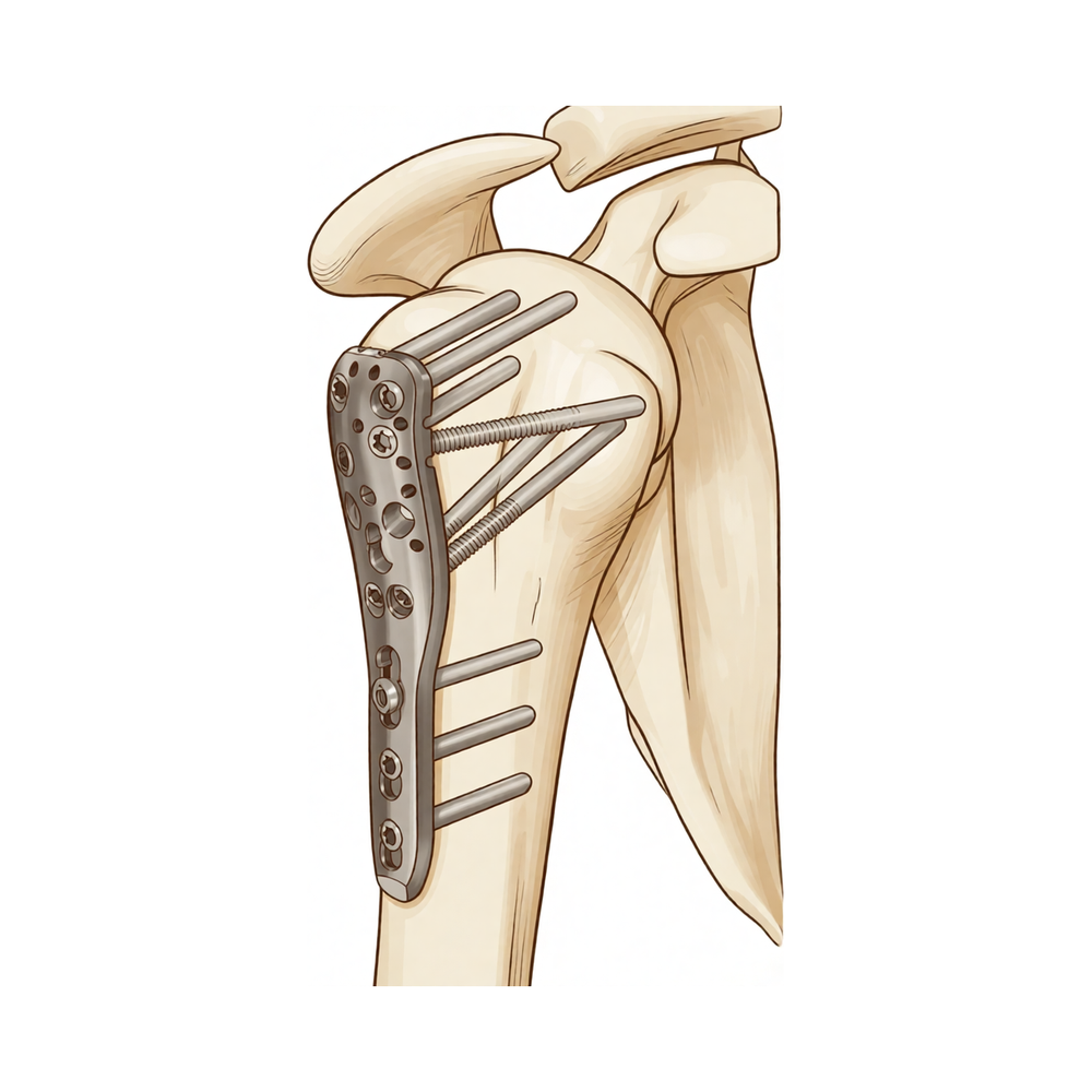
یہ پروٹوکول بازو کی ہڈی کے اوپری سرے کے فریکچر (ہڈی کا ٹوٹنا، proximal humerus fracture) کو آپریشن کے ذریعے جوڑنے کے بعد کی بحالی کے بارے میں ہے (یعنی کندھے کے قریب بازو کے اوپری حصے کی ہڈی کا ٹوٹنا، جسے لاکنگ پلیٹ اور پیچوں سے جوڑا جاتا ہے: ہڈی کو کھول کر سیدھا کرنا اور اندر سے جوڑنا، open reduction and internal fixation, ORIF)، جو ڈاکٹر کیرن ہِرپرا Mater Private Hospital Rockhampton میں کرتے ہیں۔ فزیوتھراپی کی اپنی پہلی ملاقات پر یہ صفحہ یا اس کی پی ڈی ایف ساتھ لائیں تاکہ آپ کی بحالی میں سب ایک ساتھ رہیں۔ آپ کا فزیوتھراپسٹ نیچے دیے گئے مراحل میں آپ کی بحالی کو انفرادی طور پر آگے بڑھاتا ہے، اس پر منحصر کہ آپ کا فریکچر کیسے جڑ رہا ہے۔
اگر آپریشن کے بعد آپ کو اپنے زخم کے بارے میں کوئی تشویش ہو تو کلینک سے رابطہ کریں۔ اکثر یہ مددگار ہوتا ہے کہ زخم کی تصویر لے کر جائزے کے لیے ای میل کر دیں۔
کیا توقع رکھیں
فریکچر کو جوڑنے کے بعد بحالی زیادہ تر پہلے سے طے شدہ کندھے کے آپریشنوں کے بعد کی بحالی سے مختلف طریقے سے ہوتی ہے۔ پلیٹ اور پیچ ٹوٹی ہوئی ہڈی کو اپنی جگہ پر تھامے رکھتے ہیں، لیکن خود ہڈی کو ابھی جڑنا ہوتا ہے، اور جڑنے کی یہ رفتار ہر شخص میں کافی مختلف ہوتی ہے۔ اسی وجہ سے نیچے دیا گیا ٹائم ٹیبل ایک عام رہنمائی ہے، کوئی مقرر شیڈول نہیں: آپ کے پروگرام میں ہر اگلا قدم آپریشن کے بعد گزرے ہفتوں کی تعداد اور اس بات، دونوں پر منحصر ہے کہ آپ کے ایکسرے پر فریکچر کیسے جڑ رہا ہے، جس کی تصدیق ڈاکٹر ہِرپرا کے ساتھ آپ کے معائنوں پر ہوتی ہے۔ صرف کیلنڈر دیکھ کر خود کو اگلے مرحلے میں نہ لے جائیں؛ اپنے معائنے کا انتظار کریں۔
ایک اور بات اس آپریشن کو مختلف بناتی ہے۔ بازو کے اوپری حصے کی ہڈی (humerus) کے اوپری سرے پر ہڈی کے ابھار (tuberosities) وہ جگہ ہیں جہاں روٹیٹر کف (rotator cuff)، یعنی ٹینڈنوں کا وہ گروپ جو کندھے کو تھامتا اور گھماتا ہے، کے ٹینڈن (tendon، وہ مضبوط ڈوریاں جو پٹھے کو ہڈی سے جوڑتی ہیں) جڑتے ہیں، اور بازو کی ہڈی کے اوپری سرے کے بہت سے فریکچرز میں یہ ٹکڑے بھی ٹوٹ جاتے ہیں اور انہیں پلیٹ یا ٹانکوں سے واپس جوڑا جاتا ہے۔ روٹیٹر کف کے پٹھوں سے بہت زیادہ اور بہت جلد کام لینا ان ٹکڑوں کو جڑنے سے پہلے کھینچ سکتا ہے۔ جدید لاکنگ پلیٹ کے ساتھ ہڈی کو جوڑنے کا انتظام (fixation) پہلے دن سے مضبوط ہوتا ہے، اس لیے ہلکی ابتدائی حرکت کی حوصلہ افزائی کی جاتی ہے: آپ شروع ہی سے آرام کی حد میں کندھے کو مدد سے فعال طور پر (active-assisted) اور ہلکے سے خود (active) حرکت دے سکتے ہیں، اور بازو کو بغیر درد کی حد میں اٹھا سکتے ہیں۔ احتیاط والا حصہ روٹیٹر کف پر بوجھ ڈالنا ہے: جہاں ابھار کے ٹکڑے فریکچر کا حصہ نہ ہوں، یا مضبوطی سے جوڑے گئے ہوں، وہاں آپ کے سرجن آپ کو خود بازو گھمانے اور روٹیٹر کف کی ورزشیں جلد شروع کرنے کی اجازت دے سکتے ہیں؛ جہاں ابھار فریکچر میں شامل ہوں اور جڑ رہے ہوں، وہاں روٹیٹر کف کی فعال ورزشیں اور بازو کو زبردستی باہر کی طرف گھمانا کچھ زیادہ عرصے تک روکے جاتے ہیں۔ ڈاکٹر ہِرپرا آپ کو بتائیں گے کہ آپ پر کون سی صورت لاگو ہوتی ہے۔
آپ کے ورزش کے پروگرام میں تین قسم کی حرکت استعمال ہوتی ہے، اور آپ کی ٹیم نشان لگائے گی کہ آپ پر کون سی لاگو ہوتی ہیں:
- فعال حرکت کی حد (active range of motion): بغیر کسی سہارے یا مدد کے حرکت کی اجازت ہے۔
- مدد سے فعال حرکت کی حد (active-assisted range of motion): بازو کو حرکت دینے کے لیے دوسرے بازو یا کسی چیز کی مدد لینا۔
- غیر فعال حرکت کی حد (passive range of motion): بازو پوری طرح ڈھیلا ہو، اور دوسرا بازو یا کوئی بیرونی قوت 100 فیصد کام کرے۔
آپریشن کے بعد جب آپ جاگیں گے تو آپ کا بازو سلنگ (sling)، یعنی گلے سے لٹکی وہ پٹی جو بازو کو سہارا دیتی ہے، میں ہوگا۔ پہلے تقریباً تین ہفتے اسے ہر وقت پہنیں، پھر اگلے ہفتوں میں آرام کے مطابق اسے بتدریج چھوڑ دیں؛ شائع شدہ پروٹوکولز میں بازو کو ساکن رکھنے کی اوسط مدت تقریباً تین ہفتے ہے، اور شواہد سلنگ میں زیادہ دیر رہنے کے بجائے جلد حرکت کی حمایت کرتے ہیں۔ آپ کو اسے پہن کر سونے کی ضرورت نہیں۔ ورزشوں، نہانے دھونے اور کپڑے پہننے کے لیے اسے دن میں کئی بار اتاریں، اور گھر پر آرام سے بیٹھے ہوئے آپ بازو کو سلنگ سے باہر آرام دے سکتے ہیں۔ کندھے کے کسی بھی آپریشن کے بعد کم از کم چھ ہفتوں تک گاڑی نہ چلائیں؛ آپ کے سرجن آپ کو گاڑی چلانے کی اجازت دیں گے، عموماً چھ ہفتے والے معائنے پر۔
سفر ایک نظر میں:
- مرحلہ I — حفاظت اور ابتدائی حرکت: ہفتے 0 سے 6
- مرحلہ II — فعال حرکت دوبارہ حاصل کرنا: ہفتے 6 سے 12
- مرحلہ III — طاقت بڑھانا: تقریباً مہینے 3 سے 4½
- مرحلہ IV — مکمل سرگرمی پر واپسی: تقریباً 4½ سے 6 ماہ کے بعد سے
مرحلہ I — حفاظت اور ابتدائی حرکت (ہفتے 0 سے 6)
Kieran Hirpara 4.0
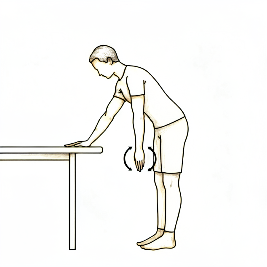
پینڈولم ورزشیں
آگے کی طرف جھکیں، اپنے دوسرے ہاتھ سے میز کا سہارا لیں، اور آپریشن والے بازو کو ڈھیلا لٹکنے دیں۔ اپنے جسم کو ہلکے سے ہلا کر چھوٹے دائرے بنائیں — پہلے گھڑی کی سوئیوں کی سمت میں، پھر الٹی سمت میں۔ بازو غیر فعال طور پر جھولتا ہے؛ حرکت آپ کے جسم سے آتی ہے، کندھے کے پٹھوں سے نہیں۔
دن میں کئی بار
Kieran Hirpara 4.0
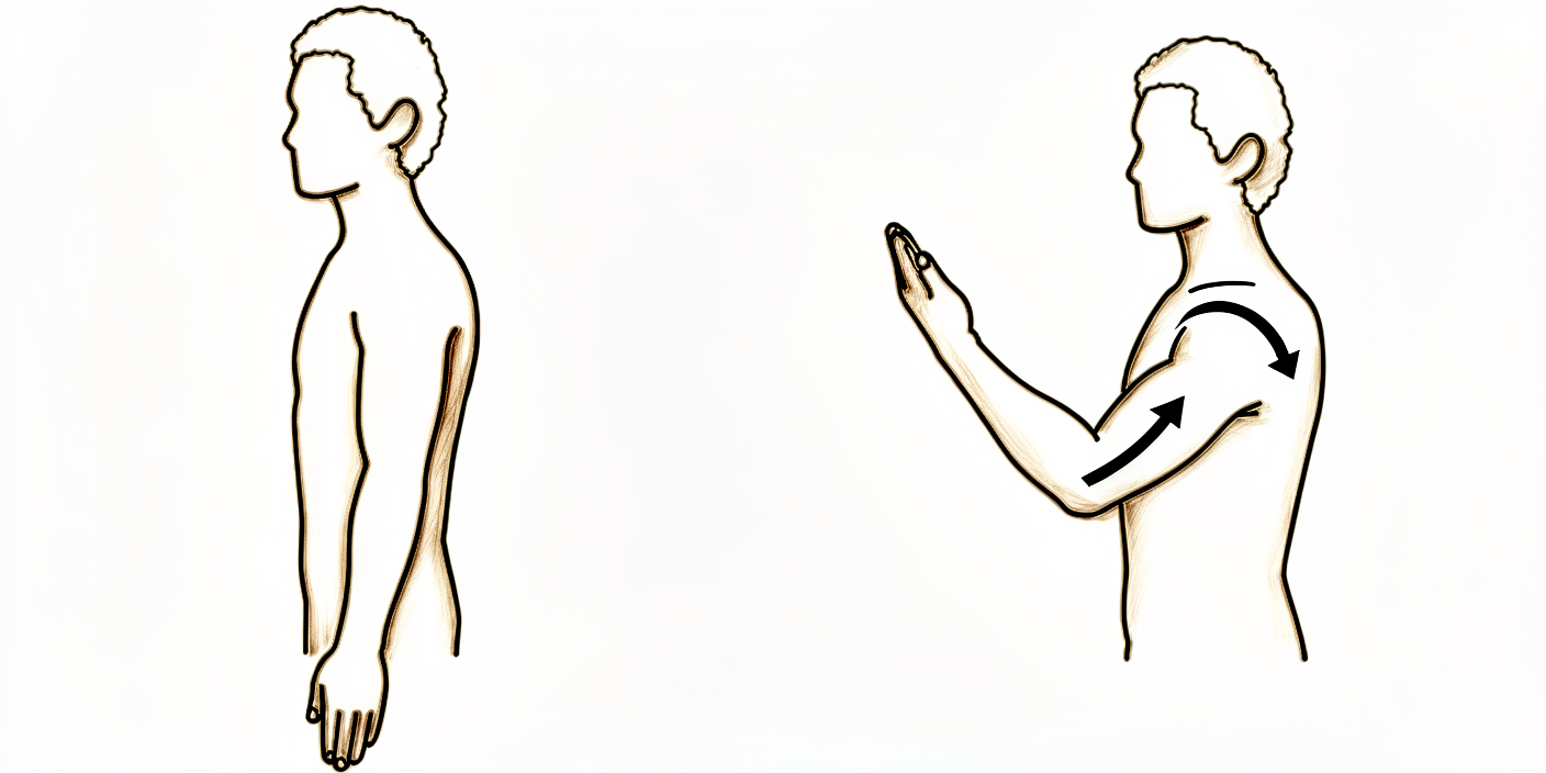
کہنی کو خود موڑنا
بازو سلنگ سے باہر ہو اور بازو کا اوپری حصہ آپ کے پہلو کے ساتھ ٹکا ہو، تو کہنی کو جتنا آرام سے ہو سکے موڑیں، پھر اسے پوری طرح سیدھا کریں۔ صرف کہنی حرکت کرے — کندھے کو ساکن اور بازو کو پہلو کے ساتھ رکھیں۔
دن میں کئی بار
Kieran Hirpara 4.0
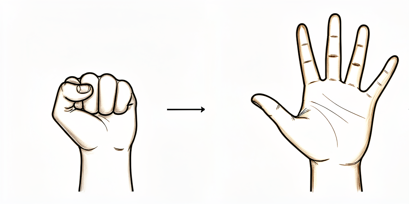
گیند دبانا
بازو کو سہارا دے کر ایک نرم گیند دبائیں یا مضبوطی سے مٹھی بنائیں، پھر ہاتھ پوری طرح کھول دیں۔ جب بھی بازو سلنگ سے باہر ہو، انگلیوں، کلائی اور ہاتھ کو حرکت دیتے رہیں۔
دن میں کئی بار
Kieran Hirpara 4.0
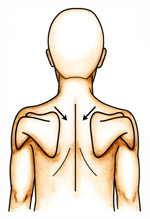
کندھے کی پچھلی ہڈی کو صحیح جگہ پر کسنا
سیدھے بیٹھ یا کھڑے ہو کر، بازوؤں کو حرکت دیے بغیر کندھے کی پچھلی ہڈیوں کو ہلکے سے نیچے اور پیچھے کی طرف آپس میں کھینچیں، چند سیکنڈ روکیں، پھر ڈھیلا چھوڑ دیں۔ اپنے کان، کندھے اور کولہے ایک سیدھ میں رکھیں۔
جیسا آپ کا فزیوتھراپسٹ بتائے
Kieran Hirpara 4.0
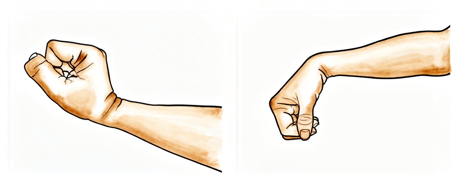
کلائی کی حرکات
بازو سلنگ سے باہر ہو تو کلائی کو آگے، پیچھے اور ہلکے سے دائیں بائیں موڑ کر حرکت میں رکھیں۔ اس سے کندھے کی حفاظت کے دوران پورا بازو کام کرتا رہتا ہے۔
دن میں کئی بار
Kieran Hirpara 4.0
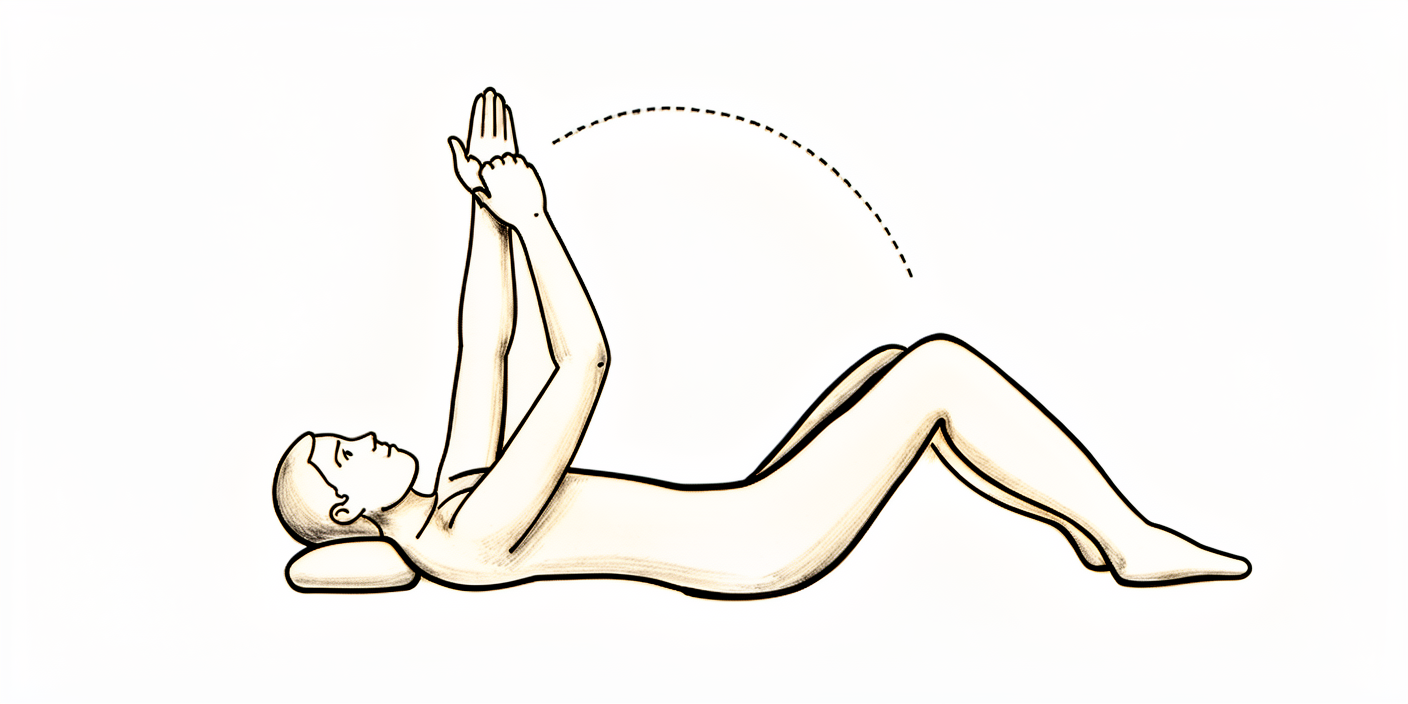
مدد سے بازو آگے اٹھانا (لیٹ کر)
پیٹھ کے بل لیٹیں اور اپنے اچھے بازو کی مدد سے آپریشن والے بازو کو اپنے سامنے اوپر اٹھائیں، پھر اچھے بازو کی مدد جاری رکھتے ہوئے اسے ہلکے سے نیچے لائیں۔ پیٹھ کے بل شروع کرنے سے کشش ثقل مدد کرتی ہے اور ابتدائی طور پر بازو اٹھانا آسان ہو جاتا ہے۔ آرام دہ، بغیر درد کی حد کے اندر رہیں اور جہاں کھنچاؤ شروع ہو وہاں رک جائیں — زبردستی نہ کریں۔
دن میں کئی بار، بغیر درد کی حد کے اندر
Kieran Hirpara 4.0
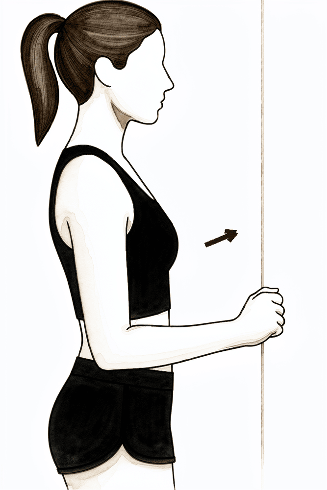
بازو آگے اٹھانے کی ہلکی آئسومیٹرک ورزش
دیوار کی طرف منہ کر کے کھڑے ہوں، آپریشن والا بازو پہلو کے ساتھ ہو اور ڈھیلی مٹھی سامنے دیوار سے لگی ہو۔ بازو کو حرکت دیے بغیر ہاتھ کو ہلکے سے آگے دیوار میں دبائیں، چند سیکنڈ روکیں، پھر ڈھیلا چھوڑ دیں۔ اس سے جڑتے ہوئے فریکچر پر زور ڈالے بغیر کندھا اٹھانے والے پٹھے متحرک ہوتے ہیں۔ یہ صرف تب کریں جب آپ کے فزیوتھراپسٹ نے آپ کو یہ ورزش شروع کروائی ہو۔
جیسا آپ کا فزیوتھراپسٹ بتائے
پہلے چھ ہفتے جوڑے گئے فریکچر کی حفاظت کے بارے میں ہیں، اور ساتھ ہی بازو کے باقی حصے (اور خود کندھے کے جوڑ) کو اکڑنے سے بچانے کے بارے میں۔ چونکہ لاکنگ پلیٹ شروع سے ہڈی کو مضبوطی سے تھامے رکھتی ہے، اس لیے چھ ہفتے انتظار کرنے کے بجائے ہلکی ابتدائی حرکت کی حوصلہ افزائی کی جاتی ہے۔ گھر جانے سے پہلے ایک فزیوتھراپسٹ آپ کو ہلکی ورزشیں شروع کروائے گا: پینڈولم ورزشیں (جسم کو ہلکے سے ہلا کر بازو کو لٹکنے اور ہلکے سے جھولنے دینا)، اور دن میں کئی بار سلنگ سے باہر کہنی، کلائی اور ہاتھ کو حرکت دیتے رہنا۔ شروع ہی سے، اور آرام دہ، بغیر درد کی حد کے اندر، آپ بازو کو اپنے دوسرے ہاتھ کی مدد سے (مدد سے فعال) اور ہلکے سے اس کی اپنی طاقت سے (فعال) حرکت دینا شروع کر سکتے ہیں، اور آرام کے مطابق اسے اپنے سامنے اٹھا سکتے ہیں۔ احتیاط والا حصہ روٹیٹر کف پر بوجھ ڈالنا ہے: بازو کو زبردستی باہر کی طرف گھمانے (external rotation) سے بچیں، اور جب تک ڈاکٹر ہِرپرا تصدیق نہ کریں کہ آپ کے ابھار جڑ گئے ہیں، روٹیٹر کف کی مزاحمت والی ورزشوں سے بچیں۔ درد میں آرام کے لیے برف استعمال کریں، اور اپنی ورزشوں اور فزیوتھراپی کی ملاقاتوں سے پہلے اپنی درد کش دوا لیں۔
آپ کے فزیوتھراپسٹ کے لیے:
اہداف
- ہڈی کو جوڑنے والے انتظام (fixation) کی حفاظت اور ہڈی کے جڑنے کو بہترین بنانا
- درد اور سوجن کم کرنا
- بغیر درد کی حد میں ابتدائی مدد سے فعال حرکت شروع کر کے فعال طور پر بازو اٹھانے (elevation) کی طرف بڑھنا
- گردن، کہنی، کلائی اور ہاتھ کی پوری حرکت برقرار رکھنا
انتظام
- سلنگ تقریباً 3 ہفتے ہر وقت، پھر آرام کے مطابق بتدریج چھوڑنا (تحقیقی لٹریچر کا عام طریقہ)؛ ورزشوں اور صفائی کے لیے دن میں کئی بار اتاری جائے؛ بستر میں ضروری نہیں
- پینڈولم / کوڈمین (Codman) ورزشیں دن میں کئی بار
- مستحکم جوڑ (stable fixation) کی صورت میں شروع سے ہی آرام دہ، بغیر درد کی حد میں ابتدائی مدد سے فعال اور ہلکی فعال طور پر بازو آگے اٹھانا (forward elevation)؛ پیٹھ کے بل لیٹ کر شروع کر کے، کنٹرول بہتر ہونے پر سیدھی پوزیشن کی طرف؛ آرام کے مطابق حد بڑھائیں
- جہاں فعال حرکت ابھی برداشت نہ ہو وہاں ضرورت کے مطابق غیر فعال حرکت: کندھے کی پچھلی ہڈی کی سطح (scapular plane) میں بازو آگے اٹھانا تقریباً 90 ڈگری تک، اندر کی طرف گھماؤ پیٹ تک (کمر کے پیچھے نہیں)
- بازو پہلو کے ساتھ ہو تو باہر کی طرف گھماؤ کو ابتدا میں تقریباً 30 سے 40 ڈگری کی ہلکی حد تک رکھیں؛ جہاں ابھار فریکچر میں شامل نہ ہوں یا مضبوطی سے جوڑے گئے ہوں وہاں سرجن اسے بڑھا سکتے ہیں
- سلنگ سے باہر گردن، کہنی، کلائی اور ہاتھ کی فعال حرکت؛ گرفت کے لیے گیند دبانا
- کندھے کی پچھلی چپٹی ہڈی (shoulder blade) کو صحیح جگہ پر کسنا (scapular setting) اور اس کی حرکت پذیری کی ورزشیں (اوپر اٹھانا، نیچے لانا، پیچھے کھینچنا، آگے لے جانا)
- آرام کے مطابق ڈیلٹائیڈ (deltoid) اور کندھے کی پچھلی ہڈی کے اردگرد کے پٹھوں (periscapular) کی ہلکی آئسومیٹرک ورزشیں (isometrics، یعنی جوڑ کو ہلائے بغیر پٹھے کو کسنا) شروع کی جا سکتی ہیں
- سیشنوں سے پہلے ٹھنڈک (برف) اور درد کش دوا؛ زخم بھرنے کے بعد زخم کے نشان کی مالش اور حساسیت کم کرنے کی ورزشیں (desensitisation)
احتیاطیں
- جب تک ابھاروں کے جڑنے کی تصدیق نہ ہو جائے (سرجن کی رہنمائی میں)، زبردستی یا مزاحمت کے خلاف باہر کی طرف گھماؤ اور روٹیٹر کف پر بھاری بوجھ سے بچیں؛ بغیر درد کی حد میں فعال طور پر بازو اٹھانے کی اجازت ہے
- کمر کے پیچھے اندر کی طرف گھماؤ بالکل نہیں؛ بازو کو سیدھا پہلو کی طرف اٹھانے (coronal plane میں abduction) سے بچیں
- ابتدائی حرکت بغیر درد کے رکھیں: ہلکی اور آرام کی حد میں، زبردستی نہیں
- آپریشن والے بازو سے تقریباً 0.5 سے 1 کلوگرام سے زیادہ وزن نہ اٹھائیں
- آپریشن والے بازو کے ذریعے وزن نہ سہاریں (کرسی یا بستر سے ہاتھوں کے زور پر نہ اٹھیں)
- چھ ہفتوں تک گاڑی نہ چلائیں (یہ کندھے کے ہر آپریشن پر لاگو ہوتا ہے)
- حرکت کی آخری حد پر نہ زبردستی حرکت اور نہ درد والی حرکت
اگلے مرحلے میں جانے کی شرائط
- ایکسرے پر ہڈی کے جڑنے میں پیش رفت کا ثبوت، جس کی تصدیق تقریباً 6 ہفتوں پر ڈاکٹر ہِرپرا کے ساتھ معائنے پر ہو
- درد اچھی طرح قابو میں ہو
- کہنی، کلائی اور ہاتھ کی پوری حرکت برقرار ہو
- بغیر درد کی حد میں مدد سے فعال سے لے کر فعال طور پر بازو کو آرام سے اٹھانا
مرحلہ II — فعال حرکت دوبارہ حاصل کرنا (ہفتے 6 سے 12)
Kieran Hirpara 4.0
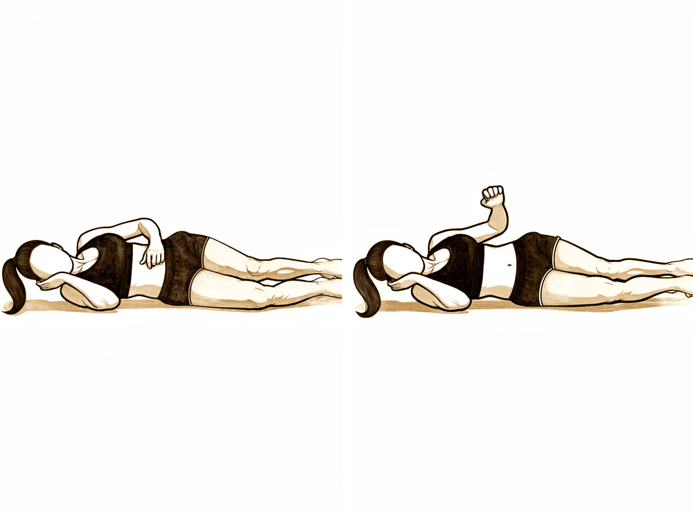
پہلو کے بل لیٹ کر باہر کی طرف گھماؤ
جس طرف آپریشن نہیں ہوا اس پہلو کے بل لیٹیں، آپریشن والا بازو اوپر ہو، کہنی 90 ڈگری پر مڑی اور جسم کے ساتھ لگی ہو۔ کہنی کو جسم کے ساتھ رکھتے ہوئے، بازو کے نچلے حصے کو پیٹ سے دور اوپر کی طرف جتنا آرام سے ہو سکے گھمائیں، پھر قابو کے ساتھ نیچے لائیں۔ کوئی اضافی وزن نہیں — بازو اپنی طاقت سے حرکت کرتا ہے۔
جیسا آپ کا فزیوتھراپسٹ بتائے
Kieran Hirpara 4.0
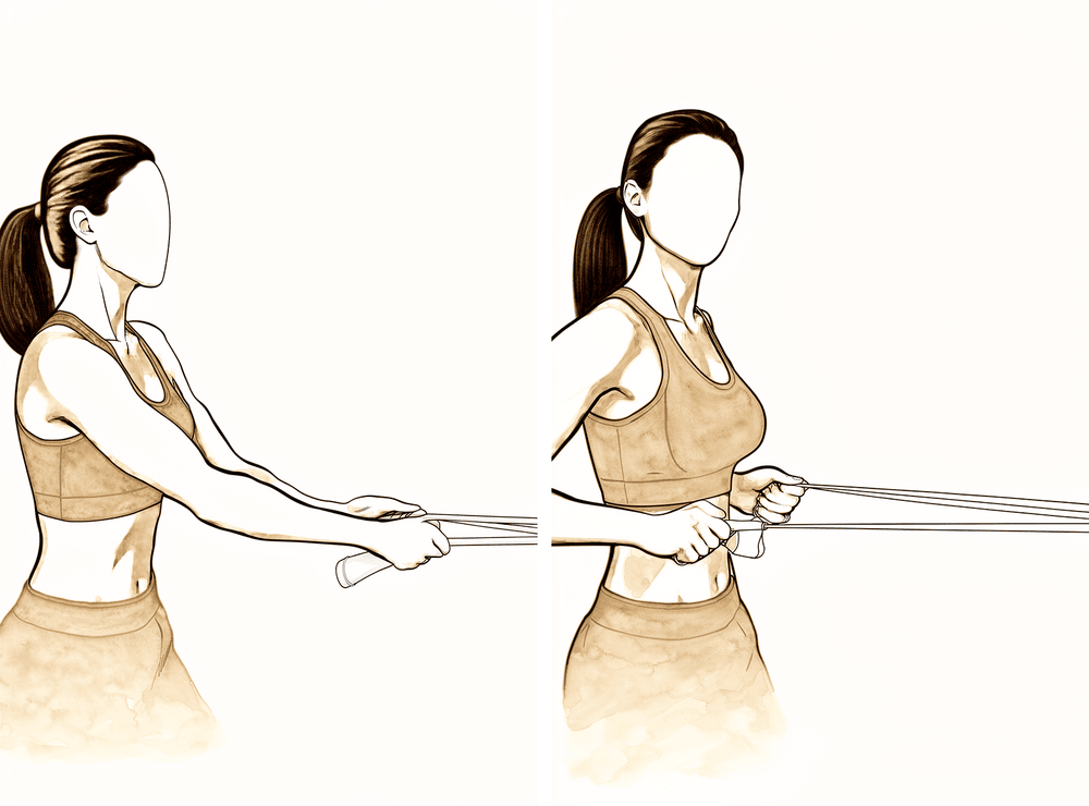
لچکدار بینڈ کے ساتھ لو رو (low row)
لچکدار بینڈ کو اپنے سامنے کمر کی اونچائی پر باندھ دیں۔ سیدھے کھڑے ہو کر بینڈ کو اپنی طرف کھینچیں، کندھے کی پچھلی ہڈی کو ہلکے سے نیچے اور پیچھے لاتے ہوئے، پھر قابو کے ساتھ چھوڑیں۔ مزاحمت ہلکی رکھیں۔
جیسا آپ کا فزیوتھراپسٹ بتائے
Kieran Hirpara 4.0
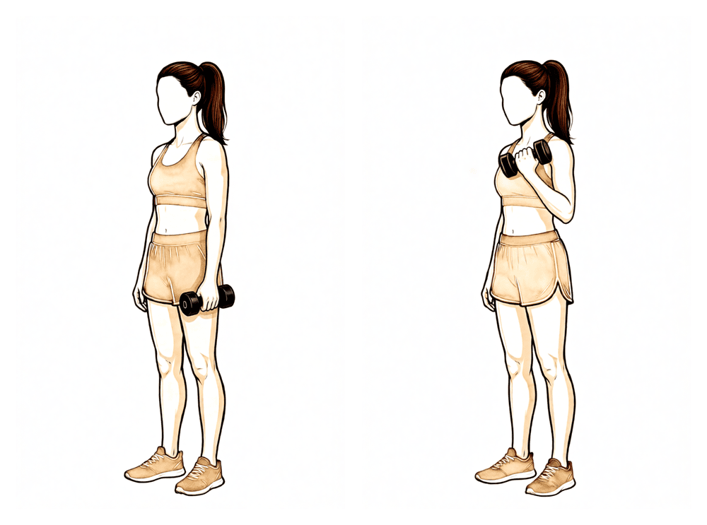
بائسیپس کرلز
ہتھیلی اوپر کی طرف رکھ کر ہلکا وزن پکڑیں، کہنی موڑ کر وزن کو اپنے کندھے کی طرف لائیں، پھر آہستہ سے نیچے کریں۔ بازو کا اوپری حصہ پہلو کے ساتھ ساکن رکھیں، اور وزن ہلکا رکھیں — اس مرحلے میں تقریباً 1 سے 2 کلوگرام سے زیادہ نہیں۔
جیسا آپ کا فزیوتھراپسٹ بتائے
Kieran Hirpara 4.0
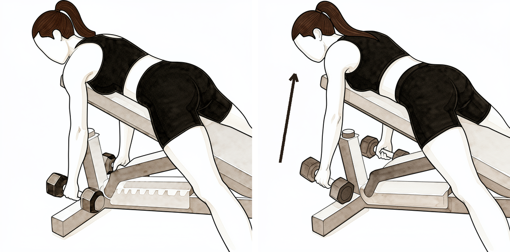
سہارے کے ساتھ رو (rows)
اپنے اچھے بازو سے میز کا سہارا لے کر آگے جھکیں، اور آپریشن والے بازو کو ہلکا وزن پکڑ کر لٹکنے دیں۔ کندھے کی پچھلی ہڈی کو پیچھے کھینچیں اور وزن کو اپنے کولہے کی طرف اوپر کھینچیں، پھر قابو کے ساتھ نیچے لائیں۔ دھڑ کو ساکن رکھیں — حرکت کندھے کی پچھلی ہڈی اور بازو سے آئے، کمر سے نہیں۔
جیسا آپ کا فزیوتھراپسٹ بتائے
Kieran Hirpara 4.0
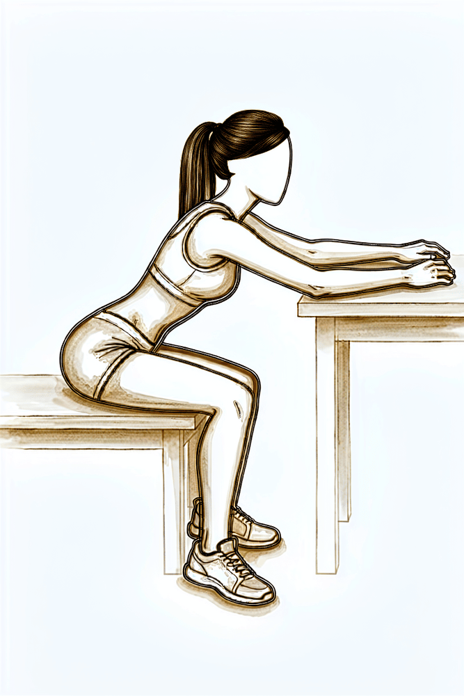
میز پر ہاتھ سرکانا
میز کے پاس بیٹھیں اور ہاتھ کسی تولیے یا کپڑے پر رکھیں۔ آگے جھکیں اور ہاتھ کو میز پر اپنے سے دور سرکائیں، تاکہ بازو سامنے کی طرف مزید آگے پہنچے، پھر اسے واپس سرکائیں۔ بازو کا وزن میز اٹھاتی ہے، اس لیے آگے کی حرکت دوبارہ حاصل کرنے کا یہ ایک نرم طریقہ ہے۔
جیسا آپ کا فزیوتھراپسٹ بتائے
Kieran Hirpara 4.0
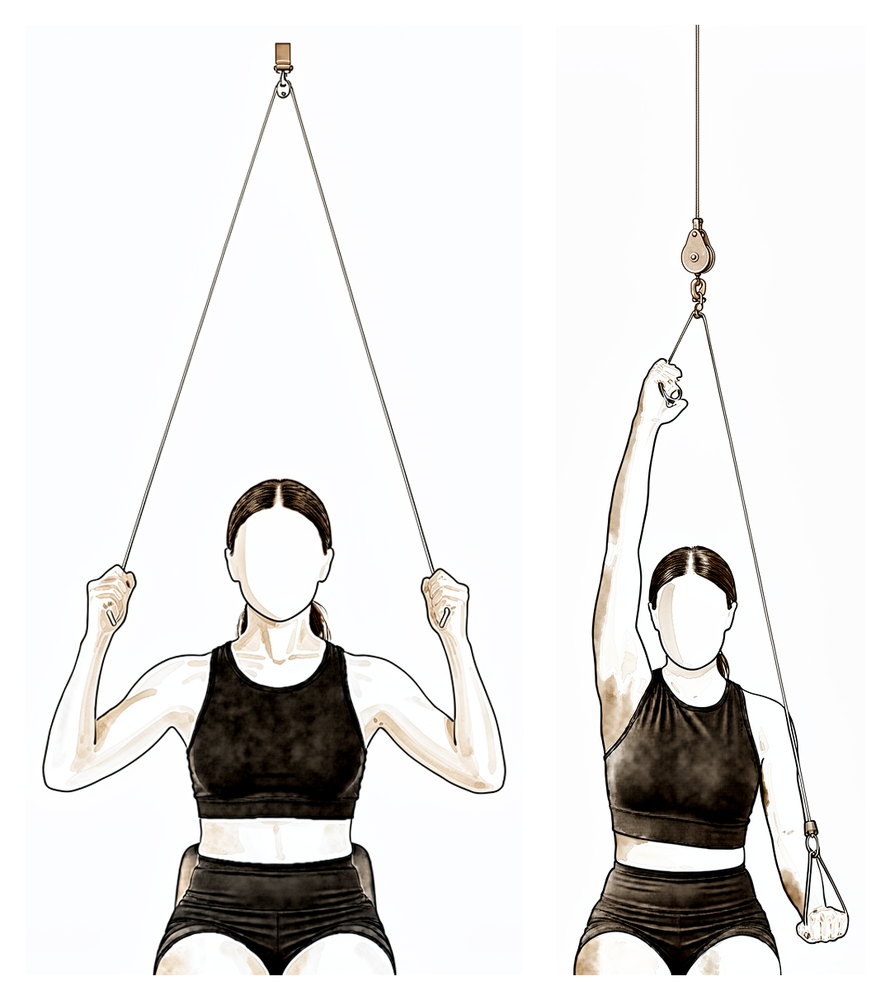
دروازے کے اوپر لگی پُلی
دروازے کے اوپر لگی پُلی کے نیچے بیٹھیں اور ہر ہاتھ میں ایک ہینڈل پکڑیں۔ اپنے اچھے بازو سے نیچے کھینچیں تاکہ آپریشن والا بازو جتنا آرام سے ہو سکے اوپر اٹھے، پھر اسے آہستہ سے نیچے لائیں۔ کام اچھے بازو کو کرنے دیں اور آرام دہ حد کے اندر رہیں۔
جیسا آپ کا فزیوتھراپسٹ بتائے
Kieran Hirpara 4.0
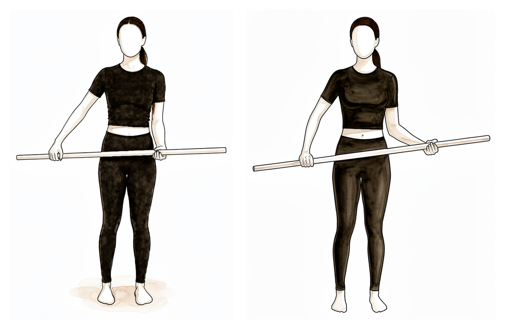
چھڑی کی مدد سے باہر کی طرف گھماؤ
پیٹھ کے بل لیٹیں، کہنی پہلو کے ساتھ لگی اور کونے کی شکل میں (right angle) مڑی ہو۔ دونوں ہاتھوں میں چھڑی پکڑیں اور اچھے بازو کی مدد سے آپریشن والے ہاتھ کو باہر کی طرف گھمائیں۔ جب ہڈی جڑنے کی تصدیق ہو جائے تو باہر کی طرف گھماؤ کو ہلکے سے تھوڑا اور بڑھائیں — لیکن اسی حد کے اندر رہیں جو آپ کے سرجن اور فزیوتھراپسٹ نے طے کی ہے۔
جیسا آپ کا فزیوتھراپسٹ بتائے
Kieran Hirpara 4.0
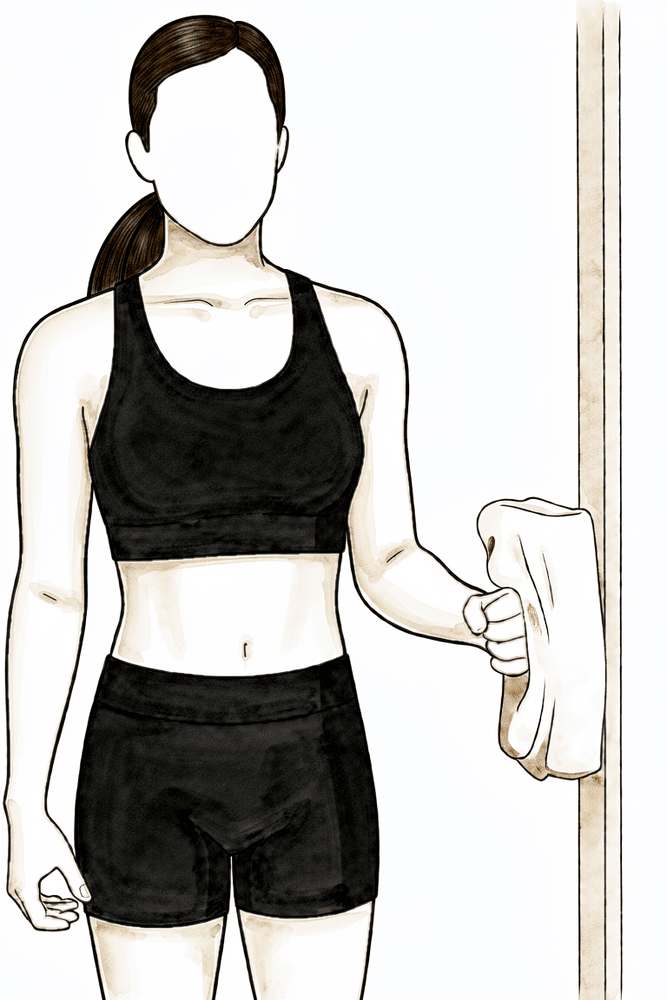
آئسومیٹرک باہر کی طرف گھماؤ
اس طرح کھڑے ہوں کہ کہنی پہلو کے ساتھ لگی اور کونے کی شکل میں (right angle) مڑی ہو، اور ہاتھ کی پشت دیوار یا دروازے کی چوکھٹ کے ساتھ ہو۔ بازو کو حرکت دیے بغیر ہلکے سے باہر کی طرف دبائیں، چند سیکنڈ روکیں، پھر ڈھیلا چھوڑ دیں۔ اس سے روٹیٹر کف بغیر کسی حرکت کے متحرک ہونا شروع ہوتا ہے — اسے صرف تب شروع کریں جب ڈاکٹر ہِرپرا ہڈی کے جڑنے کی تصدیق کر دیں۔
جیسا آپ کا فزیوتھراپسٹ بتائے
Kieran Hirpara 4.0
ٹرائسیپس ایکسٹینشنز
ہلکا وزن پکڑ کر کہنی کو موڑیں اور پھر سیدھا کریں، تاکہ بازو کے اوپری حصے کے پچھلے پٹھے پر کام ہو۔ بازو کا اوپری حصہ ساکن اور وزن ہلکا رکھیں — اس مرحلے میں تقریباً 1 سے 2 کلوگرام سے زیادہ نہیں۔
جیسا آپ کا فزیوتھراپسٹ بتائے
Kieran Hirpara 4.0
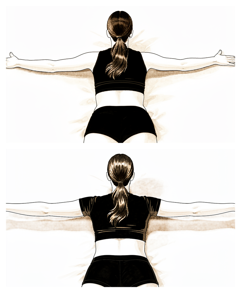
پیٹ کے بل لیٹ کر کندھے کی پچھلی ہڈی کی رو
پیٹ کے بل لیٹیں اور اپنا بازو بستر یا بینچ کے کنارے سے نیچے لٹکنے دیں۔ کندھے کی پچھلی ہڈی کو پیچھے کھینچیں اور بازو کو تھوڑا سا پہلو کی طرف اٹھائیں، کندھے کی پچھلی ہڈیوں کو آپس میں کستے ہوئے، پھر قابو کے ساتھ نیچے لائیں۔ اس سے وہ پٹھے مضبوط ہوتے ہیں جو کندھے کی پچھلی ہڈی کو ٹھہراتے ہیں۔
جیسا آپ کا فزیوتھراپسٹ بتائے
تقریباً چھ ہفتے والے معائنے پر ڈاکٹر ہِرپرا آپ کے ایکسرے دیکھتے ہیں۔ اگر ہڈی توقع کے مطابق جڑ رہی ہو تو سلنگ کا باقی ماندہ استعمال بند ہو جاتا ہے اور آپ بازو کو خود حرکت دینا شروع کرتے ہیں، پہلے مدد کے ساتھ (دوسرے بازو، چھڑی یا پُلی (pulley، رسی اور چرخی والا آلہ) کے ذریعے)، پھر خود (فعال طور پر)۔ عموماً بہتر یہ ہوتا ہے کہ بازو کو خود اٹھانا پیٹھ کے بل لیٹ کر شروع کیا جائے، جہاں کشش ثقل کم رکاوٹ بنتی ہے، اور کنٹرول بہتر ہونے پر بیٹھنے اور کھڑے ہونے کی طرف بڑھا جائے۔ روٹیٹر کف کے لیے پٹھوں کو ہلائے بغیر کسنے والی ہلکی (آئسومیٹرک) ورزشیں اس مرحلے میں تب شروع ہوتی ہیں جب ڈاکٹر ہِرپرا ہڈی کے جڑنے کی تصدیق کر دیں؛ مزاحمت کے خلاف ورزشیں بعد میں آتی ہیں۔ آپ دوبارہ گاڑی چلانا تب شروع کر سکتے ہیں جب آپ سلنگ سے باہر ہوں، آپ کے بازو میں محفوظ طریقے سے گاڑی چلانے کے لیے کافی حرکت اور کنٹرول ہو، اور آپ اب تیز اثر والی درد کش دوائیں نہ لے رہے ہوں؛ اگر شک ہو تو اپنے معائنے پر اس بارے میں بات کریں۔
آپ کے فزیوتھراپسٹ کے لیے:
اہداف
- پوری غیر فعال حرکت کی حد بحال کرنا
- تمام سمتوں میں مدد سے فعال سے فعال حرکت کی طرف بڑھنا
- کندھے کی پچھلی ہڈی اور بازو کی ہڈی کی معمول کی ہم آہنگ حرکت (scapulohumeral rhythm) دوبارہ قائم کرنا اور متبادل غلط انداز کم سے کم کرنا
- معمول کی ہلکی روزمرہ سرگرمیوں پر واپسی
انتظام
- سلنگ زیادہ سے زیادہ 6 ہفتے والے معائنے تک پوری طرح ترک کر دی جائے
- مدد سے فعال حرکت: لان چیئر پروگریشن (lawn-chair progression)، میز / دیوار پر ہاتھ سرکانا، پُلی، چھڑی کی ورزشیں، آرام کے مطابق مرحلہ I کی حدود سے آگے بڑھتے ہوئے
- تقریباً 6 سے 8 ہفتوں سے فعال حرکت: پیٹھ کے بل لیٹ کر بازو آگے اٹھانا (supine flexion) اور پھر سیدھی پوزیشن میں بازو اٹھانا؛ پہلو کے بل لیٹ کر باہر کی طرف گھماؤ اور بازو آگے اٹھانا؛ لو رو / لو پنچ (low rows / low punch)
- تقریباً 6 سے 8 ہفتوں سے، ہڈی جڑنے کی تصدیق کے بعد، بازو پہلو کے ساتھ رکھ کر روٹیٹر کف اور ڈیلٹائیڈ کی پورے زور سے کم (submaximal) آئسومیٹرک ورزشیں
- کہنی کی ہلکی آئسوٹونک (isotonic) ورزشیں (بائسیپس کرلز، ٹرائسیپس ایکسٹینشنز) اور کندھے کی پچھلی ہڈی کی طاقت کی ورزشیں (پیچھے کھینچنا، پیٹ کے بل لیٹ کر رو)
- ضرورت کے مطابق کندھے کے جوڑ (glenohumeral) اور کندھے کی پچھلی ہڈی و پسلیوں کے درمیان (scapulothoracic) موبلائزیشن، ہڈی کے جڑنے کے ساتھ درجے بڑھاتے ہوئے
- جسم کے انداز کی درستگی؛ سیشنوں کے آس پاس اپنی پسند کے مطابق گرمائش / برف اور درد کش دوا جاری رکھیں
احتیاطیں
- جب تک ہڈی کے جڑ جانے (union) کی تصدیق نہ ہو، روٹیٹر کف کی مزاحمت والی (isotonic) طاقت کی ورزشیں نہیں، عموماً 8 سے 12 ہفتوں سے پہلے نہیں
- حرکت کی آخری حد پر نہ زور دار اضافی دباؤ اور نہ جارحانہ غیر فعال اسٹریچنگ
- آپریشن والے بازو سے وزن اٹھانا تقریباً 1 سے 2 کلوگرام تک محدود
- بازو اٹھاتے وقت کندھا اچکانے اور دھڑ کو ایک طرف جھکانے جیسے متبادل انداز پر نظر رکھیں اور انہیں درست کریں
اگلے مرحلے میں جانے کی شرائط
- پوری، یا تقریباً پوری، غیر فعال حرکت کی حد
- کم از کم کندھے کی اونچائی سے نیچے، اچھے انداز کے ساتھ فعال طور پر بازو اٹھانا
- آئسومیٹرک ورزشیں درد بڑھائے بغیر اچھی طرح برداشت ہوں
- ایکسرے پر ہڈی کے جڑنے میں پیش رفت، جس کی تصدیق ڈاکٹر ہِرپرا کے ساتھ آپ کے معائنے پر ہو
مرحلہ III — طاقت بڑھانا (تقریباً مہینے 3 سے 4½)
Kieran Hirpara 4.0
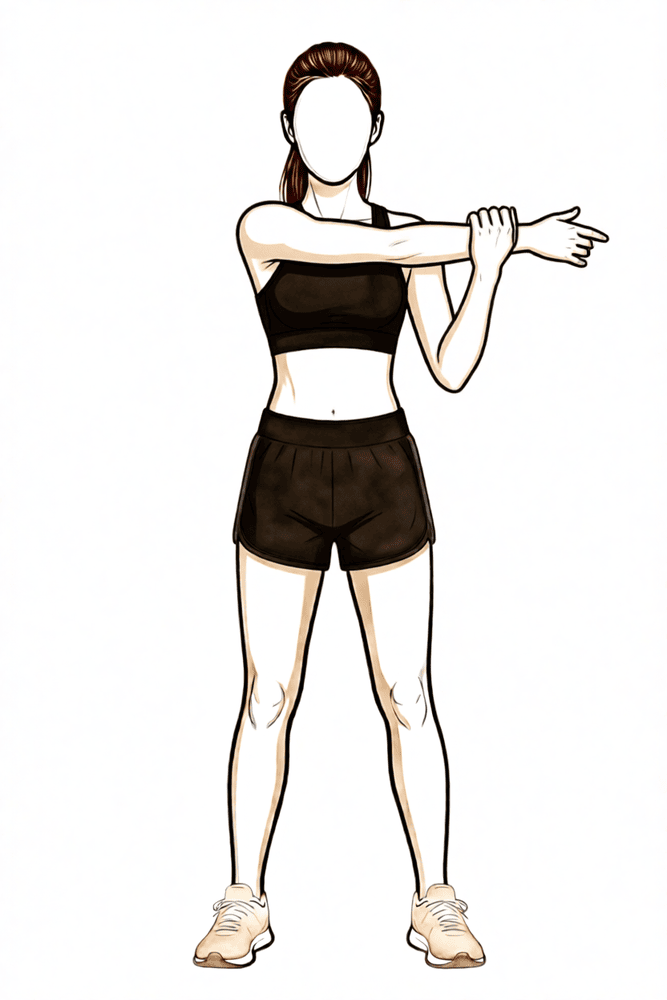
سینے کے آر پار اسٹریچ
اپنے دوسرے ہاتھ سے آپریشن والے بازو کو اپنے سینے کے آر پار کھینچیں یہاں تک کہ کندھے کے پچھلے حصے میں کھنچاؤ محسوس ہو۔ تھوڑی دیر روکیں، پھر چھوڑ دیں۔ صرف واضح کھنچاؤ اور بے آرامی تک اسٹریچ کریں — تیز درد تک کبھی نہیں۔
جیسا آپ کا فزیوتھراپسٹ بتائے
Kieran Hirpara 4.0
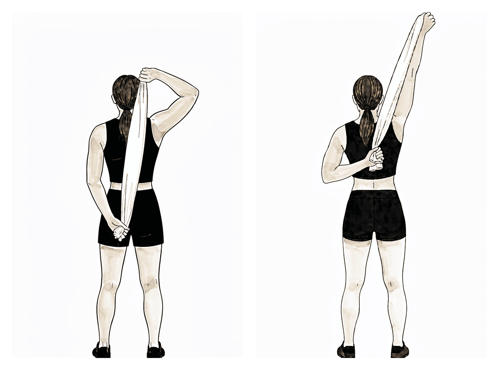
کمر کے پیچھے اسٹریچ
کمر کے پیچھے ایک تولیہ پکڑیں، اچھا بازو اوپر اور آپریشن والا بازو نیچے ہو۔ اوپر والے ہاتھ سے نیچے والے ہاتھ کو ہلکے سے کمر پر اوپر کھینچیں یہاں تک کہ آرام دہ کھنچاؤ محسوس ہو، روکیں، پھر چھوڑ دیں۔ حرکت کی حد بتدریج بڑھائیں — اسٹریچ کو کبھی زبردستی نہ کریں اور نہ تیز درد تک لے جائیں۔
جیسا آپ کا فزیوتھراپسٹ بتائے
Kieran Hirpara 4.0
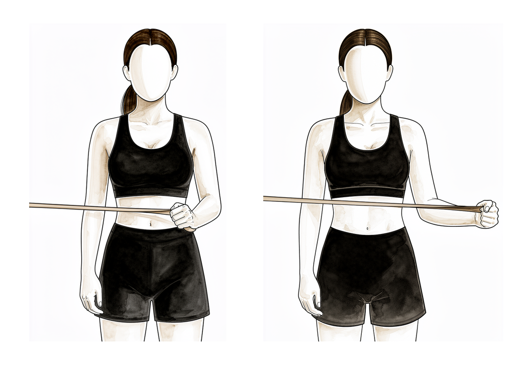
بینڈ کے ساتھ باہر کی طرف گھماؤ
کہنی کو پہلو کے ساتھ لگا کر اور کونے کی شکل میں (right angle) موڑ کر کھڑے ہوں، اور کمر کی اونچائی پر بندھا لچکدار بینڈ پکڑیں۔ کہنی کو پہلو کے ساتھ رکھتے ہوئے، بازو کے نچلے حصے کو بینڈ کے خلاف باہر کی طرف گھمائیں، پھر آہستہ سے واپس لائیں۔ اس سے بازو پہلو کے ساتھ رکھ کر روٹیٹر کف مضبوط ہوتا ہے، جو گھماؤ کی طاقت بڑھانے کا نقطۂ آغاز ہے۔
8 سے 12 بار کے 2 سے 3 سیٹ، کم مزاحمت
Kieran Hirpara 4.0
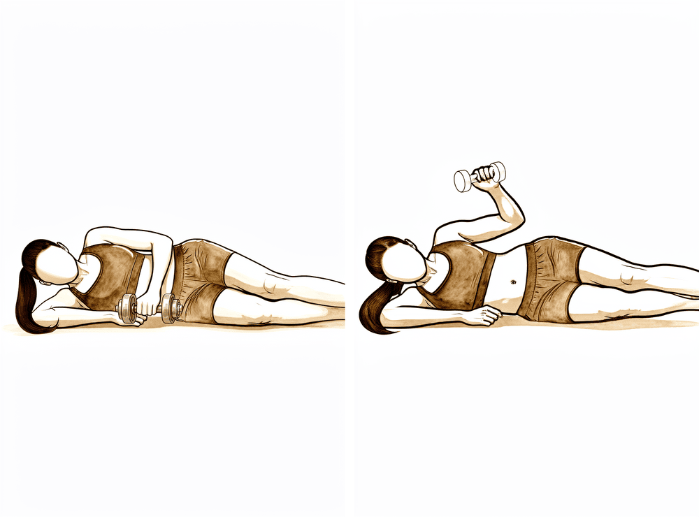
پہلو کے بل لیٹ کر وزن کے ساتھ باہر کی طرف گھماؤ
جس طرف آپریشن نہیں ہوا اس پہلو کے بل لیٹیں، اوپر والی کہنی کونے کی شکل میں (right angle) مڑی اور جسم کے ساتھ لگی ہو۔ ہلکا وزن پکڑ کر بازو کے نچلے حصے کو چھت کی طرف اوپر گھمائیں، پھر آہستہ سے نیچے لائیں۔ روٹیٹر کف کی طاقت دوبارہ بنتے وقت بوجھ ہلکا اور تکرار زیادہ رکھیں۔
8 سے 12 بار کے 2 سے 3 سیٹ، کم بوجھ
Kieran Hirpara 4.0
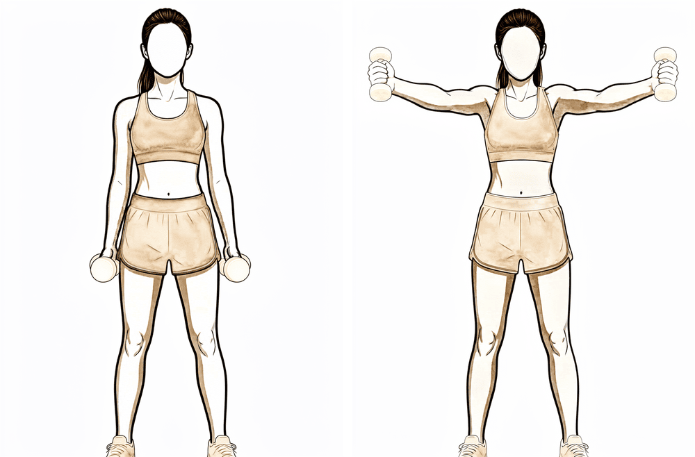
بازو ترچھا اٹھانا (اسکیپشن، full can)
انگوٹھا اوپر کی طرف رکھ کر ہلکا وزن پکڑیں، اور بازو کو سامنے اور باہر کی ترچھی سمت میں — سیدھا پہلو کی طرف نہیں — تقریباً کندھے کی اونچائی تک اٹھائیں، پھر آہستہ سے نیچے لائیں۔ اس سے کندھے کا اگلا حصہ اور روٹیٹر کف ایک ساتھ کام کرتے ہیں۔ وزن ہلکا رکھیں اور اگر ایسا درد ہو جو دیر تک رہے تو رک جائیں۔
8 سے 12 بار کے 2 سے 3 سیٹ، کم بوجھ
Kieran Hirpara 4.0
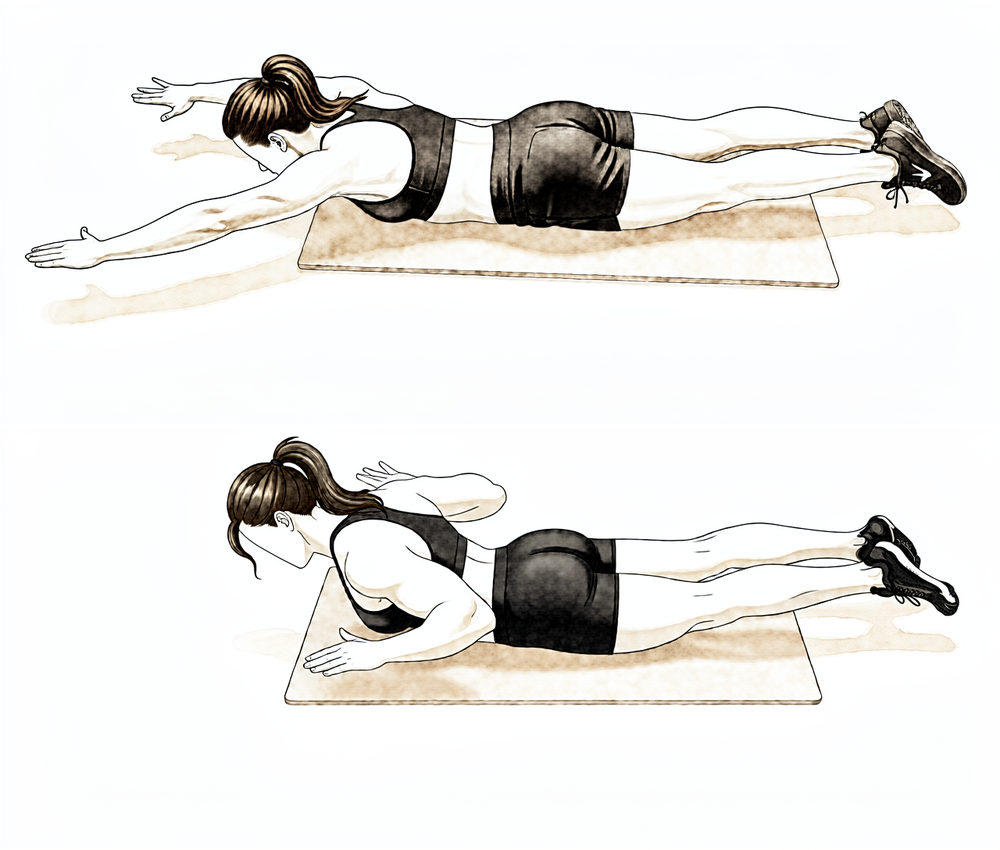
پیٹ کے بل لیٹ کر رو
پیٹ کے بل لیٹیں اور آپریشن والا بازو فرش کی طرف نیچے لٹکنے دیں۔ کہنی کو چھت کی طرف اوپر کھینچیں، کندھے کی پچھلی ہڈی کو ریڑھ کی ہڈی کی طرف کستے ہوئے، پھر آہستہ سے نیچے لائیں۔ بغیر وزن کے شروع کریں اور حرکت قابو میں رکھیں۔
جیسا آپ کا فزیوتھراپسٹ بتائے
Kieran Hirpara 4.0
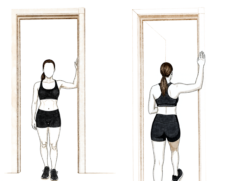
دروازے میں سینے کا اسٹریچ
دروازے میں کھڑے ہوں، بازوؤں کے نچلے حصے چوکھٹ پر ٹکے ہوں اور کہنیاں تقریباً کندھے کی اونچائی پر ہوں۔ ایک پاؤں آگے دروازے سے باہر رکھیں اور ہلکے سے آگے جھکیں یہاں تک کہ سینے کے اگلے حصے میں کھنچاؤ محسوس ہو، پھر روکیں۔ اسے ہلکا رکھیں اور کبھی زبردستی نہ کریں۔
تقریباً 20 سے 30 سیکنڈ روکیں، جیسا آپ کا فزیوتھراپسٹ بتائے
Kieran Hirpara 4.0
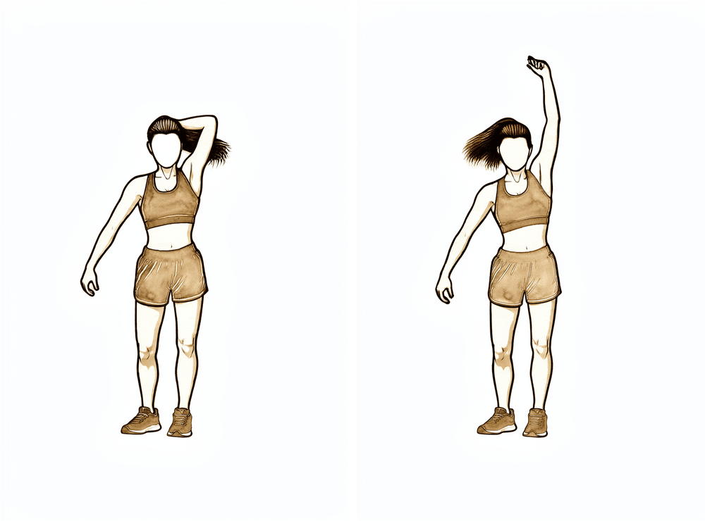
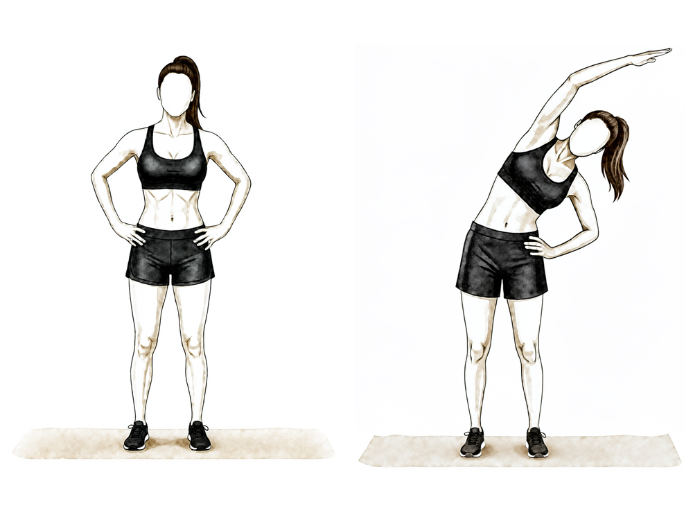
کمر کے پہلو کے پٹھے (latissimus) کا اسٹریچ
آپریشن والے بازو کو سر کے اوپر لے جائیں — ضرورت ہو تو اچھے ہاتھ سے اسے راستہ دکھائیں — اور ہلکے سے مخالف سمت میں جھکیں یہاں تک کہ کمر کے پہلو میں نیچے تک کھنچاؤ محسوس ہو، پھر روکیں۔ اسے آرام دہ اور اپنی حد کے اندر رکھیں۔
تقریباً 20 سے 30 سیکنڈ روکیں، جیسا آپ کا فزیوتھراپسٹ بتائے
فریکچر جڑ جانے اور فعال حرکت واپس آنے کے ساتھ، توجہ طاقت دوبارہ بنانے کی طرف مڑتی ہے۔ مزاحمت والی ورزشیں ہلکے سے شروع ہوتی ہیں (روٹیٹر کف، ڈیلٹائیڈ اور کندھے کی پچھلی ہڈی کے پٹھوں کے لیے لچکدار بینڈ اور ہلکے وزن) اور بتدریج بڑھتی ہیں۔ ساتھ ساتھ اسٹریچنگ جاری رہتی ہے، تمام سمتوں میں پوری حرکت کی طرف، جس میں ہاتھ کمر کے پیچھے لے جانا بھی شامل ہے۔ اس مرحلے میں روزمرہ کی عام سرگرمیاں زیادہ تر معمول پر آ جانی چاہییں، اور ہلکی تفریحی سرگرمیاں عموماً دوبارہ شروع ہو جاتی ہیں، جیسا کہ آپ کا فزیوتھراپسٹ رہنمائی کرے۔
آپ کے فزیوتھراپسٹ کے لیے:
اہداف
- تمام سمتوں میں معمول کے انداز کے ساتھ پوری فعال حرکت کی حد
- روٹیٹر کف، ڈیلٹائیڈ اور کندھے کی پچھلی ہڈی کے پٹھوں کی طاقت اور برداشت کی بتدریج بحالی
انتظام
- روٹیٹر کف، ڈیلٹائیڈ اور کندھے کی پچھلی ہڈی کو مستحکم رکھنے والے پٹھوں کے لیے آئسومیٹرک سے لچکدار بینڈ اور پھر ہلکے آزاد وزن (تقریباً 0.5 سے 2 کلوگرام) کی طاقت کی ورزشوں کی طرف بڑھیں: کم بوجھ، زیادہ تکرار (مثلاً 8 سے 12 بار کے 2 سے 3 سیٹ)، ضرورت سے زیادہ بوجھ سے بچنے کے لیے مزاحمت کے سیشن ہفتے میں تقریباً 3 بار
- گھماؤ کی طاقت کی ورزشیں شروع میں بازو پہلو کے ساتھ رکھ کر، کندھے کی اونچائی سے نیچے
- کندھے کی پچھلی ہڈی کو مستحکم بنیاد دینے کے لیے سامنے والے ڈیلٹائیڈ (anterior deltoid) اور ٹریپیزیئس اور سیریٹس اینٹیریئر پٹھوں کے مل کر کام کرنے (force couple) پر زور دیں
- تمام سمتوں میں آخری حد تک لچک کا پروگرام: کندھے کی پچھلی جھلی کا اسٹریچ (posterior capsule، بازو کو سینے کے آر پار کھینچ کر)، کمر کے پیچھے اندر کی طرف گھماؤ، سینے کے اگلے حصے / پیکٹورالس مائنر (pectoralis minor) کے اسٹریچ، دروازے میں کھڑے ہو کر اسٹریچ
- حرکت کی حد کی اجازت کے مطابق کمر کے پیچھے اندر کی طرف گھماؤ اور درجہ III سے IV کی موبلائزیشن شروع کریں
- کم مزاحمت کے ساتھ بازوؤں سے چلنے والی سائیکل (upper-body ergometer)؛ عمومی ایروبک ورزش
احتیاطیں
- طاقت کی ورزشیں آرام دہ حد کے اندر رہیں اور ایسا درد پیدا نہ کریں جو دیر تک رہے
- اس مرحلے کے دوران آپریشن والے بازو سے تقریباً 4 سے 5 کلوگرام سے زیادہ وزن نہ اٹھائیں
- بازو کے ذریعے ضرورت سے زیادہ وزن سہارنے سے بچیں (پش اپس اور اس جیسی ورزشیں بعد میں آتی ہیں)
اگلے مرحلے میں جانے کی شرائط
- کسی متبادل انداز کے بغیر پوری فعال حرکت کی حد
- طاقت کی ورزشوں کا پروگرام درد بڑھے بغیر یا حرکت کم ہوئے بغیر برداشت ہو
مرحلہ IV — مکمل سرگرمی پر واپسی (تقریباً 4½ سے 6 ماہ سے)
آخری مرحلہ بھاری وزن اٹھانے، جسمانی مشقت والے کام، سر سے اوپر کے کاموں اور کھیل پر بتدریج واپسی ہے۔ طاقت کی ورزشیں زیادہ مزاحمت اور مرکب حرکات (compound movements) کی طرف بڑھتی ہیں، اور جہاں آپ کے کام یا کھیل کے لیے ضروری ہو، تیز اور زیادہ متحرک ورزشوں کی طرف۔ زیادہ تر لوگ تقریباً چھ ماہ تک اپنی معمول کی سرگرمیوں پر واپس آ جاتے ہیں، اگرچہ طاقت اور اعتماد عموماً ایک سال تک بہتر ہوتے رہتے ہیں۔ صحیح منزل اس پر منحصر ہے کہ آپ کو اپنے بازو سے کیا کام لینا ہے، اس لیے بھاری جسمانی مشقت والے کام، یا ایسے کھیلوں جن میں کھلاڑی آپس میں ٹکراتے ہیں (contact sport) اور سر سے اوپر والے کھیلوں پر واپسی کیلنڈر کے بجائے ڈاکٹر ہِرپرا اور آپ کے فزیوتھراپسٹ کے ساتھ مل کر طے کی جاتی ہے۔
آپ کے فزیوتھراپسٹ کے لیے:
اہداف
- کام، تفریح اور کھیل کی مکمل سرگرمیوں پر واپسی
- آپریشن والے بازو کی طاقت دوسری طرف کے قریب پہنچنا
انتظام
- بینڈز، آزاد وزن اور جم میں مرکب حرکات کے ذریعے بتدریج بڑھتی مزاحمت
- برداشت کے مطابق پش اپ کی بتدریج ترقی (دیوار، پھر بینچ، پھر گھٹنوں کے بل، پھر مکمل) اور بند زنجیر (closed-chain) استحکام کی ورزشیں
- تقریباً 4½ ماہ سے: ایکسینٹرک (eccentric) بوجھ، پلائیومیٹرکس (plyometrics، وزنی گیند کے ساتھ ورزش)، اور جہاں ضروری ہو جوڑ کی پوزیشن کے احساس (proprioception) اور ردھمک استحکام (rhythmic stabilisation) کی مشقیں
- بازو کو 90 ڈگری تک اٹھا کر مزاحمت کے خلاف گھماؤ، اور جہاں مناسب ہو کھیل یا پیشے سے متعلق مخصوص مرحلہ وار (interval) پروگرام
احتیاطیں
- پیش رفت علامات کے مطابق رہتی ہے: اگر درد یا حرکت میں کمی دوبارہ ہو تو رفتار کم کریں اور پہلے آرام دہ حرکت بحال کریں
فزیوتھراپی سے فارغ کرنے کی شرائط
- جہاں ناپی گئی ہو، آپریشن والے بازو کی طاقت دوسری طرف کی کم از کم تقریباً 80 فیصد ہو
- بتدریج بڑھتی طاقت کی ورزشوں کے ساتھ کوئی درد نہ ہو
- گھر پر طاقت برقرار رکھنے والے پروگرام پر خود سے عمل
آپ کے پروٹوکول کے بعد
اوپر دیے گئے مراحل بازو کی ہڈی کے اوپری سرے کے فریکچر کو جوڑنے کے شائع شدہ بحالی کے پروٹوکولز سے اخذ کیے گئے ہیں: Massachusetts General Brigham Sports Medicine، Twin Cities Orthopedics، UConn Musculoskeletal Institute، NYU Langone Orthopedic Center اور South Bend Orthopaedics، اور ساتھ ہی بازو کی ہڈی کے اوپری سرے کے فریکچر کے بعد بحالی کا ایک منظم جائزہ (systematic review)۔ اس آپریشن کے شائع شدہ پروٹوکولز میں کندھے کی زیادہ تر سرجریوں کے مقابلے میں زیادہ فرق پایا جاتا ہے، کیونکہ صحیح رفتار اس پر منحصر ہے کہ ہر فریکچر کو کس طرح جوڑا گیا اور وہ کیسے جڑ رہا ہے؛ اس لیے مراحل میں آپ کی پیش رفت کی سمت ڈاکٹر ہِرپرا آپ کے معائنوں پر طے کرتے ہیں اور معائنوں کے درمیان آپ کا فزیوتھراپسٹ اس میں ردوبدل کرتا ہے۔ یہ صفحہ پریکٹس کے آپریشن کے بعد صحت یابی سے متعلق عمومی مشوروں کے ساتھ کام کرتا ہے؛ دیکھیں آپریشن کے بعد درد پر قابو پانا اور زخم کی دیکھ بھال۔ خود آپریشن کے بارے میں بازو کی ہڈی کے اوپری سرے کے فریکچر کو جوڑنا دیکھیں۔
اگر آپ اس پروٹوکول کے پیچھے موجود شواہد پڑھنا چاہیں (پلیٹ سے جوڑنے کے بعد جلد بمقابلہ تاخیر سے حرکت پر تحقیق، سرجری بمقابلہ سلنگ کی بحث، اور وہ پیچیدگیاں جن سے بچنے کے لیے مرحلہ وار پیش رفت ترتیب دی گئی ہے)، تو مکمل حوالوں کے ساتھ شواہد کا خلاصہ اس صفحے کے ساتھ پی ڈی ایف کی صورت میں دستیاب ہے۔
Evidence & references
This is the clinical evidence summary written for health professionals. It is technical, and it lists the research this page was built from. You do not need to read it to understand your treatment or to make a decision about it.
Proximal Humerus Fracture Fixation (Locking-Plate ORIF) — Post-operative Rehabilitation: Evidence
Topic scope: Post-operative rehabilitation after open reduction and internal fixation (ORIF) of a proximal humerus fracture with a locking (angular-stable) plate and screws. This page covers the evidence behind the phased rehabilitation program — early protected/passive motion, deferred active and resisted motion, and graded strengthening — and the surgical-outcome facts that shape it. It does not cover the separate pathways of non-operative fracture management, intramedullary nailing, hemiarthroplasty or reverse total shoulder arthroplasty, although the operative-versus-non-operative debate is summarised because it frames who is offered this operation at all.
Defining principle of the rehab here (mobilise early, protect the tuberosities): A locking plate is an angular-stable construct — the screws lock into the plate, so the fixation holds even in osteoporotic bone without relying on bone-to-screw friction. Because that stability is present from day one, this protocol now permits early active (and active-assisted) shoulder elevation in a pain-free range for stable fixation, consistent with the early-active-motion RCT evidence (Loew 2025), rather than holding the shoulder passive-only for six weeks. The element still keyed to biological healing is rotation and rotator-cuff loading: where the tuberosity fragments to which the rotator cuff attaches are part of the fracture, active/resisted rotation and cuff work are held back until those fragments unite, whereas where the tuberosities are uninvolved or solidly fixed the surgeon may clear cuff loading sooner. The brake is therefore biological, not mechanical — the plate is strong immediately — and it is now selective (rotation/cuff), not a blanket movement ban. Progression of cuff loading remains governed by fracture stability and radiographic healing, not the calendar. This places the protocol close to the early-active-movement end of the spectrum while still being more tuberosity-aware than a pure debridement/decompression, and far less tuberosity-dependent than a fracture arthroplasty, where healing of the tuberosities to the prosthesis dictates a slower, stricter cuff-loading timetable.
The operation
A proximal humerus fracture is a break of the upper end of the arm bone, near the shoulder. In ORIF the fragments are realigned (reduced) and held with a pre-contoured locking plate on the outer surface of the bone, fixed with multiple locking screws into the humeral head. Where the tuberosities (the bony knobs carrying the rotator-cuff attachments) are part of the fracture, they are reduced and secured to the plate, often reinforced with heavy sutures through the cuff. The plate provides immediate mechanical stability; the rehabilitation then protects the biological healing of the fracture and the tuberosity fragments.
Evidence by theme
1. Modern locking plates permit early active motion — supported by RCT evidence and adopted here for stable fixation
The historical "restrictive" protocol kept the arm immobilised with no active movement for ~6 weeks, with passive limits of flexion to ~90°, external rotation to ~20° and internal rotation to the belly. The rationale for early motion is to prevent the shoulder stiffening (adhesive capsulitis is a recognised complication of these fractures) while still protecting the bone. Because an angular-stable locking plate is mechanically strong from day one, early active elevation can be permitted for stable fixation; the element kept keyed to biological healing is rotation and rotator-cuff loading, because that is what pulls on the tuberosity fragments.
The more aggressive question — can patients move actively from the start? — has now been tested. A prospective randomised controlled trial (Loew et al., J Orthop, 2025) compared a conventional 4-week sling-immobilisation group against an early functional group with no movement or force restrictions after locking-plate ORIF (both groups avoided heavy lifting and impact for 3 months). At 24 months there was no significant difference in DASH or Constant score: Constant score averaged 81.3 (conventional) vs 78.4 (early functional), with relative Constant score 89.8% of the uninjured side in both groups — i.e. early active motion was non-inferior. Moderate (single RCT). This is consistent with the broader signal that early intensive mobilisation yields similar outcomes to conventional later mobilisation after operative treatment. In line with this evidence, Dr Hirpara's protocol now permits early active elevation in a pain-free range for stable fixation; the literature still has no consensus on the optimal regimen, so the one element kept deliberately cautious and keyed to radiographic healing is active/resisted rotation and rotator-cuff loading, because that is the movement that stresses the healing tuberosity fragments.
2. Post-operative protocols are highly heterogeneous, but converge on short immobilisation and early passive ROM
The best summary of practice is a systematic review of 45 cohorts (40 articles, 3,507 patients, 3,519 fractures) (Budharaju et al., Shoulder Elbow, 2024). Across studies:
- Sling immobilisation averaged 3.1 weeks (most commonly 3 weeks; range 0–6).
- Passive ROM began at ~0.9 weeks on average (most commonly at 2 days).
- Active ROM began at ~2.5 weeks on average (most commonly at 3 weeks).
- Strengthening began at ~5.5 weeks on average (most commonly at 6 weeks).
The authors emphasised substantial variability regardless of management, concluded that this heterogeneity limits cross-study comparison, and noted that early mobilisation may produce superior function — supporting consideration of shorter immobilisation. The synthesis page's "~3 weeks in the sling, then wean" reflects this averaged convention, not a trial-proven optimum. Moderate for the descriptive pattern; weak/consensus for any specific timetable.
3. Progression is governed by fracture stability and healing, not the calendar — because of the tuberosities and biological complications
The locking plate is strong immediately; what limits the rehab is the bone. Two facts anchor the "wait-for-healing" rule:
- Tuberosity / cuff loading. When the tuberosities are part of the fracture, loading the rotator cuff (forced/resisted external rotation and resisted cuff work) too early risks displacing fragments before they unite. This is the explicit reason the early phases keep rotation cautious and defer cuff loading until healing, even though early active elevation in a pain-free range is permitted; where the tuberosities are uninvolved or solidly fixed, the surgeon may clear cuff loading sooner.
- The major complications of locking-plate ORIF are largely biological and mechanical, and several are loading- and reduction-sensitive. A systematic review (Thanasas / Brorson-class series) reports the commonest complications as intra-articular screw perforation (~9–12%), varus collapse (~6.8%), loss of reduction, avascular necrosis of the humeral head (~4.6%, reported range 0–15%), subacromial impingement (~5%), adhesive capsulitis (~4%), nonunion (~1.5%) and deep infection (~1.4%). Screw perforation and avascular necrosis frequently coincide, because a head that collapses or undergoes AVN lets fixed-length locked screws migrate into the joint. Moderate (pooled observational series).
This complication profile is why progression waits on radiographs: premature loading risks tipping a borderline reduction into varus collapse or screw cut-out. Some surgeons even advocate early planned plate removal to avoid secondary screw penetration once the head shows AVN/collapse (Dimitriou et al., J Orthop, 2019) — a salvage strategy, not part of routine rehab, but it illustrates how mechanical and biological failure interact.
4. Who is offered ORIF at all — the operative-versus-non-operative debate (PROFHER and after)
The single most influential trial is PROFHER (Rangan et al., JAMA, 2015; 250 patients, 32 UK centres, displaced fractures involving the surgical neck): surgery showed no important difference in Oxford Shoulder Score versus sling-based non-operative care over 2 years, and was more expensive. The 5-year follow-up (Handoll/Keding et al., Bone Joint J, 2017) confirmed no significant difference in shoulder function or quality of life persisting to 5 years. A smaller RCT in displaced 3-part fractures in the elderly (Fjalestad et al., J Shoulder Elbow Surg, 2012) similarly found no functional advantage to internal fixation over non-operative care in that group, and a systematic review and meta-analysis (Beks et al., J Shoulder Elbow Surg, 2018) found no clear superiority of operative treatment across observational and randomised data combined. Strong (multiple RCTs + SR-MA).
The clinical upshot — and the reason this matters to a rehab page — is that ORIF is selectively indicated, typically in younger patients, in fractures where reduction and stable fixation are achievable and worthwhile, and where the alternative (non-operative care or arthroplasty) is judged less favourable. The decision is individualised; "difficulty in decision-making" for displaced fractures is itself documented as affecting outcomes (Okike et al., J Shoulder Elbow Surg, 2018). Patients should understand that being offered ORIF is a considered judgement, not an automatic consequence of the fracture.
5. Adjacent rehabilitation evidence (non-operative immobilisation duration)
Although it concerns non-operatively treated fractures, a relevant randomised controlled trial (Tanji et al., J Bone Joint Surg Am, 2021) compared 1 versus 3 weeks of immobilisation and supports the broad theme that earlier movement is at least as good as longer immobilisation for many proximal humerus fractures. It does not directly govern the post-ORIF protocol but reinforces the same direction-of-travel away from prolonged slings. Moderate (RCT, non-operative population).
Phased post-operative timeline (consistent with the synthesis page)
| Phase | Window | Sling | Shoulder motion | Strengthening | Governing rule |
|---|---|---|---|---|---|
| I — Protection & early passive motion | Weeks 0–6 | Full-time ~3 weeks, then weaned; off for exercises/hygiene; not in bed | Early active-assisted/active elevation permitted in a pain-free range from the outset (supine-start → upright); passive as needed; pendulums; elbow/wrist/hand active. ER kept to a gentle ~30–40° default; rotation cautious — defer active/resisted cuff work until tuberosities heal (surgeon-guided) | None at the shoulder (deltoid/periscapular isometrics as comfort allows) | Protect fixation + tuberosities; settle pain/swelling |
| II — Regaining active movement | Weeks 6–12 | Discarded by the 6-week review at the latest | Active-assisted → active in all planes (supine-start elevation); submaximal cuff/deltoid isometrics once healing confirmed | Light elbow/scapular work; no resisted cuff work until union (typically not before 8–12 weeks) | Radiographic healing at the ~6-week review gates active motion |
| III — Strengthening | ~Months 3–4½ | Off | Full active ROM in all planes, including behind-the-back | Graded bands → light free weights (~0.5–2 kg) for cuff, deltoid, scapula | Union confirmed; symptom-guided load progression |
| IV — Return to full activity | ~4½–6 months | Off | Full, with dynamic/overhead drills as relevant | Heavier resistance, compound + sport/work-specific | Return-to-task agreed with surgeon/physio, not the calendar |
The phase boundaries are individualised at Dr Hirpara's reviews on the basis of X-ray healing — this is the operative consequence of the "stability- and healing-governed, not calendar-governed" principle.
Key controversies / evidence quality
-
Early active versus delayed (protected) mobilisation after ORIF. A single RCT (Loew 2025) found unrestricted early active motion non-inferior to 4-week immobilisation at 24 months, and the broader literature leans toward shorter immobilisation. The protocol here aligns with that early-active-motion evidence: early active and active-assisted elevation in a pain-free range is permitted for stable fixation, rather than holding the shoulder passive-only for six weeks. Because there is still no consensus and no large confirmatory trial (protocols remain heterogeneous — Budharaju 2024), the protocol keeps one deliberate, evidence-aware caution: active/resisted rotation and cuff loading is keyed to radiographic tuberosity healing rather than released wholesale on day one. Moderate evidence, unsettled.
-
Operative versus non-operative treatment of displaced fractures. PROFHER (2015) and its 5-year follow-up (2017), plus an elderly-3-part RCT (Fjalestad 2012) and a meta-analysis (Beks 2018), found no clear functional benefit of surgery on average — which is why ORIF is selectively, not routinely, offered. The trials enrolled broad/older populations; the subgroup most likely to benefit from fixation (younger patients, good bone, reconstructable head-preserving fractures) is exactly where this operation is concentrated. Strong evidence overall; subgroup benefit remains debated.
-
The post-operative rehab protocol itself is consensus/expert. No high-level RCT defines the optimal phase structure, ROM limits or strengthening onset after ORIF. The timings here are drawn from published surgeon protocols and the systematic-review averages, individualised at review. Weak/consensus.
Evidence-strength flags (summary)
- STRONG (RCT / SR-MA): operative versus non-operative equivalence on average for displaced fractures (PROFHER 2-yr JAMA 2015 + 5-yr Bone Joint J 2017; Fjalestad 2012 RCT; Beks 2018 SR-MA).
- MODERATE (single RCT / pooled series): early active motion non-inferior to immobilisation after ORIF at 24 months (Loew 2025 RCT); 1-vs-3-week immobilisation non-operatively (Tanji 2021 RCT); locking-plate complication profile — screw perforation, varus collapse, AVN, etc. (pooled observational series); descriptive practice pattern of short sling + early passive ROM (Budharaju 2024 SR of 45 cohorts).
- WEAK / CONSENSUS: the specific phased rehabilitation timetable after ORIF (no defining rehab RCT; published surgeon protocols + systematic-review averages; progression individualised by radiographic healing).
Citations
RAG corpus (180,000+ Orthopaedic articles) — real DOIs
- Five-year follow-up results of the PROFHER trial comparing operative and non-operative treatment of adults with a displaced fracture of the proximal humerus. Bone Joint J. 2017. DOI: 10.1302/0301-620x.99b3.bjj-2016-1028
- Operative versus nonoperative treatment of proximal humeral fractures: a systematic review, meta-analysis, and comparison of observational studies and randomized controlled trials. J Shoulder Elbow Surg. 2018. DOI: 10.1016/j.jse.2018.03.009
- Internal fixation versus nonoperative treatment of displaced 3-part proximal humeral fractures in elderly patients: a randomized controlled trial. J Shoulder Elbow Surg. 2012. DOI: 10.1016/j.jse.2010.12.018
- One Versus 3-Week Immobilization Period for Nonoperatively Treated Proximal Humeral Fractures: a randomized controlled trial. J Bone Joint Surg Am. 2021. DOI: 10.2106/jbjs.20.02137
- Contemporary Management of Proximal Humeral Fractures. J Am Acad Orthop Surg. 2024. DOI: 10.5435/jaaos-d-24-01073
- The Use of Precontoured Humeral Locking Plates in the Management of Displaced Proximal Humerus Fracture. J Am Acad Orthop Surg. 2009. DOI: 10.5435/00124635-200909000-00005
- Use of locking plates in the treatment of proximal humerus fractures. J Shoulder Elbow Surg. 2010. DOI: 10.1016/j.jse.2010.01.001
- Functional results and unfavorable events after treatment of proximal humerus fractures using a new locking plate system. BMC Musculoskelet Disord. 2023. DOI: 10.1186/s12891-023-06176-5
- Fracture site augmentation with calcium phosphate cement reduces screw penetration after open reduction–internal fixation of proximal humeral fractures. J Shoulder Elbow Surg. 2012. DOI: 10.1016/j.jse.2011.09.017
- Difficulty in decision making in the treatment of displaced proximal humerus fractures: the effect of uncertainty on surgical outcomes. J Shoulder Elbow Surg. 2018. DOI: 10.1016/j.jse.2017.09.033
Literature (URLs)
- Loew M, et al. Postoperative treatment of proximal humerus fractures with an early active motion protocol: a prospective randomized controlled trial. J Orthop. 2025. https://www.sciencedirect.com/science/article/pii/S1058274625001867 (Constant 81.3 conventional vs 78.4 early-functional at 24 months; early active motion non-inferior)
- Budharaju A, Hones KM, Hao KA, et al. Rehabilitation protocols in proximal humerus fracture management: a systematic review. Shoulder Elbow. 2024;16(4):449–458. https://pmc.ncbi.nlm.nih.gov/articles/PMC11437559/ (45 cohorts; sling 3.1 wk, passive 0.9 wk, active 2.5 wk, strengthening 5.5 wk; early mobilisation may improve function)
- Rangan A, et al. (PROFHER). Surgical vs nonsurgical treatment of adults with displaced fractures of the proximal humerus: the PROFHER randomized clinical trial. JAMA. 2015;313(10):1037–1047. https://pubmed.ncbi.nlm.nih.gov/25756440/ (250 patients; no important difference in Oxford Shoulder Score at 2 years)
- Complications associated with locking plate of proximal humerus fractures (systematic review of complication rates). PMC. https://pmc.ncbi.nlm.nih.gov/articles/PMC5858203/ (screw perforation ~9–12%, varus collapse ~6.8%, AVN ~4.6%)
- Avascular necrosis and posttraumatic arthritis after proximal humerus fracture internal fixation: evaluation and management. PMC. https://pmc.ncbi.nlm.nih.gov/articles/PMC9889581/ (AVN reported range 0–15%)
- Late screw-related complications in locking plating of proximal humerus fractures: a systematic review. Injury. https://www.sciencedirect.com/science/article/abs/pii/S0020138319306989
- Dimitriou D, et al. Early locking plate removal following ORIF of proximal humeral fractures could prevent secondary implant-related complications. J Orthop. 2019;17:106–109. https://pmc.ncbi.nlm.nih.gov/articles/PMC6919395/
Published rehabilitation protocols (patient-guidance — basis for the phase structure)
- Massachusetts General Brigham Sports Medicine. Rehabilitation Protocol for Proximal Humeral Fracture Open Reduction Internal Fixation (ORIF). https://www.massgeneral.org/assets/MGH/pdf/orthopaedics/sports-medicine/physical-therapy/rehabilitation-protocol-for-proximal-humeral-fracture-with-ORIF.pdf
- LaPrade CM. Post-Surgical Physical Therapy Protocol: Proximal Humerus Fracture ORIF. Twin Cities Orthopedics. https://tcomn.com/wp-content/uploads/2024/08/CML_Proximal-Humerus-ORIF-PT_10-2024.pdf
- Coyner KJ. ORIF Proximal Humerus Fractures Protocol. UConn Musculoskeletal Institute. https://www.drcoyner.com/pdf/orif-proximal-humerus-fractures-protocol.pdf
- Jazrawi LM. Rehabilitation Protocol: Proximal Humerus Open Reduction & Internal Fixation (ORIF). NYU Langone Orthopedic Center. https://www.newyorkortho.com/pdf/proximal-humerus-fracture-orif-post-op-instructions-and-rehab.pdf
- South Bend Orthopaedics. ORIF Proximal Humerus Fracture Rehab Protocol. https://www.sbortho.com/wp-content/uploads/2023/09/br-pt-fracture-orif-proximal-humerus.pdf