Patients › Wrist
ٹی ایف سی سی (TFCC) کی چوٹ
TFCC injuries — pain on the ulnar side of the wrist, often with clicking, and treatment options.
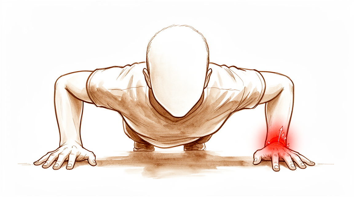
آپ کو کیا محسوس ہوتا ہے
درد آپ کی کلائی کی چھوٹی انگلی والی طرف ہوتا ہے، جسے النا والی طرف (ulnar side) کہا جاتا ہے۔ یہ اکثر اس وقت بڑھ جاتا ہے جب آپ اس طرف بوجھ ڈالتے ہیں: کرسی سے اٹھنے کے لیے زور لگانا، مرتبان کو مضبوطی سے پکڑنا، یا کوئی بھاری چیز اٹھائے ہوئے دروازے کا ہینڈل گھمانا۔ کپڑا نچوڑنے یا پیچ کس استعمال کرنے جیسی مروڑنے والی حرکتیں بھی درد شروع کر سکتی ہیں۔
کچھ لوگ محسوس کرتے ہیں کہ کلائی غیر مستحکم ہے، جیسے وہ جواب دے جائے گی، یا بازو کا نچلا حصہ (forearm) گھومنے پر اس میں کھٹکا ہوتا ہے۔ کچھ دوسرے لوگوں کو ایسا درد رہتا ہے جو دیر تک رہتا ہے اور اسکین پر اس کی کوئی واضح وجہ نظر نہیں آتی، جو کلائی کے اس حصے میں عام بات ہے۔ دکھن سرگرمی کے بعد زیادہ ہو سکتی ہے، اور رات کو یا صبح جاگنے پر آپ کو تنگ کر سکتی ہے۔
روزمرہ زندگی میں وہ کام مشکل ہو جاتے ہیں جن میں مضبوط گرفت اور مروڑنا دونوں شامل ہوں: پھنسا ہوا ڈھکن کھولنا، اسی طرف سودے کا بھاری تھیلا اٹھا کر لے جانا، یا نیچی نشست سے کھڑے ہونے کے لیے ہاتھ پر ٹیک لگانا۔ ایسا کام جس میں اوزار استعمال ہوں یا کلائی پر بار بار زور پڑے، درد کو جاری رکھ سکتا ہے۔
اگر آپ کی علامات کم نہیں ہو رہیں، ہفتوں کے دوران بدتر ہو رہی ہیں، رات کو آپ کو جگا دیتی ہیں، یا آپ کو کام کرنے یا اپنا ہاتھ استعمال کرنے سے روکتی ہیں، تو اپنے فیملی ڈاکٹر (GP) سے ملیں یا ماہر سے معائنہ کروانے کا کہیں۔
دراصل کیا ہو رہا ہے
آپ کی کلائی کے اندر، چھوٹی انگلی والی طرف، ایک ڈھانچہ ہوتا ہے جسے ٹرائینگولر فائبروکارٹلیج کمپلیکس (triangular fibrocartilage complex)، یا ٹی ایف سی سی (TFCC)، کہتے ہیں۔ اسے ایک جھٹکا جذب کرنے والا پرزہ سمجھیں جس کے ساتھ رسیاں بندھی ہوں۔ جھٹکا جذب کرنے والا پرزہ سخت اور چکنے کارٹلیج (cartilage) کی ایک ڈسک ہے جو آپ کی النا (ulna)، یعنی اس طرف کی بازو کے نچلے حصے کی ہڈی، کے سرے اور کلائی کی چھوٹی ہڈیوں کے درمیان ہوتی ہے۔ رسیاں لیگامنٹ (ligament)، یعنی وہ مضبوط بندھن جو ایک ہڈی کو دوسری ہڈی سے جوڑتا ہے، ہیں جو اس ڈسک کو النا سے باندھے رکھتے ہیں اور اس جوڑ کو مستحکم رکھتے ہیں جہاں بازو کے نچلے حصے کی دونوں ہڈیاں کلائی پر ملتی ہیں۔
ٹی ایف سی سی ایک ساتھ دو کام کرتی ہے۔ یہ آپ کی کلائی کی النا والی طرف سے گزرنے والے بوجھ کے لیے گدی کا کام کرتی ہے، اور جب آپ اپنی ہتھیلی اوپر اور نیچے گھماتے ہیں تو بازو کے نچلے حصے کے اس جوڑ کو اکٹھا رکھتی ہے۔ یہی دوہرا کردار ان دو اندازوں کی وضاحت کرتا ہے جن کے بارے میں آپ نے اوپر پڑھا۔ جب گدی والا حصہ پھٹ جاتا ہے، تو اس طرف بوجھ ڈالنے سے درد ہوتا ہے: کرسی سے اٹھنے کے لیے زور لگانا، پکڑنا، مروڑنا۔ جب رسیاں النا پر اپنی جڑنے کی جگہ سے پھٹ کر الگ ہو جاتی ہیں، تو خود جوڑ کا سہارا ختم ہو جاتا ہے، اس لیے کلائی ایسی محسوس ہوتی ہے جیسے جواب دے جائے گی، یا بازو کا نچلا حصہ گھومنے پر اس میں کھٹکا ہوتا ہے۔ دوسرا انداز زیادہ اہم ہے، کیونکہ لیگامنٹ والا حصہ ہی جوڑ کو اکٹھا رکھتا ہے۔
اس جگہ کے بارے میں ایک اور بات جاننے کے قابل ہے۔ ٹی ایف سی سی کے صرف بیرونی کنارے کو خون کی فراہمی ملتی ہے، جو تقریباً اس کا بیرونی 10 سے 40 فیصد حصہ ہے۔ اندرونی حصے کو تقریباً کوئی خون نہیں ملتا۔ خون ہی سے زخم بھرتا ہے، اس لیے کنارے کے قریب کے پھٹاؤ کے جڑنے کا حقیقی امکان ہوتا ہے، جبکہ درمیان کے پھٹاؤ عموماً خود سے ٹھیک نہیں ہوتے۔ اسی لیے یہاں کا پھٹاؤ دکھن پیدا کرتا رہ سکتا ہے: خراب بافت ایسی جگہ پر ہوتی ہے جہاں جسم کو اس کی مرمت میں مشکل ہوتی ہے، اور ہر گرفت یا مروڑ اس پر بوجھ ڈالتا رہتا ہے۔
لہٰذا درد، کھٹکا اور کلائی کا جواب دے جانا الگ الگ مسائل نہیں ہیں۔ یہ اس وقت ہوتے ہیں جب گدی اور سہارے کا کام کرنے والا یہ ڈھانچہ، جو آپ کے ہاتھ کی ہر حرکت کے ساتھ بوجھ اٹھاتا ہے، اپنے دونوں کام نہیں کر پاتا۔
ہم اس کے لیے کیا کر سکتے ہیں
ڈاکٹر کیرن ہِرپرا، جو Mater Private Hospital Rockhampton میں بازو اور ہاتھ کے سرجن (upper-limb surgeon) ہیں، سب سے کم چیر پھاڑ والے ان طریقوں سے آغاز کرتے ہیں جو آپ کی حالت کے لیے موزوں ہوں۔ مریض عموماً اپنے فیملی ڈاکٹر (GP) کے ذریعے ہمارے کلینک بھیجے جاتے ہیں؛ اگر کسی فزیوتھراپسٹ نے آپ کو ہمیں دکھانے کا مشورہ دیا ہے، تب بھی میڈیکیئر (Medicare) کی رقم واپسی کا حق دار ہونے کے لیے آپ کو اپنے فیملی ڈاکٹر سے ریفرل (referral) کی ضرورت ہوگی۔ اس پہلی ملاقات میں ہم آپ کی تکلیف کی تاریخ لیتے ہیں، آپ کی کلائی کا معائنہ کرتے ہیں، اور اگر ضرورت ہو تو اسکین کرواتے ہیں تاکہ معلوم ہو سکے کہ کیا پھٹا ہے۔
ٹی ایف سی سی کے کچھ پھٹاؤ آپریشن کے بغیر ٹھیک ہو جاتے ہیں۔ اسپلنٹ کلائی کو بے حرکت رکھ سکتا ہے اور دکھتی جگہ سے بوجھ ہٹا سکتا ہے، جبکہ بیرونی کنارے کو، یعنی اس حصے کو جسے خون ملتا ہے، جڑنے کا موقع ملتا ہے۔ ہینڈ تھراپی (ہاتھ کی بحالی کا علاج) کا مقصد درد کم کرنا اور طاقت اور گرفت دوبارہ بنانا ہے تاکہ کلائی پھر سے روزمرہ کے بوجھ سنبھال سکے۔ یہ اندازہ لگانے سے پہلے کہ یہ کام کر رہا ہے یا نہیں، ہم عموماً اسے کئی ہفتوں کا مناسب وقت دیتے ہیں۔
درد کش گولیاں اور سوزش کم کرنے والی دوائیں پھٹاؤ کے ٹھیک ہونے کے دوران آپ کو حرکت میں رہنے میں مدد دے سکتی ہیں۔ یہ بافت کی مرمت نہیں کرتیں، بلکہ دنوں کو زیادہ آرام دہ بناتی ہیں۔
اگر اس کے بعد بھی درد یا کھٹکا آپ کو محدود کرتا ہو، تو ہم سرجری کے بارے میں بات کرتے ہیں۔ معیاری پہلا قدم دوربین کے ذریعے آپریشن (keyhole) ہے، جسے کلائی کی آرتھروسکوپی (wrist arthroscopy، دوربین سے جوڑ کے اندر دیکھ کر علاج) کہتے ہیں۔ سرجن ایک ننھے کیمرے کی مدد سے چھوٹے چیروں کے ذریعے جوڑ کے اندر دیکھتے ہیں، جو ٹی ایف سی سی کے پھٹاؤ کی تصدیق کرنے اور ساتھ ہی لیگامنٹوں اور جوڑ کی سطحوں کو جانچنے کا سب سے درست طریقہ ہے۔ کچھ پھٹاؤ انہی چھوٹے چیروں کے ذریعے تراش کر صاف کیے جا سکتے ہیں یا ان کی مرمت کی جا سکتی ہے۔ جہاں گدی گھس چکی ہو اور کلائی کی چھوٹی انگلی والی طرف پر بہت زیادہ بوجھ پڑ رہا ہو، وہاں ایک طریقے سے النا کے سرے سے تھوڑا سا حصہ تراشا جا سکتا ہے تاکہ اس طرف سے دباؤ کم ہو۔ پھٹاؤ کو صاف کرنے، اس کی مرمت کرنے، یا ٹینڈن (tendon)، یعنی وہ مضبوط ڈوری جو پٹھے کو ہڈی سے جوڑتی ہے، کے گرافٹ (graft، جسم کے کسی دوسرے حصے سے لیا گیا ٹکڑا) سے اسے دوبارہ بنانے کے درمیان انتخاب اس بات پر منحصر ہے کہ پھٹاؤ کہاں ہے اور کتنے عرصے سے ہے۔
یہ فیصلہ ہم آپ کے ساتھ مل کر کرتے ہیں۔ ہم آپ کو بتاتے ہیں کہ ہر طریقے میں کیا شامل ہے اور یہ کیا بدل سکتا ہے اور کیا نہیں، اور آپ فیصلہ کرتے ہیں کہ آپ کی کلائی اور آپ کی زندگی کے لیے کیا موزوں ہے۔
کیا توقع رکھیں
بہت سے لوگوں میں ٹی ایف سی سی کا پھٹاؤ وقت اور درست دیکھ بھال سے ٹھیک ہو جاتا ہے۔ بیرونی کنارے کے قریب کے پھٹاؤ، جہاں خون کی فراہمی ہوتی ہے، خود سے یا اسپلنٹ کی مدد سے جڑنے کا حقیقی امکان رکھتے ہیں۔ گدی میں زیادہ گہرائی والے پھٹاؤ عموماً خود سے ٹھیک نہیں ہوتے، لیکن کلائی کو آرام دینے اور اس پر کم بوجھ ڈالنے سے درد پھر بھی کم ہو سکتا ہے۔
صحت یابی اچانک کے بجائے بتدریج ہوتی ہے۔ زیادہ تر لوگ دیکھتے ہیں کہ دکھن دنوں کے بجائے ہفتوں میں کم ہوتی ہے، اور کلائی کے سنبھلنے کے ساتھ گرفت اور مروڑنے کی طاقت واپس آتی ہے۔ اگر چھوٹی انگلی والی طرف کے صرف ایک الگ تھلگ پھٹاؤ کے لیے آپ کا دوربین کے ذریعے آپریشن ہو، تو کام پر واپسی کا اوسط وقت 5.5 ہفتے ہے۔ کھلاڑیوں کو اپنے کھیل پر واپسی میں زیادہ وقت لگ سکتا ہے، خاص طور پر اگر کلائی کی اسی طرف دوسری چوٹیں بھی ہوں۔ ایک بات جاننے کے قابل ہے: اگر النا کا سرا کلائی میں دب کر درد پیدا کر رہا ہو، تو کھیل پر واپسی کے بعد یہ درد عموماً لوٹ آتا ہے۔
اگر پھٹاؤ کو ایسے ہی چھوڑ دیا جائے، تو آگے کی صورتحال اس بات پر منحصر ہے کہ کون سا حصہ خراب ہوا ہے۔ کنارے کا پھٹاؤ پرسکون ہو سکتا ہے۔ درمیان کا پھٹاؤ، یا ایسا پھٹاؤ جس میں لیگامنٹ کی رسیاں ڈھیلی ہو کر الگ ہو گئی ہوں، اکثر ہر گرفت اور مروڑ کے ساتھ دکھن پیدا کرتا رہتا ہے، کیونکہ وہ بافت ایسی جگہ پر ہوتی ہے جہاں جسم کو اس کی مرمت میں مشکل ہوتی ہے۔ کلائی کے جواب دے جانے کا احساس بھی برقرار رہ سکتا ہے۔
سرجری درد سے مکمل پاک کلائی کا وعدہ نہیں کرتی۔ جن لوگوں کا کلائی کے پرانے درد کے لیے دوربین کے ذریعے آپریشن ہوا، ان میں ایک سال پر اوسطاً تقریباً 50 فیصد بہتری آئی، اور اس وقت بھی زیادہ تر کو کچھ درد اور اکڑاؤ تھا۔ سرجری جو کام اچھی طرح کرتی ہے وہ یہ ہے کہ ٹھیک ٹھیک تصدیق کرے کہ کیا پھٹا ہے اور جس کی مرمت ہو سکتی ہے اس کی مرمت کرے، جس کا مطلب عموماً کم درد اور ایسی کلائی ہوتا ہے جو پھر سے روزمرہ کے بوجھ سنبھال سکے۔
اگر آپ کی علامات کم نہیں ہو رہیں، ہفتوں کے دوران بدتر ہو رہی ہیں، رات کو آپ کو جگا دیتی ہیں، یا آپ کو کام کرنے یا اپنا ہاتھ استعمال کرنے سے روکتی ہیں، تو اپنے فیملی ڈاکٹر سے ملیں یا ماہر سے معائنہ کروانے کا کہیں۔
ڈاکٹر کو کب دکھائیں
ٹی ایف سی سی کے زیادہ تر پھٹاؤ ایمرجنسی نہیں ہوتے۔ چند صورتیں ایمرجنسی ہوتی ہیں۔ اگر آپ کی کلائی گرم، سرخ، سوجی ہوئی اور دردناک ہو جائے، خاص طور پر بخار کے ساتھ، تو اسی دن ہسپتال کی ایمرجنسی جائیں، کیونکہ یہ ایسے انفیکشن کی طرف اشارہ ہے جس کا اسی دن علاج ضروری ہے۔ یہی بات اس وقت بھی لاگو ہوتی ہے اگر آپ کی انگلیاں یا ہاتھ سفید پڑ جائیں، ٹھنڈے ہو جائیں، بالکل سفید یا نیلے ہو جائیں، یا اگر کسی چوٹ کے بعد اچانک ہاتھ میں احساس یا حرکت ختم ہو جائے۔ باقی تمام صورتوں میں، وقت کا تعلق رفتار سے نہیں بلکہ اس بات سے ہے کہ تکلیف کتنی دیر سے برقرار ہے۔ اگر کلائی کی چھوٹی انگلی والی طرف درد 3 ماہ سے زیادہ عرصے سے ہو اور آرام، اسپلنٹ یا ہینڈ تھراپی سے ٹھیک نہ ہوا ہو تو اپنے فیملی ڈاکٹر سے ملیں۔ اگر کلائی غیر مستحکم محسوس ہو، جواب دے جائے، یا ہتھیلی اوپر اور نیچے گھماتے وقت اس میں کھٹکا ہو، تو اس سے پہلے ہی ماہر سے معائنہ کروانے کا کہیں، کیونکہ اس سے لگتا ہے کہ ٹی ایف سی سی کا لیگامنٹ والا حصہ پھٹ کر الگ ہو گیا ہے۔ ایسا درد جو آپ کو کام کرنے سے روکے، رات کو جگا دے، یا دکھن جاری رہنے کے باوجود اسکین صاف آئیں، اس کے لیے بھی ماہر کی رائے لینا مناسب ہے، کیونکہ اس جگہ کو اکثر اس سے زیادہ قریب سے دیکھنے کی ضرورت ہوتی ہے جتنا صرف اسکین دکھا سکتے ہیں۔
مزید تفصیل
Advanced reading: the deeper science (optional)
یہ حصہ آپ کے اپنے علاج کے فیصلوں کے لیے ضروری معلومات سے آگے جاتا ہے۔ ٹرائینگولر فائبروکارٹلیج کمپلیکس کی چوٹ کے بارے میں اضافی مطالعہ اس لیے فائدہ مند ہے کہ آپریشن کی تکنیکوں کے بارے میں جو بحثیں زیادہ زیرِ بحث رہتی ہیں ان میں کوئی واضح فرق سامنے نہیں آیا، جبکہ آپریشن کے بعد کے طریقۂ کار کی ایک تفصیل، جس پر بہت کم توجہ دی جاتی ہے، اہم معلوم ہوتی ہے۔
یہ ڈھانچہ دراصل کیا کام کرتا ہے
ٹی ایف سی سی (TFCC) کارٹلیج کی ایک ڈسک ہے جس کے گرد لیگامنٹ کا ایک جھولا نما گھیرا ہوتا ہے، اور یہ النا (ulna)، چھوٹی انگلی والی طرف کی ہڈی، کے سرے اور کلائی کی ہڈیوں (carpal bones) کے درمیان ہوتی ہے۔ یہ ایک ہی وقت میں دو کام کرتی ہے: یہ کلائی کی النا والی طرف سے گزرنے والے وزن کے جھٹکے کو جذب کرتی ہے، اور کلائی پر بازو کی دونوں ہڈیوں کے درمیان کے جوڑ کو مستحکم رکھتی ہے، یعنی ڈسٹل ریڈیو النر جوڑ (distal radioulnar joint)۔
یہ دوہرا کردار بتاتا ہے کہ یہاں کی چوٹیں دو مختلف طریقوں سے کیوں ظاہر ہوتی ہیں۔ وہ پھٹاؤ جو زیادہ تر ڈسک کو متاثر کرے، وزن ڈالنے، کرسی سے زور لگا کر اٹھنے، پکڑنے اور مروڑنے پر درد پیدا کرتا ہے۔ وہ پھٹاؤ جو گہرے ریشوں کو النا پر ان کی جڑنے کی جگہ، یعنی فوویئل (foveal) جڑاؤ، سے الگ کر دے، عدم استحکام پیدا کرتا ہے، جس میں محسوس ہوتا ہے کہ بازو گھماتے وقت کلائی جواب دے جاتی ہے یا اس میں کھٹکے کی آواز آتی ہے۔ دوسری قسم زیادہ اہم ہے، کیونکہ لیگامنٹ کا یہی جڑاؤ جوڑ کو اکٹھا رکھتا ہے۔
ایم آر آئی درست ہے، مگر ایک شرط کے ساتھ جو جاننے کے قابل ہے
تشخیص کا بڑا انحصار اسکین پر ہوتا ہے۔ 1,298 مریضوں میں ایم آر آئی کی مجموعی درستگی قابلِ قبول تھی، اور کنارے کے پھٹاؤ (peripheral tears) کے لیے مجموعی درستگی نسبتاً زیادہ تھی، اور مناسب ترتیبات کے ساتھ کیے گئے ایم آر آئی کو پھٹاؤ کی مختلف اقسام کی تشخیص کا ایک مثالی طریقہ قرار دیا گیا [1]۔
شرط لفظ "کنارے" میں ہے۔ ایم آر آئی اس کمپلیکس کے بیرونی حصے میں سب سے اچھا کام کرتا ہے، جہاں خون کی فراہمی بہتر ہے، اور یہی وہ جگہ ہے جہاں مرمت کے قابل پھٹاؤ ہوتے ہیں۔ درمیانی اور گھساؤ والے پھٹاؤ، اور فوویئل جڑاؤ کی صحیح حالت، کو واضح کرنا زیادہ مشکل ہے، اسی لیے اسکین کے ساتھ ساتھ معائنے کے نتائج اور کبھی کبھی آرتھروسکوپی بھی اہمیت رکھتے ہیں۔
تکنیکوں کے موازنے میں فرق سامنے نہیں آتا
آپریشن کے بارے میں دو بحثیں بار بار سامنے آتی ہیں، اور ان میں سے کوئی بھی حل نہیں ہوئی۔
النا والی طرف کے عام، کنارے کے پھٹاؤ کے لیے، 240 مریضوں کے ایک منظم جائزے (systematic review) میں دوربین کے ذریعے اور کھلے آپریشن سے مرمت کے بارے میں ٹھوس نتیجہ اخذ کرنے کے لیے اعلیٰ معیار کے شواہد کی کمی پائی گئی، اور کوئی سائنسی ثبوت نہیں ملا کہ ایک تکنیک دوسری سے بہتر ہے [2]۔
فوویئل مرمت کے لیے، 904 مریضوں میں سیوچر اینکر (suture anchor)، یعنی ہڈی میں لگایا جانے والا چھوٹا پرزہ جو ٹانکوں کے دھاگے کو تھامتا ہے، اور ہڈی کے اندر سے گزارے جانے والے ٹانکوں (transosseous suture) کا موازنہ کیا گیا، اور دونوں سے کام کرنے کی صلاحیت، درد اور گرفت کی طاقت میں بہتری آئی اور دوبارہ آپریشن کی شرح کم رہی، اگرچہ حرکت کی حد کا موازنہ غیر حتمی رہا [3]۔
مستقل پیغام یہ ہے کہ مرمت کو جڑاؤ بحال کرنا ضروری ہے؛ اس کے لیے استعمال ہونے والے پرزوں سے نتیجہ بدلتا ہوا ثابت نہیں ہوا۔
آپریشن کے بعد کی وہ تفصیل جو واقعی اہم معلوم ہوتی ہے
یہاں شواہد زیادہ فرق واضح کرتے ہیں، اور عملی طور پر مفید ہیں۔ فوویئل ٹی ایف سی سی مرمت کے بعد حرکت روکنے کے طریقوں کا 288 مریضوں میں موازنہ کرنے پر معلوم ہوا کہ آپریشن کے بعد حرکت روکنے میں کہنی کی حرکت روکنے کے مقابلے میں بازو کے گھماؤ کو روکنا زیادہ فائدہ مند ہو سکتا ہے، اور کہنی کو موڑنے اور سیدھا کرنے پر اضافی پابندی نے کوئی مستقل فائدہ نہیں دکھایا [4]۔
یہ براہِ راست جسم کی ساخت سے نکلتا ہے۔ مرمت شدہ حصے پر زور بازو کے گھماؤ سے پڑتا ہے، کہنی موڑنے سے نہیں، اس لیے اسپلنٹ کو ہتھیلی کے اوپر اور نیچے گھومنے کو قابو میں رکھنا چاہیے۔ اکثر کہنی سے اوپر تک کا پلستر اس مقصد کو بالواسطہ طور پر پورا کرنے کے لیے استعمال کیا جاتا ہے، کیونکہ یہ کہنی کو گھومنے سے روکتا ہے، اور یہ شواہد بتاتے ہیں کہ کہنی والا حصہ وہ نہیں جو اصل کام کرتا ہے۔ مریض کے لیے، چھ ہفتے ایسی بریس میں گزارنا جس میں کہنی آزاد رہے، کہنی سے اوپر تک کے پلستر میں چھ ہفتے گزارنے سے کافی مختلف تجربہ ہے۔
حوالہ جات
[1] Wang ZX, Chen SL, Wang QQ, Liu B, Zhu J, Shen J. The performance of magnetic resonance imaging in the detection of triangular fibrocartilage complex injury: a meta-analysis. J Hand Surg Eur Vol. 2015;40(5):477-84. https://doi.org/10.1177/1753193414567425
[2] Robba V, Fowler A, Karantana A, Grindlay D, Lindau T. Open versus arthroscopic repair of 1B ulnar-sided triangular fibrocartilage complex tears: a systematic review. Hand (N Y). 2019;15(4):456-64. https://doi.org/10.1177/1558944718815244
[3] Ma H, Wang J, Yang C. Effectiveness of suture anchor and transosseous suture technique in arthroscopic triangular fibrocartilage complex foveal repair: a systematic review and meta-analysis. J Orthop Surg Res. 2024;19(1). https://doi.org/10.1186/s13018-024-04530-4
[4] Lee J, Lee T, Lee S, Lim H, Chang E, Park MO, et al. Postoperative immobilization after foveal triangular fibrocartilage complex repair: a systematic review and meta-analysis. J Hand Surg Am. 2026;51(5):512.e1-512.e11. https://doi.org/10.1016/j.jhsa.2026.01.029
Evidence & references
This is the clinical evidence summary written for health professionals. It is technical, and it lists the research this page was built from. You do not need to read it to understand your treatment or to make a decision about it.
Overview
Diagnosis and Assessment
- Careful history and physical examination are required to determine whether a TFCC tear is symptomatic [6].
- It is important to quantify the severity of symptoms related to TFCC pathology to determine whether surgical treatment is necessary [6].
- Acute TFCC injuries require differentiation between those causing distal radioulnar joint instability and those that do not [7].
- TFCC injury should be considered with all distal radial fractures [16].
- 1B TFCC injury is most common in patients with distal radius fractures and concomitant TFCC injury [5].
Natural History and Prognosis
- About 40% of patients sustaining a TFCC tear without distal radioulnar joint instability still had pain and disability at 1 year [8].
Surgical Outcomes
- Surgical treatment of TFCC tears and concomitant pathology in the pediatric and adolescent population results in decreased pain, improved motion and stability, and excellent functional outcomes in the majority of patients [3].
- In a series of pediatric and adolescent patients, there was marked improvement in pain, function, and range of motion in patients with TFCC tears and coexisting pathology [1].
- Arthroscopic treatment of TFCC lesions leads to satisfactory functional outcomes [4].
- Current evidence demonstrates that TFCC repair achieves good clinical outcomes, with low complication rates [23].
- There was no statistical difference in clinical outcomes after open versus arthroscopic TFCC repair [27].
- Arthroscopic debridement alone appears to be an effective and safe initial treatment for patients with traumatic central TFCC tears [32].
- With appropriate indications and patient selection, arthroscopic foveal repair of the TFCC may restore distal radioulnar joint stability and provide satisfactory results without significant complications [68].
- In high-demand athletes, arthroscopic repair of TFCC tears is becoming the treatment of choice to obtain optimum physiologic strength, complete range of motion, stability, and the shortest possible postoperative period [44].
- Open surgical TFCC repair is a safe and reliable therapeutic procedure in children and adolescent populations [56].
- TFCC capsular reattachment could be performed with an arthroscopically assisted technique, providing good long-term results [19].
Surgical Techniques and Variability
- Arthroscopic-assisted repair techniques have revolutionized surgical management, providing detailed visualization and facilitating the repair of TFCC injuries and associated pathologies with minimally invasive techniques [9].
- TFCC repair varies substantially from surgeon-to-surgeon, suggesting repairs are discretionary and preference sensitive [15].
- A systematic review demonstrates a current lack of high-quality evidence required to draw firm conclusions on the merits of arthroscopic versus open repair of 1B TFCC tears [17].
- A systematic review did not demonstrate strong evidence regarding the association between ulnar variance and outcomes after TFCC repair [31].
Anatomy & Pathophysiology
Anatomical Structures and Blood Supply
- The TFCC is a group of interrelated anatomic structures that are integral to stability of the distal radioulnar joint (DRUJ) [35].
- The TFCC acts in forearm rotation as the primary stabilizer of the DRUJ while also providing a smooth articular surface and partially absorbing the axial load from the radiocarpal joint [63].
- The central 80–85% of the articular disk is avascular, resulting in minimal healing potential for tears in this area [55].
- The outer 10% to 40% of the articular disk is well perfused, suggesting a healing potential for injured areas upon repair [49].
- The ulnar attachment of the TFCC is a three-dimensional complex consisting of proximal radioulnar ligaments, a distal hammock structure (fibrocartilage disk, meniscus homologue, and ulnocarpal ligaments), and a functional ulnar collateral ligament (UCL) [49].
- The distal hammock structure and the UCL constitute the distal component of the TFCC (dc-TFCC), while the radioulnar ligament represents the proximal component (pc-TFCC) [49].
- The palmar and dorsal radioulnar ligaments span from the dorsal and volar corners of the distal radius to a broad area of the fovea at the base of the ulnar styloid [49].
- The deep foveal components of the radioulnar ligaments are considered the true stabilizers of the DRUJ [49].
- The triangular fibrocartilage is less stiff in neutral forearm rotation, and its palmar and dorsal parts have material properties similar to those of the radiocarpal ligaments [88].
Classification and Pathology
- Palmer classification categorizes TFCC tears into traumatic (Class 1) or degenerative (Class 2) based on mechanism and location [20, 21, 49, 55].
- Class 1A injuries are central perforations or tears of the articular disk [20, 21, 55].
- Class 1B injuries are ulnar avulsions of the TFCC with or without an ulnar styloid fracture [20, 21, 55].
- Class 1C injuries are distal avulsions involving the origins of the ulnolunate and ulnotriquetral ligaments [20, 21].
- Class 1D injuries are radial avulsions involving the dorsal and/or volar radioulnar ligaments [20, 21].
- Class 2 injuries represent degeneration of the central portion of the TFCC and are associated with ulnocarpal impaction syndrome [55].
- Class 2A involves TFCC wear or thinning, progressing through Class 2E which includes ulnocarpal and DRUJ arthritis [20, 21].
- A foveal tear is a partial tear of the ulnar insertion where the foveal insertion is disrupted but some styloid insertion remains intact [132].
- The foveal insertion has a greater effect on DRUJ stability than the styloid insertion due to its closer relationship to the rotational axis of the forearm [132].
- Degenerative TFCC tears occur as a result of chronic excessive loading through the ulnocarpal joint along with natural tissue degeneration [126].
- Cadaveric examinations found TFCC perforations and chondromalacia of the ulnar head, lunate, and triquetrum in 30% to 70% of specimens [126].
Injury Mechanisms and Clinical Presentation
- Traumatic TFCC injuries may occur from falls on an outstretched hand or hyper-rotational injuries to the forearm [35].
- Repetitive forceful movement of the wrist from supination to pronation can cause overload stress affecting components of the TFCC [35].
- Injuries to the TFCC typically occur with extension and pronation of the axially loaded wrist [49].
- The most common mechanism of injury is a fall on an outstretched hand [49].
- Traumatic radial-sided tears of the TFCC typically occur during acute rotational injuries involving combined axial load with a distraction injury to the ulnar border of the forearm [126].
- Patients with peripheral TFCC tears complain of deep diffuse pain across the ulnar side of the wrist, weakness when gripping, and a clicking sensation with forearm rotation [49].
- Pain in peripheral TFCC tears is accentuated by passive pronation and supination of the wrist [49].
- A dorsally prominent ulnar head in relation to the radius can be seen in foveal detachments [49].
- Ballottement of the DRUJ reveals increased translation of the joint associated with pain in peripheral tears [49].
- A class 1A tear typically manifests as ulnar-sided wrist pain aggravated by power grip, especially with ulnar deviation or forearm rotation [43].
- Class 1A tears are confined to the disc, located 2 to 3 mm ulnar to its radial attachment, and oriented volar to dorsal [43].
- Class 1B injuries may present with point tenderness volar to the ulnar styloid, coinciding anatomically with the fovea (fovea sign) [60].
- A fracture through the base of the ulnar styloid that disrupts both deep and superficial limbs of the TFCC is more predictive of DRUJ instability than fractures through the shaft or tip [60].
- Deep TFCC fiber tears may contribute to decreased wrist rotational positioning sense and have biomechanical importance in DRUJ stability [50].
Biomechanical Effects of Deformity
- Radial shortening causes the greatest disturbance in DRUJ kinematics and the most distortion of the triangular fibrocartilage [74].
- Decreased radial inclination and dorsal angulation cause intermediate changes in DRUJ kinematics, while dorsal displacement produces minimal changes [74].
- Radial deformity does not produce DRUJ dislocation but provides biomechanical evidence of a relationship between radial malunion and persistent symptoms [74].
- Changes in ulnar variance due to ulnar lengthening or radial shortening dramatically alter force transmission through the distal ulna [90].
Classification
Palmer Classification System
- The Palmer classification divides TFCC injuries into traumatic (Class 1) and degenerative (Class 2) categories [20].
- Subtypes within the Palmer classification are based on the specific location of the tear within the TFCC [20].
- The class and location of a TFCC tear have important implications for treatment [20].
- Palmer created a classification system that divides TFCC injuries into Type I (traumatic) or Type II (degenerative) lesions [53].
- Traumatic injuries are categorized based on the location of the pathology [55].
- Degenerative tears represent the spectrum of lesions found with ulnocarpal impaction syndrome [55].
Class 1 (Traumatic) Subtypes
- Class 1A injuries are characterized by central perforation or tear of the TFCC [20].
- Class 1A injuries are characterized by central articular disc perforation [53].
- Class 1B injuries are characterized by ulnar avulsion with or without ulnar styloid fracture [20].
- Class 1B injuries are characterized by ulnar-sided avulsion with or without ulnar styloid fracture [53].
- Class 1C injuries are characterized by distal avulsion involving the origins of the ulnolunate and ulnotriquetral ligaments [20].
- Class 1C injuries are characterized by distal avulsion with disruption from distal ulnocarpal ligaments [53].
- Class 1D injuries are characterized by radial avulsion involving the dorsal and/or volar radioulnar ligaments [20].
- Class 1D injuries are characterized by radial-sided avulsion with or without sigmoid notch fracture [53].
- Class 1D injuries involve avulsion of the articular disk and the dorsal and palmar radioulnar ligaments [55].
- Class 1D injuries render the distal radioulnar joint (DRUJ) unstable [55].
- Type IA lesions are perforations of the thin, avascular articular disc and represent the most common type of traumatic tear [53].
- Traumatic tears of the articular disk commonly occur along the origin of the articular disk from the radius, which are class 1A injuries [55].
- Class 1B lesions describe traumatic avulsion of the TFCC insertion onto the ulna [55].
- Class 1B injuries are seen with or without fractures of the ulnar styloid [55].
- Avulsions of the TFCC ulnar attachment often create DRUJ instability [55].
- Class 1C lesions involve disruption of the TFCC distally through the ulnolunate and ulnotriquetral ligaments [55].
- The area of the TFCC involved in Class 1C lesions is well vascularized, giving these lesions greater potential to heal [55].
- Class 1D injuries are less common and involve radial avulsion of the entire TFCC from its attachment to the radius at the sigmoid notch [55].
Class 2 (Degenerative) Subtypes
- Class 2A is characterized by TFCC wear or thinning [20].
- Class 2B is characterized by TFCC wear plus lunate and/or ulnar chondromalacia [20].
- Class 2C is characterized by TFCC perforation plus lunate and/or ulnar chondromalacia [20].
- Class 2D is characterized by TFCC perforation, lunate and/or ulnar chondromalacia, and lunotriquetral ligament disruption [20].
- Class 2E is characterized by TFCC perforation, lunate and/or ulnar chondromalacia, lunotriquetral ligament disruption, and ulnocarpal and DRUJ arthritis [20].
- Type IIA is characterized by TFCC wear [53].
- Type IIB is characterized by TFCC wear with lunate and/or ulnar chondromalacia [53].
- Type IIC is characterized by TFCC perforation with lunate and/or ulnar chondromalacia [53].
- Type IID is characterized by TFCC perforation with lunate and/or ulnar chondromalacia and lunotriquetral ligament perforation [53].
- Type IIE is characterized by TFCC perforation with lunate and/or ulnar chondromalacia, lunotriquetral ligament perforation, and ulnocarpal arthritis [53].
Alternative Classifications
- Atzei proposed a treatment-oriented classification for Palmer 1B type peripheral tears [109].
- The Atzei classification distinguishes repairable lesions of the single distal component of the TFCC (Class 1) from those of the proximal component, which includes foveal insertions of DRUJ ligaments [121].
- Atzei Class 2 represents a complete tear pattern involving both the distal and proximal components of the TFCC [121].
- Atzei Class 3 represents an isolated tear of the proximal component of the TFCC [121].
- Atzei Class 4 represents nonrepairable lesions [121].
- Atzei Class 5 represents tears associated with DRUJ arthritis [121].
- The Atzei classification highlights clinical and arthroscopic criteria to categorize five classes of TFCC peripheral tears on a treatment-oriented system [47].
- In the pediatric population, traumatic (Class 1) injuries represent the vast majority of TFCC tears, while degenerative (Class 2) tears are far less common [24].
- There is an apparent increased prevalence of Palmer 1B tears in pediatric patients compared with adults [24].
- Palmer 1B tears represent the most common variety of TFCC injuries in children and adolescents [24].
- Tears of the radial attachment (Palmer 1D) represent the second most common type of TFCC injuries in children and adolescents [24].
- The Melone classification system does not predict the presence of TFCC lesions [102].
- Frykman Type VI and VIII fractures show a significantly higher incidence of TFCC tears [102].
Diagnostic Correlates for Classification
- Arthroscopy is the gold standard for detection of TFCC tears [20].
- The arthroscopic trampoline test is performed to assess TFCC resiliency by balloting the central portion with a small probe [20].
- The arthroscopic hook test can be used to demonstrate peripheral detachment of the TFCC [20].
- The arthroscopic suction test can show laxity of the TFCC when peripherally scarred in or foveal detachment when the DRUJ is clinically unstable [20].
- Classification of central triangular fibrocartilage complex lesions as traumatic or degenerative depends on the information provided upon viewing the lesion at arthroscopy [28].
- In more detailed classification of TFCC injuries, such as pc-TFCC tears classified by Atzei's classification, the diagnostic accuracy of MRI remains lower compared to wrist arthroscopy [36].
- Diagnostic accuracy for TFCC injuries was highest for central TFCC injuries [39].
- The presence of ulnar styloid fracture associated with distal radius fracture predicted the presence of frequently occurring traumatic triangular fibrocartilage complex injury and TFCC 1B injury [14].
Clinical Presentation
General Presentation and Diagnosis
- TFCC tears are a common cause of ulnar-sided wrist pain in athletes [10].
- Ulnar-sided wrist pain in athletes can be caused by injury to the TFCC [35].
- Wrist arthroscopy is considered the benchmark for the diagnosis and management of TFCC injuries [110].
- Wrist arthroscopy allows the direct evaluation of the TFCC and surrounding ligaments [110].
- Arthroscopy is effective in obtaining both correct diagnosis and treatment of peripheral TFCC tear [33].
- The more experienced observer had more sensitive, specific, and accurate interpretations than the less experienced observer in predicting the presence and location of a TFCC tear [13].
- There is a high rate of abnormal TFCC identified on MRI in patients without corresponding ulnar-sided wrist symptoms [34].
- The presence of an abnormal TFCC on MRI may be of questionable clinical meaning, because there is a high incidence of TFCC abnormalities in asymptomatic subjects, particularly those over the age of 50 [62].
- MR arthrography is more sensitive and specific method in terms of the diagnosis of TFCC tears compared to conventional wrist MRI [54].
- Diagnostic accuracy was highest for central TFCC injuries [39].
- The technique is best used in young patients with indeterminate physical examinations and normal radiography in whom intrinsic ligament or TFCC tears are suspected to assist decision making regarding the performance of arthroscopy [64].
Pediatric and Adolescent Presentation
- In the pediatric population, traumatic (class 1) injuries represent the vast majority of TFCC tears [24].
- Degenerative (class 2) tears of the TFCC are far less common in the pediatric population [24].
- Palmer 1B tears from the ulnar attachment with or without associated ulnar styloid fractures represent the most common variety of TFCC injuries in children and adolescents [24].
- Tears of the radial attachment (Palmer 1D tears) represent the second most common type of TFCC injuries in children and adolescents [24].
- If radial-sided tears (Palmer 1D) are mistaken for a central traumatic tear (Palmer 1A), simple debridement alone may result in persistent pain, instability, and functional limitations [24].
Association with Distal Radius Fractures
- 1B TFCC injury is most common in patients with distal radius fracture and concomitant TFCC injury [5].
- Disability outcomes were worse in patients with distal radial fracture where TFCC was injured [41].
Natural History and Chronic Findings
- About 40% of patients sustaining TFCC tear without DRUJ instability still had pain and disability at 1 year [8].
- Nonsurgical treatment is moderately successful for treating patients with TFCC tears without DRUJ instability [22].
- A higher frequency of accompanying ECU tendon and/or DRUJ disorders was found in patients with chronic TFCC tears as compared to the control group [40].
- Deep TFCC fiber tear may contribute to decreased wrist rotational positioning sense and may have biomechanical importance in distal radioulnar joint stability [50].
Investigations
Clinical Examination and Provocative Tests
- Careful history and physical examination are required to determine whether a TFCC tear is symptomatic and to quantify the severity of symptoms related to TFCC pathology to determine whether surgical treatment is necessary [6].
- A positive ulnar fovea sign was reported to be 90% sensitive and 88% specific in detecting a split tear of the ulnotriquetral ligament [70].
- Load-bearing RaUl measurement is a simple method to diagnose an unstable distal radioulnar joint in patients with TFCC injury [59].
Magnetic Resonance Imaging (MRI)
- MRI interpretation of TFCC lesions is more sensitive, specific, and accurate when performed by a more experienced observer compared to a less experienced observer [13].
- The sensitivity, specificity, and accuracy of 3.0T wrist MRI for the TFCC is consistently higher compared with those of 1.5T wrist MRI [119].
- High-resolution magnetic resonance imaging permits accurate depiction and localization of tears of the triangular fibrocartilage complex [133].
- High-resolution MRI using a microscopy surface coil allowed assessment of each TFCC component and showed a higher accuracy for diagnosing injuries to the radial attachment and the disc of the TFC compared with previous studies [113].
- MRI is controversial, but newer innovations suggest value in detection and localization of TFCC pathology [20].
MR Arthrography
- MR arthrography is a more sensitive and specific method in terms of the diagnosis of TFCC tears compared to conventional wrist MRI [54].
Computed Tomography (CT) and Arthrography
- CT scan is effective for evaluating DRUJ instability [135].
- Since ulnar-sided contrast leakage is more common in patients with peripheral TFCC injuries, distinction between an atypical configuration of the prestyloid recess and actual leakage is important in CT arthrography of the wrist [42].
- Radial multiplanar reconstructions are important for the assessment of triangular fibrocartilage complex injury in CT arthrography of the wrist [61].
- Single-injection wrist arthrography is superior to routine MRI for the detection of full-thickness triangular fibrocartilage tears [136].
Arthroscopy
Association with Distal Radius Fractures
Treatment
Non-Operative Management
- All Class 1 (acute traumatic) TFCC injuries are initially managed with immobilization and NSAIDs [20].
- Initial treatment for Class 1B injuries involves protective above-elbow immobilization for 4 to 6 weeks toward the forearm in neutral rotation [60].
- Nonoperative management of traumatic TFCC injuries with above-elbow immobilization is a viable treatment method, particularly in patients without DRUJ subluxation [123].
- Moritomo and colleagues reported that 46% of patients with avulsion of the TFCC from the fovea were pain-free after conservative treatment [49].
- Non-operative treatment is the first line of treatment for most DRUJ problems, with exceptions including some unstable distal ulnar fractures and early marked painful block to forearm rotation [125].
- In the year of natural recovery following a distal radius fracture, management options for ongoing ulnar-sided wrist pain include activity modification, splints or straps, and oral analgesics, typically NSAIDs [125].
- Steroid injections are given to treat ongoing wrist pain after distal radius fractures, with 90% of patients reporting some improvement in symptoms when reviewed at 6–8 weeks after injection [125].
- Patients with persistent pain after distal radius fractures should be supported and encouraged to wait for spontaneous improvement in symptoms over at least 1 year from injury, assuming there is no gross bone malalignment [125].
- There is no good evidence that early treatment improves the outcome of DRUJ instability, so it is worth waiting for a prolonged period, typically at least 12 months, before addressing it [125].
- The mean DASH scores showed no difference across all groups, calling into question the clinical need to pursue operative treatment via K-wire stabilization or TFCC repair over conservative treatment via cast immobilization for acute DRUJ instability following distal radius fracture [129].
Surgical Indications and Decision Making
- Careful history and physical examination are required to determine whether a TFCC tear is symptomatic, and it is important to quantify the severity of symptoms related to TFCC pathology to determine whether surgical treatment is necessary [6].
- Acute TFCC injuries require differentiation between those causing distal radioulnar joint instability and those that do not, with management ranging from nonsurgical immobilization to arthroscopic or open surgical repair depending on the specific injury pattern and stability [7].
- Surgical treatment is indicated for Class 1 TFCC injuries with failure of nonoperative treatment [20].
- Indications for surgical intervention include specific ulnar-sided wrist pain not relieved by conservative management for 3 months, especially in the presence of symptomatic instability of the DRUJ [49].
- Surgery is indicated for Class 1B injuries with persistent symptoms or evidence of DRUJ instability [60].
- Arthroscopy remains the gold standard in diagnosing and assessing the type and extent of the TFCC tear [49].
- A recent study in magnetic resonance arthrography performed with wrist traction applied demonstrated high accuracy of TFCC tear detection of 98% [49].
- Some studies have demonstrated that MRI missed over half of TFCC tears, as later confirmed on wrist arthroscopy [122].
- An MRI finding showing greater than 10% subluxation of the DRUJ on axial MRI has been shown to have increased sensitivity and specificity for TFCC tear and DRUJ instability [122].
- The arthroscopic trampoline test is performed to assess TFCC resiliency by balloting central portion with a small probe [20].
Arthroscopic Debridement (Class 1A and Degenerative)
- Class 1A (central) TFCC tears are inherently stable and treated with débridement if persistently symptomatic because this area of the TFCC is devoid of vascularity and unable to heal [20].
- A 2-mm peripheral rim should be maintained during debridement of Class 1A tears [20].
- Indications for arthroscopic TFCC debridement include acute traumatic Palmer type 1A TFCC, degenerative central tear of TFCC with ulnar neutral or negative variance, and failure to respond to conservative treatment with splint and medication for more than 3 months [26].
- Resection of unstable flaps is sufficient when the remaining margins are smooth and stable, and the peripheral 2 to 3 mm must be preserved to protect the radioulnar ligaments [26].
- A thorough synovectomy of the ulnocarpal joint and DRUJ is essential for early pain control as the pain mainly originates from the inflamed synovium over the capsule which is richly innervated [26].
- Excessive use of RF energy can lead to thermal chondral damage [26].
- Overaggressive debridement can cause DRUJ instability [26].
- Failure to diagnose ulnar impaction syndrome may lead to continued pain [26].
- No restriction of wrist and forearm motion is required postoperatively after arthroscopic TFCC debridement [26].
- Although clinical outcome scores showed significant improvement after arthroscopic debridement for central TFCC tears, wearing type tears and greater ulnar plus variance were associated with dissatisfaction and poorer postoperative outcomes after the procedure [117].
- Removal of a perforated TFC is contraindicated in the presence of radiocarpal or distal radioulnar arthritis [58].
- Patients who underwent debridement for any Type 1 Palmer class returned to work at a rate of 92%, but only 44% were free of pain [63].
Arthroscopic and Open Repair (Class 1B, 1C, 1D)
- Class 1B (peripheral) TFCC tears are amenable to arthroscopic or open repair because the rim is well vascularized [20].
- Concurrent fractures of ulnar styloid with persistent instability are either excised or fixed during repair of Class 1B tears [20].
- Class 1C (rare) TFCC tears are amenable to arthroscopic or open repair [20].
- Class 1D TFCC tears are frequently associated with distal radius fractures and often respond to reduction of the radius [20].
- Repair of a traumatic TFCC tear within 3 months of injury allows a patient to regain 80% of wrist ROM and grip strength [20].
- Arthroscopic-assisted repair techniques provide detailed visualization and facilitate the repair of TFCC injuries and associated pathologies with minimally invasive techniques [9].
- Arthroscopic transosseous foveal repair of the TFCC resulted in improvements in grip strength and functional outcomes with low complication and reoperation rates [45].
- The refixation of the TFCC after shortening of the ulnar head for traumatic avulsion class 1B of the TFCC is effective in restoring its function [29].
- Arthroscopic exploration and surgical treatment of peripheral ulnar TFCC lesions allow for satisfactory results regarding mobility and patient satisfaction [116].
- For 1B lesions that underwent repair, 68.3% were able to return to work and 41% had persistent pain [63].
- Coexisting type 2 TFCC tears significantly increased the risk of index surgery failure in patients with peripheral ulnar-side TFCC tears [30].
- The technique of arthroscopic transosseous repair using a modified outside-in technique allows for low-profile fixation for distal radioulnar joint instability and provides excellent biomechanical fixation [48].
- The combined HS and suture repair effectively restores stability to both the DRUJ and UCJ in patients with TFCC-related ulnocarpal instability, considerably reducing pain and preserving range of motion [111].
- This technique provides a minimally invasive and anatomically guided solution for restoring DRUJ stability in chronic TFCC injuries [71].
Pediatric and Adolescent Considerations
- If Palmer 1D tears are mistaken for a central traumatic tear (Palmer 1A), simple debridement alone may result in persistent pain, instability, and functional limitations [24].
- In this series, there was marked improvement in pain, function, and range of motion in patients with TFCC tears and coexisting pathology [1].
- Unlike in adults, in whom Palmer 1A (central) tears are most common, pediatric patients commonly have Palmer 1B (ulnar peripheral) tears, in some studies up to 79% of the patients [122].
- Given the improved vascularity at the periphery of the TFCC, many of these tears are amenable to repair, rather than solely débridement [122].
- 1B TFCC injury is most common in patients with DRF and concomitant TFCC injury [5].
- The most frequent ligament tear in distal fractures of the radius in young adults was the triangular fibrocartilage complex in 39 cases (78%), with a statistical correlation to ulnar styloid fractures [72].
Reconstruction and Salvage Procedures
- TFCC reconstruction with tendon graft is indicated in irreparable TFCC injuries with symptomatic DRUJ instability, neglected chronic injuries, or after suboptimal healing following nonoperative or surgical repair [38].
- Skeletal malalignment that may be responsible for the DRUJ instability should be addressed concomitantly, including ulnar incongruity, malunion of the sigmoid notch, and dorsal tilt or coronal shift of the radius [38].
- Osteoarthritis of the DRUJ and an axial instability of the forearm due to interosseous membrane injury are contraindications to TFCC reconstruction [38].
- The results of a study suggest that arthroscopic TFCC reconstruction using the free PL tendon is an effective method for treating chronic and irreparable TFCC tears with DRUJ instability [66].
- Hemiresection or interposition arthroplasty maintains the ulnar insertion of the TFCC and prevents radioulnar impingement by soft tissue (ECU tendon or capsular flap) interposition [20].
- The Sauvé-Kapandji procedure involves DRUJ arthrodesis with creation of a proximal pseudarthrosis at the ulnar neck [20].
- Ulnar head or total joint implant arthroplasty maintains the relationship between the radius and the ulnar [20].
- Results of ulnar head or total joint implant arthroplasty show good pain relief at the risk of ulnar head instability, aseptic loosening, and no appreciable change in pronosupination in comparison with preoperative values [20].
- One-bone forearm through elimination of forearm rotation represents the ultimate salvage operation for persistent pain/complications by fusing the proximal ulnar to the distal radius shaft [20].
- A complication of TFCC treatment is painful proximal ulna stump instability and convergence of the radius upon the ulna [20].
Postoperative Rehabilitation
- Traumatic tears of the peripheral portions of the TFCC may be surgically repaired and are immobilized immediately postoperatively in a long arm cast to prevent forearm rotation and wrist motion that would stress the repair [127].
- After the first 10 days to 2 weeks following surgery, the cast may be changed to a thermoplastic splint that continues the immobilization of the forearm and wrist [127].
- Splinting generally continues for 4 to 6 weeks following surgery [127].
- Elbow range of motion may be permitted at 4 weeks, generally under the supervision of the therapist [127].
- At 6 weeks, immobilization is discontinued and the patient may use a wrist and forearm splint for intermittent support and protection [127].
- The focus of therapy at 6 weeks is on the gradual mobilization of the forearm and wrist [127].
- A reverse sugar tong cast with forearm in neutral rotation is preferred to long-arm cast to allow early elbow motion after TFCC reconstruction with tendon graft [37].
- Change to reverse sugar tong splint after 3 weeks and allow early midrange forearm rotation after TFCC reconstruction with tendon graft [37].
- Allow full range active forearm rotation after full 6 weeks, start passive motion after full 8 weeks after TFCC reconstruction with tendon graft [37].
- Specific neuromuscular potential training exercises and proprioceptive hand and wrist exercises are included in the rehabilitation program to recuperate functional wrist range of motion in association with fine and gross motor prehensile strength without provoking wrist instability due to overstretching of the newly reconstructed ligament [118].
Complications
Post-traumatic and Associated Pathology
- TFCC tears are associated with positive ulnar variance in up to 81% of cases [69].
- TFCC tears are associated with lunotriquetral ligament perforation in 70% to 76% of cases [69].
- TFCC tears are associated with chondromalacia of the distal ulna or of the ulnar carpal bones in up to 100% of cases [69].
- In patients with distal radius fractures, the most frequent ligament tear was the triangular fibrocartilage complex, occurring in 39 of 50 cases (78%) [72].
- In patients with distal radius fractures, there was a statistical correlation between TFCC tears and ulnar styloid fractures [72].
- In patients with distal radius fractures, chondral lesions were found in 16 of 50 patients (32%) [72].
- In patients with distal radius fractures, the scapholunate ligament was partially or totally torn in 27 of 50 cases (54%) [72].
- In patients with chronic TFCC tears, there is a higher frequency of accompanying extensor carpi ulnaris tendon and/or distal radioulnar joint disorders compared to a control group [40].
- Disability outcomes were worse in patients with distal radial fracture where the TFCC was injured [41].
- Coexisting type 2 TFCC tears significantly increased the risk of index surgery failure in patients undergoing arthroscopic repair of peripheral ulnar-side TFCC tears [30].
Natural History and Untreated Outcomes
- Nonsurgical treatment is moderately successful for treating patients with TFCC tears without distal radioulnar joint instability [22].
- In a prospective study of 43 patients with arthroscopically proven TFCC tears after distal radius fractures who were not initially treated, instability of the distal radioulnar joint was found in 19 patients at 1-year follow-up [130].
- In a prospective study of 43 patients with arthroscopically proven TFCC tears after distal radius fractures who were not initially treated, distal radioulnar joint instability was painful in 13 patients at 1-year follow-up [130].
- In a prospective study of 43 patients with arthroscopically proven TFCC tears after distal radius fractures who were not initially treated, 10 of 11 patients with complete peripheral TFCC tears had distal radioulnar joint instability at 1-year follow-up [130].
- In a prospective study of 43 patients with arthroscopically proven TFCC tears after distal radius fractures who were not initially treated, 7 of 32 patients with partial or no peripheral tears had distal radioulnar joint instability at 1-year follow-up [130].
- In a prospective study of 43 patients with arthroscopically proven TFCC tears after distal radius fractures who were not initially treated, patients with distal radioulnar joint instability had a worse Gartland and Werley wrist score [130].
- Traumatic linear TFCC tears do not necessarily lead to significant degenerative arthritis of the ulnar head or lunate [73].
Surgical Complications and Contraindications
- Removal of a perforated triangular fibrocartilage is contraindicated in the presence of radiocarpal or distal radioulnar arthritis [58].
- Arthroscopic transosseous foveal repair of the TFCC resulted in low complication and reoperation rates [45].
Recovery
Natural History and Non-Operative Outcomes
- About 40% of patients sustaining a TFCC tear without distal radioulnar joint (DRUJ) instability still had pain and disability at 1 year [8].
Surgical Outcomes and Functional Recovery
- Arthroscopic TFCC reconstruction using the free PL tendon is an effective method for treating chronic and irreparable TFCC tears with DRUJ instability [66].
Return to Work and Activity
- In the first year after open TFCC reinsertion, 91% of the patients returned to work, including 50% within 12 weeks [137].
- Eighty-four per cent of patients had a good or excellent range of motion of the injured wrist and this resulted in an early return to former activity [140].
Complications and Associated Pathology
- Coexisting type 2 TFCC tears significantly increased the risk of index surgery failure in patients with ulnar-plus variance [30].
- Ulnar variance may be changed after a TFCC tear; it decreased after TFCC foveal repair but increased again over time, which could be one of the causes of ulnar impaction syndrome and ulnar-sided wrist pain [139].
Key Evidence
- [L4] In this series, there was marked improvement in pain, function, and range of motion in patients with TFCC tears and coexisting pathology. [1] (10.1016/s0363-5023(98)80048-8)
- [L4] Surgical treatment of TFCC tears and concomitant pathology in the pediatric and adolescent population results in decreased pain, improved motion and stability, and excellent functional outcomes in the majority of patients. [3] (10.1016/j.jhsa.2019.06.019)
- [L4] Arthroscopic treatment of TFCC lesions leads to satisfactory functional outcomes. [4] (10.1055/s-0039-3400454)
- [L3] 1B TFCC injury is most common in patients with DRF and concomitant TFCC injury. [5] (10.1186/s13018-023-04438-5)
- [L4] Careful history and physical examination are required to determine whether a TFCC tear is symptomatic, and it is important to quantify the severity of symptoms related to TFCC pathology to determine whether surgical treatment is necessary. [6] (10.5435/jaaos-d-20-00998)
- [L5] Acute TFCC injuries require differentiation between those causing distal radioulnar joint instability and those that do not, with management ranging from nonsurgical immobilization to arthroscopic or open surgical repair depending on the specific injury pattern and stability. [7] (10.5435/00124635-200806000-00004)
- [L4] About 40% of patients sustaining TFCC tear without DRUJ instability still had pain and disability at 1 year. [8] (10.1016/j.jhsa.2018.06.064)
- [L5] Arthroscopic-assisted repair techniques have revolutionized surgical management, providing detailed visualization and facilitating the repair of TFCC injuries and associated pathologies with minimally invasive techniques. [9] (10.1016/j.jhsg.2024.03.011)
- [L5] Tears of the triangular fibrocartilage complex (TFCC) are a common cause of ulnar-sided wrist pain in athletes. [10] (10.1016/s0894-1130(12)80104-9)
- [L3] The more experienced observer had more sensitive, specific, and accurate interpretations than the less experienced observer in predicting the presence and location of a TFCC tear. [13] (10.1053/jhsu.2001.24966)
- [L4] The presence of ulnar styloid fracture associated with distal radius fracture predicted the presence of frequently occurring traumatic triangular fibrocartilage complex injury and TFCC 1B injury. [14] (10.1016/j.arthro.2020.05.025)
- [L4] TFCC repair varies substantially from surgeon-to-surgeon, suggesting repairs are discretionary and preference sensitive. [15] (10.1055/s-0038-1625953)
- [L4] TFCC injury should be considered with all distal radial fractures. [16] (10.1177/1558944716660555bh)
- [L4] This SR demonstrates a current lack of high-quality evidence required to draw firm conclusions on the merits of arthroscopic versus open repair of 1B TFCC tears. [17] (10.1177/1558944718815244)
- [L5] TFCC capsular reattachment could be performed with an arthroscopically assisted technique, providing good long-term results. [19] (10.1016/j.hcl.2017.06.005)
- [L3] Nonsurgical treatment is moderately successful for treating patients with TFCC tears without DRUJ instability. [22] (10.1097/corr.0000000000000533)
- [L4] Current evidence demonstrates that TFCC repair achieves good clinical outcomes, with low complication rates. [23] (10.1055/s-0040-1718913)
- [L5] [24] (10.1016/j.hcl.2005.09.002)
- [L3] There was no statistical difference in clinical outcomes after open versus arthroscopic TFCC repair. [27] (10.1016/j.jhsa.2008.01.020)
- [L2] Classification of central triangular fibrocartilage complex lesions as traumatic or degenerative depends on the information provided upon viewing the lesion at arthroscopy. [28] (10.1177/1753193416684658)
- [L4] The refixation of the TFCC after shortening of the ulnar head for traumatic avulsion class 1B of the TFCC is effective in restoring its function. [29] (10.1016/s0266-7681(05)80046-4)
- [L4] However, coexisting type 2 TFCC tears significantly increased the risk of index surgery failure in these patients. [30] (10.1016/j.arthro.2020.05.012)
- [L4] This systematic review did not demonstrate strong evidence regarding the association between ulnar variance and outcomes after TFCC repair. [31] (10.1142/s2424835518300025)
- [L3] Arthroscopic debridement alone appears to be an effective and safe initial treatment for patients with traumatic central TFCC tears. [32] (10.1302/0301-620x.106b4.bjj-2023-0642.r3)
- [L4] Arthroscopy is effective in obtaining both correct diagnosis and treatment of peripheral TFCC tear. [33] (10.2174/1874325001711010525)
- [L4] There is a high rate of abnormal TFCC identified on MRI in patients without corresponding ulnar-sided wrist symptoms. [34] (10.1177/15589447241277846)
- [L5] [35] (10.1016/j.csm.2019.12.008)
- [L4] In more detailed classification of TFCC injuries, such as pc-TFCC tears classified by Atzei's classification, the diagnostic accuracy of MRI remains lower compared to wrist arthroscopy. [36] (10.1186/s12891-023-07140-z)
- [L1] Diagnostic accuracy was highest for central TFCC injuries. [39] (10.1055/s-0038-1629911)
- [L3] We found a higher frequency of accompanying ECU tendon and/or DRUJ disorders in patients with chronic TFCC tears as compared to the control group. [40] (10.1016/j.jhsa.2016.07.040)
- [L2] Disability outcomes were worse in patients with distal radial fracture where TFCC was injured. [41] (10.1016/j.jht.2017.09.002)
- [L4] Since ulnar-sided contrast leakage is more common in patients with peripheral TFCC injuries, distinction between an atypical configuration of the prestyloid recess and actual leakage is important in CT arthrography of the wrist. [42] (10.1186/s12891-022-05241-9)
- [L4] In high-demand athletes, arthroscopic repair of TFCC tears is becoming the treatment of choice to obtain optimum physiologic strength, complete range of motion, stability, and the shortest possible postoperative period. [44] (10.1016/j.hcl.2009.05.011)
- [L1] Arthroscopic transosseous foveal repair of the TFCC resulted in improvements in grip strength and functional outcomes with low complication and reoperation rates. [45] (10.1016/j.arthro.2020.12.209)
- [L2] The classification highlights clinical and arthroscopic criteria to categorize five classes of TFCC peripheral tears on a treatment-oriented system. [47] (10.1177/1753193416687479)
- [L5] The technique allows for low-profile fixation for distal radioulnar joint instability and provides excellent biomechanical fixation. [48] (10.1016/j.eats.2024.103349)
- [L3] Deep TFCC fiber tear may contribute to decreased wrist rotational positioning sense and may have biomechanical importance in distal radioulnar joint stability. [50] (10.1016/j.jhsa.2018.01.022)
- [L5] [53] (10.1016/j.hcl.2012.05.014)
- [L3] MR arthrography is more sensitive and specific method in terms of the diagnosis of TFCC tears compared to conventional wrist MRI. [54] (10.1016/j.injury.2019.07.032)
- [L5] [55] (10.1016/s0894-1130(96)80071-8)
- [L4] This study emphasizes that open surgical TFCC repair is a safe and reliable therapeutic procedure in children and adolescent populations. [56] (10.1142/s2424835518500509)
- [L4] Removal of a perforated TFC is contraindicated in the presence of radiocarpal or distal radioulnar arthritis. [58] (10.1016/s0363-5023(84)80104-5)
- [L2] Load-bearing RaUl measurement is a simple method to diagnose an unstable distal radioulnar joint in patients with TFCC injury. [59] (10.1016/j.jhsa.2022.01.008)
- [L3] [61] (10.1186/s12891-020-03321-2)
- [L3] The presence of an abnormal TFCC on MRI may be of questionable clinical meaning, because there is a high incidence of TFCC abnormalities in asymptomatic subjects, particularly those over the age of 50. [62] (10.1016/j.jhsa.2011.10.006)
- [Paper] [63] (10.1055/s-0040-1713580)
- [L5] The technique is best used in young patients with indeterminate physical examinations and normal radiography in whom intrinsic ligament or TFCC tears are suspected to assist decision making regarding the performance of arthroscopy. [64] (10.1016/j.jhsa.2006.03.015)
- [L4] The results of this study suggest that arthroscopic TFCC reconstruction using the free PL tendon is an effective method for treating chronic and irreparable TFCC tears with DRUJ instability. [66] (10.1186/s13018-021-02827-2)
- [L4] With appropriate indications and patient selection, arthroscopic foveal repair of the TFCC may restore DRUJ stability and provide satisfactory results without significant complications. [68] (10.1055/s-0035-1544226)
- [L4] [69] (10.1016/0363-5023(92)90325-j)
- [L5] This technique provides a minimally invasive and anatomically guided solution for restoring DRUJ stability in chronic TFCC injuries. [71] (10.1002/atn2.70005)
- [L4] [72] (10.1016/s0266-7681(97)80364-6)
- [L5] Traumatic linear TFCC tears do not necessarily lead to significant degenerative arthritis of the ulnar head or lunate. [73] (10.1016/0266-7681(94)90078-7)
- [L5] [74] (10.1016/0363-5023(93)90098-n)
- [Paper] The study showed that the triangular fibrocartilage is less stiff in neutral forearm rotation and that the palmar and dorsal parts have material properties similar to those of the radiocarpal ligaments. [88] (10.1016/s0363-5023(10)80075-9)
- [L5] Changes in ulnar variance of a forearm due to ulnar lengthening or radial shortening do, however, dramatically alter the force transmission. [90] (10.1016/0363-5023(92)90342-m)
- [L3] The Melone classification system does not predict the presence of TFCC lesions, while Frykman Type VI and VIII fractures show a significantly higher incidence of TFCC tears. [102] (10.1177/1753193408090106)
- [L5] [109] (10.1177/1753193409100120)
- [L5] [110] (10.5435/jaaos-d-16-00407)
- [L4] The combined HS and suture repair effectively restores stability to both the DRUJ and UCJ in patients with TFCC-related ulnocarpal instability, considerably reducing pain and preserving range of motion. [111] (10.1016/j.jhsg.2025.100806)
- [L3] High-resolution MRI using a microscopy surface coil allowed assessment of each TFCC component and showed a higher accuracy for diagnosing injuries to the radial attachment and the disc of the TFC compared with previous studies. [113] (10.1016/j.jhsa.2006.05.001)
- [L4] Arthroscopic exploration and surgical treatment of peripheral ulnar TFCC lesions allow for satisfactory results regarding mobility and patient satisfaction. [116] (10.1016/j.hansur.2016.10.110)
- [L4] Although clinical outcome scores showed significant improvement after arthroscopic debridement for central TFCC tears, wearing type tears and greater ulnar plus variance were associated with dissatisfaction and poorer postoperative outcomes after the procedure. [117] (10.1016/j.arthro.2018.05.044)
- [L4] [118] (10.1016/j.jhse.2007.02.170)
- [L3] The sensitivity, specificity, and accuracy of 3.0T wrist MRI for the TFCC is consistently higher compared with those of 1.5T wrist MRI, suggesting improved capability for detection of TFCC injuries. [119] (10.1016/j.jhsa.2008.02.028)
- [L2] [121] (10.1016/j.arthro.2021.03.005)
- [L5] [122] (10.5435/jaaos-d-21-01029)
- [L3] Nonoperative management of traumatic TFCC injuries with above-elbow immobilization is a viable treatment method, particularly in patients without DRUJ subluxation. [123] (10.1302/0301-620x.103b8.bjj-2020-2310.r2)
- [L5] [125] (10.1177/17531934221140238)
- [L4] [126] (10.1177/15589447221084125)
- [L5] [127] (10.1097/00130911-199709000-00009)
- [L2] However, the mean DASH scores showed no difference across all groups, calling into question the clinical need to pursue operative treatment via K-wire stabilization or TFCC repair over conservative treatment via cast immobilization. [129] (10.1016/j.jhsg.2021.02.005)
- [Paper] [130] (10.1016/j.jhsa.2006.08.015)
- [L5] [132] (10.1097/bth.0b013e3181bd8319)
- [L2] High-resolution magnetic resonance imaging permits accurate depiction and localization of tears of the triangular fibrocartilage complex. [133] (10.2106/00004623-199711000-00009)
- [L2] This finding confirms the efficacy of CT scan for evaluating DRUJ instability. [135] (10.1016/j.ijsu.2011.08.005)
- [L3] Although future advances in MRI technology will probably improve its usefulness, single-injection wrist arthrography currently is superior to routine MRI for the detection of fullthickness triangular fibrocartilage tears. [136] (10.1016/s0266-7681(98)80068-5)
- [L3] In the first year after open TFCC reinsertion, 91% of the patients returned to work, including 50% within 12 weeks. [137] (10.1016/j.hansur.2021.03.012)
- [L4] Ulnar variance may be changed after a TFCC tear; it decreased after TFCC foveal repair but increased again over time, which could be one of the causes of ulnar impaction syndrome and ulnar-sided wrist pain. [139] (10.1055/s-0038-1668153)
- [L4] Eighty-four per cent of patients had a good or excellent range of motion of the injured wrist and this resulted in an early return to former activity. [140] (10.1016/j.jhsb.2006.03.101)
References
[1] Triangular fibrocartilage injuries in pediatric and adolescent patients. The Journal of Hand Surgery. 1998. DOI: 10.1016/s0363-5023(98)80048-8
[3] Early Results of Surgical Treatment of Triangular Fibrocartilage Complex Tears in Children and Adolescents. The Journal of Hand Surgery. 2020. DOI: 10.1016/j.jhsa.2019.06.019
[4] Patient-Reported Outcomes following Arthroscopic Triangular Fibrocartilage Complex Repair. Journal of Wrist Surgery. 2019. DOI: 10.1055/s-0039-3400454
[5] Association between imaging parameter changes and triangular fibrocartilage complex injury after distal radius fractures. Journal of Orthopaedic Surgery and Research. 2023. DOI: 10.1186/s13018-023-04438-5
[6] Open and Arthroscopic Triangular Fibrocartilage Complex (TFCC) Repair. Journal of the American Academy of Orthopaedic Surgeons. 2021. DOI: 10.5435/jaaos-d-20-00998
[7] Management of Acute Triangular Fibrocartilage Complex Injury of the Wrist. Journal of the American Academy of Orthopaedic Surgeons. 2008. DOI: 10.5435/00124635-200806000-00004
[8] The Natural Course of Triangular Fibrocartilage Complex Tear without Distal Radioulnar Joint Instability. The Journal of Hand Surgery. 2018. DOI: 10.1016/j.jhsa.2018.06.064
[9] Arthroscopic-Assisted Repair of the Triangular Fibrocartilage Complex. Journal of Hand Surgery Global Online. 2024. DOI: 10.1016/j.jhsg.2024.03.011
[10] Arthroscopic management of the athlete. Journal of Hand Therapy. 1991. DOI: 10.1016/s0894-1130(12)80104-9
[13] The effect of observer experience on magnetic resonance imaging interpretation and localization of triangular fibrocartilage complex lesions. The Journal of Hand Surgery. 2001. DOI: 10.1053/jhsu.2001.24966
[14] The Presence and the Location of an Ulnar Styloid Fracture Associated With Distal Radius Fracture Predict the Presence of Triangular Fibrocartilage Complex 1B Injury. Arthroscopy: The Journal of Arthroscopic & Related Surgery. 2020. DOI: 10.1016/j.arthro.2020.05.025
[15] Variation in Repair of the Triangular Fibrocartilage Complex. Journal of Wrist Surgery. 2018. DOI: 10.1055/s-0038-1625953
[16] The Correlation of Fracture Patterns of Distal Radius Fractures and Triangular Fibrocartilage Complex Injures. HAND. 2016. DOI: 10.1177/1558944716660555bh
[17] Open Versus Arthroscopic Repair of 1B Ulnar-Sided Triangular Fibrocartilage Complex Tears: A Systematic Review. HAND. 2019. DOI: 10.1177/1558944718815244
[19] Arthroscopic Management of Triangular Fibrocartilage Complex Peripheral Injury. Hand Clinics. 2017. DOI: 10.1016/j.hcl.2017.06.005
[20] Miller S Review Of Orthopaedics. DISTAL RADIOULNAR JOINT, TRIANGULAR FIBROCARTILAGE COMPLEX, AND WRIST ARTHROSCOPY > 2. TFCC tears.
[21] Miller S Review Of Orthopaedics. SECTION 16 PATELLAR TRACKING IN TOTAL KNEE ARTHROPLASTY > DISTAL RADIOULNAR JOINT, TRIANGULAR FIBROCARTILAGE COMPLEX, AND WRIST ARTHROSCOPY > 2. TFCC tears.
[22] What is the Natural History of the Triangular Fibrocartilage Complex Tear Without Distal Radioulnar Joint Instability?. Clinical Orthopaedics & Related Research. 2018. DOI: 10.1097/corr.0000000000000533
[23] Surgical Repair of Triangular Fibrocartilage Complex Tears: A Systematic Review. Journal of Wrist Surgery. 2020. DOI: 10.1055/s-0040-1718913
[24] Pediatric Distal Radius Fractures and Triangular Fibrocartilage Complex Injuries. Hand Clinics. 2006. DOI: 10.1016/j.hcl.2005.09.002
[26] Green S Operative Hand Surgery. AUTHOR'S PREFERRED METHOD OF TREATMENT: ARTHROSCOPIC PARTIAL WRIST FUSION > Arthroscopic TFCC Debridement.
[27] Clinical Comparison of Arthroscopic Versus Open Repair of Triangular Fibrocartilage Complex Tears. The Journal of Hand Surgery. 2008. DOI: 10.1016/j.jhsa.2008.01.020
[28] Diagnosing central lesions of the triangular fibrocartilage as traumatic or degenerative: a review of clinical accuracy. Journal of Hand Surgery (European Volume). 2017. DOI: 10.1177/1753193416684658
[29] A New Technique of Reattachment after Traumatic Avulsion of the TFCC at Its Ulnar Insertion. Journal of Hand Surgery. 1995. DOI: 10.1016/s0266-7681(05)80046-4
[30] What Is the Effect of the Ulnar‐Plus Variance on the Outcomes of Arthroscopic Repair of the Peripheral Ulnar‐Side Triangular Fibrocartilage Complex Tear?. Arthroscopy. 2020. DOI: 10.1016/j.arthro.2020.05.012
[31] Triangular Fibrocartilage Complex Repair and Ulna Variance: A Systematic Review. The Journal of Hand Surgery (Asian-Pacific Volume). 2018. DOI: 10.1142/s2424835518300025
[32] Prognostic factors for clinical outcomes after arthroscopic treatment of traumatic central tears of the triangular fibrocartilage complex. The Bone & Joint Journal. 2024. DOI: 10.1302/0301-620x.106b4.bjj-2023-0642.r3
[33] Results of Arthroscopic Repair of Peripheral Triangular Fibrocartilage Complex Tear With Exploration of Dorsal Sensory Branch of Ulnar Nerve. The Open Orthopaedics Journal. 2017. DOI: 10.2174/1874325001711010525
[34] Incidental Triangular Fibrocartilage Complex Changes on Wrist MRI. HAND. 2024. DOI: 10.1177/15589447241277846
[35] Ulnar-Sided Wrist Pain in the Athlete. Clinics in Sports Medicine. 2020. DOI: 10.1016/j.csm.2019.12.008
[36] Diagnostic value of MRI in traumatic triangular fibrocartilage complex injuries: a retrospective study. BMC Musculoskeletal Disorders. 2024. DOI: 10.1186/s12891-023-07140-z
[37] Green S Operative Hand Surgery. AUTHOR'S PREFERRED METHOD OF TREATMENT: ARTHROSCOPIC PARTIAL WRIST FUSION > Arthroscopic TFCC Reconstruction With Tendon Graft.
[38] Green S Operative Hand Surgery. AUTHOR'S PREFERRED METHOD OF TREATMENT: ARTHROSCOPIC PARTIAL WRIST FUSION > TFCC Reconstruction with Tendon Graft.
[39] TFCC Injuries: Meta-Analysis and Comparison of Diagnostic Imaging Modalities. Journal of Wrist Surgery. 2018. DOI: 10.1055/s-0038-1629911
[40] The Factors Correlated with the Extensor Carpi Ulnaris Tendon and Distal Radioulnar Joint Disorder in Patients with Chronic Triangular Fibrocartilage Tears. The Journal of Hand Surgery. 2016. DOI: 10.1016/j.jhsa.2016.07.040
[41] Outcomes of surgically treated distal radial fractures with associated triangular fibrocartilage complex injury. Journal of Hand Therapy. 2019. DOI: 10.1016/j.jht.2017.09.002
[42] Evaluation of prestyloid recess morphology and ulnar-sided contrast leakage in CT arthrography of the wrist. BMC Musculoskeletal Disorders. 2022. DOI: 10.1186/s12891-022-05241-9
[43] Green S Operative Hand Surgery. Chronic Perilunate Instability (SL Plus LT Instability) > Palmer’s Classification of TFCC Lesions > Class 1A Tear.
[44] Repair of Arthroscopic Triangular Fibrocartilage Complex Tears in Athletes. Hand Clinics. 2009. DOI: 10.1016/j.hcl.2009.05.011
[45] Arthroscopic Transosseous Repair of Foveal Tears of the Triangular Fibrocartilage Complex: A Systematic Review of Clinical Outcomes. Arthroscopy. 2020. DOI: 10.1016/j.arthro.2020.12.209
[47] Classification of ulnar triangular fibrocartilage complex tears. A treatment algorithm for Palmer type IB tears. Journal of Hand Surgery (European Volume). 2017. DOI: 10.1177/1753193416687479
[48] Arthroscopic Triangular Fibrocartilage Complex Repair Using Modified Outside‐In Technique. Arthroscopy Techniques. 2025. DOI: 10.1016/j.eats.2024.103349
[49] Green S Operative Hand Surgery. AUTHOR'S PREFERRED METHOD OF TREATMENT: ARTHROSCOPIC PARTIAL WRIST FUSION > TFCC Repair > Classification, Functional Anatomy, Clinical Features, and Indication for Repair.
[50] The Effect of Triangular Fibrocartilage Complex Tear on Wrist Proprioception. The Journal of Hand Surgery. 2018. DOI: 10.1016/j.jhsa.2018.01.022
[53] Triangular Fibrocartilage Complex Injuries in the Elite Athlete. Hand Clinics. 2012. DOI: 10.1016/j.hcl.2012.05.014
[54] Comparison between conventional MRI and MR arthrography in the diagnosis of triangular fibrocartilage tears and correlation with arthroscopic findings. Injury. 2019. DOI: 10.1016/j.injury.2019.07.032
[55] The Distal Radiou1nar joint. Journal of Hand Therapy. 1996. DOI: 10.1016/s0894-1130(96)80071-8
[56] TFCC Lesions in Children and Adolescents: Open Treatment. The Journal of Hand Surgery (Asian-Pacific Volume). 2018. DOI: 10.1142/s2424835518500509
[58] Isolated tears of the triangular fibrocartilage of the wrist: Results of partial excision. The Journal of Hand Surgery. 1984. DOI: 10.1016/s0363-5023(84)80104-5
[59] Load-Bearing Radioulnar Distances to Evaluate an Unstable Distal Radioulnar Joint in Patients With Triangular Fibrocartilage Complex Tears. The Journal of Hand Surgery. 2022. DOI: 10.1016/j.jhsa.2022.01.008
[60] Green S Operative Hand Surgery. Chronic Perilunate Instability (SL Plus LT Instability) > Palmer’s Classification of TFCC Lesions > Class 1B Tear.
[61] The importance of radial multiplanar reconstructions for assessment of triangular fibrocartilage complex injury in CT arthrography of the wrist. BMC Musculoskeletal Disorders. 2020. DOI: 10.1186/s12891-020-03321-2
[62] Prevalence of Triangular Fibrocartilage Complex Abnormalities on MRI Scans of Asymptomatic Wrists. The Journal of Hand Surgery. 2012. DOI: 10.1016/j.jhsa.2011.10.006
[63] A Systematic Review and Analysis of Palmer Type I Triangular Fibrocartilage Complex Injuries: Outcomes of Treatment. Journal of Hand and Microsurgery. 2020. DOI: 10.1055/s-0040-1713580
[64] Magnetic Resonance Arthrography of the Wrist: Case Presentation and Discussion. The Journal of Hand Surgery. 2006. DOI: 10.1016/j.jhsa.2006.03.015
[66] Arthroscopic triangular fibrocartilage complex reconstruction with free tendon graft for chronic distal radioulnar joint instability. Journal of Orthopaedic Surgery and Research. 2021. DOI: 10.1186/s13018-021-02827-2
[68] Arthroscopic Foveal Repair of the Triangular Fibrocartilage Complex. Journal of Wrist Surgery. 2015. DOI: 10.1055/s-0035-1544226
[69] Wafer distal ulna resection for triangular fibrocartilage tears and/or ulna impaction syndrome. The Journal of Hand Surgery. 1992. DOI: 10.1016/0363-5023(92)90325-j
[70] Green S Operative Hand Surgery. AUTHOR'S PREFERRED METHOD OF TREATMENT: ARTHROSCOPIC PARTIAL WRIST FUSION > Arthroscopic Repair of Class 1B TFCC Tears.
[71] Arthroscopic‐Assisted Suture Tape–Reinforced Triangular Fibrocartilage Complex Repair and Distal Radioulnar Ligament Augmentation Using a Modified Adams‐Berger Technique. Arthroscopy Techniques. 2026. DOI: 10.1002/atn2.70005
[72] Intraarticular Lesions in Distal Fractures of the Radius in Young Adults. Journal of Hand Surgery. 1997. DOI: 10.1016/s0266-7681(97)80364-6
[73] Wear Patterns of the Articular Cartilage and Triangular Fibrocartilaginous Complex of the Wrist: A Cadaveric Study. Journal of Hand Surgery. 1994. DOI: 10.1016/0266-7681(94)90078-7
[74] Effects of radial deformity on distal radioulnar joint mechanics. The Journal of Hand Surgery. 1993. DOI: 10.1016/0363-5023(93)90098-n
[88] The distal radioulnar ligaments: A biomechanical study. The Journal of Hand Surgery. 1991. DOI: 10.1016/s0363-5023(10)80075-9
[90] Force transmission through the distal ulna: Effect of ulnar variance, lunate fossa angulation, and radial and palmar tilt of the distal radius. The Journal of Hand Surgery. 1992. DOI: 10.1016/0363-5023(92)90342-m
[102] The Value of Plain X-Rays in Predicting TFCC Injury after Distal Radial Fractures. Journal of Hand Surgery (European Volume). 2008. DOI: 10.1177/1753193408090106
[109] New trends in arthroscopic management of type 1-B TFCC injuries with DRUJ instability. Journal of Hand Surgery (European Volume). 2009. DOI: 10.1177/1753193409100120
[110] Evaluation of Ulnar-sided Wrist Pain. Journal of the American Academy of Orthopaedic Surgeons. 2017. DOI: 10.5435/jaaos-d-16-00407
[111] Extensor Retinaculum Capsulorrhaphy and Suture Repair for Ulnocarpal and Distal Radioulnar Joint Instability: One-Year Results. Journal of Hand Surgery Global Online. 2025. DOI: 10.1016/j.jhsg.2025.100806
[113] Comparison Between High-Resolution MRI With a Microscopy Coil and Arthroscopy in Triangular Fibrocartilage Complex Injury. The Journal of Hand Surgery. 2006. DOI: 10.1016/j.jhsa.2006.05.001
[116] Résultats des réinsertions du TFCC sous arthroscopie. Hand Surgery and Rehabilitation. 2016. DOI: 10.1016/j.hansur.2016.10.110
[117] RETRACTED: Prognostic Factors of Arthroscopic Debridement for Central Triangular Fibrocartilage Complex Tears in Adults Younger Than 45 Years: A Retrospective Case Series Analysis. Arthroscopy. 2018. DOI: 10.1016/j.arthro.2018.05.044
[118] A-0263Session 16:Ulnar sided wrist pain. Journal of Hand Surgery: European Volume. 2007. DOI: 10.1016/j.jhse.2007.02.170
[119] Diagnostic Comparison of 1.5 Tesla and 3.0 Tesla Preoperative MRI of the Wrist in Patients With Ulnar-Sided Wrist Pain. The Journal of Hand Surgery. 2008. DOI: 10.1016/j.jhsa.2008.02.028
[121] The Hook Test Is More Accurate Than the Trampoline Test to Detect Foveal Tears of the Triangular Fibrocartilage Complex of the Wrist. Arthroscopy: The Journal of Arthroscopic & Related Surgery. 2021. DOI: 10.1016/j.arthro.2021.03.005
[122] Pediatric Ulnar-sided Wrist Pain: A Review of the Current Literature. Journal of the American Academy of Orthopaedic Surgeons. 2022. DOI: 10.5435/jaaos-d-21-01029
[123] Predictors for poor outcome for conservatively treated traumatic triangular fibrocartilage complex tears. The Bone & Joint Journal. 2021. DOI: 10.1302/0301-620x.103b8.bjj-2020-2310.r2
[125] The distal radioulnar joint after distal radial fractures: when and how do we need to treat pain, stiffness or instability?. Journal of Hand Surgery (European Volume). 2023. DOI: 10.1177/17531934221140238
[126] Radial Sided Triangular Fibrocartilage Complex Tears: A Comprehensive Review. HAND. 2022. DOI: 10.1177/15589447221084125
[127] Rehabilitation Following Surgery for the Distal Radioulnar Joint. Techniques in Hand & Upper Extremity Surgery. 1997. DOI: 10.1097/00130911-199709000-00009
[129] Management of Acute Distal Radioulnar Joint Instability Following a Distal Radius Fracture: A Systematic Review and Meta-Analysis. Journal of Hand Surgery Global Online. 2021. DOI: 10.1016/j.jhsg.2021.02.005
[130] Arthroscopic Management of Distal Radius Fractures. The Journal of Hand Surgery. 2006. DOI: 10.1016/j.jhsa.2006.08.015
[132] Advantages of Open Repair of a Foveal Tear of the Triangular Fibrocartilage Complex Via a Palmar Surgical Approach. Techniques in Hand & Upper Extremity Surgery. 2009. DOI: 10.1097/bth.0b013e3181bd8319
[133] The Utility of High-Resolution Magnetic Resonance Imaging in the Evaluation of the Triangular Fibrocartilage Complex of the Wrist. The Journal of Bone and Joint Surgery (American Volume)*. 1997. DOI: 10.2106/00004623-199711000-00009
[135] DRUJ instability after distal radius fracture: A comparison between cases with and without ulnar styloid fracture. International Journal of Surgery. 2011. DOI: 10.1016/j.ijsu.2011.08.005
[136] Arthrography is Superior to Magnetic Resonance Imaging for Diagnosing Injuries of the Triangular Fibrocartilage. Journal of Hand Surgery. 1998. DOI: 10.1016/s0266-7681(98)80068-5
[137] Factors associated with return to work after open reinsertion of the triangular fibrocartilage. Hand Surgery and Rehabilitation. 2021. DOI: 10.1016/j.hansur.2021.03.012
[139] Changes in Ulnar Variance after a Triangular Fibrocartilage Complex Tear. Journal of Wrist Surgery. 2018. DOI: 10.1055/s-0038-1668153
[140] 5.3 Volar locking compression plate technique for dorsally displaced fractures of the distal radius – our first clinical results. The Journal of Hand Surgery: Journal of the British Society for Surgery of the Hand. 2006. DOI: 10.1016/j.jhsb.2006.03.101