Clinicians › Shoulder
Proximal Humerus Fracture ORIF (Plate and Nail Fixation)
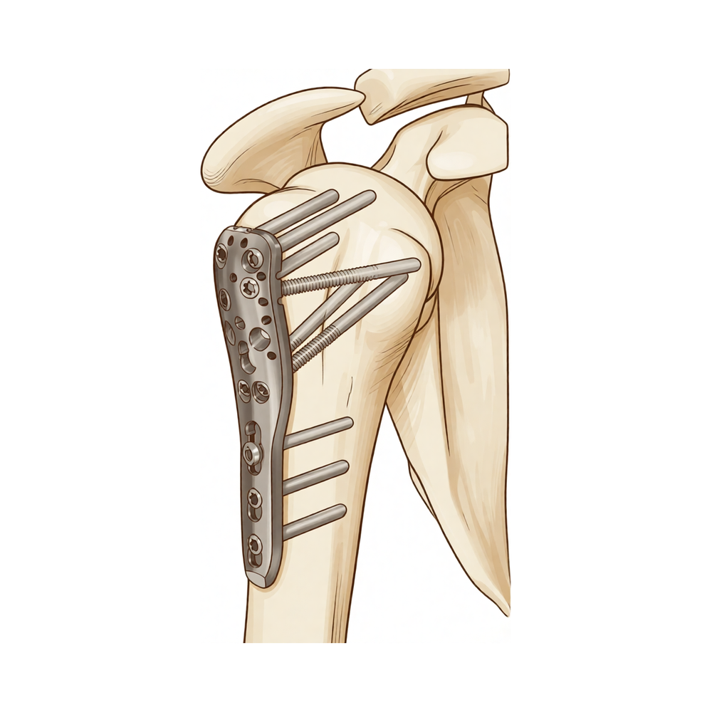
For patients: a plain-language version of this topic is available. See the patient guide.
Overview
Open reduction and internal fixation (ORIF) of proximal humeral fractures is indicated for medically fit patients with severely displaced fractures or fracture-dislocations, provided the treating center possesses the requisite expertise [4]. Both locking plates and intramedullary nails are valuable surgical options, with limited evidence suggesting they produce similar clinical and radiologic results [7, 12]. While intramedullary nailing may offer advantages in reducing total complications, intraoperative blood loss, operative time, and postoperative humeral head necrosis rates [14], the available evidence indicates that both techniques effectively restore shoulder function without clear superiority of either method [23, 28]. For three-part fractures, similar one-year outcomes suggest both techniques are useful for internal fixation [1], and Zifko nailing represents a cost-effective minimally invasive alternative with complication rates and clinical outcomes comparable to angular stable plate fixation in two-part fractures [38].
Long-term data supports the durability of these implants; ORIF of nonosteoporotic fractures with locking plates yielded favorable functional and radiologic outcomes at a minimum of 10 years of follow-up [3]. The locking plate is an excellent device for displaced unstable fractures, expanding indications for ORIF [18], while modern nail designs provide stable fixation with specific biologic advantages [16, 17]. However, complication profiles vary significantly by patient age and fracture complexity. In patients aged over 60 years, ORIF with locking plates resulted in a 44% complication and 34% failure rate [2], whereas second-generation locked plating in very elderly patients with complex fractures provided satisfactory outcomes with minimal complications [9]. Minimally invasive fixation in elderly populations shows satisfactory to excellent results in 80% of patients [5]. A randomized controlled trial demonstrated that locking plate fixation offers advantages in functional outcome and health-related quality of life over nonoperative treatment for displaced three-part fractures in the elderly, though this benefit comes at the cost of additional surgery in 30% of patients [73].
Patients undergoing ORIF for proximal humerus fracture dislocations achieve reasonable functional outcomes but face relatively high rates of avascular necrosis and reoperation [11]. The timing of surgery does not impact outcomes for patients undergoing ORIF [40]. Treatment remains controversial, as nonsurgical management also demonstrates successful outcomes and union rates greater than 90% [15]. Currently, there are no level 1 studies in the English language literature comparing ORIF with hemiarthroplasty for proximal humeral fractures [6].
Anatomy & Pathophysiology
Bony Anatomy
The proximal humerus comprises four main parts: the humeral head, greater tuberosity (GT), lesser tuberosity (LT), and humeral shaft [76]. The articular head is spherical with a diameter of 37 to 57 mm [76] and an arc of approximately 160 degrees covered by articular cartilage [88]. The radius of curvature is approximately 25 mm, slightly larger in men than in women [88]. The most superior portion of the articular surface averages 8 mm above the greater tuberosity [76], with the superior margin normally positioned 8 to 10 mm superior to the top of the GT [88].
Humeral version averages 29.8 degrees, ranging from 10 to 55 degrees [76]. The humeral head is inclined approximately 130 degrees with respect to the humeral shaft [76], while the neck-shaft angle measures an average of 135 degrees [77]. Alternative measurements report the humeral head as having 19° of retroversion and 41° of inclination [79], or an average neck-shaft angle of 45 degrees (±5 degrees) with a range of 30 to 50 degrees [88]. Retroversion averages 30 degrees relative to the transepicondylar axis [90, 77]. The anatomic neck lies at the junction of the articular surface and the tuberosities [76, 77], whereas the surgical neck represents an indistinct metadiaphyseal junction below the tuberosities but above the humeral shaft [76, 77].
The greater tuberosity is located posterior-superior to the humeral shaft [76], while the lesser tuberosity is on the anterior aspect [76]. The bicipital groove lies between the tuberosities, serving as a pathway for the long head of the biceps; its distal aspect is internally rotated relative to the proximal portion [76]. The glenoid is a convex, shallow structure shaped like an inverted pear [76]. The acromion, coracoacromial ligament, and coracoid process form the coracoacromial arch, a rigid bony-ligamentous structure imparting stability to the shoulder girdle [76]. The lateral humeral offset is defined as the distance from the lateral base of the coracoid process to the lateral margin of the greater tuberosity [88].
Ossification occurs via three centers: the humeral head (4 to 6 months), greater tuberosity (1 to 3 years), and lesser tuberosity (3 to 5 years) [79]. These centers fuse to the shaft at age 17 to 20 years [79]. The proximal humeral physis closes by 14 to 17 years in girls and 16 to 18 years in boys [83]. Eighty percent of subsequent humeral growth originates from this physis, accounting for approximately 40% of total upper extremity growth [83]. The periosteum is thicker and stronger in the posteromedial portion compared to the often thin anterolateral portion [83].
Vascular Supply
The proximal humerus receives blood supply from the anterior and posterior humeral circumflex branches of the third division of the axillary artery [76]. The anterior humeral circumflex artery (AHCA) arises at the inferior border of the subscapularis [76]. Its ascending branch courses parallel to the lateral aspect of the long head biceps tendon, entering the humeral head at the interface of the bicipital groove and greater tuberosity [76, 79]. This branch provides primary vascular inflow via its terminal anterolateral branch, known as the artery of Laing or arcuate artery [76, 79]. The terminal intraosseous portion enters at the proximal aspect of the intertubercular groove [79]. Injury to the arcuate artery may result in osteonecrosis of the humeral head, although additional extraosseous collateral branches can permit perfusion despite complete ligation [76]. Quantitative assessment indicates that 64% of humeral head blood supply arises from the posterior humeral circumflex artery [83]. The posterior humeral circumflex artery travels with the axillary nerve, enters the quadrilateral space posteriorly, and anastomoses with an anterior circumflex branch to supply the posterior cuff [76].
Muscular Attachments and Deforming Forces
The rotator cuff consists of the subscapularis, supraspinatus, infraspinatus, and teres minor [77]; the teres major is not a rotator cuff muscle [77]. The greater tuberosity serves as the attachment site for the supraspinatus, infraspinatus, and teres minor tendons [76, 83], while the lesser tuberosity anchors the subscapularis tendon [76, 83]. The subscapularis originates from the anterior scapula and inserts anteriorly onto the lesser tuberosity [83]. The supraspinatus, infraspinatus, and teres minor originate from the posterior scapula [83]. The rotator cuff muscles act as depressors of the humeral head, allowing the deltoid to efficiently abduct the humerus [77]. The infraspinatus and teres minor are external rotators, while the subscapularis is an internal rotator [77].
Displacement of proximal humeral fracture fragments follows predictable patterns based on deforming forces from tendinous insertions [76, 77]. The subscapularis causes medial displacement [76]. The supraspinatus and infraspinatus cause superior and posterior displacement [76]. The pectoralis major inserts on the humeral shaft and displaces it medially [76]. The deltoid, coursing from the clavicle and acromion to a common insertion on the lateral upper third of the humeral shaft, forward flexes and abducts the shoulder [83]. The pectoralis major powers adduction and internal rotation via its anterior insertion on the lateral wall of the bicipital groove [83]. It also forms the roof of the distal continuation of the bicipital tunnel, a closed space extending proximally to the glenohumeral joint [83].
Pathophysiology and Clinical Implications
Proximal humerus fractures alter complex shoulder girdle interactions, resulting in pain, decreased range of motion, stiffness, and disability [76]. Fractures involving the anatomic neck carry a worse prognosis than other regions due to potential disruption of the vascular supply and subsequent avascular necrosis [76, 77]. In contrast, surgical neck fractures are common and typically preserve the blood supply to the head [77]. Displaced fractures can impede normal movement of the rotator cuff, subacromial bursa, and subdeltoid bursa, causing impingement and disrupted glenohumeral motion [76]. In both displaced and nondisplaced fractures, the subdeltoid and subacromial bursae may become thickened and fibrotic, forming adhesions that limit motion; early range of motion exercises are hypothesized to decrease such adhesion formation [76].
The brachial plexus is prone to injury during proximal humeral fractures, dislocations, or traction [83]. The axillary nerve circles the humeral neck just inferior to the glenohumeral joint as it courses posteriorly [83]. Injury to the axillary nerve results in deltoid paralysis and anesthesia over the “badge” region at the lateral proximal arm [77]. The rate of non-union following closed treatment is estimated at 1.1% to 10% [21]. Prevention of local complications, particularly those leading to severe varus deviation, is essential to improve shoulder function [134]. The quality of reconstruction is closely connected to the final result, with particular importance attached to restoring the subacromial and subcoracoidal gliding mechanism [53]. Proper patient selection, thorough knowledge of anatomy and biomechanical principles, and understanding of osseous and neurovascular anatomy are pre-requisites for successful surgery [58, 113].
Biomechanical studies show that locked plating provides stable fixation for proximal humerus fractures [113]. The Humerusblock NG allows for angular stable dynamic fixation of two-part proximal humeral fractures [112]. Varus-displacing forces to the humeral head were superiorly reduced in the setting of a "proximal anchoring point" for straight antegrade nailing [108]. Lateral wall integrity of the greater tuberosity is important for the stability of osteoporotic proximal humeral fractures after plate fixation [140]. Medialization of the humeral shaft to provide medial buttress and prevent varus collapse in 3- and 4-part fractures offers a biomechanically sound approach to enhance fixation stability [141]. The short intramedullary nail violates the rotator cuff and is unable to resist deforming forces that can lead to loss of fixation and varus collapse [135]. In 38.5% of humeral heads, the predicted offset of the entry point for antegrade humeral nailing would encroach on the insertion of the supraspinatus tendon [151]. There is no biomechanical difference between locked smooth pegs and locked threaded screws for proximal fragment fixation in an unstable 2-part proximal humeral fracture model [144]. Applying biomechanical optimization criteria can assist biomedical engineers in designing or evaluating proximal humerus locked plates [150]. Simulation results predict that a novel adjustable locking plate design is sufficiently strong to not fail under regular anatomical loading scenarios, with close bending strength and fatigue life properties to clinically used locking compression plates [133]. Computer simulation results for patient-specific computational optimization of locking plates require biomechanical and clinical corroboration [121].
Classification
Neer: The Neer classification is one of the most generally accepted systems for proximal humeral fractures [37]. It categorizes fractures into displaced and minimally displaced groups, with the minimally displaced Type I accounting for 49% of all proximal humerus fractures [56]. Conservative treatment is commonly recommended for Neer Type I fractures [56], while operative treatment is indicated for displaced 2- to 4-part fractures [56]. The decision for operative treatment may be based on primary fracture displacement and subsequent instability using Neer criteria, which include 45° humeral head angulation and 1-cm displacement of the proximal humeral head fragments [132]. Additionally, a 5-mm displacement of the tubercles is considered a sign of fracture instability and an indication for surgery [132].
AO/OTA: The AO Proximal Humeral Fracture Classification is the other most generally accepted system for these injuries [37]. Fractures are classified using the OTA Compendium, with inclusion criteria for specific displacement patterns such as initial varus (OTA types 11.A2.2, A3.1, A3.3, B1.2, B2.2, C1.2, C2.2, or C2.3) or valgus (OTA types 11.A2.3, B1.1, C1.1, or C2.1) [52]. A neck-shaft angle of 130° is considered normal when classifying proximal humerus fractures [52]. Initial varus displacement is defined as a neck-shaft angle of less than 130° [52].
Other Considerations: Proximal humeral fractures are the third most prevalent fracture in patients aged over 65 years, following hip and distal radius fractures [37]. The Non-Union Scoring System (NUSS) can aid the clinician in planning the correct type of reconstructive strategy based on the evaluation of the non-union type [21]. Several observer studies have reported low agreement amongst doctors classifying proximal humeral fractures according to the AO classification, with mean kappa values for interobserver agreement varying between 0.26 and 0.53 [138]. Mean kappa values for interobserver agreement on AO classification decreased from 0.53 for AO Types to 0.2 for AO Groups, suggesting decreased agreement with an increasing number of classification units [138]. Classification type and group seem to be of minor importance for clinical outcome in most studies regarding locking plate osteosynthesis in AO/OTA Type C fractures [138].
Clinical Presentation
Proximal humerus fractures (PHFs) represent a common injury in the older adult population [1, 2]. In the United States, the estimated annual incidence is 6 per 10,000 persons [22]. Person-based incidence data indicate rates of 175 per 100,000 person-years in women and 68 per 100,000 person-years in men [2]. The risk of sustaining a PHF increases with age, particularly after 60 years [2]. The majority of these injuries result from a low-energy fall [2].
Fracture morphology varies in location and complexity, potentially involving any combination of the surgical and anatomic necks of the humerus, as well as the greater and lesser tuberosities [22]. Minimally displaced or 2-part fractures constitute between 77% and 84% of all proximal humerus fractures [2]. Additionally, 76% of proximal humerus fractures are classified as AO Foundation/Orthopaedic Trauma Association (AO/OTA) A-class fractures [2]. Fracture comminution and displacement, especially in 3- and 4-part fractures, are considered potential indications for operative treatment [2].
Most proximal humeral fractures in elderly patients can be treated nonoperatively with good functional outcomes [43]. Nonsurgical management of proximal humerus fractures demonstrates successful outcomes and union rates greater than 90% [15].
Investigations
Plain radiography: The recommended initial imaging series for proximal humerus fractures is the Neer trauma series, comprising an anteroposterior (AP) view, a lateral view in the scapular plane, and a Velpeau modified axillary view [95]. This combination allows evaluation of the shoulder joint in three separate perpendicular planes [95]. The AP view is taken in the plane of the scapula so that the x-ray beam passes through the glenohumeral joint [49]. The axillary view, often termed the “truth view,” is obtained with the arm in the functional position of elevation in the plane of the scapula, oriented to visualize both the spinoglenoid notch and the scapular neck [49]. This view is critical for assessing the glenoid articular surface and the anterior-posterior relationship of the humeral head [49, 95]. At minimum, two views are required: an AP in the plane of the glenoid and an axillary projection with the arm in abduction to demonstrate the humeral head-glenoid relationship [93]. Imaging-based assessment of fracture stability does not reliably predict outcomes in patients with two-part proximal humeral fractures and may lead to unnecessary surgeries [189].
CT: Computed tomography (CT) is frequently used to evaluate shoulder fractures and may be necessary to detail bony anatomy [95, 100]. CT is helpful for planning both fracture surgery and shoulder joint replacement [93]. All available open reduction and internal fixation (ORIF) techniques require careful analysis of fracture type, fragment displacement, and bone quality, making preoperative CT extremely valuable [199].
MRI: Magnetic resonance imaging (MRI) is the modality of choice for evaluating the rotator cuff, biceps, and subacromial/subdeltoid bursa [100]. T2-weighted MRI provides better visualization of full-thickness rotator cuff tears, while T1-weighted MRI can reveal Hill-Sachs lesions and is often used with magnetic resonance (MR) arthrograms to provide a more detailed picture of joint surfaces [100]. MRI is useful for identifying osteonecrosis of the humeral head or bone tumours [93]. It can also identify labral and rotator cuff tears, although accuracy for these is enhanced by combining the scan with arthrography [93]. MR arthrography (MRA) involves MRI of a joint injected with an intra-articular contrast agent, such as diluted gadolinium or saline solution [98]. MRA has proven utility by increasing both sensitivity and specificity in detecting injuries to the capsulolabral–ligamentous complex compared to traditional MRI [98]. In a meta-analysis of 6 studies including 4,667 shoulders, MRA demonstrated greater diagnostic test accuracy for glenoid labral lesions than MRI, with MRA sensitivity of 88% and specificity of 93% versus MRI sensitivity of 76% and specificity of 87% [98]. Abduction and external rotation (ABER) of the arm is an alternative position utilized to increase the sensitivity and specificity for detecting anteroinferior labroligamentous injury [98]. The sensitivity of MRA with the ABER position for detecting anteroinferior labral lesions was significantly higher than that of MRA in the neutral position and more effective in identifying Perthes lesions [98].
Ultrasonography: Ultrasound is a simple, accurate, and low-cost alternative to MRI and arthrography for evaluating both skeletal and soft-tissue structures of the shoulder [93, 100]. It provides immediate, real-time visualization of the rotator cuff, biceps tendon, and calcific deposits [100]. Ultrasound is useful for identifying rotator cuff tears and calcific tendinitis [93]. It can be used to measure the subacromial space, detect atrophy of rotator cuff muscles, and guide injections or barbotage (aspirating calcific deposits in the rotator cuff) [93, 100]. However, ultrasonography is highly operator dependent and is not as useful for evaluating labral tears or rotator cuff tears that are very small or larger than 3 cm [100]. Evaluation of the integrity of the rotator cuff may be difficult in the acute setting, and ultrasound, MRI, arthrogram, or arthroscopy may be valuable in making this diagnosis [95].
Vascular Assessment: Axillary artery injuries generally result from fractures or fracture-dislocations in which a medial bone spike injures or penetrates the axillary artery [95]. The index of suspicion for axillary artery injury is high if the arm shows significant color differences compared with the uninjured arm [95]. Pulses should be palpated and evaluated by Doppler studies when axillary artery injury is suspected [95].
Treatment
Non-Operative
Nonsurgical treatment should assume a more prominent role in the management of proximal humeral fractures [163]. In the geriatric population, nonoperative treatment for displaced proximal humeral fractures, particularly multipart fractures, appears to be a viable alternative to surgical intervention [183]. Over the past decade, most older adults sustaining these injuries have continued to receive nonoperative care [157]. A multicenter randomized controlled trial provides no evidence that surgery is superior to non-operative treatment for displaced 2-part proximal humerus fractures involving the surgical neck in older adults [20]. Consequently, the current practice of performing surgery on the majority of such displaced fractures in this demographic may not be beneficial [20]. Multiple studies comparing nonoperative and operative treatment for displaced proximal humeral fractures in the geriatric population have demonstrated minimal differences in functional outcomes [22].
Operative
Indications: Surgical intervention is indicated when nonoperative management is insufficient or when specific fracture patterns warrant fixation. For 2 and 3-part fractures involving the surgical neck, intramedullary nailing has demonstrated functional outcomes comparable to open reduction and internal fixation (ORIF) [22]. The decision to operate must account for the fact that multiple studies show minimal functional differences between operative and nonoperative strategies in older adults [22].
Surgical Approach / Technique: Optimizing proximal humerus fracture fixation involves key steps including preoperative planning, understanding indications, understanding the design features of modern implants, obtaining exposure effectively, obtaining proper intraoperative imaging, using a variety of reduction modalities depending on the fracture type, ensuring proper implant placement, and using supplemental fixation methods when helpful [175]. Factors such as surgeon experience as well as the quality and maintenance of the reduction may influence operative outcomes [22]. Loss of fixation following ORIF was not more frequently observed when surgery was performed 3-5 days after the incident in comparison to early intervention (<5 days) [25].
Implant Selection: Plate Fixation: The PHILOS plate provides sufficient fracture stabilization in the treatment of proximal humeral fractures of elderly patients [24]. Internal fixation with the Philos plate seems to be a reliable option in the operative treatment of upper end humeral fractures, especially in osteoporotic bone [153]. The long PHILOS plate fixation provides reliable secure fixation for the treatment of complex humeral fractures, especially long segment periarticular fractures, segmental fractures involving proximal humerus and shaft, periprosthetic fractures around well-fixed humeral resurfacing prosthesis and pathological fractures [160]. The combined application of medial anatomical locking plate fixation and minimally invasive lateral locking plate fixation is effective in maintaining operative reduction and preventing varus collapse and implant failure in proximal humerus fractures with an unstable medial column [61]. The use of MIPO with a locking compression plate in the management of proximal humerus fractures is a safe and superior option compared to ORIF [192]. The locked plate may prove useful in earlier pain-free rehabilitation of proximal humeral fractures [66].
Nail Fixation: In a prospective clinical multicentre study of 160 patients, the MultiLoc nail allowed for anatomical reduction and stable fixation of two- to four-part proximal humerus fractures [13]. The intramedullary nail is superior to locking plate in reducing the total complication, intraoperative blood loss, operative time, postoperative fracture healing time and postoperative humeral head necrosis rate of proximal humerus fractures [14]. Surgical treatment of humeral shaft fractures with cerclage wire and intramedullary nailing is a safe technique to improve fracture reduction [149]. In all cases the fracture was stabilized without pain and early return of shoulder and elbow function [69].
Pain Management: Regional anaesthesia is a good option for postoperative analgesia in patients undergoing surgical repair of a proximal humerus fracture and is associated with fewer adverse events, a shorter recovery time, and a better functional outcome than those achieved by general anaesthesia alone [173].
Adjuncts: Several authors have demonstrated the negative effect of osteopenia on outcomes after ORIF of proximal humeral fractures [22]. Augmentative procedures, including cortical strut augmentation, are being investigated to address the issue of osteopenia in proximal humerus fractures, but their role in the treatment of these fractures is unclear at this time [22].
Revision: Treatment options for proximal humerus non-union include revision of fixation and biological enhancement using osteoprogenitor cells, growth factors and scaffolds [21]. The use of the Non-Union Scoring System (NUSS) can aid the clinician to reach the right decision regarding the type of reconstructive strategy for proximal humerus non-union [21]. The results in the management of delayed or nonunion of humeral fractures after previous treatment has been satisfactory in terms of achieving fracture union, with only one fracture failing to unite [41].
Complications
General Complication Rates and Outcomes: Fixation of proximal humerus fractures with proximal humerus locking plates is associated with a high rate of complications and reoperation [70]. High complication rates were found in 4-part fractures in both open plating and minimally invasive plate osteosynthesis methods [217]. Initial varus displacement of proximal humerus fractures treated with ORIF utilizing locked plating results in higher complication rates compared to valgus displacement [52]. When uncertainty occurs in the treatment of displaced proximal humerus fractures, patients may have reduced outcomes as seen in the ORIF treatment group [205]. The intramedullary nail is superior to locking plate in reducing the total complication rate of proximal humeral fractures [14]. Fixation of proximal humeral fractures in elderly patients using locked plates with or without cement augmentation has no significant difference in revision rate, but the implant failure and total complication rates may be lesser on using the cement-augmented locked plate for fixation than on using a locked plate alone [201]. There is a low rate of loss of fracture fixation and high union rate with favorable patient reported outcome measures for proximal humerus ORIF augmented with calcium sulfate hydroxyapatite bio-composite [215].
Avascular Necrosis and Humeral Head Complications: Patients undergoing ORIF for proximal humerus fracture dislocations have relatively high avascular necrosis rates [11]. The intramedullary nail is superior to locking plate in reducing the postoperative humeral head necrosis rate of proximal humeral fractures [14]. Complications reported for proximal humeral fracture fixation include avascular necrosis (AVN) of the humeral head [37].
Nonunion and Malunion: Reported non-union rates after surgical fixation of humeral shaft fractures can be as high as 8–13% in large series [117]. Complications reported for proximal humeral fracture fixation include non-union [37]. Complications reported for proximal humeral fracture fixation include malunion [37].
Implant Failure and Hardware Complications: Complications reported for proximal humeral fracture fixation include cut-out or back-out of the screws and plates [37]. A total of 7 patients (6.4%) underwent second surgeries after percutaneous antegrade intramedullary nailing for humeral diaphyseal fractures, including 5 minor surgeries for hardware removal due to nail protrusion or screw back-out [50]. Despite careful selection of patients, the complication rate, including the non-union rate, has been very high with Halder nail fixation of humeral fractures [204].
Nerve Injury: Injury of the radial nerve was present in 3 patients treated with percutaneous antegrade intramedullary nailing for humeral diaphyseal fractures, with 1 postoperative injury that did not recover [50]. Radial nerve palsy was a complication observed in patients treated with compression plating for humeral nonunions [126].
Infection and Wound Complications: A stitch abscess was treated in a patient with humeral nonunion managed with dual plating [126]. A post-op hematoma was a complication observed in a patient treated with 4.5 mm plate fixation for humeral nonunion [126].
Shoulder-Specific Complications: Complications reported for proximal humeral fracture fixation include rotator cuff impairment and impingement syndrome [37]. Plating may reduce the occurrence of shoulder problems compared to intramedullary nailing in the treatment of humeral shaft fractures [51]. Shoulder problems are a primary reason for the higher total complication rate with interlocking intramedullary nailing compared to plate fixation in humeral shaft fractures [50]. Adhesive capsulitis requiring capsular release was a complication in one patient treated with percutaneous antegrade intramedullary nailing for humeral diaphyseal fractures [50].
Vascular Complications: Venous thromboembolism (VTE) was the most frequently reported complication after shoulder arthroplasty when compared to ORIF, with reverse shoulder arthroplasty having the highest VTE rate [185].
Reoperation Rates: Patients undergoing ORIF for proximal humerus fracture dislocations have relatively high reoperation rates [11]. ORIF of proximal humerus fracture dislocations carries a high rate of reoperation [71]. Patients 60+ years show a statistically significant increase in the odds of undergoing revision surgery or experiencing complications following an ORIF for humeral diaphyseal fractures compared to patients under 60 [212].
Recovery
Light activity (weeks): The evidence provided does not specify a typical week range for the initiation of desk work, driving, or light activities of daily living.
Full activity (months): The evidence provided does not specify a month range for the return to manual work, sport, or full range of motion and strength.
Complete recovery / outcome plateau (months): The evidence provided does not specify a month range for the stabilization of pain, strength, and final functional outcomes.
Rehabilitation protocol: Differences in rehabilitation following proximal humerus fracture treatment require further evaluation to determine the role rehabilitation may play in the outcomes of treatment studies [159].
Functional milestones: Fracture fixation with the PHILOS demonstrated good to excellent longer-term results in three fourths of patients, with outcomes partially still improving after the first postoperative year [211].
Other Considerations: A pre-contoured anatomic locking plate is a reliable option for treating humerus split type greater tuberosity fractures, as it provides stable fixation with an early return to function [176].
Key Evidence
- [L2] The similar 1-year outcomes for nail versus plate fixation of three-part proximal humeral fractures suggest that both techniques may be useful for internal fixation of these fractures. [1] (10.1007/s11999-011-2056-y)
- [L4] ORIF of proximal humerus fractures with locking plates in patients aged >60 years resulted in a 44% complication and 34% failure rate. [2] (10.1016/j.jse.2019.11.026)
- [L3] ORIF of nonosteoporotic proximal humeral fractures with locking plates led to favorable functional and radiologic outcomes at a minimum of 10 years of follow-up. [3] (10.1097/corr.0000000000002895)
- [L4] Our results support the use of primary ORIF in medically fit patients with a severely displaced fracture or a fracture-dislocation of the proximal part of the humerus in centers where the expertise to carry out such treatment exists. [4] (10.2106/jbjs.19.00595)
- [L4] Minimally invasive fixation of displaced 2-part and 3-part humeral fractures in an elderly population shows satisfactory to excellent results in 80% of patients. [5] (10.1007/s00402-009-1027-1)
- [L5] There are no level 1 studies in the English language literature comparing ORIF with hemiarthroplasty for proximal humeral fractures. [6] (10.1016/j.jhsa.2010.07.019)
- [L1] Fixation of proximal humeral fractures with locking plates or locking intramedullary nails produces similar clinical and radiologic results. [7] (10.1016/j.jse.2016.02.003)
- [Paper] Osteosynthesis with second generation locked plating techniques provide satisfactory outcome in very elderly patients with complex proximal humerus fractures with minimal complications. [9] (10.1016/j.injury.2016.08.010)
- [L4] Patients undergoing ORIF for proximal humerus fracture dislocations have reasonable functional outcomes but relatively high avascular necrosis and reoperation rates. [11] (10.1016/j.jse.2022.04.018)
- [L1] Limited evidence suggests that locking plate and intramedullary nail are both valuable options for the treatment of proximal humeral fractures. [12] (10.1186/s13018-015-0242-4)
- [L4] In a prospective clinical multicentre study of 160 patients, the MultiLoc nail allowed for anatomical reduction and stable fixation of two- to four-part proximal humerus fractures. [13] (10.1007/s00064-011-0085-z)
- [L1] The intramedullary nail is superior to locking plate in reducing the total complication, intraoperative blood loss, operative time, postoperative fracture healing time and postoperative humeral head necrosis rate of PHF. [14] (10.1186/s13018-019-1345-0)
- [L5] Treatment for proximal humerus fractures remains controversial, with nonsurgical management demonstrating successful outcomes and union rates greater than 90%. [15] (10.5435/jaaos-d-24-01073)
- [L4] Intramedullary fixation represents an alternative treatment option for proximal humeral fractures with specific fixation and biologic advantages, including reported outcomes comparable with other techniques. [16] (10.5435/jaaos-d-18-00360)
- [L5] Modern proximal humeral nail designs and techniques have demonstrated promising outcomes and can provide stable fixation. [17] (10.1016/j.jse.2015.11.016)
- [L2] The locking plate is an excellent device in the management of displaced unstable proximal humeral fractures and is expanding the indications for ORIF in these fractures. [18] (10.1016/j.jse.2009.08.008)
- [L1] [20] (10.1371/journal.pmed.1002855)
- [L5] [21] (10.1016/s0020-1383(15)30046-2)
- [L5] [22] (10.2106/jbjs.20.00665)
- [L1] Intramedullary nailing and plating demonstrate equivalent clinical outcomes for the surgical management of displaced proximal humerus fractures in adults. [23] (10.1016/j.jse.2026.02.016)
- [L4] The results demonstrate that the PHILOS plate provides sufficient fracture stabilization in the treatment of proximal humeral fractures of elderly patients. [24] (10.3944/aott.2010.2270)
- [L4] Loss of fixation following open reduction and internal fixation of proximal humeral fractures was not more frequently observed when surgery was performed 3-5 days after the incident in comparison to early intervention (<5 days). [25] (10.1016/s0020-1383(15)30019-x)
- [L5] The available evidence suggests that both intramedullary nails and locking plates can effectively restore shoulder function in the treatment of displaced proximal humeral fractures, with unclear superiority of either method. [28] (10.1016/j.xrrt.2024.01.001)
- [L4] [37] (10.1016/j.injury.2014.10.022)
- [L3] Zifko nailing represents a cost-effective minimally invasive surgical method with a complication rate and clinical outcome comparable to that after angular stable osteosynthesis by angular stable plate fixation in the treatment of two-part fractures of the proximal humerus. [38] (10.1016/j.injury.2010.04.017)
- [L3] Timing of surgery did not impact outcomes of patients who underwent ORIF for proximal humerus fractures. [40] (10.1016/j.jse.2025.02.019)
- [L4] The results in the management of delayed or nonunion of humeral fractures after previous treatment has been satisfactory in terms of achieving fracture union, with only one fracture failing to unite. [41] (10.1016/0020-1383(95)93492-z)
- [L5] Most proximal humeral fractures in elderly patients can be treated nonoperatively with good functional outcomes. [43] (10.2106/jbjs.l.01293)
- [L4] [50] (10.1016/j.jse.2023.04.012)
- [L1] Both plating and nailing can achieve a similar treatment effect on humeral shaft fractures, but plating may reduce the occurrence of shoulder problems. [51] (10.1016/j.jse.2012.06.007)
- [L3] [52] (10.1016/j.injury.2016.01.021)
- [L4] It demonstrates the close connection between the quality of the actual reconstruction and the final result, with particular importance attached to restoration of the subacromial and subcoracoidal gliding mechanism. [53] (10.1016/s1058-2746(05)80071-1)
- [L4] [56] (10.1007/s00068-007-7093-6)
- [L4] However, proper patient selection, thorough knowledge of the anatomy and biomechanical principles are the pre-requisites for a successful surgery. [58] (10.7860/jcdr/2017/26782.10304)
- [L4] The combined application of medial anatomical locking plate fixation and minimally invasive lateral locking plate fixation is effective in maintaining operative reduction and preventing varus collapse and implant failure in proximal humerus fractures with an unstable medial column. [61] (10.1186/s13018-020-02094-7)
- [L5] The locked plate may prove useful in earlier pain-free rehabilitation of proximal humeral fractures. [66] (10.1016/j.jse.2006.03.013)
- [L4] In all cases the fracture was stabilized without pain and early return of shoulder and elbow function. [69] (10.1016/s0020-1383(98)00028-x)
- [L4] Fixation of proximal humerus fractures with proximal humerus locking plates is associated with a high rate of complications and reoperation. [70] (10.1016/j.injury.2010.11.058)
- [L4] ORIF of proximal humerus fracture dislocations carries a high rate of reoperation. [71] (10.1016/j.jse.2021.01.025)
- [L1] The results of our study indicate an advantage in functional outcome and HRQoL in favor of the locking plate compared to nonoperative treatment in elderly patients with a displaced 3-part fracture of the proximal humerus, but at the cost of additional surgery in 30% of the patients. [73] (10.1016/j.jse.2010.12.018)
- [L5] Varus-displacing forces to the humeral head were superiorly reduced in this setting. [108] (10.1007/s00264-017-3498-y)
- [L5] The Humerusblock NG allows for angular stable dynamic fixation of two-part proximal humeral fractures. [112] (10.1007/s00402-012-1503-x)
- [L5] Proper technique for internal fixation of the proximal humerus requires an understanding of osseous and neurovascular anatomy, with biomechanical studies showing that locked plating provides stable fixation. [113] (10.5435/jaaos-d-20-00558)
- [L4] [117] (10.1016/s0020-1383(17)30492-8)
- [L5] These computer simulation results require biomechanical and clinical corroboration. [121] (10.1016/j.jse.2021.06.012)
- [L4] [126] (10.1007/s00402-020-03577-2)
- [L3] [132] (10.1007/s11999-009-0916-5)
- [L5] Simulation results predict that the new design is sufficiently strong to not fail under regular anatomical loading scenarios with close bending strength and fatigue life properties to clinically used locking compression plates. [133] (10.1016/j.injury.2019.08.034)
- [L2] Prevention of local complications, in particular those leading to severe varus deviation, appears essential to improve shoulder function after a proximal humeral fracture. [134] (10.1016/j.jse.2011.06.009)
- [L4] The device violates the rotator cuff and is unable to resist the deforming forces that can lead to loss of fixation and varus collapse. [135] (10.1016/j.jse.2010.12.010)
- [L2] [138] (10.1016/j.injury.2011.08.025)
- [L5] [140] (10.2106/jbjs.23.00480)
- [L4] Despite limitations, this technique offers a biomechanically sound approach to enhance fixation stability, potentially improving the outcomes in proximal humerus fractures without the need for any additional fibular grafting procedures. [141] (10.1016/j.xrrt.2024.10.002)
- [L5] There is no biomechanical difference between locked smooth pegs and locked threaded screws for proximal fragment fixation in an unstable 2-part proximal humeral fracture model. [144] (10.1016/j.jse.2009.11.003)
- [L4] Surgical treatment of humeral shaft fractures with cerclage wire and intramedullary nailing is a safe technique to improve fracture reduction. [149] (10.1016/j.injury.2018.11.042)
- [L5] Applying biomechanical optimization criteria can assist biomedical engineers in designing or evaluating PHLPs, so orthopaedic surgeons can have superior PHLP constructs for clinical use. [150] (10.1016/j.injury.2023.111247)
- [L4] 38.5% of humeral heads were categorised as 'critical types' where the predicted offset of the entry point would encroach on the insertion of the supraspinatus tendon. [151] (10.1302/0301-620x.96b2.31293)
- [L4] Internal fixation with the Philos plate seems to be a reliable option in the operative treatment of upper end humeral fractures, especially in osteoporotic bone. [153] (10.1016/j.injury.2009.03.008)
- [L4] Over the past decade, most older adults who sustain proximal humerus fractures continue to receive nonoperative treatment. [157] (10.1016/j.jseint.2021.08.006)
- [L4] Differences in rehabilitation after proximal humerus fracture treatment should be further evaluated to determine the role it may play in the outcomes of treatment studies. [159] (10.1016/j.jse.2021.12.045)
- [L4] The long PHILOS plate fixation provides reliable secure fixation for the treatment of complex humeral fractures, especially long segment periarticular fractures, segmental fractures involving proximal humerus and shaft, periprosthetic fractures around well-fixed humeral resurfacing prosthesis and pathological fractures. [160] (10.1111/j.1758-5740.2010.00085.x)
- [L3] Nonsurgical treatment should have a more prominent role in the treatment of proximal humeral fractures. [163] (10.1016/j.jse.2011.01.025)
- [L1] This systematic review suggests that RA is a good option for postoperative analgesia in patients undergoing surgical repair of a proximal humerus fracture and is associated with fewer adverse events, a shorter recovery time, and a better functional outcome than those achieved by general anaesthesia alone. [173] (10.1007/s00402-019-03253-0)
- [L5] [175] (10.5435/jaaos-d-22-01211)
- [L4] Pre-contoured anatomic locking plate is a reliable option in treating humerus split type greater tuberosity fractures as it provides stable fixation with an early return to function. [176] (10.1016/j.injury.2018.04.013)
- [L4] Nonoperative treatment in proximal humeral fractures, particularly in multipart fractures, appears to be an alternative to the surgical option. [183] (10.1177/2309499019875169)
- [L4] Among the various procedures, VTE was the most frequently reported after SA when compared to ORIF, with RSA having the highest VTE rate. [185] (10.1016/j.xrrt.2023.06.003)
- [L5] Imaging-based assessment of fracture stability does not reliably predict outcomes in patients with two-part proximal humeral fractures and may lead to unnecessary surgeries. [189] (10.1530/eor-2026-0043)
- [L3] The use of MIPO with a locking compression plate in the management of proximal humerus fractures is a safe and superior option compared to ORIF. [192] (10.1186/1471-2474-15-206)
- [L4] All available ORIF techniques require careful analysis of fracture type, fragment displacement, and bone quality, making preoperative CT extremely valuable. [199] (10.1016/j.otsr.2012.12.006)
- [L1] Fixation of proximal humeral fractures in elderly patients using locked plates with or without cement augmentation has no significant difference in revision rate, but the implant failure and total complication rates may be lesser on using the cement-augmented locked plate for fixation than on using a locked plate alone. [201] (10.1186/s12891-024-07502-1)
- [L4] Despite careful selection of patients, the complication rate, including the non-union rate has been very high. [204] (10.1016/s0020-1383(02)00112-2)
- [L4] When uncertainty occurs, patients may have reduced outcomes as seen in the ORIF treatment group. [205] (10.1016/j.jse.2017.09.033)
- [L2] Fracture fixation with the PHILOS showed good to excellent longer-term results in three fourths of patients with outcome partially still improving after the first postoperative year. [211] (10.1097/bot.0b013e3181f2b20e)
- [L3] IMN and ORIF for humeral diaphyseal fractures are comparable regarding complications and revision rates in patients under the age of 60, while patients 60+ years show a statistically significant increase in the odds of undergoing revision surgery or experiencing complications following an ORIF. [212] (10.1186/s13018-023-03663-2)
- [L3] There is a low rate of loss of fracture fixation and high union rate with favorable patient reported outcome measures. [215] (10.1016/j.injury.2025.112683)
- [L1] High complication rates were found in 4-part fractures in both surgical methods. [217] (10.1016/j.injury.2017.03.027)
See Also
- Internal Fixation
- Fractures
- Proximal Humerus Fracture
- Rotator Cuff
- Calcific Tendinitis
- Capsular Release for Frozen Shoulder
- Shoulder Arthroplasty
- Reverse Shoulder Arthroplasty
References
[1] Similar Outcomes for Nail versus Plate Fixation of Three-part Proximal Humeral Fractures. Clinical Orthopaedics & Related Research. 2012. DOI: 10.1007/s11999-011-2056-y
[2] Locking plate fixation of proximal humerus fractures in patients older than 60 years continues to be associated with a high complication rate. Journal of Shoulder and Elbow Surgery. 2020. DOI: 10.1016/j.jse.2019.11.026
[3] What Are the Long-term Outcomes of Locking Plates for Nonosteoporotic Three-part and Four-part Proximal Humeral Fractures With a Minimum 10-year Follow-up Period?. Clinical Orthopaedics & Related Research. 2023. DOI: 10.1097/corr.0000000000002895
[4] Complications and Long-Term Outcomes of Open Reduction and Plate Fixation of Proximal Humeral Fractures. Journal of Bone and Joint Surgery. 2019. DOI: 10.2106/jbjs.19.00595
[5] Minimal invasive fixation of proximal humeral fractures with an intramedullary nail: good results in elderly patients. Archives of Orthopaedic and Trauma Surgery. 2009. DOI: 10.1007/s00402-009-1027-1
[6] Three- and Four-Part Proximal Humerus Fractures: Open Reduction and Internal Fixation Versus Arthroplasty. The Journal of Hand Surgery. 2010. DOI: 10.1016/j.jhsa.2010.07.019
[7] Locking intramedullary nails compared with locking plates for two- and three-part proximal humeral surgical neck fractures: a randomized controlled trial. Journal of Shoulder and Elbow Surgery. 2016. DOI: 10.1016/j.jse.2016.02.003
[9] Second generation locked plating for complex proximal humerus fractures in very elderly patients. Injury. 2016. DOI: 10.1016/j.injury.2016.08.010
[11] Fracture dislocations of the proximal humerus treated with open reduction and internal fixation: a systematic review. Journal of Shoulder and Elbow Surgery. 2022. DOI: 10.1016/j.jse.2022.04.018
[12] Meta-analysis of locking plate versus intramedullary nail for treatment of proximal humeral fractures. Journal of Orthopaedic Surgery and Research. 2015. DOI: 10.1186/s13018-015-0242-4
[13] Internal fixation of fractures of the proximal humerus with the MultiLoc nail. Operative Orthopädie und Traumatologie. 2012. DOI: 10.1007/s00064-011-0085-z
[14] Effect of intramedullary nail and locking plate in the treatment of proximal humerus fracture: an update systematic review and meta-analysis. Journal of Orthopaedic Surgery and Research. 2019. DOI: 10.1186/s13018-019-1345-0
[15] Contemporary Management of Proximal Humeral Fractures. Journal of the American Academy of Orthopaedic Surgeons. 2025. DOI: 10.5435/jaaos-d-24-01073
[16] Intramedullary Fixation for Proximal Humeral Fractures. Journal of the American Academy of Orthopaedic Surgeons. 2020. DOI: 10.5435/jaaos-d-18-00360
[17] Intramedullary nailing of the proximal humerus: evolution, technique, and results. Journal of Shoulder and Elbow Surgery. 2016. DOI: 10.1016/j.jse.2015.11.016
[18] The results of ORIF of displaced unstable proximal humeral fractures using a locking plate. Journal of Shoulder and Elbow Surgery. 2010. DOI: 10.1016/j.jse.2009.08.008
[20] Operative versus non-operative treatment for 2-part proximal humerus fracture: A multicenter randomized controlled trial. PLOS Medicine. 2019. DOI: 10.1371/journal.pmed.1002855
[21] From plate to nail: a case-report of proximal humerus non-union. Injury. 2015. DOI: 10.1016/s0020-1383(15)30046-2
[22] Current Controversies in the Treatment of Geriatric Proximal Humeral Fractures. Journal of Bone and Joint Surgery. 2021. DOI: 10.2106/jbjs.20.00665
[23] Effectiveness of intramedullary nailing vs. locked plating (open reduction and internal fixation) in adult displaced proximal humerus fractures: a systematic review and meta-analysis. Journal of Shoulder and Elbow Surgery. 2026. DOI: 10.1016/j.jse.2026.02.016
[24] Functional outcome and complications following PHILOS plate fixation in proximal humeral fractures. Acta Orthopaedica et Traumatologica Turcica. 2020. DOI: 10.3944/aott.2010.2270
[25] Timing of surgery for open reduction and internal fixation of displaced proximal humeral fractures. Injury. 2015. DOI: 10.1016/s0020-1383(15)30019-x
[28] Intramedullary versus locking plate fixation for proximal humerus fractures: indications and technical considerations. JSES Reviews, Reports, and Techniques. 2024. DOI: 10.1016/j.xrrt.2024.01.001
[37] Fractures and fracture-dislocations of the proximal humerus: A retrospective analysis of 82 cases treated with the Philos® locking plate. Injury. 2014. DOI: 10.1016/j.injury.2014.10.022
[38] Surgical treatment of two-part fractures of the proximal humerus: Comparison of fixed-angle plate osteosynthesis and Zifko nails. Injury. 2010. DOI: 10.1016/j.injury.2010.04.017
[40] Delays beyond 5 days to surgery does not affect outcome following plate and screw fixation of proximal humerus fractures. Journal of Shoulder and Elbow Surgery. 2025. DOI: 10.1016/j.jse.2025.02.019
[41] The Seidel locking humeral nail: the Nottingham experience. Injury. 1995. DOI: 10.1016/0020-1383(95)93492-z
[43] Proximal Humeral Fracture Treatment in Adults. Journal of Bone and Joint Surgery. 2014. DOI: 10.2106/jbjs.l.01293
[49] Rockwood And Matsen S The Shoulder. Arthroscopic Management of Prearthritic and Arthritic Conditions of the Shoulder and the Postarthroplasty Shoulder > Radiographic Evaluation.
[50] Shoulder problems after percutaneous antegrade intramedullary nailing in humeral diaphyseal fractures using contemporary straight third-generation nail. Journal of Shoulder and Elbow Surgery. 2023. DOI: 10.1016/j.jse.2023.04.012
[51] Plate versus intramedullary nail fixation in the treatment of humeral shaft fractures: an updated meta-analysis. Journal of Shoulder and Elbow Surgery. 2013. DOI: 10.1016/j.jse.2012.06.007
[52] Initial varus displacement of proximal humerus fractures results in similar function but higher complication rates. Injury. 2016. DOI: 10.1016/j.injury.2016.01.021
[53] Reconstruction of the valgus-impacted humeral head fracture. Journal of Shoulder and Elbow Surgery. 1995. DOI: 10.1016/s1058-2746(05)80071-1
[56] Osteosynthesis of Proximal Humeral Fractures with the Fixed Angle PHILOS-plate. European Journal of Trauma and Emergency Surgery. 2007. DOI: 10.1007/s00068-007-7093-6
[58] Treatment of Proximal Humerus Fractures using PHILOS Plate. JOURNAL OF CLINICAL AND DIAGNOSTIC RESEARCH. 2017. DOI: 10.7860/jcdr/2017/26782.10304
[61] A novel surgical approach and technique and short-term clinical efficacy for the treatment of proximal humerus fractures with the combined use of medial anatomical locking plate fixation and minimally invasive lateral locking plate fixation. Journal of Orthopaedic Surgery and Research. 2021. DOI: 10.1186/s13018-020-02094-7
[66] Biomechanical evaluation of locked plating in proximal humeral fractures. Journal of Shoulder and Elbow Surgery. 2007. DOI: 10.1016/j.jse.2006.03.013
[69] Retrograde internal fixation of humeral fractures with ‘Halder humeral nail’. Injury. 1998. DOI: 10.1016/s0020-1383(98)00028-x
[70] A systematic review of locking plate fixation of proximal humerus fractures. Injury. 2011. DOI: 10.1016/j.injury.2010.11.058
[71] Outcomes of open reduction and internal fixation of proximal humerus fracture dislocations. Journal of Shoulder and Elbow Surgery. 2021. DOI: 10.1016/j.jse.2021.01.025
[73] Internal fixation versus nonoperative treatment of displaced 3-part proximal humeral fractures in elderly patients: a randomized controlled trial. Journal of Shoulder and Elbow Surgery. 2011. DOI: 10.1016/j.jse.2010.12.018
[76] Rockwood And Matsen S The Shoulder. Shoulder and Elbow Specialty Clinic Workers’ Survey > ANATOMY.
[77] A Lange Medical Book Current Diagnosis Treatment In Orthopedics Fifth Edition. 2Musculoskeletal Trauma Surgery > SHOULDER AND ARM INJURIES.
[79] Aaos Comprehensive Orthopaedic Review 3. Anatomy of the Shoulder, Arm, and Elbow > I. Shoulder.
[83] Rockwood And Matsen S The Shoulder. Fractures, Dislocations, and Acquired Problems of the Shoulder in Children > FRACTURES OF THE PROXIMAL HUMERUS.
[88] Campbell S Operative Orthopaedics 4 Volume Set. RECONSTRUCTIVE PROCEDURES OF THE SHOULDER AND ELBOW IN ADULTS > ANATOMY AND BIOMECHANICS.
[90] Miller S Review Of Orthopaedics. Genetics of musculoskeletal conditions and abnormalities are summarized in Table 1.27 > UPPER EXTREMITY > SHOULDER.
[93] Apley And Solomon S Concise System Of Orthopaedics And Trauma. INVESTIGATION.
[95] A Lange Medical Book Current Diagnosis Treatment In Orthopedics Fifth Edition. 2Musculoskeletal Trauma Surgery > FRACTURES AND DISLOCATIONS AROUND THE SHOULDER.
[98] Rockwood And Green S Fractures In Adults. 29: Principles of Nonunion and Bone Defect Treatment > Magnetic Resonance Imaging and Arthrography.
[100] Orthopaedic Knowledge Update 13 Ebook Without Multimedia. Shoulder Anatomy and Biomechanics, Clinical Evaluation, Imaging > Clinical Evaluation > Imaging.
[108] Biomechanical evaluation of straight antegrade nailing in proximal humeral fractures: the rationale of the “proximal anchoring point”. International Orthopaedics. 2017. DOI: 10.1007/s00264-017-3498-y
[112] The Humerusblock NG: a new concept for stabilization of proximal humeral fractures and its biomechanical evaluation. Archives of Orthopaedic and Trauma Surgery. 2012. DOI: 10.1007/s00402-012-1503-x
[113] Principles of Locking Plate Fixation of Proximal Humerus Fractures. Journal of the American Academy of Orthopaedic Surgeons. 2021. DOI: 10.5435/jaaos-d-20-00558
[117] Recalcitrant aseptic atrophic non-union of the shaft of the humerus after failure of surgical treatment: management by excision of non-union, bone grafting and stabilization by LCP in different modes. Injury. 2017. DOI: 10.1016/s0020-1383(17)30492-8
[121] One size may not fit all: patient-specific computational optimization of locking plates for improved proximal humerus fracture fixation. Journal of Shoulder and Elbow Surgery. 2022. DOI: 10.1016/j.jse.2021.06.012
[126] Management of humeral nonunions following failed surgical fixation. Archives of Orthopaedic and Trauma Surgery. 2020. DOI: 10.1007/s00402-020-03577-2
[132] Is Locking Nailing of Humeral Head Fractures Superior to Locking Plate Fixation?. Clinical Orthopaedics & Related Research. 2009. DOI: 10.1007/s11999-009-0916-5
[133] A novel adjustable locking plate (ALP) for segmental bone fracture treatment. Injury. 2019. DOI: 10.1016/j.injury.2019.08.034
[134] Path analysis of factors for functional outcome at one year in 463 proximal humeral fractures. Journal of Shoulder and Elbow Surgery. 2011. DOI: 10.1016/j.jse.2011.06.009
[135] Surgical treatment of displaced proximal humerus fractures with a short intramedullary nail. Journal of Shoulder and Elbow Surgery. 2011. DOI: 10.1016/j.jse.2010.12.010
[138] Benefits and harms of locking plate osteosynthesis in intraarticular (OTA Type C) fractures of the proximal humerus: A systematic review. Injury. 2012. DOI: 10.1016/j.injury.2011.08.025
[140] Lateral Wall Integrity of the Greater Tuberosity Is Important for the Stability of Osteoporotic Proximal Humeral Fractures After Plate Fixation. Journal of Bone and Joint Surgery. 2024. DOI: 10.2106/jbjs.23.00480
[141] Medialization of shaft of humerus to provide medial buttress and prevent varus collapse in 3- and 4-part proximal humerus fractures: review of the surgical technique. JSES Reviews, Reports, and Techniques. 2025. DOI: 10.1016/j.xrrt.2024.10.002
[144] Biomechanical comparison of a proximal humeral locking plate using two methods of head fixation. Journal of Shoulder and Elbow Surgery. 2010. DOI: 10.1016/j.jse.2009.11.003
[149] Cerclage wiring and intramedullary nailing, a helpful and safe option specially in proximal fractures. A multicentric study. Injury. 2019. DOI: 10.1016/j.injury.2018.11.042
[150] Biomechanical design optimization of proximal humerus locked plates: A review. Injury. 2024. DOI: 10.1016/j.injury.2023.111247
[151] Lack of fifth anchoring point and violation of the insertion of the rotator cuff during antegrade humeral nailing. The Bone & Joint Journal. 2014. DOI: 10.1302/0301-620x.96b2.31293
[153] Mid-term results of internal fixation of proximal humeral fractures with the Philos plate. Injury. 2009. DOI: 10.1016/j.injury.2009.03.008
[157] Trending a decade of proximal humerus fracture management in older adults. JSES International. 2022. DOI: 10.1016/j.jseint.2021.08.006
[159] Understanding postoperative rehabilitation preferences in operatively managed proximal humerus fractures: do trauma and shoulder surgeons differ?. Journal of Shoulder and Elbow Surgery. 2022. DOI: 10.1016/j.jse.2021.12.045
[160] Long Philos Plate Fixation for Complex Humeral Fractures. Shoulder & Elbow. 2010. DOI: 10.1111/j.1758-5740.2010.00085.x
[163] Locking plate versus nonsurgical treatment for proximal humeral fractures: better midterm outcome with nonsurgical treatment. Journal of Shoulder and Elbow Surgery. 2011. DOI: 10.1016/j.jse.2011.01.025
[173] Regional anaesthesia for surgical repair of proximal humerus fractures: a systematic review and critical appraisal. Archives of Orthopaedic and Trauma Surgery. 2019. DOI: 10.1007/s00402-019-03253-0
[175] Technical Tips for Reduction and Stable Fixation of Proximal Humerus Fractures. Journal of the American Academy of Orthopaedic Surgeons. 2023. DOI: 10.5435/jaaos-d-22-01211
[176] Application of pre-contoured anatomic locking plate for treatment of humerus split type greater tuberosity fractures: A prospective review of 68 cases with an average follow-up of 2.5 years. Injury. 2018. DOI: 10.1016/j.injury.2018.04.013
[183] PHILOS plate versus nonoperative treatment in 2-, 3-, and 4-part proximal humeral fractures: Comparison with healthy control subjects. Journal of Orthopaedic Surgery. 2019. DOI: 10.1177/2309499019875169
[185] Venous thromboembolism following surgical management of proximal humerus fractures: a systematic review. JSES Reviews, Reports, and Techniques. 2023. DOI: 10.1016/j.xrrt.2023.06.003
[189] Imaging-based assessment of fracture stability does not reliably predict outcomes in patients with two-part proximal humeral fractures and may lead to unnecessary surgeries. EFORT Open Reviews. 2026. DOI: 10.1530/eor-2026-0043
[192] Minimally invasive plate osteosynthesis with a locking compression plate is superior to open reduction and internal fixation in the management of the proximal humerus fractures. BMC Musculoskeletal Disorders. 2014. DOI: 10.1186/1471-2474-15-206
[199] Surgical treatment of three and four-part proximal humeral fractures. Orthopaedics & Traumatology: Surgery & Research. 2013. DOI: 10.1016/j.otsr.2012.12.006
[201] Cement-augmented locked plate fixation proximal humerus fractures in elderly patient: a systematic review and meta-analysis. BMC Musculoskeletal Disorders. 2024. DOI: 10.1186/s12891-024-07502-1
[204] Problems with Halder nail fixation of humeral fractures. Injury. 2003. DOI: 10.1016/s0020-1383(02)00112-2
[205] Difficulty in decision making in the treatment of displaced proximal humerus fractures: the effect of uncertainty on surgical outcomes. Journal of Shoulder and Elbow Surgery. 2018. DOI: 10.1016/j.jse.2017.09.033
[211] Clinical Longer-Term Results After Internal Fixation of Proximal Humerus Fractures With a Locking Compression Plate (PHILOS). Journal of Orthopaedic Trauma. 2011. DOI: 10.1097/bot.0b013e3181f2b20e
[212] Outcome and complication comparison for intramedullary nail versus open reduction internal fixation in humeral diaphyseal fractures for 2800 matched patient pairs utilizing the Nationwide Readmissions Database. Journal of Orthopaedic Surgery and Research. 2023. DOI: 10.1186/s13018-023-03663-2
[215] Clinical, patient-reported, and radiographic outcomes of proximal humerus open reduction internal fixation augmented with calcium sulfate hydroxyapatite bio-composite (CERAMENT BONE VOID FILLER). Injury. 2025. DOI: 10.1016/j.injury.2025.112683
[217] Clinical comparison between open plating and minimally invasive plate osteosynthesis for displaced proximal humeral fractures: A prospective randomized controlled trial. Injury. 2017. DOI: 10.1016/j.injury.2017.03.027