What you're feeling
The pain sits right at the last joint of your finger, the one closest to the fingernail. Wear-and-tear arthritis (osteoarthritis) wears away the smooth cartilage that lets the joint glide. New bone then forms at the edges of the joint, and that is what causes the aching, stiffness and swelling you notice.
The joint is usually stiffest in the morning or after you have been using your hand. Gripping, pinching and bending the fingertip can make the pain worse. Resting the finger often settles it. Some people find the ache nags at night as well.
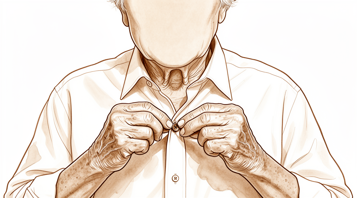
Everyday tasks that use the fingertip become harder. Buttoning a shirt, picking up small coins, typing, or turning a key can all be uncomfortable. Because this joint helps with fine pinch, tasks like threading a needle or picking up a piece of paper may feel clumsy.
You may also notice a small, firm lump or cyst near the joint, sometimes with a groove or ridging in the fingernail itself. These cysts are linked to the same arthritic changes seen on X-ray. Over time, the joint can develop a bend that does not straighten fully, and that curve can slowly increase as the stiffness worsens.
If an old injury to this joint, such as a mallet finger, has left it stiff or bent, the same wear-and-tear changes can appear years later.
What's actually happening
Each finger is a chain of small bones. The joint right at the fingertip, next to the nail, is called the end finger joint. In this condition, wear-and-tear arthritis affects that joint more than any other joint in the hand.
Healthy cartilage works like a shock absorber between the bones. It lets the joint glide smoothly as you bend and straighten. When the cartilage wears away, bone rubs on bone. The body responds by growing extra bone at the joint edges. That new bone, along with the roughened joint surface, is what causes the aching, swelling and stiffness you feel.
Tendons also matter here. A tendon is a strong cord that joins muscle to bone. Cords run across the top and the underside of the finger, and they need to stay balanced for the fingertip to straighten and bend normally. Arthritis can disturb that balance, so the joint may rest in a bent position that slowly worsens over time.
An old injury can set this up too. If you once had a mallet finger, where the tendon that straightens the fingertip tore or pulled away, the joint can lose its balance. The fingertip droops, and the middle joint behind it can then over-straighten into a swan neck shape. A joint left in that position for years wears out early, giving pain and stiffness.
There are two broad patterns of this arthritis. In one, the joint looks and behaves like ordinary wear and tear. In the other, called erosive arthritis, the joint surface itself is gradually eaten away, which tends to be more painful and more destructive.
The good news is that this joint has limited jobs. It helps with fine pinch, but it does not carry most of your grip. That is why treatment options range from settling symptoms to stiffening or replacing the joint, depending on how much the arthritis is troubling you.
What we can do about it
Dr Kieran Hirpara, an upper-limb surgeon at Mater Private Hospital Rockhampton, starts with the least invasive options that suit your condition. Patients are generally referred to our clinic by their GP; if a physiotherapist has suggested you see us, you will still need a referral from your GP in order to be eligible for the Medicare rebate. At your visit we take a history, examine your finger and arrange X-rays if they are needed to confirm the diagnosis.
For a long-standing problem like this, we usually begin with non-operative care. A splint made to fit your finger can settle the pain and help the joint straighten better. Wearing a splint does not make the joint stiffer or limit how far it moves, and most people keep wearing it without trouble. Hand therapy aims to keep the joint moving and ease the strain on it during daily tasks. We usually ask you to give these simple measures a fair go before thinking about anything further.
If those steps have not settled things enough, surgery may be considered. The standard operation for this joint is a fusion, where the worn joint surfaces are removed and the bones are joined so they heal into one solid piece. The finger ends up stiff at that joint, but the pain is gone. Fusion can also stop a mucous cyst from coming back. In some people, keeping the joint moving matters more, so a joint replacement or a procedure that trims the worn bone edges may be an option instead. These choices depend on your symptoms, your work and what you want from your hand. We will talk through the options with you and decide together which path suits you.
What to expect
For most people, this arthritis is a long-term condition rather than a passing one. The ache and stiffness tend to come and go, often worse after you have used the finger a lot. Resting it usually settles things down. Over time the joint may stiffen further, and a bend that does not straighten fully can slowly increase.
If the arthritis came from an old injury, such as a mallet finger, the outlook is similar. The joint may lose some movement, but that loss does not always change how your finger feels or works day to day. Some people keep good function even with limited bending at the fingertip.
Without treatment, the main risks are ongoing pain and a joint that stiffens or bends further. If a deep injury to the joint is not treated quickly, within 8 hours, the joint can become very stiff. Leaving a bent joint alone for years can also let it wear out early.
With treatment, most people find the pain settles. Simple measures like a splint and hand therapy are often enough. If surgery is needed, a fusion removes the pain but leaves that joint stiff. If keeping movement matters more to you, trimming the worn bone edges or a joint replacement can ease pain while preserving some motion. These options carry their own risks, and your surgeon will talk these through with you.
One thing worth knowing: if both the middle joint and the fingertip joint of the same finger are severely painful, they can sometimes be treated in the one operation.
When to see someone
See your GP if the fingertip joint stays painful despite rest, or if a lump or cyst near the nail keeps growing or changes the nail. Ask for a specialist review if the joint is becoming stiffer or more bent over time, if fine tasks like buttoning or picking up coins are getting harder, or if an old mallet finger injury has left the joint stiff or drooping. Go to an emergency department if you have crushed or deeply injured the fingertip joint, as a delay of more than 8 hours can leave it very stiff.
In more depth
Advanced reading: the deeper science (optional)
This section goes further than you need for your own treatment decisions. Arthritis of the fingertip joint is worth the extra reading because fusion, the standard operation, usually described as straightforward, has a higher complication rate than its reputation suggests, and because a motion-preserving alternative exists that is rarely mentioned.
Fusion is not as uneventful as it sounds
Fusing the fingertip joint is presented as a reliable operation, and in terms of pain relief it is. The complication profile is less benign. A review of risk factors across 173 patients states it directly: arthrodesis of the distal interphalangeal joint often leads to complications, with osteoarthritis, revision arthrodesis and smoking identified as risk factors [1].
Smoking appearing on that list is worth acting on, because it is the one factor a patient controls. Bone union depends on blood supply, and this is a small joint with a thin soft-tissue envelope at the end of the finger's blood supply.
The implant that unites better also causes problems the cheaper ones do not
The fixation debate has an unusually clean answer, and it is a trade rather than a winner. Across 1,125 patients, headless compression screws appear to have increased union rates but are associated with complications not seen with other well-established and cheaper techniques, and other than union, there is insufficient evidence to show the screw is superior [2].
The complications specific to the screw follow from the anatomy. The screw runs down the axis of the fingertip, so it passes close to the nail bed and can produce nail deformity, and in a very small distal phalanx there may be inadequate bone to hold it. Wires are cheaper and avoid those particular problems while uniting slightly less reliably.
Where union alone is the priority, a revision fusion, or a smoker, the screw's advantage is most relevant. Where the bone is small and the nail matters, it is less clearly the right choice.
The alternative that preserves movement
Fusion is not the only option, and the alternative is not widely discussed. In patients whose main complaint is the bony lumps and the pain from them rather than the arthritis throughout the joint, the prominent osteophytes can be removed while leaving the joint intact.
Open cheilectomy of the fingertip joint is described as a safe and effective alternative to arthrodesis in patients with symptomatic osteoarthritis who wish to preserve joint motion, across 78 patients [3].
This matters because the fingertip joint contributes little to grip strength but a good deal to fine manipulation and to the appearance of the hand. For someone whose symptoms are driven by the Heberden's nodes themselves, trading them for a permanently stiff fingertip is a larger concession than it may sound, and there is a middle option.
The cyst that often accompanies it
Arthritis at this joint frequently produces a mucous cyst, a small fluid-filled swelling arising from the arthritic joint, usually beside the nail. Because it is driven by the underlying joint, it behaves like the wrist ganglion in that respect: draining it addresses the swelling and not the source. It is covered separately, but the connection is worth knowing, since a recurring cyst is a sign of the arthritis beneath rather than an isolated problem.
References for the advanced reading
- Runkel A, Bonaventura B, Sundermann B, Zajonc H, Eisenhardt S, Leibig N. Risk factors in distal interphalangeal joint arthrodesis in the hand: a retrospective study. J Hand Surg Eur Vol. 2022;47(9):907-14.
- Dickson D, Mehta S, Nuttall D, Ng C. A systematic review of distal interphalangeal joint arthrodesis. J Hand Microsurg. 2014;6(2):74-84.
- Lin EA, Papatheodorou LK, Sotereanos DG. Cheilectomy for treatment of symptomatic distal interphalangeal joint osteoarthritis: a review of 78 cases. J Hand Surg Am. 2017;42(11):889-93.
Evidence & references
This is the clinical evidence summary written for health professionals. It is technical, and it lists the research this page was built from. You do not need to read it to understand your treatment or to make a decision about it.
Overview
- In select patients, percutaneous DIP joint arthrodesis is advantageous in comparison with open fusion techniques [1].
- Swan neck deformity can progress significantly over time due to increasing DIPJ flexion contracture [2].
- Simultaneous surgical intervention is recommended for severe painful osteoarthritis of the PIP and DIP joints of the same digit [3].
- Denervation with cheilectomy presents a motion-preserving alternative to arthrodesis for symptomatic DIP joint osteoarthritis [4].
- A lateral approach and plate fixation for DIP joint arthrodesis yields results equivalent to traditional methods but with fewer major complications [5].
- The combination of DIP arthrodesis and PIP Swanson arthroplasty results in a favourable outcome regarding simultaneous bony union and flexibility [7].
- Silicone interpositional arthroplasty of the DIP joint is an acceptable alternative to arthrodesis, achieving excellent pain relief and a range of movement of 30–40 degrees with a low overall complication rate of 5% [8].
- The smile incision and reverse shotgun approach is a surgical option for DIPJ arthrodesis when more volar part joint preparation and more volar implant insertion sites are necessary [9].
- The nonaxial multiple small screws (NMSS) technique is a feasible option for DIPJ and thumb IPJ arthrodesis, especially when a small finger is indicated and a significant flexion angle is required [11].
- A customized structural bone graft addresses issues of bone stock loss and medullary absence in failed DIPJ silicone arthroplasty, achieving reliable union rates and high patient satisfaction [14].
- There is no difference in biomechanical performance between K-wires and compression screws for DIPJ arthrodesis, so cost and complication profiles should be considered when choosing an implant [26].
Anatomy & Pathophysiology
Bony Anatomy & Dimensions
- A substantial number of distal phalanges are too small to accommodate commonly available headless compression screws, particularly in females and the small finger [25].
- The dorsal integument of the distal phalanx is characterized by the presence of the nail bed with its matrix [28].
- The hand consists of 19 bones, 17 articulations, and 19 muscles situated entirely within the hand [27].
Soft Tissue Anatomy
- The terminal tendon of the extensor mechanism inserts at the base of the distal phalanx to extend it [29].
- The dorsal covering of the interphalangeal articulations of the digits forms a unique cutaneous unit characterized by a considerable excess of skin when the digits are in extension [28].
- The fine tight skin of the dorsal aspect of the middle phalanx forms a distinct functional cutaneous unit [28].
Pathophysiology & Biomechanics
- Osteoarthritis occurs with the highest prevalence in the distal interphalangeal joint of the hand and has been divided into an erosive and a nonerosive form [12].
- The pathogenesis of the early stages of osteoarthritis is poorly understood, but considerable emphasis has been placed on the role of cartilage and subchondral bone as well as soft tissue structures such as collateral ligaments and tendons [12].
- Palmar subluxation of a DIP joint without preexisting arthritic deformity is expected when more than one half of the dorsal articular surface is injured [6].
- Fracture fragments that are less than 43% of the distal phalanx articular surface usually do not allow volar subluxation, whereas fragments involving larger than 52% of the joint surface consistently allow subluxation [48].
- Mallet fractures associated with large fragments may result in volar subluxation of the distal phalanx as the collateral ligaments remain attached to the fracture fragment [48].
- If left untreated, mallet fractures with volar subluxation may lead to a secondary swan neck deformity of the finger, premature osteoarthritis, pain, or stiffness [48].
- Immobilization of the distal interphalangeal joint of any finger reduces the overall grip strength of the hand, with the effect becoming progressively more pronounced from the index to the little fingers [19].
- Positioning the middle finger DIP joint in either extension or 20° of flexion did not significantly affect grip strength or dexterity [38].
- Irreducibility was more commonly seen in dorsal than in volar dislocations of the distal interphalangeal joint [49].
- Volar dislocations of the distal interphalangeal joint carried a higher risk of instability immediately after reduction compared to dorsal dislocations [49].
Classification
- Osteoarthritis of the distal interphalangeal joint has been divided into an erosive and a nonerosive form [12].
- The interrater reliability of the Kellgren & Lawrence and OARSI classification systems for post-traumatic osteoarthritis in the distal interphalangeal joint after mallet finger fractures is considerably lower than initially assumed [50].
Clinical Presentation
- Osteoarthritis occurs with the highest prevalence in the distal interphalangeal joint of the hand [12].
- The pathogenesis of the early stages of osteoarthritis is poorly understood [12].
- Considerable emphasis has been placed on the role of cartilage and subchondral bone in the pathogenesis of distal interphalangeal joint osteoarthritis [12].
- Soft tissue structures such as collateral ligaments and tendons play a role in the pathogenesis of distal interphalangeal joint osteoarthritis [12].
- Radiographic evaluation represents the most standardized method to quantify disease progression in distal interphalangeal joint osteoarthritis [12].
- Different systems have been developed for defining and grading radiographic features of distal interphalangeal joint osteoarthritis [12].
- Osteoarthritis at the distal interphalangeal joint often results in pain and deformity [15].
- Swan neck deformity can progress significantly with time due to increasing distal interphalangeal joint flexion contracture [2].
- Palmar subluxation of a distal interphalangeal joint without preexisting arthritic deformity is expected when more than one half of the dorsal articular surface is injured [6].
- Radiological osteoarthritis after a mallet finger fracture is similar to the natural degenerative process in the distal interphalangeal joint [13].
- Radiological osteoarthritis after a mallet finger fracture is accompanied by a decrease in range of motion of the distal interphalangeal joint [13].
- The decrease in range of motion following mallet finger fracture does not clinically affect patient-reported outcome measures [13].
- Primary synovial chondromatosis of the distal interphalangeal joint is an extremely rare entity [43].
- Primary synovial chondromatosis of the distal interphalangeal joint requires accurate diagnosis to distinguish from other arthropathies [43].
- Floating distal interphalangeal joint injuries can be misdiagnosed initially due to minimal deformity [21].
Investigations
- A distinct collagen septum exists between the extensor tendon and skin at the DIP joint, confirmed using MRI and histology [55].
- The curvatures of the DIP joints may lend insight into the biomechanics and disease progression within the DIP joints [10].
Treatment
Non-Operative
- DIP joint splinting reduces pain and improves extension at the joint [17].
- DIP joint splinting does not result in non-compliance, increased stiffness, or restriction of range of motion [17].
- Collagenase Clostridium histolyticum injection is an option for treating DIP joint contractures in Dupuytren disease [54].
- The potential risk for recurrence must be carefully weighed prior to using Collagenase Clostridium histolyticum for DIP joint contractures [54].
Operative
- Arthrodesis is generally the most accepted surgical option for treatment of degenerative and traumatic conditions involving the DIP joint of the fingers or the IP joint of the thumb [20].
- The ideal position for DIP joint arthrodesis is slightly flexed to improve power, fine pinch, and grip [20].
- Most surgeons use straight intramedullary implants for DIP joint arthrodesis, which obligates the joint to be positioned in neutral extension [20].
- Dorsal plate fixation allows for DIP joint arthrodesis in slight flexion [20].
- Indications for DIP joint fusion include rheumatoid arthritis, posttraumatic arthritis, and chronic conditions [20].
- Percutaneous DIP joint arthrodesis is advantageous in comparison with open fusion techniques in select patients [1].
- A lateral approach with plate fixation for DIP joint arthrodesis yields results equivalent to traditional methods but with fewer major complications [5].
- Diabetes and surgeon experience are factors that increase the risk of postoperative complications in DIP/thumb IP joint arthrodeses [16].
- Open cheilectomy and debridement of the DIP joint is a safe and effective alternative to DIP joint arthrodesis in patients with symptomatic osteoarthritis who wish to preserve joint motion [15].
- In a review of 78 patients, open DIP joint cheilectomy resulted in a significant improvement in mean visual analog scale pain scores from 8 to 1 [15].
- In a review of 78 patients, open DIP joint cheilectomy improved DIP joint flexion contracture by a mean of 6 degrees and DIP joint range of motion by a mean of 20 degrees [15].
- No postoperative infections or other complications were noted in a review of 78 patients undergoing open DIP joint cheilectomy [15].
- No reoperations were required during the follow-up period in a review of 78 patients undergoing open DIP joint cheilectomy [15].
- Denervation with cheilectomy presents a compelling motion-preserving alternative to arthrodesis for symptomatic DIP joint osteoarthritis [4].
- For patients prioritizing hand aesthetics or with unstable joints, DIP joint arthrodesis is preferable to silicone arthroplasty [18].
- The combination of DIP arthrodesis and PIP Swanson arthroplasty resulted in a favourable outcome in terms of simultaneous bony union and flexibility [7].
- The authors recommend simultaneous surgical intervention in case of severe painful OA of the PIP and DIP joints of the same digit [3].
- Open reduction and internal fixation is a viable treatment option for chronic floating DIP joint injuries, though osteoarthritis may develop [21].
Complications
- Arthrodesis of the distal interphalangeal joint often leads to complications [22].
- Diabetes and surgeon experience were identified as factors increasing the risk of postoperative complications in DIP and thumb IP joint arthrodeses [16].
- The complication rate for DIP joint arthrodesis using the AcuTwist headless compression screw was 9% [56].
- In a series of 48 primary arthrodeses using the AcuTwist device, three arthrodeses failed to fuse, including two asymptomatic nonunions and one fixation loss requiring revision with autograft [56].
- There were no cases of nail deformity, wound complications, tip hypersensitivity, or clinically notable malalignment in a series of 48 primary arthrodeses using the AcuTwist device [56].
- The overall complication rate for silicone interpositional arthroplasty of the DIP joint was 5% [8].
- No postoperative infections or other complications were noted in a review of 78 patients undergoing open cheilectomy and debridement of the DIP joint [15].
- No reoperations were required or performed during the follow-up period for patients undergoing open cheilectomy and debridement of the DIP joint [15].
- Swan neck deformity can progress significantly with time due to increasing DIPJ flexion contracture [2].
Recovery
Operative
- The results of lateral approach and plate fixation for DIP joint arthrodesis are equivalent to traditional methods but with fewer major complications [5].
- A customized structural bone graft using the described technique addresses issues of bone stock loss and medullary absence in failed DIPJ silicone arthroplasty, achieving reliable union rates and high patient satisfaction [14].
- Diabetes and surgeon experience were identified as factors increasing the risk of postoperative complications in DIP/thumb IP joint arthrodeses [16].
Non-Operative
- Splinting of the DIP joint does not give rise to non-compliance, increased stiffness or restriction of range of motion [17].
Key Evidence
- [L4] In select patients, this percutaneous DIP joint arthrodesis is advantageous in comparison with open fusion techniques. [1] (10.1007/s11552-010-9265-9)
- [L5] The swan neck deformity in this individual progressed significantly with time because of increasing DIPJ flexion contracture. [2] (10.1016/j.jht.2009.11.005)
- [L3] The authors recommend simultaneous surgical intervention in case of severe painful OA of the PIP and DIP joints of the same digit. [3] (10.1177/17531934231191255)
- [L4] It presents a compelling motion-preserving alternative to arthrodesis for symptomatic DIP joint osteoarthritis. [4] (10.1016/j.jhsa.2026.01.027)
- [L4] The results obtained in this small series are equivalent to the traditional methods of DIP joint arthrodesis but with fewer major complications. [5] (10.1016/j.jhsa.2007.09.004)
- [L5] Palmar subluxation of a DIP joint without preexisting arthritic deformity is expected when more than one half of the dorsal articular surface is injured. [6] (10.1016/j.jhsa.2007.09.006)
- [L4] The combination of DIP arthrodesis and PIP Swanson arthroplasty resulted in a favourable outcome in terms of simultaneous bony union and flexibility. [7] (10.1177/17531934231215790)
- [L4] The study confirms that silicone interpositional arthroplasty of the DIP joint is an acceptable alternative to arthrodesis, achieving excellent pain relief and a range of movement of 30–40 degrees with a low overall complication rate of 5%. [8] (10.1177/1753193411422679)
- [L4] This technique may be a good surgical option for DIPJ arthrodesis when more volar part joint preparation and more volar implant insertion sites are necessary. [9] (10.1186/s12891-024-08016-6)
- [L5] Our understanding of morphology may lend insight into the biomechanics and disease progression within the DIP joints. [10] (10.1007/s11552-014-9605-2)
- [L4] Thus, the NMSS technique could be used as a feasible option in DIPJ and thumb IPJ arthrodesis, especially when a small finger is indicated and a significant flexion angle is required. [11] (10.1186/s12891-022-05473-9)
- [L5] [12] (10.1016/j.jhsa.2010.09.003)
- [L4] Radiological OA after an MFF is similar to the natural degenerative process in the DIP joint and is accompanied by a decrease in range of motion of the DIP joint, which does not clinically affect PROMs. [13] (10.1016/j.jhsa.2023.03.027)
- [L4] A customized structural bone graft using the described technique addresses issues of bone stock loss and medullary absence in failed DIPJ silicone arthroplasty, achieving reliable union rates and high patient satisfaction. [14] (10.1177/17531934231151217)
- [L4] [15] (10.1016/j.jhsa.2017.07.006)
- [L3] Diabetes and surgeon experience were identified as factors increasing the risk of postoperative complications in these DIP/thumb IP joint arthrodeses. [16] (10.1186/s12891-024-07361-w)
- [L2] It does not give rise to non-compliance, increased stiffness or restriction of range of motion. [17] (10.1016/j.jht.2013.08.004)
- [L3] For patients prioritizing hand aesthetics or with unstable joints, distal interphalangeal joint arthrodesis is preferable. [18] (10.1177/1753193420917818)
- [L4] Immobilization of the distal interphalangeal joint of any finger reduces the overall grip strength of the hand, with the effect becoming progressively more pronounced from the index to the little fingers. [19] (10.1177/1753193418765068)
- [L4] [20] (10.1016/j.jhsa.2018.03.049)
- [Case_report] Floating DIP joint injuries can be misdiagnosed initially due to minimal deformity; open reduction and internal fixation is a viable treatment option for chronic cases, though osteoarthritis may develop. [21] (10.1016/j.jhsa.2010.05.025)
- [L3] Arthrodesis of the distal interphalangeal joint often leads to complications. [22] (10.1177/17531934221111641)
- [L4] A substantial number of distal phalanges are too small to accommodate commonly available headless compression screws, particularly in females and the small finger. [25] (10.1007/s11552-014-9679-x)
- [L5] Given the lack of difference in biomechanical performance between K-wires and compression screws, consideration should be given to other factors such as cost and complication profiles when choosing an implant for DIPJ fusion. [26] (10.1177/1558944715627211)
- [L2] Positioning the middle finger DIP joint in either extension or 20° of flexion did not significantly affect grip strength or dexterity, allowing other considerations such as appearance to be prioritized. [38] (10.1016/j.jhsa.2014.06.021)
- [Case_report] Primary synovial chondromatosis of the distal interphalangeal joint is an extremely rare entity that requires accurate diagnosis to distinguish from other arthropathies. [43] (10.1177/15589447211049520)
- [L5] [48] (10.1177/1753193414554772)
- [L4] Irreducibility was more commonly seen in dorsal than in volar dislocations, while volar dislocations carried a higher risk of instability immediately after reduction. [49] (10.1177/1753193415616957)
- [L4] The interrater reliability of the Kellgren & Lawrence and OARSI classification systems for post-traumatic osteoarthritis in the distal interphalangeal joint after mallet finger fractures is considerably lower than initially assumed. [50] (10.1016/j.jhsa.2024.03.012)
- [L4] Injection with CCH is an option for the treatment of DIP joint contractures in Dupuytren disease, though the potential risk for recurrence should be carefully weighed prior to its use. [54] (10.1016/j.jhsa.2018.07.004)
- [L5] We confirmed the existence of a distinct collagen septum between the extensor tendon and skin at the DIP joint using MRI and histology. [55] (10.1016/j.jhsa.2008.11.030)
- [L4] [56] (10.1016/j.jhsa.2013.09.040)
References
[1] Treatment of Symptomatic Distal Interphalangeal Joint Arthritis with Percutaneous Arthrodesis: A Novel Technique in Select Patients. HAND. 2010. DOI: 10.1007/s11552-010-9265-9
[2] Swan Neck Deformity after Distal Interphalangeal Joint Flexion Contractures: A Biomechanical Analysis. Journal of Hand Therapy. 2010. DOI: 10.1016/j.jht.2009.11.005
[3] Does distal interphalangeal joint arthrodesis affect proximal interphalangeal joint arthroplasty outcomes in the same finger?. Journal of Hand Surgery (European Volume). 2023. DOI: 10.1177/17531934231191255
[4] Denervation with Cheilectomy of the Distal Interphalangeal Joint: Technique and Medium-Term Results. The Journal of Hand Surgery. 2026. DOI: 10.1016/j.jhsa.2026.01.027
[5] Alternative to the Distal Interphalangeal Joint Arthrodesis: Lateral Approach and Plate Fixation. The Journal of Hand Surgery. 2008. DOI: 10.1016/j.jhsa.2007.09.004
[6] A Biomechanical Study of Distal Interphalangeal Joint Subluxation After Mallet Fracture Injury. The Journal of Hand Surgery. 2008. DOI: 10.1016/j.jhsa.2007.09.006
[7] Simultaneous anterograde screw arthrodesis of distal interphalangeal joint and silastic proximal interphalangeal joint replacement for osteoarthritis. Journal of Hand Surgery (European Volume). 2023. DOI: 10.1177/17531934231215790
[8] Joint replacement in 131 painful osteoarthritic and post-traumatic distal interphalangeal joints. Journal of Hand Surgery (European Volume). 2011. DOI: 10.1177/1753193411422679
[9] Smile incision and reverse shotgun approach in distal interphalangeal joint arthrodesis. BMC Musculoskeletal Disorders. 2024. DOI: 10.1186/s12891-024-08016-6
[10] Curvatures of the DIP Joints of the Hand. HAND. 2014. DOI: 10.1007/s11552-014-9605-2
[11] Distal interphalangeal joint arthrodesis with nonaxial multiple small screws: a biomechanical analysis with axial headless compression screw and clinical result of 15 consecutive cases. BMC Musculoskeletal Disorders. 2022. DOI: 10.1186/s12891-022-05473-9
[12] Osteoarthritis of the Distal Interphalangeal Joint. The Journal of Hand Surgery. 2010. DOI: 10.1016/j.jhsa.2010.09.003
[13] Posttraumatic Osteoarthritis of the Distal Interphalangeal Joint: A Follow-Up Study of 12 Years After Nonsurgical Treatment of Mallet Finger Fractures. The Journal of Hand Surgery. 2023. DOI: 10.1016/j.jhsa.2023.03.027
[14] Salvage of failed Swanson’s arthroplasty of the distal interphalangeal joint. Journal of Hand Surgery (European Volume). 2023. DOI: 10.1177/17531934231151217
[15] Cheilectomy for Treatment of Symptomatic Distal Interphalangeal Joint Osteoarthritis: A Review of 78 Patients. The Journal of Hand Surgery. 2017. DOI: 10.1016/j.jhsa.2017.07.006
[16] Arthrodesis of distal interphalangeal and thumb interphalangeal joint: a retrospective cohort study of 149 cases. BMC Musculoskeletal Disorders. 2024. DOI: 10.1186/s12891-024-07361-w
[17] Splinting of the Distal Interphalangeal Joint Reduces Pain and Improves Extension at the Joint; Results Front the Splint-OA Study. Journal of Hand Therapy. 2014. DOI: 10.1016/j.jht.2013.08.004
[18] Silicone arthroplasty versus screw arthrodesis in distal interphalangeal joint osteoarthritis. Journal of Hand Surgery (European Volume). 2020. DOI: 10.1177/1753193420917818
[19] Effect of immobilization of the distal interphalangeal joint of fingers on grip strength. Journal of Hand Surgery (European Volume). 2018. DOI: 10.1177/1753193418765068
[20] Dorsal Plate Fixation for Distal Interphalangeal Joint Arthrodesis of the Fingers and Thumb. The Journal of Hand Surgery. 2018. DOI: 10.1016/j.jhsa.2018.03.049
[21] Floating Distal Interphalangeal Joint Injury: Case Report. The Journal of Hand Surgery. 2010. DOI: 10.1016/j.jhsa.2010.05.025
[22] Risk factors in distal interphalangeal joint arthrodesis in the hand: a retrospective study of 173 cases. Journal of Hand Surgery (European Volume). 2022. DOI: 10.1177/17531934221111641
[25] Dimensional Analysis of the Distal Phalanx with Consideration of Distal Interphalangeal Joint Arthrodesis Using a Headless Compression Screw. HAND. 2014. DOI: 10.1007/s11552-014-9679-x
[26] Biomechanical Analysis of Internal Fixation Methods for Distal Interphalangeal Joint Arthrodesis. HAND. 2016. DOI: 10.1177/1558944715627211
[27] Exam Of The Hand Wrist 2Ed. INTRODUCTION.
[28] Exam Of The Hand Wrist 2Ed. Functional cutaneous units.
[29] Green S Operative Hand Surgery. Interosseous and Hypothenar Muscles.
[38] Simulated Distal Interphalangeal Joint Fusion of the Index and Middle Fingers in 0° and 20° of Flexion: A Comparison of Grip Strength and Dexterity. The Journal of Hand Surgery. 2014. DOI: 10.1016/j.jhsa.2014.06.021
[43] Primary Distal Interphalangeal Joint Tenosynovial Chondromatosis of the Small Finger: A Case Report With Literature Review. HAND. 2022. DOI: 10.1177/15589447211049520
[48] Mallet fingers with bone avulsion and DIP joint subluxation. Journal of Hand Surgery (European Volume). 2014. DOI: 10.1177/1753193414554772
[49] Differences between dorsal and volar dislocations of the distal interphalangeal joint of fingers: a report of 30 cases. Journal of Hand Surgery (European Volume). 2016. DOI: 10.1177/1753193415616957
[50] Rater Agreement of Post-Traumatic Osteoarthritis of the Distal Interphalangeal Joint 12 Years After a Mallet Finger Fracture. The Journal of Hand Surgery. 2024. DOI: 10.1016/j.jhsa.2024.03.012
[54] Collagenase Clostridium histolyticum for the Treatment of Distal Interphalangeal Joint Contractures in Dupuytren Disease. The Journal of Hand Surgery. 2019. DOI: 10.1016/j.jhsa.2018.07.004
[55] Dorsal Digital Septum of the Distal Interphalangeal Joint. The Journal of Hand Surgery. 2009. DOI: 10.1016/j.jhsa.2008.11.030
[56] Arthrodesis of the Thumb Interphalangeal Joint and Finger Distal Interphalangeal Joints With a Headless Compression Screw. The Journal of Hand Surgery. 2014. DOI: 10.1016/j.jhsa.2013.09.040