Clinicians › Hand
Flexor tendon repair
For patients: a plain-language version of this topic is available. See the patient guide.
Overview
The primary objective of surgical intervention for flexor tendon injuries is to achieve sufficient repair strength to facilitate early motion, prevent adhesions within the tendon sheath, and restore normal range of motion and function [17]. Consistent management relies on a comprehensive understanding of tendon anatomy, zone-specific characteristics, repair techniques, potential complications, rehabilitation protocols, and emerging advances such as tissue engineering and biologic modification [8]. Despite these foundational principles, no gold standard has been established for the optimal flexor tendon repair algorithm [10]. Consequently, the selection of a specific repair technique is usually driven by surgeon familiarity, popularity, and technical difficulty rather than a single definitive protocol [10].
Outcomes are influenced by numerous factors that cannot be controlled intraoperatively [3]. While current randomized controlled trials are expected to provide data for domains requiring better evidence in repair and rehabilitation [6], continued therapy utilization after primary repair remains an independent predictor of the need for reoperation [9]. Identifying factors associated with tenolysis allows surgeons to risk-stratify patients prior to surgery and helps guide postoperative expectations if complications arise [7]. Active motion regimens are considered desirable following repair of flexor tendons in all zones [19].
Current practice shows that the majority of surgeons perform zone II flexor tendon repairs in accordance with the best currently available evidence [42]. However, significant variability persists regarding suture material, surgical technique, and rehabilitation protocols specifically for zone II injuries [42].
Anatomy & Pathophysiology
Extrinsic Flexor Muscles
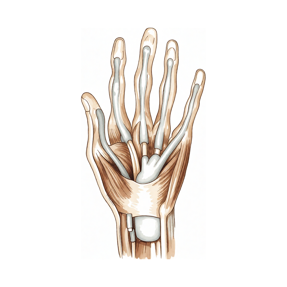
The extrinsic finger flexors comprise the flexor digitorum profundus (FDP) and the flexor digitorum superficialis (FDS) [48]. The FDP originates from the proximal ulna and interosseous membrane, dividing in the forearm into a radial component for the index finger and an ulnar component for the middle, ring, and little fingers [82]. It inserts on the proximal volar aspect of the distal phalanx, providing flexion at both the proximal and distal interphalangeal joints [48, 82]. Innervation of the FDP to the index and middle fingers is via the anterior interosseous branch of the median nerve, while the ring and little fingers receive innervation from the ulnar nerve [82].
The FDS possesses a radial head originating from the proximal radius and a humeral ulnar head from the medial humeral epicondyle and coronoid process [82]. Its tendon bifurcates around the FDP at the A2 pulley, with slips reuniting distally at the Camper chiasm; approximately half of the fibers remain ipsilateral while half cross contralaterally [82]. The FDS inserts via radial and ulnar slips into the proximal metaphysis of the middle phalanx, primarily flexing the proximal interphalangeal joint [82]. The entire FDS muscle is innervated by the median nerve [82].
The flexor pollicis longus (FPL) originates from a radial head on the proximal radius and interosseous membrane, plus an accessory head on the ulnar coronoid process and medial humeral epicondyle [82]. It inserts into the proximal base of the thumb distal phalanx and is innervated by the anterior interosseous branch of the median nerve [82]. The FPL flexes both the interphalangeal and metacarpophalangeal joints of the thumb [82].
Tendon Sheath and Pulley System
Distal to the metacarpal neck, flexor tendons enter the fibroosseous tunnel, or digital flexor sheath, which extends to the proximal aspect of the distal phalanx [82]. The sheath contains annular pulleys for mechanical stability and cruciate pulleys for flexibility [82]. The A1, A3, and A5 pulleys are located over the metacarpophalangeal, proximal interphalangeal, and distal interphalangeal joints, respectively, while the A2 and A4 pulleys sit over the middle portions of the proximal and middle phalanges [82]. The A2 and A4 pulleys are most essential for maintaining the mechanical advantage of the flexor tendons [82].
The tenosynovium lining the tunnel supplies nutrition and lubrication to the poorly vascularized tendons within the sheath [82]. Proximal to the sheath, tendons are well vascularized by the peritenon, whereas within the sheath, vascularity is supplied via the vincula system, specifically the vinculum longus and brevis [82]. The A0 pulley is a unique anatomic structure of the thumb [120].
Vascular Anatomy
The tenosynovial sheath of the FPL is continuous with the radial bursa, and the sheath to the little finger is continuous with the ulnar digital bursa [82]. In some patients, these bursae communicate, allowing a horseshoe abscess to spread between the thumb and little finger if infection occurs in either flexor tendon sheath [82]. An arcade deep in the flexor tendon joins the two palmar arteries of the thumb at the distal metaphysis of the first phalanx [79]. Vessels from this subtendinous arcade enter the vincula to irrigate the flexor tendon [79].
Healing Pathophysiology
Flexor tendon healing involves both extrinsic and intrinsic mechanisms [82]. Extrinsic healing occurs via capillary and fibroblast ingrowth, leading to adhesion formation at the repair site [82]. Intrinsic healing arises from tenocytes within the tendon, involving proliferation and extracellular matrix production [82, 149]. However, tendons have minimal capacity for intrinsic healing due to poor cell density [149]. Extrinsic healing also involves invasion of cells from the surrounding sheath and synovium, which envelop the tendon and contribute to adhesion formation [149].
Tendon healing comprises three phases: inflammatory, fibroblastic, and remodelling [57]. During the inflammatory phase, repair strength relies solely on the surgical suture and possibly the fibrin clot [57]. In the fibroblastic phase, strength increases through extracellular matrix synthesis, with collagens deposited in a random, disorganized manner [57]. Adhesions form between the tendon and sheath during this phase, inhibiting natural gliding [57]. The main complications following repair are rupture from inadequate healing and adhesion formation from excessive fibrosis [57]. Up to 30% to 40% of tendon injuries end with postsurgical adhesion formation [149].
Biomechanics and Function
Flexor tendons function as cables transmitting forces to move and stabilize joints [100]. A satisfying functional outcome requires reestablishing both tendon continuity and the gliding mechanism of the tendon and surrounding structures [16]. The goal of repair and postoperative care is to encourage intrinsic and extrinsic healing without thick adhesions that limit tendon excursion and restrict finger motion [82].
In a modified Kessler repair, the biomechanically optimal location for the core suture knot is under the dorsal surface of the flexor tendon [66]. The tensile strength of a flexor tendon repair increases with suture purchase, likely due to the mechanical characteristics of the suture polymer rather than improved grip [45].
Classification
IFSSH: Flexor tendon injuries are divided into five zones according to the International Federation of Societies for Surgery of the Hand (IFSSH) classification system [154]. Zone III injuries are located between the distal border of the transverse carpal ligament and the proximal edge of the fibroosseous sheath [154].
Other Considerations: Zone I flexor tendon injuries traditionally have not yielded results as good as other flexor tendon injuries [11]. Full motion is rarely regained in Zone I flexor tendon injuries [11]. Good or excellent results are reported in only up to 67% of Zone I flexor tendon injury cases [11]. Flexor tendon repair in Zone II is a technically demanding procedure [4]. Outcomes for flexor tendon repair in Zone II have become more predictable and satisfying [4]. Tendon grafting is the treatment of choice for flexor tendon injuries in zones I and II when direct repair is not possible or delayed [13]. Many of the principles of flexor tendon repair and rehabilitation can be applied to zones III–V [44]. The review aims to highlight differences in the classification of flexor tendon injuries between children and adults [56].
Clinical Presentation
Flexor tendon injuries occur with an annual incidence of 12 per 100,000 in men and 2 per 100,000 in women [33]. Spontaneous ruptures of the hand are more frequent than commonly recognized, with the majority involving the profundus tendon of the small finger in the palm [52]. Pediatric presentations differ from adult cases in both diagnosis and rehabilitation, often necessitating surgical exploration due to uncooperative patients [29].
Preoperative factors: The type of trauma (e.g., cut, crush), degree of wound contamination, and associated injuries significantly affect functional outcomes [16]. Tearing injuries caused by saws result in poorer outcomes for Zone II repairs compared with sharp injuries at an average follow-up of 4 years [38]. An accompanying volar plate injury in Zone 2 also worsens results [43].
Risk stratification: Multiple variables influence post-repair results in Zone 2, including the mechanism of injury (clean-cut versus crush), associated fractures, the number of digits or tendons injured, patient age, surgeon experience, and the type of postoperative mobilization programme [22]. A large part of outcome variance is attributed to the psychological and biologic characteristics of the patient [22]. Adverse outcomes have previously been reported in association with age, smoking, zone of injury, multiple finger injuries, associated flexor digitorum superficialis (FDS) tendon injury, nerve injuries, and delayed treatment [59]. Identifying factors associated with tenolysis allows surgeons to risk-stratify patients prior to surgery and guide postoperative expectations if complications arise [7]. Understanding risk factors for reoperation provides important guidance to surgeons and therapists when treating patients with flexor tendon injuries [32].
Outcome assessment: Objective functional outcomes, such as range of motion or grip strength, do not always align with patients’ perceived activity levels [33]. The DASH appears to be an appropriate outcome tool after flexor tendon repair, and its combination with measures of impairment such as TAM and TPM gives a more comprehensive picture of functional outcome [39]. The use of patient-reported outcomes, in addition to clinician-reported outcomes, provides deeper insight into patients' perceptions of their recovery after flexor tendon injury [51]. The large heterogeneity in the outcome domains being assessed or measured across studies highlights the need for a consistent core outcome set to be measured in future clinical research on hand flexor tendon injuries [18].
Investigations
Physical Examination: A careful physical examination is essential to direct care and future testing if indicated [47]. Diagnostic tests such as imaging and serum laboratory studies are useful in the determination of pathology but can be expensive, time consuming, and often nonspecific [47].
MRI: MRI is recommended for the evaluation of gaps after flexor tendon repair [20]. 1.5T MRI has slightly better sensitivity and specificity for distinguishing clinically intact from clinically impaired repairs than 3T MRI [20].
Other Considerations: The Disabilities of the Arm, Shoulder and Hand (DASH) questionnaire appears to be an appropriate outcome tool after flexor tendon repair [39]. The combination of the DASH with measures of impairment such as total active motion (TAM) and total passive motion (TPM) gives a more comprehensive picture of functional outcome [39]. Pediatric flexor tendon injuries often require surgical exploration due to uncooperative patients [29].
Treatment
Non-Operative
The provided evidence does not detail specific conservative management protocols such as weight loss, physical therapy regimens, NSAIDs, or injections for flexor tendon injuries.
Operative
Indications: Secondary reconstruction remains an important and useful technique for complicated flexor tendon injuries or those that have failed primary repair [12]. Single-stage tendon grafting for reconstruction of zone I and II flexor tendon injuries requires careful patient selection, strict indications, and adherence to sound surgical principles [126].
Surgical Approach / Technique: Principles of surgical treatment include early primary repair with a strong core stitch (4-6 strand) combined with an epitendinous suture [16]. The epitendinous suture has been demonstrated to contribute considerably to the strength of the repair [16]. No gold standard has been determined for the optimal flexor tendon repair algorithm, and repairs are usually chosen based on familiarity, popularity, and technical difficulty [10]. Flexor tendon repair in Zone II is a technically demanding procedure, but outcomes have become more predictable and satisfying [4]. Repair of the divided flexor tendon to achieve normal or near-normal function consistently remains a problem which has not yet been solved [15]. A modified protocol for primary flexor tendon repair in zones 1 and 2 utilizes a 6-strand core suture without circumferential suturing, selective pulley division, and partial FDS resection to facilitate early active motion [24]. The biomechanically optimal location of the core suture knot in a modified Kessler repair was under the dorsal surface of flexor tendon [66]. Tensile strength of a flexor tendon repair increases with suture purchase, likely due to the mechanical characteristics of the suture polymer rather than improved grip [45]. In an in vivo model, current modifications to suture techniques for intrasynovial flexor tendon repair demonstrated significant improvements in gap formation and rupture compared with a similar technique using shorter purchase lengths and shallower purchase depth [170]. The combination of figure-of-eight peripheral sutures and a 2-strand core suture provided the repaired rat flexor tendon with enough strength to prevent rerupture without cast fixation or immobilization after surgery [172]. The bulky 'figure of eight' technique can be used in isolated profundus tendon injuries in zones IIA/IIB [157]. Although repair of both flexor digitorum profundus and flexor digitorum superficialis tendons is slightly more preferable based on increased grip strength, the repair of the flexor digitorum superficialis together with flexor digitorum profundus is not mandatory [36]. Endoscope-assisted repair of flexor tendon rupture is a useful alternative surgical technique that enables a minimally invasive approach and provides favorable range of motion [96]. The volar plate flap technique may take its place in flexor tendon surgery based on encouraging initial clinical experience [95]. A vascularized dorsal digital fascial flap combined with a standard modified Kessler technique is used to prevent restrictive adhesions between the superficialis and profundus tendons in zone II [148]. A challenging step of digital flexor tendon repair is retrieving proximal ends of the cut tendons without damaging annular pulleys or extending the exposure excessively [152]. A technique of tendon retrieval using a twisted loop of 26-gauge wire to snare sutures holding the tendons has been described [152]. A retracted flexor digitorum profundus tendon can be retrieved by pushing the tendon distally through a palm incision using two forceps to feed the stump distally bit by bit [155].
Implant Selection: Polyvinylidene fluoride sutures offer a possible alternative to polypropylene sutures for flexor tendon repair [167]. Appropriate absorbable core sutures can be used safely for flexor tendon repair when combined with a controlled active motion regime [163]. Barbed sutures may have appealing attributes for flexor tendon reconstruction, and further investigation is required to evaluate and optimize these materials for use in this context [60]. Novel flexor tendon repair techniques, including barbed suture, should be tested cyclically in a manner that closely mimics in vivo conditions during postoperative rehabilitation before clinical adoption [23]. A novel flexor tendon repair technique with improved biomechanical performance may be available for use in flexor tendon repairs [21]. Suture anchors are commonly used in patients with good bone quality and may be combined with a pull-out suture for the increased strength of a multistrand repair when sufficient profundus tendon is not available [102].
Adjuncts: Tendon surgery can be performed under local anesthesia without a tourniquet by injecting epinephrine mixed with lidocaine to achieve vasoconstriction [158]. The authors no longer perform flexor tendon repair with tourniquet, sedation, or muscle paralysis [133]. Incorporating intraoperative mobilization using 'wide-awake' surgery could emerge to further improve tendon outcomes [173].
Other Considerations: The most common donor tendon for palm-to-fingertip reconstruction is the palmaris longus tendon, and the most common donor tendon for forearm-to-fingertip reconstruction is the plantaris tendon [102]. The palmaris longus tendon is present in only 75% to 85% of people [102]. The plantaris tendon is present in about 80% of people [102]. Intrasynovial grafts are associated with fewer adhesions in animal models [102]. Tendon grafts will tolerate early motion therapy if the proximal and distal tenorrhaphy junctures are strong enough to withstand these forces of active finger motion [126]. The described proximal junction techniques for active tendon implants were strong enough to resist early active motion in the immediate postoperative period without significant elongation [171].
Rehabilitation and Postoperative Management
Postoperatively, an early motion protocol should be used in compliant patients [16]. There is not sufficient evidence to support true active motion as an effective or preferable choice for flexor tendon rehabilitation at this time based on a lack of superior benefits following true active motion regimens [144]. A Cochrane review from 2021 concluded that evidence remains insufficient to determine the superiority of any current rehabilitation method following flexor tendon repair [33]. There is currently limited evidence informing use of relative motion flexion orthoses following flexor tendon repair [165]. Treatment guidelines varied across UK hand therapy departments, suggesting that patients receive differing advice about how to protect, move and use their hand after zone I/II flexor tendon repair [141]. Patient selection, cooperation, and a rational goal are as key to success as the operative procedure itself for flexor tenolysis [61]. MRI is recommended for evaluation of gaps after flexor tendon repair, as the 1.5T has slightly better sensitivity and specificity for distinguishing clinically intact from clinically impaired repairs than the 3T [20]. Despite a decline in opioid quantity per patient, most patients still receive opioids after zone II flexor tendon repair [161].
General Considerations and Evidence Gaps
Consistent, successful management of flexor tendon injuries relies on understanding the anatomy, characteristics and repair of tendons in the different zones, potential complications, rehabilitation protocols, recent advances in treatment, and future directions [8]. Flexor tendon injuries are complex, and management requires consideration of surgical timing, injury location, approach, and soft tissue handling [31]. Better evidence is required in many domains of flexor tendon repair and rehabilitation, hoping that current RCTs will provide this data [6]. Despite significant advances in flexor tendon repair and reconstruction, the drive towards perfection continues [46]. The majority of respondents are performing zone II flexor tendon repairs in accordance with the best currently available evidence, although there is variability with respect to suture material, surgical technique, and rehabilitation protocols [42]. Factors affecting the results after flexor tendon repair in zone 2 include the mechanism of injury, associated fractures, the number of digits/tendons injured, age, the experience of the surgeon, and the type of postoperative mobilization programme [22]. A large part of the variance in the outcome of zone 2 flexor tendon repair is thought to be related to the psychological and biologic characteristics of the patient [22].
Complications
Repair Rupture
Repair rupture rates in the literature range from 4% to 10% for finger flexors and 3% to 17% for the flexor pollicis longus (FPL) of the thumb [25]. A specific reported rate for repair rupture is 4% [129]. In a study of 45 patients undergoing Zone 2 repairs with a six-strand figure-of-eight suture, one repair (2%) ruptured on the seventh postoperative day [99]. In a multicentre cohort of 218 primary single-finger repairs, rupture causes were often miscellaneous and unclear, precluding the definition of a single major cause [53]. Outcomes have improved over the last decade, with minimal or no rupture during postoperative active motion [177]. Zone 2 repair outcomes are comparable to other zones, with a very low to zero incidence of rupture [182].
Adhesions and Tenolysis
Secondary complications such as tendon adhesions and reduced gliding amplitude commonly arise following Zone II flexor tendon repair, potentially leading to fixed deformities and decreased function [34]. Ruptures and adhesions requiring tenolysis are found in approximately 5% of cases each [59]. The reported rate of tendon adhesions is 4% [129]. Most reoperations after primary flexor tendon repair involve tenolysis alone, accounting for 58% of cases [106]. Flexor tenolysis is effective for improving motion following tendon repair, particularly in primary procedures and isolated tendon injuries [63]. Improved outcomes have been reported over the last decade with lower rates of tenolysis [177].
Reoperation Rates
The overall rate of reoperation after flexor tendon repair across all zones is 6% [129]. Further surgery for rupture or adhesions is required in around 6% of cases [59]. In New York state, the rate of reoperation for repair rupture is 2.3%, and the rate of tenolysis is 3.6% [129]. The median time to reoperation in New York state was 140 days [129]. Incidence of reoperation ranged from 5% (23 patients) in 2002 to 7% (61 patients) in 2004 [106]. Patients with workers’ compensation as a primary payer were 48% more likely to undergo reoperation than those with any other type of primary payer [106]. Conversely, patients who had a concomitant nerve repair during the index procedure were 26% less likely to undergo reoperation [106].
Infection
Despite aggressive and prompt antibiotic therapy and surgical intervention, otherwise healthy patients can expect some residual digital stiffness following flexor tendon sheath infection [72]. Repair of flexor digitorum profundus (FDP) avulsions via all-inside suture fixation is associated with an incidence of infection (24%) at final follow-up [187]. Infection is more common during flexor tendon pulley reconstruction procedures, as these are often associated with two-stage flexor tendon reconstructions and the placement of a tendon implant into the flexor sheath [162]. An infected implant in pulley reconstruction is usually preceded by synovitis, which is due either to excessive activity or poor implant gliding resulting in implant buckling [162].
Other Complications
Other possible complications after flexor tendon repair include triggering, pulley failure, quadriga, and lumbrical plus deformity [129]. Transosseous repair of Zone I flexor digitorum profundus injuries with a buried dorsal suture is associated with a high rate of clinical complications [190]. Repair of FDP avulsions via all-inside suture fixation is associated with limited distal interphalangeal joint motion [187]. Late fracture of the phalanx beneath the pulley is a complication of flexor tendon pulley reconstruction [162]. Over-tightening the graft during flexor tendon pulley reconstruction commonly results in poor finger flexion and resultant stiffness [162]. Prolonged incubation of arthroscopic suture knots influences slippage, which could result in unsuccessful primary attachment of the tendon during the healing phase [186]. In a case report of simultaneous dislocation of both interphalangeal joints and flexor tendon tear, active flexion was limited by adherence of the flexor tendon at the fracture site when last seen 4 months after the accident [184].
Recovery
Rehabilitation protocol: Primary flexor tendon repair in zones 1 and 2 utilizes a modified protocol featuring a 6-strand core suture without circumferential suturing, selective pulley division, and partial FDS resection to facilitate early active motion [24]. This approach aligns with the standard practice of strong multistrand core sutures and modified pulley preservation in the digital sheath area [27]. Both PTFE and FW provide for a robust tendon repair so that early active motion regimens for rehabilitation can be applied [109]. The partial-range active flexion protocol is recommended as a safe, efficient, and generalizable framework for rehabilitation after flexor tendon repair and other hand disorders, particularly where therapist assistance is unavailable [114]. Early passive motion is an effective technique to improve the results of flexor tendon repairs in Zone II by limiting adhesions and improving tensile strength at an earlier stage of healing [115].
Functional milestones: The effects of an early active motion protocol may improve the grip strength and upper limb mobility of individuals after zone II flexor tendon repair [58]. Repeated administration of sodium hyaluronate at the tendon repair site may be effective in improving postoperative active finger motion after primary hand flexor tendon repair in the mid-term [64].
Other Considerations: Zone I flexor tendon injuries traditionally have not yielded results as good as other flexor tendon injuries, with full motion rarely regained and good/excellent results reported in only up to 67% of cases [11]. Delayed primary flexor tendon repair under WALANT can achieve excellent functional outcomes, although longer follow-up is needed for extension deficit recovery [40]. Late direct repair is possible in a small proportion of patients (approximately 1 in 10 to 1 in 15) where tendon ends can be approximated with acceptable tension [68].
Key Evidence
- [L4] The outcome of a flexor tendon repair is influenced by many factors that cannot be controlled intraoperatively. [3] (10.1016/j.jhsa.2022.01.015)
- [L5] Flexor tendon repair in Zone II is a technically demanding procedure, but outcomes have become more predictable and satisfying. [4] (10.1016/j.hcl.2004.11.001)
- [L5] The authors conclude that better evidence is required in many domains of flexor tendon repair and rehabilitation, hoping that current RCTs will provide this data. [6] (10.1177/17531934261445904)
- [L3] Identification of factors associated with tenolysis following flexor tendon repair allows surgeons to risk-stratify patients prior to surgery and help guide postoperative expectations if complications arise. [7] (10.1177/15589447221142890)
- [L5] Consistent, successful management of flexor tendon injuries relies on understanding the anatomy, characteristics and repair of tendons in the different zones, potential complications, rehabilitation protocols, recent advances in treatment, and future directions, including tissue engineering and biologic modification of the repair site. [8] (10.5435/jaaos-d-16-00316)
- [L3] Continued therapy utilization after primary flexor tendon repair is an independent predictor of reoperation need. [9] (10.1016/j.jht.2021.04.015)
- [L5] No gold standard has been determined for the optimal flexor tendon repair algorithm, and repairs are usually chosen based on familiarity, popularity, and technical difficulty. [10] (10.1016/j.jhsa.2014.06.025)
- [L5] Zone I flexor tendon injuries traditionally have not yielded results as good as other flexor tendon injuries, with full motion rarely regained and good/excellent results reported in only up to 67% of cases. [11] (10.1016/j.hcl.2004.12.004)
- [L5] Secondary reconstruction remains an important and useful technique for complicated flexor tendon injuries or those that have failed primary repair. [12] (10.1016/j.jhsa.2007.08.018)
- [L5] Tendon grafting is the treatment of choice for flexor tendon injuries in zones I and II when direct repair is not possible or delayed. [13] (10.1016/j.hcl.2004.12.003)
- [L5] Repair of the divided flexor tendon to achieve normal or near-normal function consistently remains a problem which has not yet been solved. [15] (10.1054/jhsb.2002.0800)
- [L5] [16] (10.1016/j.hcl.2009.11.004)
- [L5] The ultimate goal of surgical intervention for flexor tendon repair is to achieve enough strength to allow early motion, prevent adhesions within the tendon sheath, and restore the finger to normal range of motion and function. [17] (10.1016/j.ocl.2015.08.019)
- [L2] The large heterogeneity in the outcome domains being assessed/measured across studies highlights the need for a consistent core outcome set to be measured in future clinical research on hand flexor tendon injuries. [18] (10.1177/17531934251342732)
- [L5] Active motion regimens are desirable after repair of the flexor tendons in all zones but are not always suitable for all extensor tendon repairs. [19] (10.1016/j.injury.2013.01.022)
- [L5] We recommend MRI for evaluation of gaps after flexor tendon repair, as the 1.5T has slightly better sensitivity and specificity for distinguishing clinically intact from clinically impaired repairs than the 3T. [20] (10.1016/j.jhsa.2020.10.031)
- [L5] A novel flexor tendon repair technique with improved biomechanical performance may be available for use in flexor tendon repairs. [21] (10.1177/1558944716685829)
- [L4] [22] (10.1177/1753193410387333)
- [Letter] The authors advocate that novel flexor tendon repair techniques, including barbed suture, should be tested cyclically in a manner that closely mimics in vivo conditions during postoperative rehabilitation before clinical adoption. [23] (10.1016/j.jhsa.2015.02.034)
- [L5] The authors describe a modified protocol for primary flexor tendon repair in zones 1 and 2 that utilizes a 6-strand core suture without circumferential suturing, selective pulley division, and partial FDS resection to facilitate early active motion and improve outcomes. [24] (10.1016/j.hcl.2017.03.001)
- [L4] Repair ruptures were documented in most reports with rates ranging from 4%-10% in finger flexors and 3%-17% in FPL of thumbs. [25] (10.1016/j.hcl.2004.11.005)
- [L5] Primary flexor tendon repair in the digital sheath area has become standard practice with a shift toward strong multistrand core sutures and modified pulley preservation. [27] (10.1016/j.hcl.2013.02.003)
- [L5] Pediatric flexor tendon injuries differ from adults in diagnosis and rehabilitation, often requiring surgical exploration due to uncooperative patients. [29] (10.1016/j.hcl.2004.11.004)
- [L5] Flexor tendon injuries are complex, and management requires consideration of surgical timing, injury location, approach, and soft tissue handling. [31] (10.1016/j.jhsa.2024.05.013)
- [L3] Understanding these risk factors may provide important guidance to surgeons and therapists when treating patients with flexor tendon injuries. [32] (10.1177/17531934221101563)
- [L1] [33] (10.1186/s12891-026-09626-y)
- [L4] Secondary complications such as tendon adhesions and reduced gliding amplitude commonly arise following flexor tendon repair in zone II, potentially leading to fixed deformities and decreased function. [34] (10.1055/s-0036-1586173)
- [L3] Although repair of both flexor digitorum profundus and flexor digitorum superficialis tendons is slightly more preferable based on increased grip strength, the repair of the flexor digitorum superficialis together with flexor digitorum profundus is not mandatory. [36] (10.1177/1753193420932446)
- [L3] Tearing types of injury, such as those caused by saws, led to poorer outcomes for Zone II flexor tendon injuries compared with sharp injuries at an average follow-up of 4 years. [38] (10.1016/j.jhsa.2012.09.021)
- [L3] The DASH appears to be an appropriate outcome tool after flexor tendon repair, and its combination with measures of impairment such as TAM and TPM gives a more comprehensive picture of functional outcome. [39] (10.1258/ht.2012.012005)
- [L4] Delayed primary flexor tendon repair under WALANT can achieve excellent functional outcomes, although longer follow-up is needed for extension deficit recovery. [40] (10.1177/17531934251315039)
- [L4] This study demonstrates that the majority of respondents are performing zone II flexor tendon repairs in accordance with the best currently available evidence, although there is variability with respect to suture material, surgical technique, and rehabilitation protocols. [42] (10.1016/j.jhsa.2016.11.022)
- [L4] The study aims to evaluate the contribution of palmar plate injury to postoperative recovery in zone 2 flexor tendon repairs, noting that outcomes were worse if there was an accompanying volar plate injury. [43] (10.1177/17531934241300510)
- [L5] Many of the principles of flexor tendon repair and rehabilitation can be applied to zones III–V. [44] (10.1016/j.hcl.2004.11.007)
- [L5] Tensile strength of a flexor tendon repair increases with suture purchase, likely due to the mechanical characteristics of the suture polymer rather than improved grip. [45] (10.1177/1753193409104555)
- [L5] Despite significant advances in flexor tendon repair and reconstruction, the drive towards perfection continues. [46] (10.1177/17531934251404821)
- [L3] The use of patient-reported outcomes, in addition to clinician-reported outcomes, provided deeper insight into patients' perceptions of their recovery after flexor tendon injury. [51] (10.1016/j.jht.2024.12.011)
- [L4] Spontaneous flexor tendon ruptures of the hand occur more often than one might recognize, with the majority involving the profundus tendon of the small finger in the palm. [52] (10.1016/j.jhsa.2007.06.012)
- [L3] The closer look at our data showed miscellaneous and often even unclear reasons for these tendon ruptures, making the definition of one major cause, such as two-strand repairs, impossible. [53] (10.1177/17531934231184737)
- [L5] [56] (10.1177/1753193413498207)
- [L5] [57] (10.1177/1753193413509231)
- [L1] The effects of an early active motion protocol may improve the grip strength and upper limb mobility of individuals after zone II flexor tendon repair. [58] (10.1016/j.jht.2023.05.012)
- [L3] [59] (10.1177/1753193416657758)
- [L5] Barbed sutures may have appealing attributes for flexor tendon reconstruction, and further investigation is required to evaluate and optimize these materials for use in this context. [60] (10.1016/j.jhsa.2011.05.040)
- [L5] The article outlines preoperative, operative, and postoperative considerations for flexor tenolysis, emphasizing that patient selection, cooperation, and a rational goal are as key to success as the operative procedure itself. [61] (10.1016/j.hcl.2004.11.008)
- [L4] Flexor tenolysis can be effective for improving motion following tendon repair, particularly in primary procedures and isolated tendon injuries. [63] (10.1016/j.jhsg.2025.100941)
- [L1] Repeated administration of sodium hyaluronate at the tendon repair site may be effective in improving postoperative active finger motion after primary hand flexor tendon repair in the mid-term. [64] (10.1016/j.jhsa.2021.07.012)
- [L5] The biomechanically optimal location of the core suture knot in a modified Kessler repair was under the dorsal surface of flexor tendon. [66] (10.1016/j.jhsa.2025.11.019)
- [L4] Late direct repair is possible in a small proportion of patients (approximately 1 in 10 to 1 in 15) where tendon ends can be approximated with acceptable tension. [68] (10.1016/j.hcl.2013.02.004)
- [L5] Despite aggressive and prompt antibiotic therapy and surgical intervention, even otherwise healthy patients can expect some residual digital stiffness following flexor tendon sheath infection. [72] (10.5435/jaaos-20-06-373)
- [L4] Initial clinical experience is encouraging and the volar plate flap technique may take its place in flexor tendon surgery. [95] (10.1016/j.jhsa.2015.11.004)
- [L4] Endoscope-assisted repair of flexor tendon rupture is a useful alternative surgical technique that enables a minimally invasive approach and provides favorable range of motion. [96] (10.1016/j.jhsa.2022.03.024)
- [L3] [99] (10.1177/1753193408099818)
- [L5] An understanding of the biomechanics of the flexor tendon system is essential to proper evaluation and treatment of disorders of the upper extremity, as the tendons function as cables transmitting forces to move and stabilize joints. [100] (10.1016/j.hcl.2004.11.002)
- [L2] [106] (10.1016/j.jhsa.2012.02.003)
- [L5] Both PTFE and FW provide for a robust tendon repair so that early active motion regimens for rehabilitation can be applied. [109] (10.1007/s00402-021-03899-9)
- [L5] The author recommends the partial-range active flexion protocol as a safe, efficient, and generalizable framework for rehabilitation after flexor tendon repair and other hand disorders, particularly where therapist assistance is unavailable. [114] (10.1177/17531934211037112)
- [L3] Early passive motion appeared to be an effective technique to improve the results of flexor tendon repairs in Zone II by limiting adhesions and improving tensile strength at an earlier stage of healing. [115] (10.1016/j.jhsa.2025.05.017)
- [L5] Based on these anatomic findings, not only can the A0 pulley be defined as a unique anatomic structure of the thumb, but its repair also has implications for successful surgical treatment of thumb pulley system pathologies. [120] (10.1177/15589447241302356)
- [L5] [126] (10.1016/j.jhsa.2015.04.016)
- [L5] [129] (10.5435/jaaos-22-12-791)
- [L5] The authors no longer perform flexor tendon repair with tourniquet, sedation, or muscle paralysis. [133] (10.1016/j.hcl.2013.02.009)
- [L4] Treatment guidelines varied across UK hand therapy departments, suggesting that patients receive differing advice about how to protect, move and use their hand after zone I/II flexor tendon repair. [141] (10.1177/17589983221089654)
- [L1] Based on a lack of superior benefits following true active motion regimens, there is not sufficient evidence to support true active motion as an effective or preferable choice for flexor tendon rehabilitation at this time. [144] (10.1016/j.jht.2018.06.001)
- [L3] [148] (10.1177/1753193413484625)
- [L5] [149] (10.1016/j.jhsa.2017.06.013)
- [L5] [152] (10.1016/j.jhsa.2011.04.008)
- [L5] [154] (10.1016/j.jht.2011.07.028)
- [L5] [155] (10.1177/1753193419868599)
- [L4] [157] (10.1177/1753193410384697)
- [L5] Tendon surgery can be performed under local anesthesia without a tourniquet by injecting epinephrine mixed with lidocaine to achieve vasoconstriction. [158] (10.4055/cios.2015.7.3.275)
- [L2] Despite a decline in opioid quantity per patient, most patients still receive opioids after zone II flexor tendon repair. [161] (10.1016/j.jhsg.2026.100971)
- [L5] [162] (10.1016/j.jhsa.2010.07.029)
- [L3] This study suggests that appropriate absorbable core sutures can be used safely for flexor tendon repair when combined with a controlled active motion regime. [163] (10.1177/1753193408090758)
- [L4] There is currently limited evidence informing use of relative motion flexion orthoses following flexor tendon repair. [165] (10.1016/j.jht.2022.11.004)
- [L5] Polyvinylidene fluoride sutures offer a possible alternative to polypropylene sutures for flexor tendon repair. [167] (10.1054/jhsb.2000.0508)
- [L5] In an in vivo model, current modifications to suture techniques for intrasynovial flexor tendon repair demonstrated significant improvements in gap formation and rupture compared with a similar technique using shorter purchase lengths and shallower purchase depth. [170] (10.1016/j.jhsa.2012.09.012)
- [L5] The described proximal junction techniques for active tendon implants were strong enough to resist early active motion in the immediate postoperative period without significant elongation. [171] (10.1016/j.jhsa.2014.10.034)
- [L5] The combination of figure-of-eight peripheral sutures and a 2-strand core suture provided the repaired rat flexor tendon with enough strength to prevent rerupture without cast fixation or immobilization after surgery. [172] (10.1016/j.jhsg.2020.08.004)
- [L4] Incorporating intraoperative mobilization using 'wide-awake' surgery could emerge to further improve tendon outcomes. [173] (10.1097/prs.0000000000002769)
- [L5] Improved outcomes have been reported over the last decade with minimal or no rupture during postoperative active motion and lower rates of tenolysis. [177] (10.1097/prs.0000000000004416)
- [L5] Outcomes of Zone 2 repairs are not dissimilar to those in other zones with very low to zero incidence of rupture. [182] (10.1177/17531934211053757)
- [L5] When last seen 4 months after the accident, there was still some swelling and aching, but no deformity and full passive movements, although active flexion was limited by adherence of the flexor tendon at the fracture site. [184] (10.1016/s0020-1383(79)80086-8)
- [L4] Prolonged incubation of arthroscopic suture knots influences slippage, which could result in unsuccessful primary attachment of the tendon during the healing phase. [186] (10.1016/j.otsr.2012.09.020)
- [L4] Repair of FDP avulsions via all-inside suture fixation is associated with limited distal interphalangeal joint motion and an incidence of infection (24%) at final follow-up. [187] (10.1016/j.jhsa.2022.08.006)
- [L4] Transosseous repair of zone I flexor digitorum profundus injuries with a buried dorsal suture is associated with a high rate of clinical complications. [190] (10.1016/j.jhsa.2020.05.025)
References
[3] Outcomes of Wide-Awake Flexor Tendon Repairs in 58 Fingers and 9 Thumbs. The Journal of Hand Surgery. 2023. DOI: 10.1016/j.jhsa.2022.01.015
[4] Acute Flexor Tendon Repairs in Zone II. Hand Clinics. 2005. DOI: 10.1016/j.hcl.2004.11.001
[6] Re: Tang et al. The IFSSH consensus and current guidelines on flexor tendon repairs and reconstruction. Journal of Hand Surgery (European Volume). 2026. DOI: 10.1177/17531934261445904
[7] Revision of Flexor Tendon Repair: Factors Associated With Flexor Tenolysis. HAND. 2022. DOI: 10.1177/15589447221142890
[8] Flexor Tendon Injuries. Journal of the American Academy of Orthopaedic Surgeons. 2018. DOI: 10.5435/jaaos-d-16-00316
[9] Patient outcomes and costs after isolated flexor tendon repairs of the hand. Journal of Hand Therapy. 2022. DOI: 10.1016/j.jht.2021.04.015
[10] Flexor Tendon Repairs: Techniques, Eponyms, and Evidence. The Journal of Hand Surgery. 2014. DOI: 10.1016/j.jhsa.2014.06.025
[11] Zone I Flexor Tendon Injuries. Hand Clinics. 2005. DOI: 10.1016/j.hcl.2004.12.004
[12] Secondary Flexor Tendon Reconstruction, A Review. The Journal of Hand Surgery. 2007. DOI: 10.1016/j.jhsa.2007.08.018
[13] Delayed Treatment of Flexor Tendon Injuries Including Grafting. Hand Clinics. 2005. DOI: 10.1016/j.hcl.2004.12.003
[15] Primary Flexor Tendon Repair – Operative Repair, Pulley Management and Rehabilitation. Journal of Hand Surgery. 2002. DOI: 10.1054/jhsb.2002.0800
[16] Complications After Flexor Tendon Injuries. Hand Clinics. 2010. DOI: 10.1016/j.hcl.2009.11.004
[17] Flexor Tendon Repair. Orthopedic Clinics of North America. 2016. DOI: 10.1016/j.ocl.2015.08.019
[18] Developing a core outcome set for hand flexor tendon injuries: a systematic review of treatment outcomes. Journal of Hand Surgery (European Volume). 2025. DOI: 10.1177/17531934251342732
[19] Rehabilitation of flexor and extensor tendon injuries in the hand: Current updates. Injury. 2013. DOI: 10.1016/j.injury.2013.01.022
[20] Comparative Accuracy of 1.5T MRI, 3T MRI, and Static Ultrasound in Diagnosis of Small Gaps in Repaired Flexor Tendons: A Cadaveric Study. The Journal of Hand Surgery. 2021. DOI: 10.1016/j.jhsa.2020.10.031
[21] A Biomechanical Study of a Novel Asymmetric 6-Strand Flexor Tendon Repair Using Porcine Tendons. HAND. 2017. DOI: 10.1177/1558944716685829
[22] Zone 2 lacerations of both flexor tendons of all fingers in the same patient. Journal of Hand Surgery (European Volume). 2010. DOI: 10.1177/1753193410387333
[23] Letter Regarding “Biomechanical Analysis of Flexor Tendon Repair Using Knotted Kessler and Bunnell Techniques and the Knotless Bunnell Technique”. The Journal of Hand Surgery. 2015. DOI: 10.1016/j.jhsa.2015.02.034
[24] Primary Flexor Tendon Repair with Early Active Motion. Hand Clinics. 2017. DOI: 10.1016/j.hcl.2017.03.001
[25] Clinical Outcomes Associated with Flexor Tendon Repair. Hand Clinics. 2005. DOI: 10.1016/j.hcl.2004.11.005
[27] Current Practice of Primary Flexor Tendon Repair. Hand Clinics. 2013. DOI: 10.1016/j.hcl.2013.02.003
[29] Pediatric Flexor Tendon Injuries. Hand Clinics. 2005. DOI: 10.1016/j.hcl.2004.11.004
[31] Flexor Tendon Injuries. The Journal of Hand Surgery. 2024. DOI: 10.1016/j.jhsa.2024.05.013
[32] Risk factors for reoperation after flexor tendon repair: a registry study. Journal of Hand Surgery (European Volume). 2022. DOI: 10.1177/17531934221101563
[33] Item-level reanalysis of DASH outcomes after flexor tendon repair using Svensson’s non-parametric method. BMC Musculoskeletal Disorders. 2026. DOI: 10.1186/s12891-026-09626-y
[34] Pathomechanics and Management of Secondary Complications Associated with Tendon Adhesions Following Flexor Tendon Repair in Zone II. Journal of Hand and Microsurgery. 2016. DOI: 10.1055/s-0036-1586173
[36] Flexor digitorum profundus with or without flexor digitorum superficialis tendon repair in acute Zone 2B injuries. Journal of Hand Surgery (European Volume). 2020. DOI: 10.1177/1753193420932446
[38] Clinical Outcomes of Zone II Flexor Tendon Repair Depending on Mechanism of Injury. The Journal of Hand Surgery. 2012. DOI: 10.1016/j.jhsa.2012.09.021
[39] Is there a correlation between patient-reported outcome assessed by the Disabilities of the Arm, Shoulder and Hand Questionnaire and total active motion after flexor tendon repair?. Hand Therapy. 2012. DOI: 10.1258/ht.2012.012005
[40] Delayed primary flexor tendon repair in zone II injuries: results of using WALANT and controlled true active motion. Journal of Hand Surgery (European Volume). 2025. DOI: 10.1177/17531934251315039
[42] Zone II Flexor Tendon Repairs in the United States: Trends in Current Management. The Journal of Hand Surgery. 2017. DOI: 10.1016/j.jhsa.2016.11.022
[43] Influence of palmar plate injury on outcomes of zone 2 flexor tendon repair. Journal of Hand Surgery (European Volume). 2024. DOI: 10.1177/17531934241300510
[44] Treatment of Acute Flexor Tendon Injury: Zones III–V. Hand Clinics. 2005. DOI: 10.1016/j.hcl.2004.11.007
[45] What is the Significance of Tendon Suture Purchase?. Journal of Hand Surgery (European Volume). 2009. DOI: 10.1177/1753193409104555
[46] The IFSSH consensus and current guidelines on flexor tendon repairs and reconstruction. Journal of Hand Surgery (European Volume). 2026. DOI: 10.1177/17531934251404821
[47] Orthopaedic Knowledge Update 13 Ebook Without Multimedia. Anatomy, Evaluation, Clinical Examination, and Imaging > Evaluation and Clinical Examination: Current Concepts.
[48] A Lange Medical Book Current Diagnosis Treatment In Orthopedics Fifth Edition. 9Hand Surgery > Image DISORDERS OF THE MUSCULATURE OF THE HAND.
[51] Finger flexor tendon injuries repaired surgically followed by an early active motion program: A prospective cohort study of clinician- and patient-reported outcomes. Journal of Hand Therapy. 2026. DOI: 10.1016/j.jht.2024.12.011
[52] Spontaneous Flexor Tendon Ruptures of the Hand: Case Series and Review of the Literature. The Journal of Hand Surgery. 2007. DOI: 10.1016/j.jhsa.2007.06.012
[53] Outcomes of 218 primary single-finger flexor tendon repairs up to 1 year after surgery: a multicentre cohort study. Journal of Hand Surgery (European Volume). 2023. DOI: 10.1177/17531934231184737
[56] Flexor tendon injuries in the child. Journal of Hand Surgery (European Volume). 2013. DOI: 10.1177/1753193413498207
[57] The growth factors involved in flexor tendon repair and adhesion formation. Journal of Hand Surgery (European Volume). 2013. DOI: 10.1177/1753193413509231
[58] Safety and efficacy of ultrasonography of tension after zone II flexor tendon repair: A randomized controlled trial. Journal of Hand Therapy. 2023. DOI: 10.1016/j.jht.2023.05.012
[59] Predictors of outcome after primary flexor tendon repair in zone 1, 2 and 3. Journal of Hand Surgery (European Volume). 2016. DOI: 10.1177/1753193416657758
[60] Biomechanical Evaluation of Flexor Tendon Repair Using Barbed Suture Material: A Comparative ex vivo Study. The Journal of Hand Surgery. 2011. DOI: 10.1016/j.jhsa.2011.05.040
[61] Flexor Tenolysis. Hand Clinics. 2005. DOI: 10.1016/j.hcl.2004.11.008
[63] Clinical Outcomes of Flexor Tenolysis Following Zone 2-3 Flexor Tendon Repair: A Retrospective Review. Journal of Hand Surgery Global Online. 2026. DOI: 10.1016/j.jhsg.2025.100941
[64] Effectiveness of Sodium Hyaluronate and ADCON-T/N for the Prevention of Adhesions in Hand Flexor Tendon Surgery: A Systematic Review and Meta-Analysis. The Journal of Hand Surgery. 2022. DOI: 10.1016/j.jhsa.2021.07.012
[66] The Effect of Core Suture Knot Location on the Mechanical Properties of Flexor Tendon Repaired With Modified Kessler Technique. The Journal of Hand Surgery. 2026. DOI: 10.1016/j.jhsa.2025.11.019
[68] Uncommon Methods of Flexor Tendon and Tendon-Bone Repairs and Grafting. Hand Clinics. 2013. DOI: 10.1016/j.hcl.2013.02.004
[72] Flexor Tendon Sheath Infections of the Hand. Journal of the American Academy of Orthopaedic Surgeons. 2012. DOI: 10.5435/jaaos-20-06-373
[79] Exam Of The Hand Wrist 2Ed. Techniques of investigation of the arterial supply by J P Melki > Vascularization of the thumb > Palmar aspect.
[82] A Lange Medical Book Current Diagnosis Treatment In Orthopedics Fifth Edition. 9Hand Surgery > FLEXOR TENDON INJURY.
[95] Use of the Volar Plate of the Distal Interphalangeal Joint as a Distally Based Flap in Flexor Tendon Surgery. The Journal of Hand Surgery. 2016. DOI: 10.1016/j.jhsa.2015.11.004
[96] Comparison of Clinical Outcomes of Endoscope-Assisted Technique and Conventional Surgery in Zone 2 Flexor Tendon Repair. The Journal of Hand Surgery. 2023. DOI: 10.1016/j.jhsa.2022.03.024
[99] Flexor Tendon Repair in Zone 2 Using A Six-Strand ‘Figure of Eight’ Suture. Journal of Hand Surgery (European Volume). 2009. DOI: 10.1177/1753193408099818
[100] Biomechanics of the Flexor Tendons. Hand Clinics. 2005. DOI: 10.1016/j.hcl.2004.11.002
[102] Green S Operative Hand Surgery. Obtaining Tendon Grafts.
[106] The Epidemiology of Reoperation After Flexor Tendon Repair. The Journal of Hand Surgery. 2012. DOI: 10.1016/j.jhsa.2012.02.003
[109] Polytetrafluoroethylene (PTFE) suture vs fiberwire and polypropylene in flexor tendon repair. Archives of Orthopaedic and Trauma Surgery. 2021. DOI: 10.1007/s00402-021-03899-9
[114] Rehabilitation after flexor tendon repair and others: a safe and efficient protocol. Journal of Hand Surgery (European Volume). 2021. DOI: 10.1177/17531934211037112
[115] Digital function following flexor tendon repair in Zone II: A comparison of immobilization and controlled passive motion techniques. The Journal of Hand Surgery. 2025. DOI: 10.1016/j.jhsa.2025.05.017
[120] Back to the Anatomy Lab to Redefine the Pulley System of the Thumb: Is There an A0 Pulley?. HAND. 2024. DOI: 10.1177/15589447241302356
[126] Single-Stage Flexor Tendon Grafting: Refining the Steps. The Journal of Hand Surgery. 2015. DOI: 10.1016/j.jhsa.2015.04.016
[129] Update on Zone II Flexor Tendon Injuries. Journal of the American Academy of Orthopaedic Surgeons. 2014. DOI: 10.5435/jaaos-22-12-791
[133] Wide-awake Flexor Tendon Repair and Early Tendon Mobilization in Zones 1 and 2. Hand Clinics. 2013. DOI: 10.1016/j.hcl.2013.02.009
[141] Variation in patient information and rehabilitation regimens after flexor tendon repair in the United Kingdom. Hand Therapy. 2022. DOI: 10.1177/17589983221089654
[144] Flexor tendon rehabilitation in the 21st century: A systematic review. Journal of Hand Therapy. 2019. DOI: 10.1016/j.jht.2018.06.001
[148] Vascularized dorsal digital fascial flap improves flexor tendon repairs. Journal of Hand Surgery (European Volume). 2013. DOI: 10.1177/1753193413484625
[149] Molecular Biology of Flexor Tendon Healing in Relation to Reduction of Tendon Adhesions. The Journal of Hand Surgery. 2017. DOI: 10.1016/j.jhsa.2017.06.013
[152] Wire Loop Technique to Retrieve Flexor Tendon. The Journal of Hand Surgery. 2011. DOI: 10.1016/j.jhsa.2011.04.008
[154] A Closer Look at the Complications of Zone III Flexor Tendon Repairs of the Hand: An Analytical Cadaveric Study. Journal of Hand Therapy. 2011. DOI: 10.1016/j.jht.2011.07.028
[155] Retrieval of a retracted flexor tendon by pushing the tendon distally through a palm incision. Journal of Hand Surgery (European Volume). 2019. DOI: 10.1177/1753193419868599
[157] Isolated flexor digitorum profundus tendon injuries in zones IIA and IIB repaired with figure of eight sutures. Journal of Hand Surgery (European Volume). 2010. DOI: 10.1177/1753193410384697
[158] Wide-Awake Primary Flexor Tendon Repair, Tenolysis, and Tendon Transfer. Clinics in Orthopedic Surgery. 2015. DOI: 10.4055/cios.2015.7.3.275
[161] Opioid Prescription Patterns Following Zone II Flexor Tendon Repairs: A National Database Study. Journal of Hand Surgery Global Online. 2026. DOI: 10.1016/j.jhsg.2026.100971
[162] Flexor Tendon Pulley Reconstruction. The Journal of Hand Surgery. 2010. DOI: 10.1016/j.jhsa.2010.07.029
[163] Comparison of Zones 1 to 4 Flexor Tendon Repairs Using Absorbable and Unabsorbable Four-strand Core Sutures. Journal of Hand Surgery (European Volume). 2008. DOI: 10.1177/1753193408090758
[165] Relative motion flexion following zone I-III flexor tendon repair: Concepts, evidence and practice.. Journal of Hand Therapy. 2023. DOI: 10.1016/j.jht.2022.11.004
[167] Comparison of Mechanical Properties of Polyvinylidene Fluoride and Polypropylene Monofilament Sutures Used for Flexor Tendon Repair. Journal of Hand Surgery. 2001. DOI: 10.1054/jhsb.2000.0508
[170] The Effect of Core and Epitendinous Suture Modifications on Repair of Intrasynovial Flexor Tendons in an In Vivo Canine Model. The Journal of Hand Surgery. 2012. DOI: 10.1016/j.jhsa.2012.09.012
[171] Proximal Tendon-Prosthesis Junction for Active Tendon Implants of the Hand: A Biomechanical Comparison of 2 Techniques. The Journal of Hand Surgery. 2015. DOI: 10.1016/j.jhsa.2014.10.034
[172] Robust Suture Combination for Rat Flexor Tendon Repair Model. Journal of Hand Surgery Global Online. 2020. DOI: 10.1016/j.jhsg.2020.08.004
[173] Improving Outcomes in Tendon Repair: A Critical Look at the Evidence for Flexor Tendon Repair and Rehabilitation. Plastic & Reconstructive Surgery. 2016. DOI: 10.1097/prs.0000000000002769
[177] New Developments Are Improving Flexor Tendon Repair. Plastic & Reconstructive Surgery. 2018. DOI: 10.1097/prs.0000000000004416
[182] Flexor tendon repair: recent changes and current methods. Journal of Hand Surgery (European Volume). 2021. DOI: 10.1177/17531934211053757
[184] Letter to the editor. Injury. 1979. DOI: 10.1016/s0020-1383(79)80086-8
[186] Influence of prolonged immersion on the resistance of arthroscopy knots in biological media. Orthopaedics & Traumatology: Surgery & Research. 2013. DOI: 10.1016/j.otsr.2012.09.020
[187] Complications and Clinical Outcomes Following Zone I Flexor Tendon Repair Using All-Inside Suture Fixation. The Journal of Hand Surgery. 2024. DOI: 10.1016/j.jhsa.2022.08.006
[190] Complications Following Transosseous Repair of Zone I Flexor Tendon Injuries. The Journal of Hand Surgery. 2020. DOI: 10.1016/j.jhsa.2020.05.025