Patients › Shoulder
Distal Clavicle Osteolysis
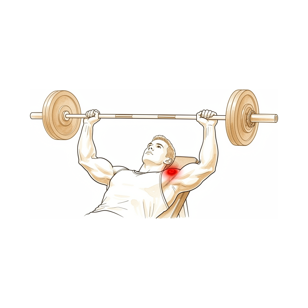
What you're feeling
The pain sits at the outer end of your collarbone, right where it meets the top of your shoulder. That spot is called the acromioclavicular joint. It tends to hurt most when you use the arm, especially with pushing, lifting or pressing movements, and it usually settles when you rest.
Weight training is a common trigger. Bench pressing and other heavy lifting loads this joint directly, and the pain often builds over weeks of training rather than coming on with one clear injury. Some people notice it after a shoulder injury too, when pain lingers long after the rest of the shoulder has settled.
The ache can flare at night or after a session in the gym. Reaching across your body, pressing up from a chair, carrying shopping on that side or pushing a lawnmower can all aggravate it. Because the soreness sits at the very top of the shoulder, it is easy to mistake for a muscle strain at first.
This condition can look a lot like other shoulder problems, so scans are used to confirm what is going on. A special X-ray view of the joint, taken on both sides for comparison, and an MRI scan can show changes at the end of the collarbone.
Watch for these signs and act on them:
Go to the emergency department the same day if your arm becomes hot, red, swollen and painful, especially with a fever. Go the same day if you lose feeling or movement in the arm suddenly after an injury.
See your GP or ask for a specialist review if the pain is not settling, is getting worse over weeks, wakes you at night, or stops you working or using your arm. If you cannot reach the clinic, out of hours or on a weekend, go to your nearest emergency department.
What's actually happening
The acromioclavicular joint is a small joint at the top of your shoulder, where the outer end of your collarbone meets a shelf of bone called the acromion. The collarbone acts as the strut that holds your arm away from your chest, and this little joint is one of the places where it connects to the shoulder blade. A tough strap of ligaments underneath keeps the two bones lined up while you lift and press.
In this condition, the outer end of the collarbone starts to break down. The medical word for bone breaking down like this is osteolysis. The body normally replaces worn bone with fresh bone, but here the breakdown outpaces the repair, so the bone at the end of the collarbone softens and erodes. The joint above it stays sound; the trouble is confined to the collarbone side.
The most common trigger is repeated heavy loading, especially bench pressing and weight training. Each press drives the two bones of the joint into each other, a bit like repeatedly stamping on the same small patch of ground until it wears away. The same process can follow a shoulder injury, where pain lingers for weeks to months after the original knock. Less often, a low-grade infection or a metal plate used to hold the collarbone down after a dislocation can set it off.
As the bone erodes, the joint becomes inflamed and swollen, which is why the ache builds over weeks of training and flares at night. The eroded end also loses its smooth fit against the acromion, so pressing, lifting and reaching across your body grind on raw bone. That is the pain you feel at the very top of your shoulder.
What we can do about it
Dr Kieran Hirpara, an upper-limb surgeon at Mater Private Hospital Rockhampton, starts with the least invasive options that suit your condition. Patients are generally referred to our clinic by their GP; if a physiotherapist has suggested you see us, you will still need a referral from your GP in order to be eligible for the Medicare rebate. Once we have confirmed the diagnosis with your history, an examination and scans, we work through the treatments in order, starting with the simplest.
The first step is changing how you load the joint. Because repeated heavy pressing is what wears the bone down, easing off or swapping the exercises that flare it gives the bone a chance to settle. Physiotherapy aims to settle the inflammation and keep your shoulder strong and moving while you do this. For athletes in season, this approach lets you keep playing while the treatment works. How long this takes depends on whether you can genuinely change the way you train, and we will talk through what that means for your sport or work.
Medication is the next layer. Anti-inflammatory tablets, sometimes called NSAIDs, reduce the swelling and ache in the joint. We can also inject a combination of numbing medicine and cortisone, a strong anti-inflammatory, directly into the joint. This injection does two jobs: it confirms the joint is the source of your pain, and it can settle that pain for a period afterwards.
If these steps have not given you enough relief, we may talk about surgery. The operation removes a small sliver of bone from the outer end of your collarbone, so the raw, eroded surfaces no longer grind against each other. Only a few millimetres of bone are taken, and if a bone spur is pressing on a nearby tendon we tidy that at the same time. We will go through what the operation involves, what recovery looks like and what it can and cannot change, so you can decide together with us whether it is right for you.
What to expect
For most people, this condition settles once the loading that caused it stops. The bone at the end of your collarbone can rebuild itself when the repeated pressing and lifting eases off. That is the honest catch: the treatment works when you can genuinely change how you train or work. If you keep loading the joint the same way, the pain tends to persist or keep coming back.
Recovery without an operation is not a quick fix. The ache builds over weeks, and it usually takes weeks to months of easing off before it settles. Some people notice the pain after a shoulder injury instead, and in that case it can linger long after the rest of the shoulder has healed. If the pain is still there after a reasonable stretch of rest and changed training, that is worth having checked rather than pushing through.
If non-operative care does not give you enough relief, surgery is an option. The operation removes a small sliver of bone from the outer end of your collarbone so the raw surfaces stop grinding. People who have this done report less pain and better shoulder movement, and they are generally satisfied with the result. The keyhole version tends to leave less lingering pain than the open version, and people get back to their activities sooner with it.
Surgery is not a promise of a pain-free shoulder. A small number of people need another operation later, somewhere between 4% and 10%. That can happen if too little or too much bone was taken, if there was another problem in the shoulder that went unnoticed, or if new bone forms in the joint as it heals. Your surgeon will talk through these risks with you before you decide anything.
Whatever path you take, the aim is the same: settle the inflammation, restore smooth movement, and get you back to using your arm without that sharp ache at the top of your shoulder. If your symptoms are not settling, are getting worse over weeks, wake you at night, or stop you working or using your arm, see your GP or ask for a specialist review.
When to see someone
This condition is easy to mistake for a muscle strain, so it often goes unnoticed at first. See your GP if pain at the outer end of your collarbone lingers after a shoulder injury, once a broken bone or an unstable joint has been ruled out. Ask for a specialist review if the ache keeps building with training, or if it is not settling despite rest and changed loading. Scans can confirm the diagnosis: a special X-ray view taken of both shoulders for comparison, and an MRI that shows swelling deep in the collarbone end. Getting this right matters, because the same pain can come from other shoulder problems, and treatment aimed at the wrong cause will not help. If your symptoms are not settling, are getting worse over weeks, wake you at night, or stop you working or using your arm, see your GP or ask for a specialist review. If you cannot reach the clinic, out of hours or on a weekend, go to your nearest emergency department.
In more depth
Advanced reading: the deeper science (optional)
This section goes further than you need for your own treatment decisions. Distal clavicle osteolysis is worth the extra reading because the cause is nearly always identifiable, most people recover without an operation, and the difficult part is whether the loading that caused it can actually be changed.
The load is usually identifiable, and it is usually the bench press
A 2026 scoping review pooled 8 studies covering 483 patients and found bench pressing was the single most common provoking activity at 49.1%, with general weight training a further 24.4% [1]. Pain at the outer end of the collarbone or the acromioclavicular joint was the presenting symptom in 69.9% of patients [1]. The series that first defined the condition found the same pattern: of 46 men diagnosed with osteolysis and no history of acute injury, 45 lifted weights as part of their training [2].
Most settle without surgery, but only if the loading actually changes
In the pooled review most patients recovered with conservative care, and 16% went on to surgery after conservative treatment failed [1]. A smaller group, 4.4% (3 patients), came to surgery because the disease progressed structurally or because they were unwilling to modify their load for sporting or occupational reasons [1]. That last group is the honest version of the problem: the non-operative treatment works when the provoking activity genuinely stops, and the difficulty is rarely in making the diagnosis.
When resection is done, little bone is removed and recovery is quick
In a series of weight lifters, a limited arthroscopic resection averaging 4.5 mm allowed return to sport at an average of 3.2 days and to the preoperative weight-training programme at 9.1 days, with all patients continuing to train and increasing their loads afterwards [3]. A modern prospective series of 59 patients followed for at least two years reported pain falling from 8.20 to 1.36 out of 10 and SPADI disability from 62.65 to 6.13 by 24 months, with return to sport at 1.72 months and to work at 3.02 months [4]. In the original series, resection was performed in 21 patients and relieved symptoms in all 19 of those who were followed [2].
Imaging confirms it; the clinical picture alone does not
The review's own conclusion is that the clinical signs and symptoms of this condition were not distinguishable from other shoulder pathologies [1]. MRI was the most frequently used investigation, showing marrow oedema and subchondral fracture at the end of the collarbone, and the authors are explicit that it should be interpreted alongside the examination rather than in place of it [1]. In practice that is why the history — what you lift, how often, and how heavy — carries as much weight as the scan.
References for the advanced reading
- Wilkinson M, Groch N, Freestone C, et al. Risk factors and management of atraumatic distal clavicular osteolysis: a scoping review. Shoulder Elbow. 2026.
- Cahill BR. Osteolysis of the distal part of the clavicle in male athletes. J Bone Joint Surg Am. 1982;64(7):1053-58.
- Auge WK, Fischer RA. Arthroscopic distal clavicle resection for isolated atraumatic osteolysis in weight lifters. Am J Sports Med. 1998;26(2):189-92.
- Leon JV, Hermans D, Venkatesha V, et al. Patient outcomes following arthroscopic distal clavicle excision: a prospective case series. JSES Int. 2023;7(6):2400-05.
Evidence & references
This is the clinical evidence summary written for health professionals. It is technical, and it lists the research this page was built from. You do not need to read it to understand your treatment or to make a decision about it.
Overview
Clinical Presentation and Diagnosis
- Clinical signs and symptoms for atraumatic distal clavicular osteolysis are not distinguishable from other shoulder pathologies [1].
- Post-traumatic osteolysis of the distal clavicle should be considered if there is persistent pain after a shoulder injury and bony lesions or instabilities have been excluded [3].
- Osteolysis of the distal end of the clavicle is a rare condition that can occur during judo [5].
- Special care must be taken to properly identify the acromioclavicular joint and rule out pre-existing distal clavicle stress fracture or osteolysis before performing the arthroscopic Mumford procedure [22].
Etiology and Pathogenesis
- Propionibacterium acnes–mediated distal clavicular osteolysis is a documented etiology [2].
- Heterotopic bone formation is a more common cause of failure of acromioplasty or distal claviculectomy than is acknowledged by the literature [13].
- Subacromial osteolysis can occur following hook plate fixation for acromioclavicular dislocation [4].
- The risk of subacromial osteolysis following hook plate fixation is minimized by removing the implant within 5.5 months and preventing severe osteolysis by removal no more than 11.9 months after surgery [15].
- Maintaining the acromion-hook angle at 10 degrees or less is recommended to minimize the risk of subacromial osteolysis following hook plate fixation [15].
Surgical Management
- The combination of distal clavicle resection and antibiotics halted osteolysis in a patient with Propionibacterium acnes–mediated disease, who remained symptom-free at 10 months after surgery [2].
- Arthroscopic distal clavicle excision results in statistically and clinically significant improvements in range of motion and patient-reported outcome measures in carefully selected patients with isolated acromioclavicular joint pathology [7].
- Arthroscopic and open distal clavicle excisions both provide significant pain reduction at 1 year with no significant difference in outcome measures between groups, except for VAS pain score improvement [9].
- Patients undergoing arthroscopic distal clavicle excision for acromioclavicular joint pathology can expect a faster return to activities while obtaining similar long-term outcomes compared with the open procedure [11].
- Limited distal clavicle excision of patients with acromioclavicular joint osteoarthritis resistant to conservative treatment reduced pain and improved shoulder function at midterm follow-up [14].
- Open and arthroscopic distal clavicle excision are both effective surgeries to treat recalcitrant acromioclavicular joint pain, providing similarly good to excellent results regarding patient satisfaction and shoulder function at intermediate-term follow-up [32].
- Less residual pain was found using the arthroscopic technique compared to the open technique for distal clavicle excision [32].
- Both the direct superior approach and the indirect subacromial approach to arthroscopic distal clavicle resection result in successful clinical outcome with clinically insignificant difference at final follow-up [24].
- A well-performed distal clavicle excision will likely perform better than a poorly performed one, regardless of whether an open or arthroscopic approach is chosen [21].
- Portal placement remains paramount in both facilitating surgery and avoiding injury to adjacent extra-articular structures regardless of the technique chosen for distal clavicle resection [19].
- Intraoperative use of ultrasound and cannulated dilators allows surgeons to perform distal clavicle excisions in a more efficient, reproducible, and safer manner [33].
- The supraspinatus fossa portal technique provides a technical option for distal clavicle resection when the patient is in the lateral decubitus position, allowing convenient removal of the posterior edge of the distal clavicle to prevent impingement [38].
- Coracoclavicular ligament reconstruction is an effective surgical approach for decreasing the incidence of subacromial osteolysis following hook plate fixation [4].
Surgical Technique and Biomechanics
- A 5 mm resection of the distal clavicle guaranteed no bone-to-bone abutment in a cadaver model, whereas excision with 2.5 mm of bone was successful in many specimens [16].
- Surgery for osteolysis of the distal clavicle progressing to osteoarthritic change in the acromioclavicular joint may involve resection of the distal clavicle and capsule, transfer of the acromial attachment, and repair of the supraspinatus tendon [10].
Indications and Contraindications
- Routine distal clavicle resection in the setting of rotator cuff repair does not result in improved outcomes for patients with no difference being observed at 24 months post surgery [23].
- Distal clavicle resection in patients with rotator cuff tears did not result in better clinical outcome scores or shoulder range of motion and was not associated with a lower risk of reoperation [26].
- Routine distal clavicle excision is not absolutely necessary, even in patients with symptomatic acromioclavicular joint osteoarthritis [29].
Anatomy & Pathophysiology
Bony Anatomy
- The clavicle is the only long bone to ossify by intramembranous ossification [39].
- The clavicle serves as the primary stabilizer between the axial skeleton via the sternoclavicular joint and the appendicular skeleton via the acromioclavicular joint [39].
- The clavicle forms a unique S-shaped curve on the axial view [39].
- The distal clavicle is flat in the AP plane [39].
- The primary blood supply to the clavicle is periosteal, with no nutrient blood supply [39].
- The clavicle is subcutaneous, and its muscular envelope includes the platysma, pectoralis major, deltoid, and some of the strap muscles of the neck [39].
- In the transverse plane, the clavicle resembles an italic S [52].
- The greater radius of curvature of the clavicle occurs at its medial curve, which is anteriorly convex [52].
- The smaller lateral curve of the clavicle is posteriorly convex [52].
- The bone is somewhat rounded in its midsection and medially and relatively flat laterally [52].
- The medial end of the clavicle has a 30% incidence of a rhomboid fossa on its inferior surface where the costoclavicular ligaments insert [52].
- The medial end of the clavicle has a 2.5% incidence of actual articular surface facing inferiorly toward the first rib [52].
- The middle portion of the clavicle contains the subclavian groove where the subclavius muscle has a fleshy insertion [52].
- The lateral portion of the clavicle has the coracoclavicular process when present [52].
- The conoid ligament attaches to the clavicle at the conoid tubercle [52].
- The trapezoid ligament attaches at the trapezoid line, which lies in an anteroposterior direction just lateral to the conoid tubercle [52].
- The distance from the lateral edge of the clavicle to the medial edge of the conoid tubercle is approximately 45 mm in male and female specimens [52].
- The distance from the lateral edge of the clavicle to the center of the trapezoid tuberosity is approximately 25 mm in male and female specimens [52].
- The trapezius inserts on the posterosuperior surface of the distal end of the clavicle [52].
- The subclavius muscle has a fleshy insertion on the inferior surface of the middle third of the clavicle [52].
- The deltoid originates on the anterior portion of the inner surface of the lateral curve of the clavicle [52].
- The pectoralis major originates from the anterior portion of the medial two-thirds of the clavicle [52].
- The sternocleidomastoid largely originates on the posterior portion of the middle third of the clavicle [52].
- The sternohyoid originates on the clavicle just medial to the origin of the sternocleidomastoid [52].
- The subclavian vein and artery and brachial plexus lie posterior to the clavicle [52].
- The medial anterior curve of the clavicle is often described as an accommodation for the subclavian vein and artery and brachial plexus [52].
- The clavicle is one of the first bones to ossify, beginning from two primary ossification centers (medial and lateral) by 5 to 6 weeks of gestation [51].
- By 7 to 8 weeks of gestation, the clavicle has already assumed its overall contour and “S” shape [51].
- Most growth (80%) of the clavicle occurs from the medial physis [51].
- The lateral epiphysis of the clavicle forms and fuses at around 18 to 19 years of age [51].
- The medial epiphysis of the clavicle is the last in the body to ossify, at the age of 18 to 20 years [51].
- The medial epiphysis of the clavicle is the last to complete ossification, at the age of 23 to 25 years [51].
Ligaments and Soft Tissue Stabilizers
- The coracoclavicular ligaments consist of the conoid (medial) and trapezoid (lateral) components [39].
- The coracoclavicular ligaments are the primary stabilizers to superior (vertical) translation of the distal clavicle [39].
- The superior shoulder suspensory complex is a bone–soft-tissue ring that provides a stable connection of the glenoid and scapula to the clavicle [39].
- The superior shoulder suspensory complex is composed of four bony landmarks: distal clavicle, acromion, coracoid process, and glenoid neck [39].
- The superior shoulder suspensory complex includes the supporting ligamentous complexes of the AC joint and the CC ligaments [39].
- The acromion has three ossification centers: the meta-acromion (base), the mesoacromion (mid), and the preacromion (tip) [50].
- The coracoid process is the site of ligament attachments for the CC ligaments [50].
- The superior shoulder suspensory complex plays an important role in shoulder stability and biomechanics [50].
- The pectoralis major muscle originates from the clavicular shaft anteroinferiorly [56].
- The sternocleidomastoid originates superiorly on the clavicle [56].
- The pectoralis origin merges with the origin of the anterior deltoid laterally [56].
- The trapezius insertion blends superiorly with the deltoid origin at the lateral margin of the clavicle [56].
- The subclavius muscle inserts on the undersurface of the clavicle [56].
- The platysma usually envelopes the anterior and superior aspects of the clavicle and runs in the subcutaneous tissues [56].
- The supraclavicular nerves originate from cervical roots C3 and C4 [58].
- The supraclavicular nerves exit from a common trunk behind the posterior border of the sternocleidomastoid muscle [58].
- There are typically three major branches of the supraclavicular nerves (anterior, middle, and posterior) that cross the clavicle superficially from medial to lateral [58].
- The subclavian vein runs directly below the subclavius muscle and above the first rib [58].
- The subclavian artery and the brachial plexus lie more posteriorly than the subclavian vein, separated from the vein and clavicle by the scalenus anterior muscle medially [58].
- The brachial plexus is closest to the clavicle in its midportion [58].
- In the middle third of the clavicle, the subclavian artery is a mean of 17 mm from the clavicle [58].
- In the middle third of the clavicle, the subclavian vein is a mean of 13 mm from the clavicle [58].
- In the middle third of the clavicle, the subclavian vessels are located at an approximate angle of 60 degrees to the horizontal [58].
- Laterally, the subclavian artery is a mean of 63 mm from the clavicle [58].
- Laterally, the subclavian vein is a mean of 76 mm from the clavicle [58].
Pathophysiology
- Clinical signs and symptoms for atraumatic distal clavicular osteolysis were not distinguishable from other shoulder pathologies [1].
- Post-traumatic osteolysis of the distal clavicle occurs from several weeks to several months after injury to the acromioclavicular joint [17].
- In post-traumatic osteolysis, radiographs show no luxation or fracture initially [17].
- Shoulder pain in post-traumatic osteolysis develops gradually with swelling of the acromioclavicular joint and limitation of shoulder motion [17].
- The process of osteolysis in post-traumatic distal clavicle osteolysis occurs only in the distal clavicle, and there is no osteolytic lesion in the acromion [17].
- Post-traumatic osteolysis of the distal clavicle was first described by Werder in 1950 [17].
- More than 100 cases of post-traumatic osteolysis have been reported [17].
- The pathogenesis of post-traumatic osteolysis remains unclear [17].
- Osteoclastic resorption has been hypothesized as a pathogenesis for post-traumatic osteolysis [17].
- Nervous system dysfunction has been hypothesized as a pathogenesis for post-traumatic osteolysis [17].
- Avascular necrosis has been hypothesized as a pathogenesis for post-traumatic osteolysis [17].
- Microfracture has been hypothesized as a pathogenesis for post-traumatic osteolysis [17].
- Synovial invasion has been hypothesized as a pathogenesis for post-traumatic osteolysis [17].
- Non-traumatic osteolysis of the acromial end of the clavicle should be considered in cases of pain in the shoulder in athletes [6].
- It is important to consider the possibility of post-traumatic osteolysis of the distal clavicle if there is persistent pain after a shoulder injury, and bony lesions or instabilities have been excluded [3].
- The combination of distal clavicle resection and antibiotics halted the osteolysis in a case of Propionibacterium acnes–mediated distal clavicular osteolysis [2].
- The patient with Propionibacterium acnes–mediated distal clavicular osteolysis remained symptom free at 10 months after surgery [2].
- For all patients with atraumatic osteolysis of the distal clavicle, return to sports activities was dependent on changes in the way of training [8].
- Distal clavicle excision is a widely accepted surgical treatment for symptomatic acromioclavicular joint pathology, including osteoarthritis, post-traumatic degeneration, and osteolysis [47].
- Mechanisms of failure following distal clavicle excision include inadequate resection [47].
- Mechanisms of failure following distal clavicle excision include excessive bone removal resulting in instability [47].
- Mechanisms of failure following distal clavicle excision include unrecognized concomitant pathology [47].
- Mechanisms of failure following distal clavicle excision include iatrogenic disruption of the coracoclavicular ligaments [47].
- The reported incidence of reoperation following distal clavicle excision ranges from 4% to 10% [47].
- A 5-mm distal clavicle resection guaranteed no abutment but decreased joint stiffness [35].
- If the resection was 2.5 mm or less, abutment was likely to occur between the acromion and the clavicle, particularly inferiorly and posteriorly [53].
- Joint stiffness diminished with the amount of bone resected [53].
- Resection of more than 7.6 mm of the distal clavicle in men can violate the superior AC ligament [53].
- Resection of more than 5.2 mm of the distal clavicle in women can violate the superior AC ligament [53].
- Resection of less than 11.0 mm should never violate any portion of the trapezoid ligament in 98% of men or women [53].
- Resection of less than 24.0 mm should never violate any of the conoid ligament [53].
- Patients with a resection of more than 1.0 cm have more pain than patients with less than 1 cm of resection [53].
- The anterior-posterior load to clinical failure of the AC joint after 5 mm of resection from the distal clavicle and medial acromion is significantly greater than 1 cm of the resected distal clavicle alone [77].
- Intact acromioclavicular ligaments protect the coracoclavicular reconstruction by decreasing the in situ graft force [75].
- Inaccurate resection from lack of depth perception and inadequate visualization has been reported to lead to failure and revision surgery [28].
Classification
- Distal clavicular osteolysis presents as isolated pain at the distal clavicle and acromioclavicular joint that tends to worsen with activity [27].
- There are two forms of distal clavicular osteolysis: posttraumatic distal clavicular osteolysis caused by a direct traumatic injury, and atraumatic distal clavicular osteolysis attributable to repetitive stress [27].
- In adults, the most common risk factor for stress-induced distal clavicular osteolysis is weightlifting [27].
- Post-traumatic osteolysis of the distal clavicle is an uncommon condition that occurs more often than is generally recognized, with many cases potentially overlooked or misinterpreted [17].
- In post-traumatic osteolysis, radiographs initially show no luxation or fracture, followed by gradual development of shoulder pain, swelling of the acromioclavicular joint, and limitation of shoulder motion [17].
- The process of post-traumatic osteolysis occurs only in the distal clavicle, with no osteolytic lesion in the acromion [17].
- Hypothesized pathogenesis for post-traumatic osteolysis includes osteoclastic resorption, nervous system dysfunction, avascular necrosis, microfracture, and synovial invasion [17].
- Propionibacterium acnes–mediated distal clavicular osteolysis is a recognized etiology for the condition [2].
- Aseptic foreign-body reaction to Gore-Tex grafts used for clavicle-to-coracoid stabilization can result in clavicle osteolysis [20].
- Prolonged implant retention and higher-grade fracture types significantly increase the risk of subacromial osteolysis following hook plate fixation [78].
Clinical Presentation
Symptoms and Signs
- Patients with distal clavicular osteolysis present with isolated pain at the distal clavicle and acromioclavicular joint that tends to worsen with activity [27, 65].
- Post-traumatic osteolysis of the distal clavicle is characterized by shoulder pain that develops gradually with swelling of the acromioclavicular joint and limitation of shoulder motion [17].
- In post-traumatic osteolysis, radiographs initially show no luxation or fracture [17].
- Persistent pain after a shoulder injury, after bony lesions or instabilities have been excluded, should prompt consideration of post-traumatic osteolysis of the distal clavicle [3].
- Non-traumatic osteolysis of the acromial end of the clavicle should be considered in cases of shoulder pain in athletes [6].
- Osteolysis of the distal end of the clavicle can occur during judo [5].
- A 58-year-old male carpenter presented with osteolysis of the distal clavicle progressing to osteoarthritic change in the acromioclavicular joint [10].
Imaging Findings
- Radiographic findings for distal clavicular osteolysis may range from mild osteopenia to overt osteolysis [27, 65].
- The bilateral Zanca view is helpful for diagnosing distal clavicular osteolysis as it profiles the acromioclavicular joint and allows for comparison with the contralateral side [27, 65].
- MRI characteristics of distal clavicular osteolysis include distal clavicular bone marrow edema that is out of proportion to the edema at the acromion [27, 65].
- MRI characteristics of distal clavicular osteolysis include subchondral cystic change [27, 65].
- In advanced cases of distal clavicular osteolysis, periostitis may be observed at the distal clavicle on MRI [27, 65].
- The process of osteolysis in post-traumatic cases occurs only in the distal clavicle, with no osteolytic lesion in the acromion [17].
Risk Factors and Etiology
- Posttraumatic distal clavicular osteolysis is caused by a direct traumatic injury [27, 65].
- Atraumatic distal clavicular osteolysis is attributable to repetitive stress [27, 65].
- Post-traumatic osteolysis of the distal clavicle is an uncommon condition that occurs more often than generally recognized, with many cases potentially overlooked or misinterpreted [17].
- More than 100 cases of post-traumatic osteolysis have been reported since Werder first described the condition in 1950 [17].
- Propionibacterium acnes infection has been identified as a cause of distal clavicular osteolysis [2].
- Aseptic foreign-body reaction to Gore-Tex grafts can cause clavicle osteolysis [20].
- Bone osteolysis can occur following acromioclavicular joint reconstruction using synthetic ligament (Surgilig™) [31].
Investigations
Clinical Presentation and Diagnosis
- In post-traumatic osteolysis, radiographs typically show no luxation or fracture initially [17].
- The process of osteolysis in post-traumatic distal clavicle injury occurs only in the distal clavicle, with no osteolytic lesion in the acromion [17].
- A high index of suspicion is needed to diagnose complications such as osteolysis early before irretrievable bone loss [31].
Imaging and Anatomical Landmarks
- Special care must be taken to properly identify the AC joint and rule out pre-existing distal clavicle stress fracture or osteolysis before performing the arthroscopic Mumford procedure [22].
- The cross-sectional A-frame morphology of the superior cortex of the distal clavicle provides a reproducible landmark that is eliminated approximately 1.0 cm medial to the distal, lateral end of the clavicle [49].
- The A-frame morphology of the distal clavicle can be used intraoperatively to determine when adequate resection has been completed [49].
Treatment
Non-Operative
- Activity modification, NSAIDs, and physical therapy are first-line treatments for distal clavicular osteolysis [27].
- Intra-articular injection of lidocaine and corticosteroids into the acromioclavicular joint is both diagnostic and therapeutic for distal clavicular osteolysis [27].
- Nonoperative treatment with injections, medication, and physical therapy is a good option for in-season athletes with distal clavicular osteolysis [27].
- The combination of distal clavicle resection and antibiotics halted osteolysis in a case of Propionibacterium acnes–mediated distal clavicular osteolysis [2].
Operative
- Distal clavicle resection is the definitive operative treatment for distal clavicular osteolysis failing nonoperative management [27].
- Patients undergoing arthroscopic distal clavicle excision through the direct approach can expect a faster return to activities while obtaining similar long-term outcomes compared with the open procedure [11].
- Open and arthroscopic distal clavicle excision provide similarly good to excellent results regarding patient satisfaction and shoulder function at intermediate-term follow-up, though less residual pain was found using the arthroscopic technique [32].
- A 5 mm resection of the distal clavicle guaranteed no bone-to-bone abutment in a cadaver model [16].
- A 5-mm distal clavicle resection guaranteed no abutment but decreased joint stiffness in a cadaveric model [35].
- Anterior acromioplasty is needed in cases with subacromial impingement during distal clavicle resection [18].
- SLAP lesion repair after initial distal clavicle resection successfully treated 15 patients, indicating that associated or underlying superior labral detachment may cause similar symptoms leading to misdiagnosis [25].
- The use of intraoperative ultrasound and cannulated dilators allows surgeons to perform distal clavicle excisions in a more efficient, reproducible, and safer manner [33].
- The introduction of the supraspinatus fossa portal technique provides a special technical option for distal clavicle resection when the patient is in the lateral decubitus position, allowing convenient removal of the posterior edge of the distal clavicle to prevent impingement [38].
- Coracoclavicular ligament reconstruction is an effective surgical approach for decreasing the incidence of subacromial osteolysis following hook plate fixation for acromioclavicular dislocation [4].
- Removing the implant within 5.5 months minimizes osteolysis risk and no more than 11.9 months prevents severe osteolysis, while maintaining the acromion-hook angle at 10 degrees or less [15].
- Foreign-body reaction and osteolysis have been reported with the use of Gore-Tex graft to stabilize the clavicle to the coracoid [20].
Complications
Diagnostic and Pathologic Complications
- Post-traumatic osteolysis of the distal clavicle is an uncommon condition that occurs more often than generally recognized, with many cases overlooked or misinterpreted [17].
- In post-traumatic osteolysis, radiographs initially show no luxation or fracture, and shoulder pain develops gradually with swelling of the acromioclavicular joint and limitation of shoulder motion [17].
- Heterotopic bone formation may be a more common cause of failure of acromioplasty or distal clavicectomy than acknowledged in the literature [30].
- Aseptic foreign-body reaction and clavicle osteolysis can occur following acromioclavicular reconstruction using Gore-Tex graft [20].
- A high index of suspicion is needed to diagnose complications such as osteolysis early before irretrievable bone loss occurs [31].
- Distal clavicle fracture is a complication of arthroscopic distal clavicle resection [22].
- Pre-existing distal clavicle stress fracture or osteolysis must be ruled out before performing the arthroscopic Mumford procedure [22].
Operative Complications and Failure Mechanisms
- Multiple mechanisms of failure following distal clavicle excision include inadequate resection, excessive bone removal resulting in instability, unrecognized concomitant pathology, and iatrogenic disruption of the coracoclavicular ligaments [47].
- A 5 mm resection of the distal clavicle guaranteed no bone-to-bone abutment in a cadaver model, whereas 2.5 mm resection was successful in many specimens [16].
- SLAP lesions can cause continued symptoms and failure of distal clavicle resection due to misdiagnosis of associated or underlying superior labral detachment [25].
- Resection of the lateral end of the clavicle following osteolysis is a described surgical management [6].
- Surgery for osteolysis of the distal clavicle progressing to osteoarthritic change can involve resection of the distal clavicle and capsule, transfer of the acromial attachment, and repair of the supraspinatus tendon [10].
Outcomes and Prognosis
- A subset of patients experience persistent pain, residual instability, or functional limitation following primary distal clavicle excision that ultimately requires additional surgical intervention [47].
- Outcomes following revision surgery for persistent pain and/or instability after distal clavicle excision remain poorly characterized and are primarily composed of small cohort studies [47].
- Return to sports activities for patients with atraumatic osteolysis of the distal clavicle was dependent on changes in the way of training [8].
- In carefully selected patients with isolated acromioclavicular joint pathology, arthroscopic distal clavicle excision results in statistically and clinically significant improvements in range of motion and patient-reported outcome measures [7].
Recovery
Post-operative Outcomes and Return to Activity
- Patients undergoing arthroscopic distal clavicle excision, specifically through the direct approach, can expect a faster return to activities while obtaining similar long-term outcomes compared with the open procedure [11].
Surgical Technique and Biomechanics
Complications and Failure Modes
- The successful treatment of 15 patients by SLAP lesion repair after initial distal clavicle resection indicates that there may be misdiagnosis due to associated or underlying superior labral detachment causing similar symptoms [25].
- A high index of suspicion is needed to diagnose complications such as bone osteolysis following acromioclavicular joint reconstruction using synthetic ligament early before irretrievable bone loss [31].
- The combination of distal clavicle resection and antibiotics halted osteolysis in a case of Propionibacterium acnes–mediated distal clavicular osteolysis, with the patient remaining symptom free at 10 months after surgery [2].
Key Evidence
- [L4] Clinical signs and symptoms for atraumatic distal clavicular osteolysis (ADCO) were not distinguishable from other shoulder pathologies. [1] (10.1177/17585732261479715)
- [Case_report] The combination of distal clavicle resection and antibiotics halted the osteolysis, and the patient has remained symptom free at 10 months after surgery. [2] (10.1016/j.jse.2015.03.004)
- [L4] It is important to consider the possibility of post-traumatic osteolysis of the distal clavicle if there is persistent pain after a shoulder injury, and bony lesions or instabilities have been excluded. [3] (10.1007/bf00573456)
- [L1] The current analysis suggests coracoclavicular ligament reconstruction as an effective surgical approach for decreasing the incidence of subacromial osteolysis. [4] (10.1016/j.jse.2024.03.018)
- [L5] Osteolysis of the distal end of the clavicle is a rare condition and it can occur during judo. [5] (10.1016/s1058-2746(96)80543-x)
- [L4] Non-traumatic osteolysis of the acromial end of the clavicle should be borne in mind in cases of pain in the shoulder in athletes. [6] (10.1016/0020-1383(87)90010-6)
- [L4] In carefully selected patients with isolated ACJ pathology, arthroscopic distal clavicle excision results in statistically and clinically significant improvements in range of motion and patient-reported outcome measures. [7] (10.1016/j.jseint.2023.07.014)
- [L4] For all patients, return to sports activities was dependent on changes in the way of training. [8] (10.1016/s1058-2746(96)80377-6)
- [L1] Arthroscopic and open distal clavicle excisions both provide significant pain reduction at 1 year with no significant difference in outcome measures between groups, except for VAS pain score improvement. [9] (10.1016/j.jse.2006.10.006)
- [L5] A 58-year-old male carpenter with osteolysis of the distal clavicle progressing to osteoarthritic change in the acromioclavicular joint underwent surgery involving resection of the distal clavicle and capsule, transfer of the acromial attachment, and repair of the supraspinatus tendon. [10] (10.1016/s1058-2746(95)80313-0)
- [L3] Among patients undergoing distal clavicle excision for acromioclavicular joint pathology, those having an arthroscopic procedure, specifically through the direct approach, can expect a faster return to activities while obtaining similar long-term outcomes compared with the open procedure. [11] (10.1016/j.arthro.2009.12.007)
- [L4] This retrospective series suggests that heterotopic bone formation is a more common cause of failure of acromioplasty or distal claviculectomy than is acknowledged by the literature. [13] (10.1016/s1058-2746(05)80050-3)
- [L4] Limited distal clavicle excision of patients with AC joint osteoarthritis resistant to conservative treatment reduced pain and improved shoulder function at midterm follow-up. [14] (10.1016/j.otsr.2016.01.008)
- [L3] The authors recommend removing the implant within 5.5 months to minimize osteolysis risk and no more than 11.9 months to prevent severe osteolysis, while maintaining the acromion-hook angle at 10 degrees or less. [15] (10.1016/j.jse.2024.09.027)
- [Abstract] Although distal clavicle excision with 2.5 mm of bone was successful in many specimens, a 5 mm resection guaranteed no bone-to-bone abutment. [16] (10.1016/j.jse.2007.02.105)
- [L5] [17] (10.1067/mse.2002.122229)
- [L4] Additionally, the anterior acromioplasty is needed in the cases with subacromial impingement. [18] (10.1016/s1058-2746(96)80520-9)
- [Case_report] Regardless of the technique chosen for distal clavicle resection, portal placement remains paramount in both facilitating surgery and avoiding injury to adjacent extra-articular structures. [19] (10.1016/j.jse.2010.08.032)
- [L5] The case presented reports foreign-body reaction and osteolysis with the use of Gore-Tex graft to stabilize the clavicle to the coracoid. [20] (10.1016/j.jse.2004.01.027)
- [L5] A well-performed distal clavicle excision will likely perform better than a poorly performed one, regardless of whether an open or arthroscopic approach is chosen. [21] (10.1016/j.arthro.2018.03.004)
- [L4] Special care must be taken to properly identify the AC joint and rule out pre-existing distal clavicle stress fracture or osteolysis before performing the arthroscopic Mumford procedure. [22] (10.1016/j.arthro.2009.02.008)
- [L2] Routine distal clavicle resection in the setting of rotator cuff repair does not result in improved outcomes for patients with no difference being observed at 24 months post surgery. [23] (10.1177/1758573217741124)
- [L2] Both the direct superior approach and the indirect subacromial approach to the arthroscopic distal clavicle resection result in successful clinical outcome with clinically insignificant difference at final follow-up. [24] (10.1177/0363546506294855)
- [L4] The successful treatment of 15 patients by SLAP lesion repair after initial distal clavicle resection indicates that there may be misdiagnosis due to associated or underlying superior labral detachment causing similar symptoms. [25] (10.1016/s1058-2746(96)80319-3)
- [L1] Distal clavicle resection in patients with rotator cuff tears did not result in better clinical outcome scores or shoulder ROM and was not associated with a lower risk of reoperation. [26] (10.1097/corr.0000000000000424)
- [Paper] [28] (10.1016/j.eats.2020.10.010)
- [L2] Routine distal clavicle excision is not absolutely necessary, even in patients with symptomatic ACJ osteoarthritis. [29] (10.1007/s00167-020-06098-y)
- [L4] Heterotopic bone formation may be a more common cause of failure of acromioplasty or distal clavicectomy than acknowledged in the literature. [30] (10.1016/s1058-2746(96)80126-1)
- [L4] A high index of suspicion is needed to diagnose such complications early before irretrievable bone loss to osteolysis. [31] (10.1111/sae.12035)
- [L3] Open and arthroscopic distal clavicle excision are both effective surgeries to treat recalcitrant acromioclavicular joint pain, providing similarly good to excellent results regarding patient satisfaction and shoulder function at intermediate-term follow-up, though less residual pain was found using the arthroscopic technique. [32] (10.1177/0363546511419633)
- [L5] The technique will allow surgeons to perform distal clavicle excisions in a more efficient, reproducible and safer manner. [33] (10.1016/j.eats.2024.103331)
- [L5] A 5-mm distal clavicle resection guaranteed no abutment but decreased joint stiffness. [35] (10.1016/j.arthro.2007.07.004)
- [Paper] The introduction of this technique provides a special technical option for distal clavicle resection when the patient is in the lateral decubitus position, allowing convenient removal of the posterior edge of the distal clavicle to prevent impingement. [38] (10.1016/j.eats.2020.08.040)
- [L4] [47] (10.1016/j.jse.2026.08.016)
- [L5] The cross-sectional A-frame morphology of the superior cortex of the distal clavicle provides a reproducible landmark that is eliminated approximately 1.0 cm medial to the distal, lateral end of the clavicle, which can be used intraoperatively to determine when adequate resection has been completed. [49] (10.1016/j.jse.2021.10.013)
- [L5] Intact acromioclavicular ligaments protect the coracoclavicular reconstruction by decreasing the in situ graft force. [75] (10.1177/0363546510374447)
- [L5] This cadaveric study demonstrates that the anterior-posterior load to clinical failure of the AC joint after 5 mm of resection from the distal clavicle and medial acromion is significantly greater than 1 cm of the resected distal clavicle alone. [77] (10.1177/0363546512469873)
- [L3] Prolonged implant retention and higher-grade fracture types significantly increase the risk of subacromial osteolysis (SAO). [78] (10.1186/s12891-026-09516-3)
References
[1] Risk factors and management of atraumatic distal clavicular osteolysis: A scoping review. Shoulder & Elbow. 2026. DOI: 10.1177/17585732261479715
[2] Propionibacterium acnes–mediated distal clavicular osteolysis: a case report. Journal of Shoulder and Elbow Surgery. 2015. DOI: 10.1016/j.jse.2015.03.004
[3] Post-traumatic osteolysis of the distal clavicle. Archives of Orthopaedic and Trauma Surgery. 1996. DOI: 10.1007/bf00573456
[4] Subacromial osteolysis following hook plate fixation for acromioclavicular dislocation: a systematic review and meta-analysis. Journal of Shoulder and Elbow Surgery. 2024. DOI: 10.1016/j.jse.2024.03.018
[5] Post-traumatic osteolysis of the distal end of the clavicle in judo athlete: Case report. Journal of Shoulder and Elbow Surgery. 1996. DOI: 10.1016/s1058-2746(96)80543-x
[6] Resection of the lateral end of the clavicle following osteolysis, with emphasis on non-traumatic osteolysis of the acromial end of the clavicle in athletes. Injury. 1987. DOI: 10.1016/0020-1383(87)90010-6
[7] Patient outcomes following arthroscopic distal clavicle excision: a prospective case series. JSES International. 2023. DOI: 10.1016/j.jseint.2023.07.014
[8] Atraumatic osteolysis of the distal clavicle. Journal of Shoulder and Elbow Surgery. 1996. DOI: 10.1016/s1058-2746(96)80377-6
[9] Arthroscopic versus open distal clavicle excision: Comparative results at six months and one year from a randomized, prospective clinical trial. Journal of Shoulder and Elbow Surgery. 2007. DOI: 10.1016/j.jse.2006.10.006
[10] Osteolysis of the distal end of the clavicle: A case report. Journal of Shoulder and Elbow Surgery. 1995. DOI: 10.1016/s1058-2746(95)80313-0
[11] Open Versus Arthroscopic Distal Clavicle Resection. Arthroscopy. 2010. DOI: 10.1016/j.arthro.2009.12.007
[13] Heterotopic ossification after acromioplasty and distal clavicle resection. Journal of Shoulder and Elbow Surgery. 1995. DOI: 10.1016/s1058-2746(05)80050-3
[14] Limited distal clavicle excision of acromioclavicular joint osteoarthritis. Orthopaedics & Traumatology: Surgery & Research. 2016. DOI: 10.1016/j.otsr.2016.01.008
[15] Predictive value of implant removal time and acromion-hook angle on subacromial osteolysis following hook plate fixation for acromioclavicular dislocation. Journal of Shoulder and Elbow Surgery. 2025. DOI: 10.1016/j.jse.2024.09.027
[16] Arthroscopic Distal Clavicle Resection: A Biomechanical Analysis In A Cadaver Model. Journal of Shoulder and Elbow Surgery. 2007. DOI: 10.1016/j.jse.2007.02.105
[17] A case of post-traumatic osteolysis of the distal clavicle: Histologic lesion of the acromion. Journal of Shoulder and Elbow Surgery. 2002. DOI: 10.1067/mse.2002.122229
[18] The usefulness of distal clavicle resection for acromioclavicular joint disease. Journal of Shoulder and Elbow Surgery. 1996. DOI: 10.1016/s1058-2746(96)80520-9
[19] Acromioclavicular dislocation after arthroscopic distal clavicle resection: a case report. Journal of Shoulder and Elbow Surgery. 2011. DOI: 10.1016/j.jse.2010.08.032
[20] Failure of acromioclavicular reconstruction using Gore-Tex graft due to aseptic foreign-body reaction and clavicle osteolysis: A case report. Journal of Shoulder and Elbow Surgery. 2004. DOI: 10.1016/j.jse.2004.01.027
[21] Editorial Commentary: The “Mumford” & Sons: For Distal Clavicle Excisions, What Are Our Young Surgeons Doing, and How Well Are They Doing It?. Arthroscopy. 2018. DOI: 10.1016/j.arthro.2018.03.004
[22] Distal Clavicle Fracture as a Complication of Arthroscopic Distal Clavicle Resection. Arthroscopy. 2009. DOI: 10.1016/j.arthro.2009.02.008
[23] Is routine distal clavicle resection necessary in rotator cuff repair surgery? A systematic review and meta-analysis. Shoulder & Elbow. 2017. DOI: 10.1177/1758573217741124
[24] Arthroscopic Distal Clavicle Resection in Athletes. The American Journal of Sports Medicine. 2007. DOI: 10.1177/0363546506294855
[25] The slap lesion as a cause of continued symptoms and failure of distal clavicle resection. Journal of Shoulder and Elbow Surgery. 1996. DOI: 10.1016/s1058-2746(96)80319-3
[26] Does Distal Clavicle Resection Decrease Pain or Improve Shoulder Function in Patients With Acromioclavicular Joint Arthritis and Rotator Cuff Tears? A Meta-analysis. Clinical Orthopaedics & Related Research. 2018. DOI: 10.1097/corr.0000000000000424
[27] Arthroscopic Treatment of Acromioclavicular Joint Osteolysis, Arthritis, and Instability. 2016.
[28] Distal Clavicle Excision for Acromioclavicular Joint Osteoarthritis Using a Fluoroscopic Kirschner Wire Guide. Arthroscopy Techniques. 2021. DOI: 10.1016/j.eats.2020.10.010
[29] Acromioclavicular joint arthritis is not an indication for routine distal clavicle excision in arthroscopic rotator cuff repair. Knee Surgery, Sports Traumatology, Arthroscopy. 2020. DOI: 10.1007/s00167-020-06098-y
[30] Heterotopic ossification after acromioplasty and distal clavicle resection. Journal of Shoulder and Elbow Surgery. 1996. DOI: 10.1016/s1058-2746(96)80126-1
[31] Bone Osteolysis Following Acromioclavicular Joint Reconstruction Using Synthetic Ligament (Surgilig™). Shoulder & Elbow. 2014. DOI: 10.1111/sae.12035
[32] Arthroscopic Versus Open Distal Clavicle Excision. The American Journal of Sports Medicine. 2011. DOI: 10.1177/0363546511419633
[33] Intraoperative Use of Ultrasound and Cannulated Dilators to Safely Identify and Access the Acromioclavicular Joint for Distal Clavicle Excision: A Technique Guide. Arthroscopy Techniques. 2024. DOI: 10.1016/j.eats.2024.103331
[35] Arthroscopic Distal Clavicle Resection: A Biomechanical Analysis of Resection Length and Joint Compliance in a Cadaveric Model. Arthroscopy. 2007. DOI: 10.1016/j.arthro.2007.07.004
[38] Arthroscopic Distal Clavicle Resection Through the Supraspinatus Fossa Portal. Arthroscopy Techniques. 2020. DOI: 10.1016/j.eats.2020.08.040
[39] Aaos Comprehensive Orthopaedic Review 3. Fractures of the Clavicle, Scapula, and Glenoid > I. Clavicular Fractures.
[47] Clinical Outcomes and Predictors of Dissatisfaction Following Revision Surgery After Distal Clavicle Excision. Journal of Shoulder and Elbow Surgery. 2026. DOI: 10.1016/j.jse.2026.08.016
[49] Distal clavicle “A-frame” morphology: a reliable intraoperative guide for arthroscopic distal clavicle excision. Journal of Shoulder and Elbow Surgery. 2022. DOI: 10.1016/j.jse.2021.10.013
[50] Aaos Comprehensive Orthopaedic Review 3. Fractures of the Clavicle, Scapula, and Glenoid > II. Scapular and Glenoid Fractures.
[51] Rockwood And Matsen S The Shoulder. Fractures, Dislocations, and Acquired Problems of the Shoulder in Children > FRACTURES OF THE CLAVICLE.
[52] Rockwood And Matsen S The Shoulder. Developmental Anatomy of the Shoulder and Anatomy of the Glenohumeral Joint > Clavicle.
[53] Rockwood And Matsen S The Shoulder. Shoulder and Elbow Specialty Clinic Workers’ Survey > Neurovascular Structures > EXCISION OF THE DISTAL CLAVICLE.
[56] Rockwood And Green S Fractures In Adults. 29: Principles of Nonunion and Bone Defect Treatment > Muscular Anatomy of the Clavicle.
[58] Rockwood And Green S Fractures In Adults. 29: Principles of Nonunion and Bone Defect Treatment > Neurovascular Anatomy of the Clavicle.
[65] Rockwood And Matsen S The Shoulder. Arthroscopic Treatment of Acromioclavicular Joint Osteolysis, Arthritis, and Instability > Distal Clavicle Osteolysis > Indications.
[75] The Effect of Distal Clavicle Excision on in Situ Graft Forces in Coracoclavicular Ligament Reconstruction. The American Journal of Sports Medicine. 2010. DOI: 10.1177/0363546510374447
[77] The Biomechanical Stability of Distal Clavicle Excision Versus Symmetric Acromioclavicular Joint Resection. The American Journal of Sports Medicine. 2012. DOI: 10.1177/0363546512469873
[78] What is the optimal implant removal timing following hook plate fixation in distal clavicle fractures: a retrospective analysis from subacromial osteolysis cases. BMC Musculoskeletal Disorders. 2026. DOI: 10.1186/s12891-026-09516-3