Clinicians › Shoulder
Rotator cuff repair
Surgeon-side topic for rotator cuff repair. Backed by 410 articles from the corpus, retrieved via combined MeSH + title-text matching.
For patients: a plain-language version of this topic is available. See the patient guide.
Overview
Rotator cuff repair has evolved into one of the most common orthopaedic surgical procedures worldwide, yet significant work remains to improve healing rates, outcomes, and long-term durability [22]. The primary indication for revision surgery is the persistence of clinical symptoms despite nonsurgical management in the absence of substantial risk factors for failure [2, 4]. While open rotator cuff repair remains a valid option in specific indications or settings with limited arthroscopic resources [58], there is no evidence of difference in effectiveness between open and arthroscopic repair of rotator cuff tears [208]. Shoulder surgeons must carefully interpret literature comparing these approaches, as differences in complications may be influenced by selection bias and narrowing indications for open repair [64]. Patient characteristics and indications for surgery were not described in a majority of clinical outcome studies [1].
Short-term clinical outcomes of patients undergoing revision rotator cuff repair were similar to those after primary rotator cuff repair [6, 8, 9]. However, prevention of complications and successful primary rotator cuff repair results in better functional outcomes despite the availability of revision strategies and treatment options [7]. Repeat cuff repair can improve pain and function when indicated, but outcomes are poorer than primary procedures if criteria such as small retear, good tissue quality, and no osteoarthritis are not met [71]. Rotator cuff repair has a low incidence of short-term complications [10]. At long-term follow-up (≥15 years), patient-reported outcomes of all-arthroscopic rotator cuff repair show significant improvement from baseline preoperative function and remain durable over a period of 15 years [5]. One-year follow-up is not the last word for rotator cuff repair outcomes; patients must live with the long-term outcomes of surgical procedures, and authors are obliged to evaluate these long-term outcomes [70]. The widely accepted 2-year benchmark for rotator cuff repair outcomes may be arbitrary and does not add clinical relevance, as 1-year follow-up appears clinically similar to 2-year outcomes [190].
Higher values for American Shoulder and Elbow Surgeons, Simple Shoulder Test, and visual analog scale pain scores should be considered when evaluating improvements of individual patients after rotator cuff repair, determining comparative effectiveness of various techniques, and determining sample sizes for prospective comparative trials [57]. It was not possible to reach any definitive conclusion regarding the most relevant predictors of outcome of rotator cuff repair due to low methodological quality of included studies [21]. There is a need for well-designed level I and level II trials to elucidate the optimal rotator cuff repair rehabilitation protocol [23]. Retrospective and prospective evaluations of the outcome of rotator cuff repair are different [74]. Critical Shoulder Angle and Acromial Index do not appear to influence 24-month functional outcomes postoperatively and hence are not contraindications to arthroscopic rotator cuff repair [75]. Routine distal clavicle resection is not recommended with arthroscopic rotator cuff repair [62]. A paucity of rigorous clinical evaluation for both effectiveness and safety prevents firm recommendations for augmented rotator cuff repair with implantable meshes, despite several studies suggesting a decreased failure rate and small improvements in shoulder function and pain [73]. Rotator cuff repair is cost-effective for all populations [197].
Anatomy & Pathophysiology
Anatomy
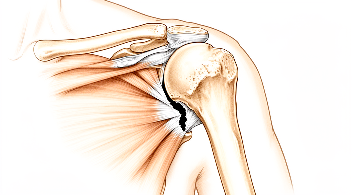
The rotator cuff comprises a sheet of conjoined tendons closely applied over the shoulder capsule, consisting of the subscapularis anteriorly, supraspinatus superiorly, and infraspinatus and teres minor posteriorly [12]. These muscles stabilize the humeral head by pulling it firmly into the glenoid whenever the deltoid lifts the arm forwards or sideways [12]. The tendons insert mainly into the greater tuberosity of the humerus, with the subscapularis inserting into the lesser tuberosity [12]. The subscapularis arises from the anterior aspect of the scapula and attaches over much of the lesser tuberosity, innervated by the upper and lower subscapular nerves [89]. The supraspinatus arises from the fossa superior to the scapular spine, passes beneath the acromion and acromioclavicular joint, and attaches to the superior aspect of the greater tuberosity, innervated by the suprascapular nerve after it passes through the suprascapular notch [89]. The infraspinatus arises from the fossa below the scapular spine and attaches to the posterolateral aspect of the greater tuberosity, innervated by the suprascapular nerve after it passes through the spinoglenoid notch [89]. The teres minor arises from the lower lateral aspect of the scapula and attaches to the lower portion of the greater tuberosity, innervated by a branch of the axillary nerve [89].
The coracoacromial arch is a fibro-osseous canopy formed by the acromion process posterosuperiorly, the coracoid process anteriorly, and the coracoacromial ligament joining them [12]. The subacromial bursa separates the rotator cuff tendons from this arch, allowing them to glide [12]. The long head of the biceps tendon attaches to the supraglenoid tubercle, runs between the subscapularis and supraspinatus, and exits through the bicipital groove under the transverse humeral ligament [89]. The coracohumeral ligament and transverse humeral ligament keep the long head of the biceps tendon aligned in the groove [89].
Histologic studies describe the rotator cuff tendons as having five distinct layers [89]. The most superficial layer is composed of coracohumeral ligament fibers oriented obliquely to the axis of the muscle [89]. The second layer consists of fibers grouped into large bundles extending from the supraspinatus tendon over the biceps tendon [89]. The third layer contains smaller, less tightly packed tendon fascicles with less uniform orientation [89]. The fourth layer is composed of loose connective tissue with thick bands of collagen fibers that merge with the coracohumeral ligament at the anterior edge of the supraspinatus [89]. The fifth and deepest layer is a continued sheet of collagen fibrils composing the superior joint capsule [89]. There is significant interdigitation and overlap of the supraspinatus and infraspinatus tendons near the footprint [89]. The infraspinatus insertion occupies the preponderance of the footprint on the greater tuberosity, while the supraspinatus insertion is smaller than previously believed [89].
The supraspinatus footprint measures 13 mm width medial-lateral and 20 mm anteroposterior [26]. A 7 mm medial-lateral tear corresponds to a 50% partial thickness tear of the supraspinatus [26]. The infraspinatus footprint measures 14 mm wide and 20 mm superoinferior [26]. The rotator cable is a thick bundle of fibers running perpendicular to the supraspinatus tendon fibers, connecting the supraspinatus and infraspinatus tendons [26]. This cable is divided into three segments: * Anterior segment: Forms the lateral part of the rotator interval [92]. * Middle segment: Runs under the supraspinatus tendon perpendicular to the longitudinal axes of the supraspinatus and infraspinatus tendons [92]. * Posterior segment: Covered by the infraspinatus tendon and ends at the insertion region between the infraspinatus and teres minor tendons [92].
The hypovascular critical zone is located on the articular side of the rotator cuff close to the insertion on the greater tuberosity [26]. The primary passive stabilizers of the glenohumeral joint are the capsule and scapulohumeral ligaments, which act as checkreins near the extremes of range of motion [98]. The glenohumeral joint capsule is thickest in the inferior pouch at 2.8 mm, 2.4 mm in the anterior portion, and 2.2 mm in the posterior portion [98]. The superior glenohumeral ligament crosses the rotator interval capsule and lies between the supraspinatus and subscapularis tendons [98]. The coracohumeral ligament originates at the base of the coracoid, blends into the cuff tendons, and inserts into the greater and lesser tuberosities [98]. The superior glenohumeral ligament and coracohumeral ligament come under tension with glenohumeral flexion, extension, external rotation, and adduction [98]. The middle glenohumeral ligament originates anterosuperiorly on the glenoid and inserts midway along the anterior humeral articular surface adjacent to the lesser tuberosity [98].
Pathophysiology
Rotator cuff pathology follows a spectrum from tendinitis (20 to 35 years old), to tendinosis (35 to 45 years old), to rotator cuff tears (>45 years old), and finally to cuff arthropathy (>65 years old) [26]. Partial-thickness rotator cuff tears have an overall prevalence of 20% in the general population, increasing to 26% in patients older than 60 years [3]. The prevalence of full-thickness rotator cuff tears is approximately 25% in patients over 60 years of age and increases to 50% in patients over 80 years of age [19]. The prevalence of rotator cuff tears is 28% in patients under 50 years and 65% in patients over 70 years [26]. If a patient has a symptomatic full-thickness rotator cuff tear in one shoulder, they have a 50% chance of an asymptomatic full-thickness rotator cuff tear in the contralateral shoulder [19]. Rotator cuff pathology constitutes up to 60% of all shoulder conditions [125].
Intrinsic degeneration involves age-related changes in collagen, proteoglycan, water content, and vascularity, usually involving the supraspinatus and infraspinatus starting on the articular side [26]. Extrinsic factors include chronic impingement on the coracoacromial arch, with tears usually starting on the bursal side of the tendon [26]. Acute traumatic rotator cuff tears typically occur after a fall and/or dislocation of the shoulder in patients under 40 years old [26]. Risk factors for rotator cuff tear development include age, smoking, female sex, family history, diabetes, and high cholesterol [26]. Biopsy specimens of retrieved tendon from rotator cuff tears show disorganized collagen, scar tissue, and minimal attempts at a healing process [13]. The confluence of age-related tendinosis and apoptosis leads to rotator cuff tendon failure [13]. Apoptosis is an organized form of cell death involving a programmed sequence of events leading to cellular elimination [13].
Rotator cuff impingement arises from repetitive compression or rubbing of the tendons, mainly the supraspinatus, under the coracoacromial arch [112]. Intrinsic factors for impingement include tendon degeneration, changes in highly sulphated glycosaminoglycans, changes in collagen composition with loading, and changes in vascularity [112]. Extrinsic factors for impingement include spurs growing down the coracoacromial ligament and osteoarthritic thickening of the acromioclavicular joint [112]. The "impingement position" is defined as abduction, slight flexion, and internal rotation of the arm [112]. Primary impingement results from encroachment of the rotator cuff in the subacromial space due to swelling or scarring, acromial morphologies (type II or type III), or selective hypomobility of noncontractile tissues [125]. Secondary impingement results from microinstability of the glenohumeral joint due to acquired ligamentous laxity, inadequate dynamic muscular stabilization, and scapular dysfunction [125]. Internal impingement is commonly involved in overhead athletes or when the arm is used in an abducted, externally rotated, and horizontally extended position [125].
The critical shoulder angle is formed by lines drawn from the inferior glenoid to the superior glenoid and from the inferior glenoid to the inferolateral acromion [235]. A critical shoulder angle above 35° to 38° has been shown to correlate with rotator cuff tears and postoperative retears [235]. A critical shoulder angle less than 30° has been shown to correlate with osteoarthritis [235]. The acromial index is the ratio of the medial/lateral length from the glenoid to the lateral acromion divided by the length from the glenoid to the lateral aspect of the greater tuberosity [235]. An acromial index above 0.7 has been associated with full-thickness rotator cuff tears and higher retear rates [235]. Increased lateralization of the acromion and increased superior glenoid inclination may increase the shear forces of deltoid contraction [235]. Rotator cuff disease may involve a genetic predisposition to intrinsic degenerative tendinopathic changes in molecular composition and vascularity [235]. Studies have shown familial hereditary patterns of rotator cuff disease and genes associated with significant pathology [235].
The rotator cuff serves multiple functions including initiating/assisting active shoulder motion and providing dynamic stability via force-couple moments [235]. Most rotator cuff tears occur in the anterior portion of the supraspinatus tendon [235]. Tears involving the rotator cable result in increased tear gap distance and strain compared with those in the crescent area [235]. The cable tissue is critical for the structural and functional integrity of the superior cuff [235]. Partial-thickness rotator cuff tears have a limited ability to spontaneously heal [3]. As many as 53% of partial-thickness rotator cuff tears will progress in tear size [3]. A portion of partial-thickness rotator cuff tears will progress to full-thickness rotator cuff tears [3]. Small full-thickness rotator cuff tears and painful partial-thickness tears become 25% to 50% larger within 3 to 4 years [26]. In a prospective study of small full-thickness tears in patients younger than 60 years, 49% of tears increased in size at 29 months [240]. Age older than 60 years, full-thickness tears, fatty infiltration, and symptoms are correlated with rotator cuff tear progression [240]. There is no correlation between pain, the size of rotator cuff tears, age, and chronicity at the time of presentation [240]. Fifty-one percent of individuals with asymptomatic rotator cuff tears began having pain at an average of 2.8 years in a longitudinal ultrasonographic study [240]. The progression of rotator cuff disease from tendinitis to degenerated full-thickness tear is a failure of biology where tissue is unable to respond productively to the insult [240].
Cuff tear arthropathy is the final stage of the shoulder impingement syndrome spectrum, affecting patients with long-term insufficient massive rotator cuff tears [30]. It involves superior migration of the humeral head toward the acromion, subchondral osteoporosis, humeral head collapse, and painful debilitating shoulder arthritis [30]. Cuff tear arthropathy affects women at a 3:1 female to male ratio, more commonly in patients over 70 years old and on the dominant shoulder [30]. Mechanical factors include insufficient cuff, superior migration of the humeral head, instability, eccentric wear of the glenoid, and humeral head deformity [30]. Nutritional factors include hypomobility-induced cartilage atrophy, poor nutrition leading to decreased glycosaminoglycans, dehydration, and subchondral osteoporosis [30]. Crystalline-induced arthropathy involves synovial-based matrix protein degradation destroying rotator cuff tendons and cartilage, with end-stage calcium-phosphate crystal deposition [30].
Muscle degeneration following rotator cuff injury is mediated by fibroadipogenic progenitor cells that differentiate into adipocytes and fibroblasts in the chronic setting [287]. Fatty infiltration and intramuscular fibrosis are known risk factors for rotator cuff retear after arthroscopic repair [287]. Disorganized connective, fatty, and fibrous tissue deposition in chronic rotator cuff tears is classified most commonly by the Goutallier staging system [240]. The Goutallier classification for fatty atrophy uses T1 sagittal MRI cuts: * Grade 0: Normal muscle [26]. * Grade 1: Some fat streaks [26]. * Grade 2: More muscle than fat [26]. * Grade 3: Fat evident on an equivalent amount as muscle [26]. * Grade 4: More fat than muscle [26].
Goutallier grades 3 and 4 indicate a long-term chronic rotator cuff tear with a higher potential for failure when surgery is undertaken and are likely deemed irreparable [19]. The tangent sign corresponds with muscle atrophy and chronicity [26]. A positive tangent sign predicts the repairability of rotator cuff tears [15]. Goutallier stages III and IV tears, if accompanied by a tendinous stump of less than 15 mm and a positive tangent sign, have a 90% failure rate [19].
A normal tendon-bone insertion site is not regenerated after tendon-to-bone repair; healing occurs by reactive scar formation [288]. The zone of calcified cartilage does not re-form after rotator cuff repair [288]. The poor healing response in rotator cuff repair likely relates to incomplete and abnormal expression of genes that direct formation of the complex structure and composition of the insertion site [288]. Factors preventing appropriate molecular signals and cell differentiation during postnatal healing include abnormal or insufficient gene expression, insufficient numbers of undifferentiated cells at the healing interface, and mechanical load on the healing tendon [288]. The presence of inflammation in the postnatal organism is a fundamental reason for the lack of appropriate molecular signals and cell differentiation during rotator cuff healing [288].
Glenohumeral decentering is significantly associated with diminished shoulder function and active range of motion in all planes [118]. Increased glenohumeral joint loads due to a full-thickness supraspinatus tear can be reversed with rotator cuff repair in a dynamic biomechanical cadaveric model [133]. Isolated supraspinatus tears did not perturb glenohumeral kinematics in a cadaveric model of the throwing shoulder, in contrast to a transosseous-equivalent repair [135]. Shoulder posture is an important determinant of passive forces during rotator cuff repair surgery [137]. The glenohumeral joint contact center gradually shifts superiorly over 24 months after rotator cuff repair, potentially reflecting altered loading patterns or loss of dynamic stability despite functional improvements [142]. Three-dimensional shoulder kinematics normalize after rotator cuff repair, with observed changes in scapular kinematics associated with increased overall range of motion [87]. More significant increases in shoulder complex kinematics were observed at the 6-month evaluation than at the 3-month evaluation following rotator cuff repair [188].
The prevalence of calcific tendinitis in a working population is 2.7%, with 35% of these shoulders being symptomatic [198]. Calcific tendinitis typically affects patients aged 30 to 60 years and women more commonly than men [198]. The supraspinatus tendon is most often involved in calcific tendinitis [198]. The precalcific stage consists of predominantly fibrocartilaginous metaplasia within less vascular areas of the tendon [198]. In the formative phase of the calcific stage, matrix vesicles unite to form calcium hydroxyapatite deposits separated by fibrocollagenous tissue [198]. The resorption phase involves an inflammatory response and is exquisitely painful [198]. Pain during the proliferative phase is correlated with macrophage activity during the resorptive phase [198].
Classification
Partial-Thickness Tears
Partial-thickness rotator cuff tears are classified as articular-sided, bursal-sided, or intratendinous tears [3]. The Ellman classification grades these tears based on location and depth observed during shoulder arthroscopy [3]. It designates tears as articular-sided (A), bursal-sided (B), or intratendinous (C) [3]. Depth is graded as follows: grade 1 involves 3 mm or less of tendon; grade 2 involves 3 to 6 mm of tendon; and grade 3 involves more than 6 mm of tendon [3]. Grade 3 tears represent tears of more than 50% of tendon width [3]. On MRI evaluation, partial-thickness tears are classified as low grade or high grade depending on whether they involve less than or more than 50% of the tendon width [3].
Full-Thickness and Massive Tears
The Cofield classification categorizes rotator cuff tears as small (0–1 cm), medium (1–3 cm), or large (3–5 cm) [50]. The Snyder classification describes the extent, location, and size of rotator cuff tears [264]. Tear location is classified as articular (type A), bursal (type B), and complete (type C) [264]. Full-thickness tears are further defined by type: type 1 is a small tear; type 2 is a moderate tear, less than 2 cm of only 1 tendon without retraction; type 3 is a large complete tear, usually 3 to 4 cm, with minimal retraction; and type 4 is a massive rotator cuff tear involving 2 or more rotator cuff tendons with retraction associated [264].
The Hamada classification is a commonly used scheme that uses a mechanistic approach to explain the radiographic changes seen with chronic massive rotator cuff tears [161]. It highlights the progressive nature of massive rotator cuff tears leading to CTA [161]. A modified Patte classification system has been developed to predict reparability and tendon healing in arthroscopic rotator cuff repair [149]. The diagnostic performance of this system was excellent for reparability and acceptable for rotator cuff healing [149].
Repair Integrity and Healing
The Sugaya classification distinguishes 5 repair categories using oblique coronal, oblique sagittal, and transverse T2-weighted images [268]. Type 1 indicates a repaired rotator cuff that has sufficient thickness with homogeneously low intensity on each image [268]. Type 2 indicates sufficient thickness with a partial high-intensity area [268]. Type 3 indicates insufficient thickness without discontinuity [268]. Type 4 indicates the presence of a minor discontinuity in more than 1 slice of each image, suggestive of a small tear [268]. Type 5 indicates the presence of a major discontinuity on each image, suggestive of a large tear [268].
The Boileau et al. grading system uses computed tomography arthrography to assess the structural integrity of the repaired tendon [260]. 'Intact' integrity indicates no leakage at the insertion or in the tendon [260]. 'Incomplete leakage' integrity indicates a small slit of contrast medium leakage into the tendon limited to one oblique coronal section [260]. 'Complete leakage' integrity indicates apparent discontinuity with contrast medium into the subacromial space [260]. Healing is defined as occurring when structural integrity is intact at both the insertion and in the tendon [260]. Incomplete healing is defined as incomplete leakage at either the insertion or in the tendon [260]. Retears are defined by the presence of complete leakage at the insertion [260]. New tears are defined by the presence of complete leakage in the tendon [260].
Clinical Presentation
Symptoms and History
Patients with symptomatic full-thickness rotator cuff tears typically report anterolateral shoulder pain radiating into the subdeltoid region [19]. Common complaints include pain during overhead activity and lifting objects with an outstretched arm, alongside frequent night pain and sleep disturbance [19]. In chronic rotator cuff disease, patients often describe an insidious onset of lateral and/or anterior shoulder pain associated with overhead activities, with night pain serving as a common presenting symptom [209]. A clear history of trauma resulting in acute pain and weakness strongly suggests an acute rotator cuff tear [209]. A family or personal history of rotator cuff disease increases the likelihood of diagnosis [209]. The combination of age >65 years, night pain, and weakness carries 95% specificity for rotator cuff tears [26]. Across all conditions comprising rotator cuff syndrome, patients complain of pain over the front and lateral aspect of the shoulder [12]. Weakness on abduction is present in supraspinatus involvement, cuff tears, and tendinitis [12]. Pain over the front of the shoulder is associated with biceps pathology, and rupture of the rotator cuff or biceps results in weakness [12]. In pediatric rotator cuff tears, pain is the most common presenting complaint, reported in 21 out of 23 patients with initial presentation data [94]. Other pediatric presenting complaints include weakness (n=6), functional impairment (n=3), decreased range of motion (n=2), dislocation (n=2), and numbness (n=2) [94].
Physical Examination
Physical examination for rotator cuff tears consists of assessing range of motion in adducted and abducted positions, evaluating strength, and examining associated structures such as the biceps and acromioclavicular joint [209]. Inspection includes checking for muscular atrophy of the deltoid and the supraspinatus and infraspinatus fossa in chronic tears [26]. In long-standing cases of partial or complete rupture, wasting of the supraspinatus and infraspinatus is usually present [219]. Palpation involves the greater tuberosity, acromioclavicular joint, bicipital groove, and coracoid process [26]. Tenderness of the acromioclavicular joint is often present in long-standing cases [219]. Cervical spine palpation, range of motion, and the Spurling test are used to rule out cervical spine pathologies as a cause of shoulder pain [26].
Resisted elevation tests, including the Jobe, empty can, and drop arm sign, assess supraspinatus function [26]. The empty can test has a sensitivity of 71.7% and a specificity of 64.6% for full-thickness supraspinatus tears [209]. External rotation tests, such as the Lag sign, Patte, and hornblower sign, assess infraspinatus and teres minor function [26]. A positive lag sign indicates a massive rotator cuff tear [26]. Patients with an external rotation lag sign at the side likely have a large posterosuperior tear involving the infraspinatus [209]. A positive hornblower sign suggests a massive posterosuperior cuff tear that prohibits active positioning of the hand in space [209]. Internal rotation tests, including the Belly press, lift-off, modified lift-off, and bear hug, assess subscapularis function [26]. The lift-off and belly-press tests have high specificity but low sensitivity for full-thickness subscapularis tears [209]. Deltoid strength and axillary nerve function are also assessed during the examination [26]. Provocative tests such as Neer, Hawkins-Kennedy, Yocum, and painful arc are impingement signs with low specificity for rotator cuff tears [26]. On testing the biceps in long-standing rotator cuff tears, there may be an old tear of the long head of the biceps tendon [219]. In a pediatric systematic review, special tests for specific rotator cuff muscle function were reported in 17 total patients, with 16 of 17 having at least one positive special test indicating rotator cuff pathology [94].
Imaging
The goal of diagnostic imaging is to determine the presence, size, and orientation of the rotator cuff tear, evaluate the healing capacity of the tendon, and assess associated pathology [209]. MRI is the benchmark for diagnosing rotator cuff tears with 94% sensitivity and 93% specificity [26]. T2-weighted MRI images best visualize rotator cuff tears [26]. T1 sagittal oblique MRI cuts reveal muscle and tendon retraction and muscle atrophy to determine chronicity, reparability, and outcome of surgical repairs [26]. MRI allows the surgeon to characterize the location, size, and amount of retraction of the tear, as well as the degree of atrophy and fatty infiltration of the musculature [19]. MRI accurately assesses muscle, bone, and cartilage, offering advantages for surgical planning [209]. An MRI is indicated in younger, active patients with acute rotator cuff tears and in patients with chronic rotator cuff tears in whom a trial of nonoperative treatment has failed [19]. Intra-articular contrast-enhanced magnetic resonance arthrography (MRA) is best for detecting partial-thickness rotator cuff tears with 95% sensitivity and 95% specificity [26].
Ultrasonography has good accuracy with 92% sensitivity and 93% specificity for rotator cuff tears [26]. For full-thickness rotator cuff tears, ultrasonography approaches the sensitivity and specificity of MRI for detecting the presence of a tear with an experienced practitioner [209]. Ultrasonography allows for dynamic assessment of the cuff insertion [26]. However, it is operator dependent, has limited assessment of chondral lesions, and has poor sensitivity to diagnose partial-thickness rotator cuff tears [26].
Radiographs are used to rule out glenohumeral and acromioclavicular arthritis [26]. The true AP radiograph view evaluates the acromiohumeral interval, critical shoulder angle, and acromion index [26]. Greater tuberosity excrescences on AP views in external and internal rotation are pathognomonic for cuff disease [26]. The supraspinatus outlet view evaluates acromial morphology according to the Bigliani classification [26]. The axillary view assesses glenohumeral joint morphology and joint space and rules out dislocations [26]. CT arthrography is useful in postoperative assessment, retear evaluation in patients with retained metallic anchors causing artifact on MRI, and when MRI is contraindicated [26].
In a pediatric systematic review, 50 out of 51 patients with reported MRI or MRA results demonstrated some indication of rotator cuff pathology [94]. All 3 patients with reported ultrasound examinations were positive for signs of rotator cuff damage [94]. Of 17 reported radiograph results, 12 were negative, while 5 showed signs of pathology [94].
Classification and Prognostic Factors
Rotator cuff tears are classified by time as acute versus chronic (>3 months from the onset of pain/injury) [26]. They are classified by size as small (0 to 1 cm), medium (1 to 3 cm), large (3 to 5 cm), and massive (>5 cm) [26]. Classification by shape includes crescent, U-shaped, L-shaped, and retracted-immobile [26]. The Ellman classification grades partial-thickness tears as grade 1 if involving 3 mm or less, grade 2 if 3 to 6 mm, and grade 3 if more than 6 mm of tendon is torn [3]. MRI evaluation classifies partial-thickness tears as low grade or high grade depending on whether they involve less than or more than 50% of the tendon width [3]. The Patte classification divides rotator cuff tears into Group I (partial or full-substance <1 cm), Group II (full-substance supraspinatus), Group III (full-substance >1 tendon), and Group IV (massive tears with secondary OA) [17]. The Goutallier-Fuchs classification for fatty atrophy uses T1 sagittal MRI cuts: Grade 0 normal muscle, Grade 1 some fat streaks, Grade 2 more muscle than fat, Grade 3 fat evident on equivalent amount as muscle, and Grade 4 more fat than muscle [26].
Factors associated with failure after rotator cuff repair include larger tears, greater retraction, advanced Goutallier grade, older age, smoking status, osteoporosis, diabetes mellitus, hypercholesterolemia, and more aggressive rehabilitation protocols [19]. Tears with Goutallier grades III and IV usually are deemed irreparable [19]. The best potential for healing was in patients younger than 70 years with acute, small tears (<3 cm) and a healthy tendon-bone interface who are compliant with sling wear and are able to undergo an extended rehabilitation program of 6 to 9 months [19]. Evidence synthesized in an umbrella review underscores the importance of patient age, expectations, and the extent of the rotator cuff tear in influencing outcomes following rotator cuff repair [117]. Teres minor hypertrophy is a common finding in the setting of rotator cuff tearing and is not a positive predictor of outcomes following rotator cuff repair [127]. At mid-term follow-up, greater preoperative rotator cuff disease severity was associated with failure to achieve clinically significant outcomes [121]. Preoperative mental health scores and achieving patient acceptable symptom state are predictive of return to work after arthroscopic rotator cuff repair [55].
Investigations
Plain radiography: Radiographs are the first imaging study obtained for rotator cuff evaluation [212]. They may demonstrate classic changes within the acromion or coracoacromial ligament, including spurring and calcification [155]. Cystic changes within the greater tuberosity may also be visible [155]. With chronic rotator cuff disease, radiographs may show superior migration of the humeral head with extensive degenerative change [155].
MRI: Magnetic resonance imaging provides the best overall evaluation of the rotator cuff [212]. It is used to define the extent of the tear, degree of tear retraction, and presence of muscular atrophy [155]. MRI is key for evaluating fatty infiltration, although the Goutallier classification was originally based on CT [155]. MRI is sensitive and specific for full-thickness rotator cuff tears, with a sensitivity of 92.1% and a specificity of 92.9% [160]. For partial-thickness tears, MRI sensitivity is 63.6% and specificity is 91.7% [160]. Articular-side thickening of the supraspinatus and infraspinatus on MRI suggests the presence of a partial-thickness articular-side rotator cuff tear [160]. The rotator cable can be reliably identified with the shoulder in the ABER position on MRI, even in the absence of a rotator cuff tear [160].
Magnetic Resonance Arthrography: MRA is sensitive and specific for full-thickness rotator cuff tears, with a sensitivity of 95.4% and a specificity of 98.9% [160]. For partial-thickness tears, MRA sensitivity is 85.9% and specificity is 96.0% [160]. MRA is more sensitive than MRI for the detection of partial-thickness, particularly articular-sided tears [160]. However, MRA is not suitable for the diagnosis of long head of biceps (LHB) lesions prior to arthroscopic rotator cuff repair [335]. MRA is insufficiently accurate to diagnose biceps lesions prior to rotator cuff repair, regardless of the gold standard used [283].
Ultrasonography: Ultrasonography is increasing in popularity as a tool for the diagnosis of rotator cuff disease [155]. It is also used for confirmation of intraarticular or subacromial location of injections [155]. Ultrasonography is more accurate for full-thickness rotator cuff tears, comparable to MRI [15]. In the community setting, ultrasound may be used to evaluate the integrity of a repaired rotator cuff tendon and constitutes a comparable alternative to MRI [324]. Ultrasonography is an adequate imaging modality to classify rotator cuff repair integrity [312].
Specific Radiographic and MRI Signs: The tangent sign is defined as failure of the supraspinatus muscle belly to cross a line from the superior border of the coracoid to the superior border of the scapular spine [155]. This sign correlates with muscle atrophy and fatty infiltration of the supraspinatus [155]. Patients with the presence of the tangent sign are more likely to have an irreparable rotator cuff tear [155]. The shift angle and medial retraction of the deep layer on MRI are associated with intraoperative reparability in large-to-massive rotator cuff tears [333]. Routine MRI assessment of the upper subscapularis muscle and coracohumeral distance can contribute to the diagnostic accuracy of subscapularis tears [338]. Higher upper subscapularis Goutallier grade and coracohumeral distance narrowing are predictive of subscapularis tears in patients undergoing arthroscopic rotator cuff repair [338].
Postoperative Imaging and Healing Assessment: MRI is useful for evaluating patients with pain and loss of function after rotator cuff repair, particularly for identifying full-thickness tears, intact cuffs, ruptures of the biceps tendon, and detachments of the origin of the deltoid muscle [319]. MRI is less effective than other modalities for identifying partial-thickness tears in the evaluation of failed repairs [319]. MRI often fails to diagnose the presence of subscapularis tears and infraspinatus tears, despite good concordance with arthroscopic findings for the supraspinatus [82]. Neither MRI nor clinical tests are sufficiently reliable for diagnosing biceps lesions prior to rotator cuff repair [283]. Metal artefacts from titanium suture anchors prevented accurate diagnosis from MRI scans of rotator cuff retear in 36% of patients studied [325]. Biocomposite suture anchors are frequently still visible on MRI 2 years after rotator cuff reconstruction [83].
Cuff integrity on follow-up MRI scans had a positive effect on the clinical outcome after arthroscopic transosseous rotator cuff repair [32]. Radiographic changes occurred in 40% of patients within 5 years after arthroscopic rotator cuff repair [78]. MRI appearance of the repaired tendon changes over time but does not correlate with function or predict clinical outcomes at 1 year after surgery [286]. Most deformities after rotator cuff repair remodel over time and do not affect the radiologic outcome of repair after 6 months of follow-up [304]. Immediate changes in the appearance of fatty infiltration and muscle atrophy on MRI should be considered when assessing rotator cuff muscle changes by comparing preoperative and postoperative MRI [284]. After successful arthroscopic rotator cuff repair, there was a slight increase in supraspinatus muscle volume from preoperatively to final follow-up, as seen on serial MRI [263]. A prospective pilot study suggests a temporal relationship of MRI and ultrasound parameters that parallels the expected phases of healing in the repaired rotator cuff [267].
Poor interobserver agreement regarding postoperative graft and rotator cuff integrity by MRI was found in patients undergoing superior capsular reconstruction augmentation [310]. The presence of clinically significant disagreements in inter-rater agreement of rotator cuff tendon and muscle MRI parameters indicates the need for improved imaging tools for precise rotator cuff evaluation [299]. In 18 of 22 patients (82%) with partial-thickness rotator cuff tears treated with tear completion followed by surgical repair, there was no evidence of a full-thickness or near full–thickness defect on follow-up MRI at a minimum of 2 years [243]. Although MRI data suggest improved healing rates in single-row repairs in the entire patient population, double-row repair showed improved radiographic healing when similar-sized tears were compared [289]. There is no MRI difference in 12-month rotator cuff retear rates between triple-loaded single-row repairs or suture-bridging double-row repairs [322]. Imaging results showed no significant difference in cuff integrity between single-row and double-row repair groups in patients with any tear size at 6-month and minimum 2-year follow-up [343].
Arthroscopic repair of medium to large rotator cuff tears using a triple-loaded single-row repair augmented with bone marrow vents resulted in a 91% healing rate by MRI [336]. Arthroscopic repair of medium to large rotator cuff tears using triple-loaded medially based single-row repair augmented with marrow vents resulted in a 92% healing rate by MRI [342]. Rotator cuff repairs augmented with a porcine dermal patch resulted in excellent clinical outcomes with a higher healing rate and close-to-normal MRI findings [278]. 50% of patients who underwent biologically enhanced demineralized bone matrix augmentation of their rotator cuff repair demonstrated MRI-determined failure of supraspinatus healing [317]. The RoHI score may not predict short-term functional recovery following rotator cuff repair, though further study including postoperative imaging may clarify its potential role in long-term prognostication [302].
Treatment
Non-Operative
Non-surgical treatment is the preferred initial approach for patients with non-traumatic rotator cuff injuries [226]. While nonoperative management may yield better short-term outcomes, surgical repair provides significantly better long-term outcomes and a higher probability of substantial improvement for symptomatic rotator cuff tears [214]. Most partial-thickness rotator cuff tears are best initially managed with nonoperative treatment [3].
Operative
Indications: Clinical decision-making for rotator cuff tears is complex and lacks consensus, as neither the American Academy of Orthopaedic Surgeons clinical practice guidelines nor Cochrane systematic reviews provide specific management guidance [11]. Patients are generally categorized into those needing urgent or early operative repair, those who can benefit from a trial of conservative treatment, and those best suited for nonoperative treatment [11]. The increasing incidence of rotator cuff repairs is questioned because there are not convincing data demonstrating the superiority of operative treatment over non-operative management in all rotator cuff tears [152]. However, operative management is increasingly cost-effective with time, given that nonrepaired cuff tears are unlikely to heal and portend worse symptomatology [184]. Arthroscopic examination and attempted repair of large and massive rotator cuff tears is considered the gold standard of treatment for nonarthritic shoulders [274]. Repair of symptomatic single-tendon rotator cuff tears in patients aged older than 65 years who do not respond to conservative treatment appears justified [238]. Severely obese patients achieve overall excellent outcomes after repair and noninferior clinical differences when compared to healthy weight patients [49]. For partial-thickness tears, surgical treatment with either rotator cuff repair or debridement is indicated for patients in whom nonoperative treatment fails [3]. Tears involving more than 50% of tendon width are best treated with repair, while those involving less than 50% are best treated with debridement and potential decompression [3]. For tears with poor-quality tendon remaining and involving more than 80% of tendon thickness, debridement is favored over repair [3]. If delamination of the tear is present in articular-side delamination-type tears, a transtendinous repair is indicated [3].
Surgical Approach / Technique: Arthroscopic rotator cuff repair has evolved into one of the most common orthopedic surgical procedures worldwide [22]. The current standard of care is arthroscopic repair, although the superiority of arthroscopic versus open or mini-open repair remains somewhat controversial [68]. Arthroscopic repair has been shown to have similar outcomes and failure rates compared to open or mini-open repair, with decreased short-term pain and more rapid return to activity [68]. Arthroscopy allows a more comprehensive assessment of intra-articular pathology and rotator cuff tear configuration by viewing from multiple angles [231]. Tendon mobilization in arthroscopic repair is facilitated by precise releases of adhesions that limit tendon excursion, leading to an improved ability to anatomically reduce the edge and create a tension-free repair [231]. Injury to the deltoid muscle is minimized in arthroscopic repair, and the acromial deltoid origin is preserved, eliminating the risk of deltoid dehiscence [231]. A key theoretical benefit of arthroscopic repair is decreased postoperative pain secondary to less soft tissue trauma, which aids in postoperative rehabilitation and leads to earlier resumption of range of motion [231]. Anatomic footprint restoration is now possible in arthroscopic repair, with fixation at both the suture-tendon interface and the anchor-bone interface that approximates traditional open transosseous repairs [231]. Arthroscopic repair of rotator cuff tears is an effective and straightforward technique when performed in an organized, well-considered manner [134]. One-stage treatment for rotator cuff tears in stiff shoulders provides comparable range of motion and patient-reported clinical outcomes as rotator cuff repair for non-stiff shoulders [257]. Delamination does not affect outcomes after arthroscopic rotator cuff repair as compared with nondelaminated rotator cuff tears [313].
Mini-open rotator cuff repair allows direct assessment of the nature of the cuff tear, the quality of the remaining tissue, and the ease with which the tendon edge can be restored to the normal insertion site [51]. The transosseous technique for attachment of the tendon to the tuberosity in mini-open repair is simple, expeditious, avoids issues related to suture anchors, and enables the surgeon to find sufficiently strong bone to hold the sutures [51]. The creation of a trough into which the tendon is inserted increases the surface area of the repair, stimulates local stem cells and growth factors, excludes joint fluid from the repair site, and places sutures laterally so that they do not rub beneath the coracoacromial arch [51]. The goal of open rotator cuff repair is strong fixation of the tendon to the humerus under normal tension with the arm at the side while leaving a smooth surface on the proximal humeral concavity to articulate with the undersurface of the coracoacromial arch [51]. The desired attachment site for open rotator cuff repair is at the sulcus near the base of the tuberosity [51]. Three stages of sequential release are required in open rotator cuff repair because the cuff is usually retracted and tissue is lost in chronic cuff disease [51]. Without sequential releases, increased tension in the repaired tendon will predispose to tightness of the glenohumeral joint and will additionally challenge the repair site [51]. The goals of mobilization in open rotator cuff repair are to obtain tissue of adequate strength, to position it anatomically for repair without damage to innervation and without compromise of deltoid function, and to decompress the subacromial space to prevent further mechanical impingement on repaired cuff tissue [31]. If the supraspinatus and infraspinatus tendons are retracted so far that adequate length cannot be obtained with tendon mobilization, the capsule is incised at its insertion into the glenoid labrum [31]. The use of a second posterior incision over the scapular spine to increase mobilization has been described, but the authors have no experience with this technique [31].
Implant Selection: The double-row technique has been advocated as a better biomechanical construct and a more anatomic repair strategy than single-row repair [231]. Controlled laboratory studies have generally shown superiority of double-row techniques over single-row in terms of initial and ultimate failure strength, decreased gap formation, decreased strain and suture cut-through, and improved vascularity in transosseous-equivalent double-row repair [68]. While biomechanical studies show double-row repair outperforms single-row repair in failure strength, superior clinical results with double-row fixation over single-row fixation are still controversial [231]. The healing of the rotator cuff using an anatomic technique was superior to that in the single row repair group [145]. The best results in open rotator cuff repair are obtained with the double-row technique, suturing the tendon to bone in a cancellous trough in combination with suture anchor fixation [31]. Using transosseous tunnels through the greater tuberosity increases the surface area of tendon-to-bone healing, which more closely restores the anatomic footprint [31]. Sutures are placed 5 to 10 mm from the free edge of the tear in a horizontal mattress manner to help push the tendon down into the trough [31]. A 3 mm wide shallow trough is created running the length of the exposed bone of the greater tuberosity to accommodate the thickness of the supraspinatus and infraspinatus [31].
Arthroscopic transosseous rotator cuff repair achieves similar clinical outcomes, patient-reported outcomes, and healing rates with 30-80% reduction in implant costs compared to traditional anchored techniques [346]. The knotless technique for rotator cuff repair required less surgical procedure time and cost less overall than the knotted technique and resulted in equivalent clinical results [350]. Both knotless and knot-tying anchor techniques may be used successfully to repair full-thickness rotator cuff tears with very good functional outcome [138]. The novel all-inside arthroscopic rotator cuff repair technique was safe and significantly faster and provided better healing rates than other repair techniques [139]. The described transosseous anchorless technique is a safe and reproducible fully arthroscopic approach for medium to large rotator cuff tears that mimics the previous gold standard [154]. Shoulder function was improved after complete rotator cuff repair and similar clinical outcomes were achieved regardless of suture anchor material and shape [72]. At a short-term follow-up, differences between arthroscopic repair of full-thickness rotator cuff tears with metal and biodegradable suture anchors were not significant [293]. Every patient presenting with shoulder pain after usage of a biodegradable fixation material should be evaluated closely [328]. Every tested knotless suture anchor presented different problems that may lead to premature failure of the rotator cuff reconstruction [329]. Gap formation between the bone and the soft tissue fixation jeopardizes the repair, and an anchor is not appropriate for rotator cuff repair if used on its own if it causes such gap formation [309]. Initial fixation strength is an essential consideration in optimizing rotator cuff repair [320]. The ideal rotator cuff repair technique should have high initial fixation strength to allow minimal gap formation between the tendon and bone [339]. Suture anchor loading findings may have implications for the type of cuff fixation device used and for rehabilitation after fixation [331]. Placement of anchors in specific regions provides stronger fixation and may prevent anchor loosening and failure of the rotator cuff repair [297]. Both 20° and 45° anchor insertion techniques resulted in acceptable shoulder clinical outcomes for patients with osteoporosis undergoing rotator cuff repair [349].
Adjuncts: Acromioplasty cannot be recommended as a routine technique in every rotator cuff repair [205]. Despite Clinical Practice Guidelines recommending the nonroutine use of acromioplasty, surgeons continue to perform acromioplasty with rotator cuff repair in most of the cases throughout all subcategorizations analyzed [239]. Routine distal clavicle resection (DCR) is not recommended with arthroscopic rotator cuff repair [62]. The results of repair without decompression are not as good as the results using the combined procedure of anterior acromioplasty and rotator cuff repair [31]. A subacromial bursectomy is performed during open rotator cuff repair, and the biceps tendon is protected unless biceps pathology is present, in which case a proximal biceps tendon release or tenodesis is indicated [31].
Revision: Partial rotator cuff repair represents a reasonable, low-morbidity salvage option for the patient with a rotator cuff tear that is not primarily repairable [60, 61, 148]. For the treatment of large to massive rotator cuff repairs, not all partial repairs are equivalent [38]. Revision rotator cuff repair is technically more difficult than primary repair due to poor tissue quality, adhesions, and retained hardware [84]. Optimization of surgical techniques and the use of appropriate biologic or tendon transfer techniques, if indicated, is the best method for the management of failed rotator cuff repair [136]. Superior capsular reconstruction with incorporation of the native rotator cuff may provide surgeons the opportunity to perform a robust repair in situations where arthroplasty or other more invasive procedures were once the only options [151]. The technique of concomitant superior capsular reconstruction with rotator cuff repair has been a reproducible method for achieving successful surgical management of patients with massive rotator cuff tears and superior capsular insufficiency [147]. The general indications for augmentation include patients with large (>3 cm), multitendon rotator cuff tears or chronic tears with poor tissue quality [194]. While there is still considerable work to be done before scaffolds are introduced into routine clinical practice, there does appear to be a clear indication for their use as an interpositional graft for large and massive retracted rotator cuff tears and when repairing a poor-quality degenerative tendon [210]. Despite the growing clinical use of scaffold devices for rotator cuff repair, numerous questions related to their indication, surgical application, safety, mechanism of action, and efficacy remain to be clarified or addressed [191]. Strategies to biologically augment rotator cuff repair can be divided into strategies that are clinically available now in humans and strategies that have largely been tested in animal models [153]. Patch augmentation is technically challenging and requires the surgeon to be patient and organized [300]. The described patch augmentation technique is specifically used to augment posterosuperior rotator cuff tears that are completely reparable and is not recommended to address either completely irreparable or partially reparable tears [300]. Acellular dermal graft augmentation is a simple and reproducible method for rotator cuff augmentation that can be easily adopted by arthroscopic shoulder surgeons to decrease surgical time [130]. The "Pull-Over" technique for all arthroscopic rotator cuff repair with extracellular matrix augmentation offers additional advantages over previously described techniques by facilitating the passage of the patch into the bursa as well as providing a more robust medial stabilization of the augment [307]. Extracellular matrix augmentation of large and massive rotator cuff tears may decrease repair failure rates [307]. Arthroscopic augmentation is technically challenging [307]. Biopsies of collagen implants retrieved from human rotator cuff repair subjects revealed cellular incorporation, tissue formation and maturation, implant resorption, and biocompatibility [351]. A synthetic interposition graft with screw fixation could not prevent cuff tear arthropathy and preserve cuff integrity in a long-term perspective [321]. Autologous bursa augmentation is a straightforward, reproducible, and effective approach to enhancing rotator cuff repair for partial-thickness bursal-site tears [97].
Other Considerations: Patients' characteristics and indications for surgery were not described in a majority of clinical outcome studies of rotator cuff repair [1]. Rotator cuff repair may not alter the natural history of the disease [18]. Every attempt should be made at the time of arthroplasty to balance the soft tissues and securely repair the rotator cuff because subsequent attempts to repair the rotator cuff are prone to failure [39]. Partial-thickness rotator cuff tears have a limited ability to spontaneously heal as shown by histological and radiographic studies [3]. As many as 53% of partial-thickness rotator cuff tears will progress in tear size, and a portion of these will progress to full-thickness rotator cuff tears [3]. A higher failure rate of debridement has been suggested for partial-thickness bursal-sided rotator cuff tears compared to articular-sided rotator cuff tears [3]. Some surgeons favor repair in even low-grade bursal-sided tears due to the higher failure rate of debridement in this subgroup [3]. There is no difference in functional outcome scores or re-tear rates between in situ rotator cuff repair and completion to a full-thickness rotator cuff tear with subsequent repair [3]. Intratendinous tears are repaired side-to-side using arthroscopic technique after the tear is opened and the edge is debrided back slightly to promote local healing [3]. Arthroscopic surgery offers more thorough visualization, diagnosis, and treatment of lesions within the joint, which is critical because a high prevalence of concomitant intra-articular pathology has been reported in patients undergoing rotator cuff repair [231]. The banana suture lasso double-row fixation technique has simplified massive rotator cuff tear repairs and offered an easier approach that is simple, reproducible, and does not cost more [115]. The Hybridge technique is a combined technique of suture bridge and tension band for an arthroscopic eco-responsible rotator cuff repair [290]. Prepassing braided, nonabsorbable sutures with a suture passer through the rotator cuff tendon during repair shortens operative time by eliminating additional suture-passing steps after medial-row anchor placement [295]. Using ETHIBOND Excel suture as a prepass suture is not recommended because of its tendency to tangle and jam in the suture passer [295]. Damaged sutures during prepassing can lead to suboptimal rotator cuff repair, potentially compromising repair integrity and increasing retear or poor healing risks [295]. A simultaneous dual-camera technique for partial-thickness, articular-sided, in situ transtendinous rotator cuff repair allows simultaneous visualization of intra-articular and subacromial spaces, improved placement of anchors and sutures, and minimizes risk of tension mismatch and tendon buckling [306]. The disadvantages of the simultaneous dual-camera technique include a more challenging setup compared with traditional techniques, possible excessive soft-tissue swelling with 2 fluid pumps, and complex visualization due to use of 2 cameras [306]. MRI often fails to diagnose the presence of subscapularis tears and infraspinatus tears, while concordance with MRI results is good for the supraspinatus [82].
Complications
General Complication Rates: Short-term complications after rotator cuff repair are rare [44], yet clinical registries indicate that adverse events may occur in almost 1 of 5 cases [358]. In a consecutive series of 263 patients undergoing primary arthroscopic repair, 28 patients (10.6%) sustained a complication [251]. The complication rate after arthroscopic revision rotator cuff repair is about twice the published rate for primary repair [229]. Open repair carries higher risks of any adverse event, readmission, and major surgical site infections within 30 days compared to arthroscopic repair [352]. Conversely, arthroscopic repair is associated with significantly lower rates of deep incisional surgical-site infections, organ space infections, wound disruptions, and return to the operating room compared with open repair [381]. Infections and stiffness were rare but slightly and significantly more frequent in open repairs compared to arthroscopic repairs [116].
Risk Factors: Complications occur at increased rates in HIV-infected patients relative to non-infected groups [356]. Nearly a quarter of solid organ transplant patients experience a perioperative complication, necessitating careful surgical consideration and increased postoperative surveillance [330]. A 15-minute incremental increase in operative duration is associated with an additional risk of complications within 30 days after arthroscopic repair [316]. Incremental increases in operative time are also associated with increased risks of surgical site infection, pulmonary embolism, transfusion, and extended hospital stay [316]. No intraoperative, perioperative, or postoperative complications were reported in patients aged younger than 45 years undergoing arthroscopic primary repair [124], nor in a single-center series of patients aged 30 years and under undergoing full-thickness arthroscopic repair [258].
Repair Failure and Re-tears: Recurrent rotator cuff tears are not uncommon after arthroscopic repair of large and massive tears [25]. Anatomical failure is the most common complication in the literature regarding arthroscopic repair [129], with re-rupture rates ranging between 11.4 and 94% in a literature review [129]. Pooled analysis demonstrates substantial retear rates in both arthroscopic and open/mini-open groups, associated with inferior shoulder function [369]. Five patients (5.5%) required revision surgery after primary arthroscopic transosseous-equivalent double-row repair [285]. Three patients (9.4%) had repair failures in a cohort aged 30 years and under [258]. Twenty patients (5.0%) underwent revision arthroscopic surgery for repair failure in a cohort with diabetes mellitus [272]. The incidence of re-interventions was 10% in patients with rotator cuff repair after traumatic shoulder dislocation, primarily due to symptomatic re-tears [372]. A decrease in retear rate was observed during the study period of 1600 consecutive repairs [388]. For approximately half of type 2 patients at 1 year after surgery, cuff integrity worsened at 10 years [377].
Stiffness / Arthrofibrosis: Stiffness was observed in 74 patients in a literature review of complications associated with arthroscopic repair [129]. Persistent stiffness was the most common complication in a series of 263 patients undergoing arthroscopic repair, usually responding to extensive physical therapy [251]. Five patients (15.6%) had postoperative shoulder stiffness in a cohort aged 30 years and under [258]. Six patients (1.5%) underwent capsular release and lysis of adhesions for postoperative stiffness in a cohort with diabetes mellitus [272]. Two patients underwent capsular release and lysis of adhesions after developing stiffness following primary arthroscopic transosseous-equivalent double-row repair [285].
Infection: The overall incidence of infection was 8.5/1000 arthroscopic rotator cuff repairs over a 10-year period [296]. Arthroscopic repair is associated with a lower rate of postoperative infection compared with open or mini-open approaches [359]. Patients undergoing open repair had a significantly higher rate of postoperative infection compared with those undergoing arthroscopic repair [360], and arthroscopic techniques reduced the risk of infection compared with open techniques [362]. An overall 0.11% rate of postoperative infection was found following primary arthroscopic repair in a single-institution retrospective review [384]. Only 4 infections were deep in a single surgeon experience of mini-open repair, suggesting deep infection approximates rates seen with arthroscopic techniques [391]. Infection is a rare but serious complication that can lead to poor outcomes [265]. Patients with deep infections after arthroscopic repair showed moderate mid- to long-term outcomes [366]. Eradication of deep infection is possible with preservation of the glenohumeral joint; however, substantial functional limitations are not unusual [380]. Arthroscopic reconstruction of the rotator cuff is a feasible goal in the setting of prior deep infection [385]. Withholding prophylactic antibiotics in low-risk patients undergoing routine repair does not increase infection rates [386]. Preoperative corticosteroid injection prior to primary arthroscopic repair did not increase the risk of reoperation or infection with a minimum follow-up of 2 years [382]. Prior shoulder arthroscopy was associated with a significantly higher risk of prosthetic joint infection (OR 2.77) in patients with concomitant glenohumeral osteoarthritis [390].
Thromboembolism: Venous thromboembolism is a rare complication following rotator cuff repair, with an incidence of 0.3% [241]. This is the first reported case of pulmonary embolism due to subclavian venous thrombosis following rotator cuff tendon repair [308]. Given that all detected cases of lower extremity deep vein thrombosis after arthroscopic repair were asymptomatic and limited to distal veins, routine screening may not be necessary [326].
Other Specific Complications: Hardware-related complications were observed in 12 patients in a literature review of complications associated with arthroscopic repair [129]. Eleven less common complications were reported in a literature review: 5 neurovascular, 3 septic, 2 thromboembolic events, and 1 anesthesiological complication [129]. A huge subacromial ossification was seen in one patient following open repair, requiring subacromial decompression 11 months after primary large cuff reconstruction [111]. Persistent postoperative shoulder pain was reported in 4 patients (12.5%) in a cohort aged 30 years and under [258]. One patient suffered repair failure secondary to infection at 38 days following primary arthroscopic transosseous-equivalent double-row repair and underwent irrigation and debridement with revision cuff repair [285]. One patient underwent 2 consecutive debridement and decompression surgeries for persistent pain and subacromial impingement following primary arthroscopic transosseous-equivalent double-row repair [285].
Other Considerations: TRT within 1 year of rotator cuff repair appears to be a risk factor for multiple postoperative complications and subsequent shoulder surgery [361]. Over two years, TRT was associated with higher rates of total shoulder arthroplasty but lower rates of lysis of adhesions, with no difference in revision rotator cuff repair [387].
Recovery
Light activity (weeks): Patients can safely return to driving at 2 weeks postoperatively with no clinically important negative impact on driving fitness [225].
Full activity (months): A vast majority of patients undergoing rotator cuff repair can expect to return to work within 8 months of surgery [55]. The majority of injured workers return to previous work at approximately 8 months after surgery [144]. In a cohort of manual laborers, 89.6% of patients were able to return to work following arthroscopic repair [181]. As much as 88.5% of patients were able to return to activities after arthroscopic rotator cuff repair with a mean duration of 6.59 months [244]. For geriatric workers' compensation patients, return to full-duty work takes about 5 months regardless of surgical approach [221].
Complete recovery / outcome plateau (months): The plateau of maximum recovery following rotator cuff repair occurs at 1 year [165, 175]. Most functional improvement and pain relief occur within the first 6 months but continue to 24 months [29]. Functional recovery based on clinical outcomes shows approximately 60% of ultimate recovery at 3 months and approximately 75% recovery at 6 months [182]. Arthroscopic repair provides functional recovery as early as 3 months, with further improvement over the first year and subsequent stabilization [327]. Recovery of the ability to sleep comfortably plateaued at 6 months, and these improvements are maintained more than 4 years without evidence for the re-emergence of sleep disturbance [354].
Rehabilitation protocol: There is no consensus on rehabilitation protocols following rotator cuff repair [167]. Early passive motion exercise is not mandatory after arthroscopic repair of small to medium-sized full-thickness tears [178]. The currently available literature did not identify any significant differences in functional outcomes and relative risks of re-tears between delayed and early motion in patients undergoing arthroscopic repairs [65]. Early rehabilitation is associated with some initial improvements in range of motion and function [179]. Water therapy provides better early results compared to traditional dry rehabilitation or self-exercise therapy [176]. A delayed postoperative physical therapy protocol was associated with improvements in outcome measures and shoulder function compared to the preoperative state, with rotator cuff healing demonstrated by MRI [141]. The healing rate at long-term follow-up is not clearly affected by the type of rehabilitation, but the early passive motion protocol might result in lower rates of tendon healing in the shoulder with large-sized tendon tears [186]. Further studies are needed to determine effective dosing of physical therapy after rotator cuff repair [164]. A healed rotator cuff repair results in a superior outcome for the patient compared with a non-healed repair, and the surgeon can maximize the chance of a healed repair by adhering to core principles regarding tear pattern recognition, repair construct selection, biological healing optimization, and avoiding aggressive early rehabilitation [67].
Functional milestones: Patients achieve milestones measured by the five most commonly asked questions on the PROMIS-UE CAT by 1-4 months postoperatively [318]. All patient-reported outcome measures except SAL demonstrate high responsiveness to rotator cuff repair surgery during the first 2 postoperative years, with the majority of gains occurring within the first 6 months [249]. Patient-reported outcomes of all-arthroscopic rotator cuff repair show significant improvement from baseline and remain durable over a period of 15 years [5].
Other Considerations: One-year follow-up does not determine the long-term outcome of rotator cuff repair [70]. The early high functional scores after primary rotator cuff repair or reconstruction did not persist, with function lost as range of motion and strength decreased to less than preoperative values at 16 years [344]. Six months after rotator cuff repair, patients who continued to work after injury but presurgery were the most likely to return to work at any level, and patients who had less strenuous preinjury levels of work were the most likely to return to their preinjury levels of work [228]. Workers' compensation recipients who underwent arthroscopic rotator cuff repair had worse outcomes while receiving workers' compensation benefits [211]. Patients who undergo staged bilateral rotator cuff repair can expect to have similarly good clinical outcomes regardless of hand dominance or chronologic incidence with excellent healing rates in both shoulders [46]. At a minimum of 1-year follow-up after rotator cuff repair, patients should expect similar improvement in pain and validated outcome measures for each side, though they may experience more pain during the middle portion of recovery on the contralateral side [345]. Despite the apparent risks associated in rotator cuff repair in upper extremity ambulators, these patients demonstrate clinically significant improvements following surgery [69]. Subacromial injection of tranexamic acid demonstrated no impairment in tendon healing or function following arthroscopic rotator cuff repair at 2 years postoperative [54]. Bone marrow stimulation in the setting of primary arthroscopic rotator cuff repair has no significant effect on functional outcomes, healing, pain, or reoperation rates [85].
Key Evidence
- [L3] The patients' characteristics and indications for surgery were not described in a majority of clinical outcome studies of rotator cuff repair. [1] (10.1007/s11999-008-0585-9)
- [L4] The major indication for revision rotator cuff repair is the persistence of clinical symptoms, despite nonsurgical management, in the absence of substantial risk factors for failure. [2] (10.1055/b-0041-179892)
- [L5] The major indication for revision rotator cuff repair is the persistence of clinical symptoms despite nonsurgical management in the absence of substantial risk factors for failure. [4] (10.5435/00124635-201111000-00002)
- [L4] At long-term follow-up (≥15 years), the patient-reported outcomes of all-arthroscopic rotator cuff repair show significant improvement from baseline preoperative function and remain durable over a period of 15 years. [5] (10.1016/j.jse.2022.01.116)
- [L3] The short-term clinical outcomes of patients undergoing revision rotator cuff repair were similar to those after primary rotator cuff repair. [6] (10.1177/0363546514560729)
- [L4] Despite the availability of revision strategies and treatment options, the prevention of complications and successful primary rotator cuff repair results in better functional outcomes. [7] (10.2106/jbjs.rvw.17.00052)
- [L3] Short-term clinical outcomes of patients undergoing revision rotator cuff repair were similar to primary rotator cuff repair. [8] (10.1016/j.jse.2015.05.015)
- [L3] The short term clinical outcomes of patients undergoing revision rotator cuff repair were similar to primary rotator cuff repair. [9] (10.1177/2325967114s00016)
- [L4] Rotator cuff repair has a low incidence of short-term complications. [10] (10.1016/j.arthro.2017.10.040)
- [L5] [13] (10.1016/j.arthro.2013.07.265)
- [L4] Thus, rotator cuff repair may not alter natural history. [18] (10.2106/jbjs.oa.17.00043)
- [L4] Despite the large number of outcomes and prognostic factors evaluated, it was not possible to reach any definitive conclusion regarding the most relevant predictors of outcome of rotator cuff repair due to low methodological quality of included studies. [21] (10.1007/s00167-015-3700-y)
- [L4] Arthroscopic rotator cuff repair has evolved into one of the most common orthopedic surgical procedures worldwide, yet there is still much work to be carried out to improve healing rates, outcomes, and long-term durability. [22] (10.1016/j.xrrt.2021.01.004)
- [L1] There is a need for well-designed level I and level II trials to elucidate the optimal rotator cuff repair rehabilitation protocol. [23] (10.1177/1941738108331200)
- [L3] Recurrent rotator cuff tears are not uncommon after arthroscopic repair of large and massive tears. [25] (10.1177/0363546511413372)
- [L2] Rotator cuff repairs provide sustained clinical improvement out past 5 years, with most functional improvement and pain relief occurring within the first 6 months but continuing to 24 months. [29] (10.1177/23259671221119222)
- [L4] Cuff integrity on follow-up MRI scans had a positive effect on the clinical outcome. [32] (10.1016/j.jse.2018.09.003)
- [L5] For the treatment of large to massive rotator cuff repairs, not all partial repairs are equivalent. [38] (10.1016/j.arthro.2018.03.041)
- [L4] Every attempt should be made at the time of the arthroplasty to balance the soft tissues and securely repair the rotator cuff because subsequent attempts to repair the rotator cuff are prone to failure. [39] (10.1016/j.jse.2005.06.002)
- [L3] Short-term complications after rotator cuff repair are rare. [44] (10.1016/j.arthro.2017.10.027)
- [L4] Patients who undergo staged bilateral rotator cuff repair can expect to have similarly good clinical outcomes regardless of hand dominance or chronologic incidence with excellent healing rates in both shoulders. [46] (10.1016/j.jse.2016.01.027)
- [L3] Severely obese patients and their associated comorbid conditions pose unique challenges in rotator cuff tear management, but they still achieve overall excellent outcomes after repair and noninferior clinical differences when compared to healthy weight patients. [49] (10.1016/j.arthro.2022.02.026)
- [L3] [50] (10.1186/s12891-021-04304-7)
- [L2] It demonstrated no impairment in tendon healing or function following arthroscopic rotator cuff repair at 2 years postoperative. [54] (10.1016/j.jisako.2025.100806)
- [L3] A vast majority of patients undergoing rotator cuff repair can expect to return to work within 8 months of surgery. [55] (10.1177/2325967119878415)
- [L4] Use of these higher values should be considered when evaluating improvements of individual patients after rotator cuff repair, to determine comparative effectiveness of various rotator cuff repair techniques and to determine sample sizes for prospective comparative trials of rotator cuff repair methods. [57] (10.1016/j.jse.2019.11.018)
- [L4] Open rotator cuff repair surgery remains a valid option and has some appeal in specific indications and in settings where arthroscopic resources are limited. [58] (10.1007/s43465-020-00345-7)
- [L4] This technique represents a reasonable, low-morbidity salvage option for the patient with a rotator cuff tear that is not primarily repairable. [60] (10.1016/j.jse.2016.12.056)
- [L4] This technique represents a reasonable, low-morbidity salvage option for the patient with a rotator cuff tear that is not primarily repairable. [61] (10.1016/j.arthro.2016.03.037)
- [L1] We do not recommend doing routine DCR with arthroscopic rotator cuff repair. [62] (10.1016/j.otsr.2020.08.006)
- [L5] Shoulder surgeons must carefully interpret literature comparing open and arthroscopic rotator cuff repair outcomes, as differences in complications may be influenced by selection bias and narrowing indications for open repair. [64] (10.1016/j.arthro.2017.11.026)
- [L4] The currently available literature did not identify any significant differences in functional outcomes and relative risks of re-tears between delayed and early motion in patients undergoing arthroscopic rotator cuff repairs. [65] (10.2174/1874325001711010154)
- [L5] A healed rotator cuff repair results in a superior outcome for the patient compared with a non-healed repair, and the surgeon can maximize the chance of a healed repair by adhering to core principles regarding tear pattern recognition, repair construct selection, biological healing optimization, and avoiding aggressive early rehabilitation. [67] (10.1016/j.arthro.2018.11.002)
- [L4] Despite the apparent risks associated in rotator cuff repair in upper extremity ambulators, these patients demonstrate clinically significant improvements following surgery. [69] (10.1016/j.jseint.2022.08.015)
- [L5] One-year follow-up is not the last word for rotator cuff repair outcomes; patients must live with the long-term outcomes of surgical procedures, and authors are obliged to evaluate these long-term outcomes. [70] (10.1016/j.arthro.2024.12.040)
- [L5] Repeat cuff repair can improve pain and function when indicated, but outcomes are poorer than primary procedures if criteria such as small retear, good tissue quality, and no osteoarthritis are not met. [71] (10.1016/j.otsr.2018.06.012)
- [L1] Shoulder function was improved after complete rotator cuff repair and similar clinical outcomes were achieved regardless of suture anchor material and shape. [72] (10.1016/j.arthro.2019.08.049)
- [L1] While several studies suggest a decreased failure rate and small improvements in shoulder function and pain following augmented rotator cuff repair, a paucity of rigorous clinical evaluation, for both effectiveness and safety, prevents firm recommendations. [73] (10.1136/bmjopen-2020-039552)
- [L3] Retrospective and prospective evaluations of the outcome of rotator cuff repair are different. [74] (10.1016/j.jse.2008.04.003)
- [L3] CSA and AI do not appear to influence 24-month functional outcomes postoperatively and hence are not contraindications to arthroscopic rotator cuff repair. [75] (10.1177/0363546517717947)
- [L2] The data support the hypothesis that the functional results of all-arthroscopic rotator cuff repairs remain stable at an intermediate follow-up of 5 years. [77] (10.1016/j.jse.2011.03.029)
- [L3] Radiographic changes occurred in 40% of patients within 5 years after arthroscopic rotator cuff repair. [78] (10.1177/23259671221126095)
- [L4] While concordance with MRI results are good for the supraspinatus, MRI often fails to diagnose the presence of subscapularis tears and infraspinatus tears. [82] (10.4103/0973-6042.91000)
- [L4] Biocomposite suture anchors are frequently still visible on MRI 2 years after rotator cuff reconstruction. [83] (10.1177/2325967117s00368)
- [L5] Revision rotator cuff repair is technically more difficult than primary repair due to poor tissue quality, adhesions, and retained hardware. [84] (10.1016/j.jse.2011.11.029)
- [L1] This meta-analysis of level I evidence demonstrates that bone marrow stimulation in the setting of primary arthroscopic rotator cuff repair has no significant effect on functional outcomes, healing, pain, or reoperation rates. [85] (10.1016/j.jse.2023.08.012)
- [L4] The observed changes in scapular kinematics are associated with an increased overall range of motion and suggest restored function of shoulder muscles. [87] (10.1016/j.jse.2015.10.021)
- [L4] [94] (10.1016/j.asmr.2021.11.010)
- [L5] This surgical technique is a straightforward, reproducible, and effective approach to enhancing rotator cuff repair. [97] (10.1016/j.eats.2025.103581)
- [L4] [111] (10.1097/01.000093903.12372.3f)
- [Paper] The technique has simplified massive rotator cuff tear repairs and offered an easier approach that is simple, reproducible, and does not cost more. [115] (10.1016/j.eats.2020.10.048)
- [L3] Infections and stiffness were rare complications that were slightly but significantly more frequent in open rotator cuff repairs. [116] (10.1177/2325967117731310)
- [L1] Evidence synthesized in this review underscores the importance of patient age, expectations, and the extent of the rotator cuff tear in influencing outcomes following rotator cuff repair. [117] (10.1186/s12891-025-08608-w)
- [L3] Glenohumeral decentering is significantly associated with diminished shoulder function and active range of motion in all planes. [118] (10.1016/j.jse.2025.03.038)
- [L4] At mid-term follow-up, greater preoperative rotator cuff disease severity was associated with failure to achieve clinically significant outcomes. [121] (10.1016/j.arthro.2023.06.031)
- [L4] [124] (10.1016/j.arthro.2013.01.015)
- [L3] Teres minor hypertrophy is a common finding in the setting of rotator cuff tearing and is not a positive predictor of outcomes following rotator cuff repair. [127] (10.1177/2325967116s00102)
- [Paper] [129] (10.1007/s12306-011-0175-y)
- [L5] The purpose of this technique is to show a simple and reproducible method for rotator cuff augmentation that can be easily adopted by arthroscopic shoulder surgeons to decrease surgical time. [130] (10.1016/j.eats.2025.103878)
- [L5] In a dynamic biomechanical cadaveric model, increased glenohumeral joint loads due to a full-thickness SSP tear can be reversed with RCR. [133] (10.1016/j.arthro.2021.10.036)
- [L5] Arthroscopic repair of rotator cuff tears is an effective and straightforward technique when performed in an organized, well-considered manner. [134] (10.5435/00124635-200707000-00008)
- [L5] In contrast to a TOE repair of the supraspinatus tendon, isolated supraspinatus tears did not perturb glenohumeral kinematics in this cadaveric model of the throwing shoulder. [135] (10.1177/0363546514547348)
- [L5] Optimization of surgical techniques and the use of appropriate biologic or tendon transfer techniques, if indicated, is the best method for the management of failed rotator cuff repair. [136] (10.5435/jaaos-d-17-00086)
- [Paper] Shoulder posture is an important determinant of passive forces during rotator cuff repair surgery. [137] (10.1016/j.clinbiomech.2011.04.005)
- [L4] Both techniques may be used successfully to repair full-thickness rotator cuff tears with very good functional outcome. [138] (10.1007/s00402-020-03584-3)
- [L4] The novel all-inside arthroscopic rotator cuff repair technique was safe and significantly faster and provided better healing rates than other repair techniques. [139] (10.1177/2325967119864088)
- [L4] The delayed postoperative physical therapy protocol was associated with improvements in the outcome measures and shoulder function compared to the preoperatory state and rotator cuff healing demonstrated by MRI. [141] (10.3390/osteology1010003)
- [L3] Shoulder strength and patient-reported outcomes improved significantly over 24 months, but the glenohumeral joint contact center gradually shifted superiorly, potentially reflecting altered loading patterns or loss of dynamic stability despite functional improvements. [142] (10.1016/j.jseint.2025.101421)
- [L4] The majority of injured workers undergoing rotator cuff repair return to previous work at approximately 8 months after surgery. [144] (10.1177/0363546520975426)
- [L3] The healing of the rotator cuff using an anatomic technique was superior to that in the single row repair group. [145] (10.1016/j.arthro.2012.04.078)
- [Paper] This technique of concomitant superior capsular reconstruction with rotator cuff repair has been a reproducible method for achieving successful surgical management of patients with massive rotator cuff tears and superior capsular insufficiency. [147] (10.1016/j.eats.2019.01.004)
- [L4] This technique represents a reasonable, low-morbidity salvage option for the patient with a rotator cuff tear that is not primarily repairable. [148] (10.1016/j.arthro.2011.03.020)
- [L3] Diagnostic performance of the modified Patte classification system was excellent for reparability and acceptable for rotator cuff healing, with high measurement reliability. [149] (10.1002/ksa.12162)
- [Paper] Superior capsular reconstruction with incorporation of the native rotator cuff may provide surgeons the opportunity to perform a robust repair in situations where arthroplasty or other more invasive procedures were once the only options. [151] (10.1016/j.eats.2018.04.005)
- [L3] This progress can be questioned, since there are not convincing data of the superiority of the operative treatment over non-operative management in all rotator cuff tears. [152] (10.1186/s12891-015-0639-6)
- [L4] Strategies to biologically augment rotator cuff repair can be divided into strategies that are clinically available now in humans and strategies that have largely been tested in animal models. [153] (10.1007/s12178-011-9095-6)
- [Paper] The described technique is a safe and reproducible fully arthroscopic approach for medium to large rotator cuff tears that mimics the previous gold standard. [154] (10.1016/j.eats.2018.10.004)
- [L5] The Hamada classification is a commonly used classification scheme that uses a mechanistic approach to explain the radiographic changes seen with chronic massive rotator cuff tears, highlighting the progressive nature of massive rotator cuff tears leading to CTA. [161] (10.1007/s11999-017-5340-7)
- [L2] Further studies are needed to determine effective dosing of physical therapy after rotator cuff repair. [164] (10.1016/j.jse.2017.02.009)
- [L4] The plateau of maximum recovery following rotator cuff repair occurred at 1 year with high satisfaction rates at all time points. [165] (10.1016/j.arthro.2017.04.033)
- [Paper] The paper is a consensus paper based on an evidence-based evaluation of literature and expert opinions, noting that there is no consensus on rehabilitation protocols following rotator cuff repair. [167] (10.1007/s11678-018-0448-2)
- [L4] The plateau of maximum recovery after rotator cuff repair occurred at 1 year with high satisfaction rates at all time points. [175] (10.1016/j.jse.2016.11.002)
- [L1] Water therapy after rotator cuff repair provides better early results compared to traditional dry rehabilitation or self-exercise therapy. [176] (10.1177/2325967121s00007)
- [L1] Early passive motion exercise is not mandatory after arthroscopic repair of small to medium-sized full-thickness rotator cuff tears. [178] (10.1177/0363546511434287)
- [Paper] Early rehabilitation after arthroscopic cuff repair is associated with some initial improvements in ROM and function. [179] (10.1080/00913847.2015.1025683)
- [L4] Arthroscopic rotator cuff repair portended favorable outcomes and high rates of return to work in this cohort of manual laborers, with 89.6% of patients able to return to work. [181] (10.1177/03635465221097102)
- [L3] Functional recovery based on clinical outcomes showed approximately 60% of ultimate recovery at 3 months and approximately 75% recovery at 6 months after rotator cuff repair. [182] (10.1007/s00167-020-06019-z)
- [L3] Operative management of cuff tears is increasingly cost-effective with time, given nonrepaired cuff tears are unlikely to heal and portend worse symptomatology. [184] (10.1016/j.jseint.2025.04.038)
- [L1] The healing rate at long-term follow-up is not clearly affected by the type of rehabilitation, but the EPM protocol might result in lower rates of tendon healing in the shoulder with large-sized tendon tears. [186] (10.1097/md.0000000000009625)
- [L4] More significant increases in kinematics were observed at the 6-month evaluation than the 3-month evaluation, which is not aligned with the standard rehabilitation endpoint. [188] (10.1016/j.jelekin.2019.07.001)
- [L5] The widely accepted 2-year benchmark for rotator cuff repair outcomes may be arbitrary and does not add clinical relevance, as 1-year follow-up appears clinically sufficient. [190] (10.1016/j.arthro.2023.11.002)
- [L4] Despite the growing clinical use of scaffold devices for rotator cuff repair, numerous questions related to their indication, surgical application, safety, mechanism of action, and efficacy remain to be clarified or addressed. [191] (10.1016/j.jse.2011.10.003)
- [L5] The general indications for augmentation include patients with large (>3 cm), multitendon rotator cuff tears or chronic tears with poor tissue quality. [194] (10.1016/j.eats.2024.103287)
- [L4] Rotator cuff repair is cost-effective for all populations. [197] (10.2106/jbjs.l.01495)
- [L3] Thus, acromioplasty cannot be recommended as a routine technique in every rotator cuff repair. [205] (10.1080/00913847.2016.1216717)
- [L1] There is no evidence of difference in effectiveness between open and arthroscopic repair of rotator cuff tears. [208] (10.1302/0301-620x.99b1.bjj-2016-0424.r1)
- [L4] While there is still considerable work to be done before scaffolds are introduced into routine clinical practice, there does appear to be a clear indication for their use as an interpositional graft for large and massive retracted rotator cuff tears and when repairing a poor-quality degenerative tendon. [210] (10.1177/2325967115587495)
- [L3] Workers' compensation recipients who underwent arthroscopic rotator cuff repair had worse outcomes while receiving workers' compensation benefits. [211] (10.1177/2309499018802507)
- [Paper] Radiographs should be the first imaging study obtained, and magnetic resonance imaging provides the best overall evaluation of the cuff. [212] (10.1016/j.csm.2012.07.010)
- [L5] This commentary highlights that while nonoperative treatment may yield better short-term outcomes, surgical repair provides significantly better long-term outcomes and a higher probability of substantial improvement for symptomatic rotator cuff tears, supporting its use in shared decision-making. [214] (10.2106/jbjs.20.00400)
- [L4] Return to full-duty work in geriatric workers' compensation patients after rotator cuff repair takes about 5 months regardless of surgical approach and costs significantly more in patients aged ≥ 65. [221] (10.1177/2151458517732209)
- [L2] Patients showed no clinically important negative impact on driving fitness as early as 2 weeks after rotator cuff repair. [225] (10.2106/jbjs.21.01436)
- [L3] Additionally, non-surgical treatment is recommended as the preferred approach for patients with non-traumatic rotator cuff injuries. [226] (10.1186/s13018-024-04858-x)
- [L3] Six months after rotator cuff repair, patients who continued to work after injury but presurgery were the most likely to return to work at any level, and patients who had less strenuous preinjury levels of work were the most likely to return to their preinjury levels of work. [228] (10.1177/03635465231152479)
- [L4] The complication rate after arthroscopic revision rotator cuff repair is about twice the published rate for primary rotator cuff repair. [229] (10.1016/j.arthro.2013.06.015)
- [L4] Repair of symptomatic single-tendon rotator cuff tears in patients aged older than 65 years who do not respond to conservative treatment appears justified. [238] (10.1016/j.jse.2012.03.012)
- [L4] Despite Clinical Practice Guidelines recommending the nonroutine use of acromioplasty, surgeons continue to perform acromioplasty with rotator cuff repair in most of the cases throughout all subcategorizations analyzed. [239] (10.5435/jaaosglobal-d-22-00075)
- [L3] Venous thromboembolism is a rare complication following rotator cuff repair (0.3%). [241] (10.1016/j.arthro.2019.05.045)
- [L4] In 18 of 22 patients (82%) with partial-thickness rotator cuff tears treated with tear completion followed by surgical repair, there was no evidence of a full-thickness or near full–thickness defect on follow-up MRI at a minimum of 2 years. [243] (10.1016/j.arthro.2010.08.017)
- [L1] As much as 88.5% of patients were able to return to activities after arthroscopic rotator cuff repair with a mean duration of 6.59 months. [244] (10.1177/2325967121s00867)
- [L2] All patient-reported outcome measures (PROMs) except SAL demonstrated high responsiveness to rotator cuff repair surgery during the first 2 postoperative years, with the majority of gains occurring within the first 6 months. [249] (10.1016/j.jse.2026.01.010)
- [L4] [251] (10.1016/j.arthro.2006.09.001)
- [L4] One-stage treatment for rotator cuff tears in stiff shoulders provides comparable range of motion and patient-reported clinical outcomes as rotator cuff repair for non-stiff shoulders. [257] (10.1186/s13018-023-04104-w)
- [L4] [258] (10.1177/17585732241312192)
- [L3] [260] (10.1007/s00167-011-1520-2)
- [L4] After successful arthroscopic rotator cuff repair, there was a slight (11.3%-13.9%) increase in muscle volume from preoperatively to final follow-up, as seen on serial MRI. [263] (10.1177/0363546515625211)
- [L1] [264] (10.1177/0363546517695789)
- [L4] [265] (10.1136/jisakos-2018-000241)
- [L4] Despite a small patient cohort, this prospective pilot study suggests a temporal relationship of MRI and ultrasound parameters that parallels the expected phases of healing in the repaired rotator cuff. [267] (10.1002/jum.15583)
- [L3] [268] (10.1016/j.arthro.2013.12.018)
- [L3] [272] (10.1016/j.jse.2023.06.034)
- [L5] The author considers arthroscopic examination and attempted repair of large and massive rotator cuff tears to be the gold standard of treatment for nonarthritic shoulders, cautioning against the high complication and reoperation rates associated with reverse total shoulder replacements in younger patients. [274] (10.1016/j.arthro.2017.10.036)
- [L2] Rotator cuff repairs augmented with a porcine dermal patch resulted in excellent clinical outcomes with a higher healing rate and close-to-normal MRI findings. [278] (10.1016/j.jse.2019.05.043)
- [L5] The authors maintain that magnetic resonance arthrography is insufficiently accurate to diagnose biceps lesions prior to rotator cuff repair, regardless of the gold standard used, and that neither MRI nor clinical tests are sufficiently reliable in this context. [283] (10.1007/s00167-019-05775-x)
- [L4] These immediate changes should be considered when assessing rotator cuff muscle changes by comparing preoperative MRI with postoperative MRI. [284] (10.1016/j.arthro.2012.10.006)
- [L4] [285] (10.1177/03635465211015419)
- [L2] MRI appearance of the repaired tendon changes over time but does not correlate with function or predict clinical outcomes at 1 year after surgery. [286] (10.2214/ajr.10.4436)
- [L5] [287] (10.5435/jaaos-d-25-00069)
- [L5] [288] (10.1016/j.jse.2007.03.012)
- [L3] Although MRI data suggest improved healing rates in SR repairs in the entire patient population, DR repair showed improved radiographic healing when similar-sized tears were compared. [289] (10.1016/j.arthro.2010.03.013)
- [L5] [290] (10.1016/j.eats.2022.08.025)
- [L1] At a short-term follow-up, differences between arthroscopic repair of full-thickness rotator cuff tears with metal and biodegradable suture anchors were not significant. [293] (10.1016/j.arthro.2010.01.030)
- [L5] [295] (10.1016/j.eats.2025.103538)
- [L3] The overall incidence of infection was 8.5/1000 arthroscopic rotator cuff repairs over a 10-year period. [296] (10.1007/s00167-016-4202-2)
- [L4] However, the presence of clinically significant disagreements, even in such favorable circumstances, indicates the need for improved imaging tools for precise rotator cuff evaluation. [299] (10.1016/j.jse.2021.04.021)
- [L4] [300] (10.1007/s12178-020-09658-4)
- [L4] These findings suggest that while RoHI may not predict short-term functional recovery following rotator cuff repair, further study-including postoperative imaging and retear analysis-may clarify its potential role in long-term prognostication. [302] (10.1177/2325967126s00267)
- [L3] Most deformities after rotator cuff repair remodel over time and do not affect the radiologic outcome of repair after 6 months of follow-up. [304] (10.1016/j.arthro.2013.07.023)
- [L5] [306] (10.1016/j.eats.2025.103754)
- [Paper] [307] (10.1016/j.eats.2016.11.007)
- [L4] This is the first reported case of pulmonary embolism due to subclavian venous thrombosis following rotator cuff tendon repair. [308] (10.1016/j.jse.2008.02.001)
- [L5] Therefore, gap formation between the bone and the soft tissue fixation jeopardizes the repair, and the anchor is not appropriate for rotator cuff repair if used on its own. [309] (10.1016/j.jse.2007.11.017)
- [L4] Poor interobserver agreement regarding postoperative graft and rotator cuff integrity by MRI was found. [310] (10.1016/j.arthro.2021.11.008)
- [L4] Ultrasonography is an adequate imaging modality to classify rotator cuff repair integrity, which could reduce economic and practical burdens of CTA, MRI or MRA. [312] (10.1007/s00167-015-3505-z)
- [L3] [313] (10.1177/0363546518817764)
- [L3] Although the rate of short-term complications after arthroscopic rotator cuff repair is low, incremental increases in operative time are associated with an increased risk of adverse events such as surgical site infection, pulmonary embolism, transfusion, and extended length of hospital stay. [316] (10.1177/2325967119860752)
- [L4] The present study found that 50% of patients who underwent biologically enhanced DBM augmentation of their rotator cuff repair demonstrated MRI-determined failure of supraspinatus healing. [317] (10.3390/jcm11112956)
- [L3] On average, patients undergoing rotator cuff repair achieved milestones measured by the five most commonly asked questions on the PROMIS-UE CAT by 1-4 months postoperatively. [318] (10.1177/2325967121s00341)
- [L3] Magnetic resonance imaging is useful for the evaluation of patients who have pain and loss of function after a repair of the rotator cuff, particularly for identifying full-thickness tears, intact cuffs, ruptures of the biceps tendon, and detachments of the origin of the deltoid muscle, though it is less effective for partial-thickness tears. [319] (10.2106/00004623-199609000-00015)
- [L5] It highlights that initial fixation strength is an essential consideration in optimizing rotator cuff repair. [320] (10.4055/cios.2013.5.2.89)
- [L4] These results indicate that a synthetic interposition graft with screw fixation could not prevent cuff tear arthropathy and preserve cuff integrity in a long-term perspective. [321] (10.1016/j.jse.2018.03.011)
- [L1] There is no MRI difference in 12 month rotator cuff retear rates between triple-loaded single-row repairs or suture-bridging double-row repairs. [322] (10.1016/j.arthro.2016.03.038)
- [L3] In the community setting, ultrasound may be used to evaluate the integrity of a repaired rotator cuff tendon and constitutes a comparable alternative to MRI when evaluating the integrity of a rotator cuff repair. [324] (10.1016/j.jse.2014.01.045)
- [L4] Metal artefacts prevented accurate diagnosis from MRI scans of rotator cuff retear in 36% of the patients studied. [325] (10.1177/1758573217710833)
- [L2] However, given that all detected cases were asymptomatic and limited to distal veins, routine screening for lower extremity deep vein thrombosis after arthroscopic rotator cuff repair may not be necessary. [326] (10.1016/j.jse.2026.06.012)
- [L4] Arthroscopic repair of rotator cuff lesions gives very good results in terms of functional recovery, with recovery as early as 3 months, further improvement over the first year, and subsequent stabilization. [327] (10.1016/j.arthro.2007.07.023)
- [L4] Every patient presenting with shoulder pain after usage of a biodegradable fixation material should be evaluated closely. [328] (10.1007/s00402-010-1125-0)
- [L5] Every tested anchor presented different problems that may lead to premature failure of the rotator cuff reconstruction. [329] (10.1016/j.arthro.2009.12.023)
- [L3] With nearly a quarter of all SOT patients experiencing a perioperative complication following rotator cuff repair, careful consideration for surgery as well as increased postoperative surveillance should be considered in this unique population. [330] (10.1016/j.jse.2020.12.024)
- [L5] This finding may have implications for the type of cuff fixation device used and for rehabilitation after fixation. [331] (10.1016/j.jse.2009.05.002)
- [L3] The shift angle and medial retraction of the deep layer on magnetic resonance imaging are associated with intraoperative reparability in large-to-massive rotator cuff tears. [333] (10.1002/ars2.70058)
- [L1] MRA is not suitable for the diagnosis of LHB lesions prior to arthroscopic rotator cuff repair. [335] (10.1007/s00167-019-05633-w)
- [L4] Arthroscopic repair of medium to large rotator cuff tears using a triple-loaded single-row repair augmented with bone marrow vents resulted in a 91% healing rate by MRI and excellent patient reported clinical outcomes comparable to similar reported results in the literature. [336] (10.1007/s00167-017-4595-6)
- [L3] Routine MRI assessment of the upper subscapularis muscle and coracohumeral distance can contribute to the diagnostic accuracy of subscapularis tears and offer valuable information regarding the severity of such tears. [338] (10.1016/j.arthro.2023.10.017)
- [L5] The ideal rotator cuff repair technique should have high initial fixation strength to allow minimal gap formation between the tendon and bone. [339] (10.1177/23259671211006040)
- [L4] Arthroscopic repair of medium to large rotator cuff tears using triple-loaded medially based single-row repair augmented with marrow vents resulted in a 92% healing rate by MRI and excellent patient-reported outcomes. [342] (10.1016/j.arthro.2020.08.003)
- [L2] However, the imaging results showed no significant difference in cuff integrity in both groups in patients with any tear size at 6-month and minimum 2-year follow-up. [343] (10.1016/j.arthro.2011.07.003)
- [L4] The early high functional scores after primary rotator cuff repair or reconstruction did not persist; function was lost as ROM and strength decreased to less than preoperative values. [344] (10.1007/s11999-010-1403-8)
- [L5] At a minimum of 1-year follow-up after rotator cuff repair, patients should expect similar improvement in pain and validated outcome measures for each side, though they may experience more pain during the middle portion of recovery on the contralateral side. [345] (10.1016/j.arthro.2018.07.025)
- [L3] Arthroscopic transosseous rotator cuff repair achieves similar clinical outcomes, patient-reported outcomes, and healing rates with 30-80% reduction in implant costs, compared to traditional anchored techniques. [346] (10.1016/j.arthro.2016.03.084)
- [L3] Both anchor insertion techniques resulted in acceptable shoulder clinical outcomes for patients with osteoporosis undergoing rotator cuff repair. [349] (10.1177/23259671251408497)
- [L4] Despite using more anchors and incurring higher implant costs, the knotless technique for rotator cuff repair required less surgical procedure time and cost less overall than the knotted technique and resulted in equivalent clinical results. [350] (10.1016/j.asmr.2019.09.005)
- [L4] Biopsies of collagen implants retrieved from human rotator cuff repair subjects revealed cellular incorporation, tissue formation and maturation, implant resorption, and biocompatibility. [351] (10.1016/j.arthro.2016.06.047)
- [L3] Patients undergoing open rotator cuff repair had higher risks of any adverse event, readmission rates, and major surgical site infections compared to arthroscopic repair within 30 days. [352] (10.1016/j.jse.2016.12.023)
- [L4] Although recovery of the ability to sleep comfortably plateaued at 6 months, the achieved improvements in sleep are maintained more than 4 years after rotator cuff repair without evidence for the re-emergence of sleep disturbance. [354] (10.1016/j.jse.2024.05.043)
- [L3] Complications following rotator cuff repair occurred at increased rates in the HIV-infected group relative to the non-infected group. [356] (10.1177/2325967120s00412)
- [L5] Clinical registries are a highly beneficial source of information showing that adverse events after rotator cuff repair may occur in almost 1 of 5 cases, but retrospective review of registry data has limitations including inconsistent reporting, lack of control of confounding variables, and loss to follow-up. [358] (10.1016/j.arthro.2020.10.045)
- [L5] Arthroscopic rotator cuff repair is associated with a lower rate of postoperative infection compared with open or mini-open approaches, but the preponderance of male patients among infections requires further research to determine effective preventive strategies. [359] (10.1016/j.arthro.2015.12.034)
- [L3] Patients undergoing open rotator cuff repair had a significantly higher rate of postoperative infection compared with those undergoing arthroscopic rotator cuff repair. [360] (10.1177/2325967117715416)
- [L3] TRT within 1 year of rotator cuff repair appears to be a risk factor for multiple postoperative complications and subsequent shoulder surgery. [361] (10.1016/j.jseint.2025.10.002)
- [L4] Arthroscopic rotator cuff repair reduced the risk of infection compared with open techniques. [362] (10.1016/j.arthro.2015.08.021)
- [L4] Patients with deep infections after arthroscopic rotator cuff repair showed moderate mid- to long-term outcomes. [366] (10.1016/j.asmr.2020.03.004)
- [L4] Pooled analysis demonstrated sustained improvement in long-term shoulder scores and pain with a substantial retear rate in both groups, which was associated with inferior shoulder function. [369] (10.1177/03635465211073332)
- [L3] The incidence of re-interventions was 10% and the main cause was a symptomatic rotator cuff re-tear. [372] (10.1016/j.jse.2021.03.051)
- [L4] For approximately half of type 2 patients at 1 year after surgery, the cuff integrity worsened at 10 years after surgery. [377] (10.1016/j.jse.2026.04.017)
- [L4] The eradication of deep infection after rotator cuff repair is possible with preservation of the glenohumeral joint; however, substantial functional limitations are not unusual. [380] (10.1016/j.jse.2006.05.013)
- [L3] Arthroscopic rotator cuff repair was associated with significantly lower rates of deep incisional surgical-site infections, organ space infections, wound disruptions, and return to the operating room compared with open repair. [381] (10.1016/j.arthro.2017.04.019)
- [L3] Preoperative corticosteroid injection prior to primary arthroscopic rotator cuff repair did not increase the risk of reoperation, infection, or influence patient-reported outcome measures with a minimum follow-up of 2 years. [382] (10.1016/j.jseint.2023.10.013)
- [L4] Through this single-institution, large cohort retrospective review, we found an overall 0.11% rate of postoperative infection following primary arthroscopic RCR. [384] (10.1016/j.asmr.2021.08.014)
- [L4] Arthroscopic reconstruction of the rotator cuff is a feasible goal in the setting of prior deep infection. [385] (10.1016/j.asmr.2020.01.006)
- [L3] Withholding prophylactic antibiotics in low-risk patients undergoing routine rotator cuff repair does not increase infection rates and is advocated to prevent potential harm. [386] (10.1007/s00167-021-06664-y)
- [L2] Over two years, TRT was associated with higher rates of total shoulder arthroplasty but lower rates of lysis of adhesions, with no difference in revision rotator cuff repair. [387] (10.1016/j.jse.2025.12.013)
- [L4] A decrease in retear rate after arthroscopic rotator cuff repair occurred during the study period. [388] (10.1016/j.jses.2019.01.002)
- [L3] Prior shoulder arthroscopy was associated with a significantly higher risk of prosthetic joint infection (OR 2.77). [390] (10.1016/j.arthro.2023.01.090)
- [L4] Only 4 infections were deep, which suggests that deep infection after mini-open RCR is uncommon and approximates infection rates seen with arthroscopic techniques. [391] (10.1016/j.jse.2024.08.021)
See Also
- Rotator Cuff
- Revision rotator cuff repair
- Cuff Arthropathy
- Shoulder Arthritis
- Calcific Tendinitis
- Shoulder Arthroscopy
- Capsular Release for Frozen Shoulder
- Subacromial Decompression
- Total shoulder arthroplasty
References
[1] Indications for Surgery in Clinical Outcome Studies of Rotator Cuff Repair. Clinical Orthopaedics & Related Research. 2009. DOI: 10.1007/s11999-008-0585-9
[2] Arthroscopic Revision Rotator Cuff Repair. Video Atlas of Arthroscopic Rotator Cuff Repair. 2021. DOI: 10.1055/b-0041-179892
[3] Campbell S Operative Orthopaedics 4 Volume Set. ARTHROSCOPIC REPAIR OF POSTERIOR HUMERAL AVULSION OF THE GLENOHUMERAL LIGAMENT > PARTIAL-THICKNESS ROTATOR CUFF TEARS.
[4] Arthroscopic Revision Rotator Cuff Repair. American Academy of Orthopaedic Surgeon. 2011. DOI: 10.5435/00124635-201111000-00002
[5] Minimum 15-year follow-up for clinical outcomes of arthroscopic rotator cuff repair. Journal of Shoulder and Elbow Surgery. 2022. DOI: 10.1016/j.jse.2022.01.116
[6] Revision Versus Primary Arthroscopic Rotator Cuff Repair. The American Journal of Sports Medicine. 2014. DOI: 10.1177/0363546514560729
[7] Complications Following Arthroscopic Rotator Cuff Repair and Reconstruction. JBJS Reviews. 2018. DOI: 10.2106/jbjs.rvw.17.00052
[8] Primary vs. Revision Arthroscopic Rotator Cuff Repair: An Analysis in 360 Consecutive Patients. Journal of Shoulder and Elbow Surgery. 2015. DOI: 10.1016/j.jse.2015.05.015
[9] Primary Versus Revision Arthroscopic Rotator Cuff Repair - An Analysis In 350 Consecutive Patients. Orthopaedic Journal of Sports Medicine. 2014. DOI: 10.1177/2325967114s00016
[10] Risk Factors for Short‐term Complications After Rotator Cuff Repair in the United States. Arthroscopy. 2017. DOI: 10.1016/j.arthro.2017.10.040
[11] Rockwood And Matsen S The Shoulder. Fractures, Dislocations, and Acquired Problems of the Shoulder in Children > Indications for Rotator Cuff Repair.
[12] Apley And Solomon S Concise System Of Orthopaedics And Trauma. DISORDERS OF THE ROTATOR CUFF.
[13] Management of Disorders of the Rotator Cuff: Proceedings of the ISAKOS Upper Extremity Committee Consensus Meeting. Arthroscopy. 2013. DOI: 10.1016/j.arthro.2013.07.265
[15] Orthopaedic Knowledge Update Sports Medicine 6. Rotator Cuff Disease > Annotated References.
[17] Classifications And Scores Of The Shoulder. 5.7 Classification of the extent of rotator cuff tears according to Patte [107].
[18] The Effect of Rotator Cuff Repair on Natural History. JBJS Open Access. 2018. DOI: 10.2106/jbjs.oa.17.00043
[19] Campbell S Operative Orthopaedics 4 Volume Set. ARTHROSCOPIC REPAIR OF POSTERIOR HUMERAL AVULSION OF THE GLENOHUMERAL LIGAMENT > FULL-THICKNESS ROTATOR CUFF TEARS.
[21] Prognostic factors influencing the outcome of rotator cuff repair: a systematic review. Knee Surgery, Sports Traumatology, Arthroscopy. 2015. DOI: 10.1007/s00167-015-3700-y
[22] Current concepts in the evolution of arthroscopic rotator cuff repair. JSES Reviews, Reports, and Techniques. 2021. DOI: 10.1016/j.xrrt.2021.01.004
[23] Rotator Cuff Repair Rehabilitation: A Level I and II Systematic Review. Sports Health: A Multidisciplinary Approach. 2009. DOI: 10.1177/1941738108331200
[25] When Do Rotator Cuff Repairs Fail? Serial Ultrasound Examination After Arthroscopic Repair of Large and Massive Rotator Cuff Tears. The American Journal of Sports Medicine. 2011. DOI: 10.1177/0363546511413372
[26] Aaos Comprehensive Orthopaedic Review 3. Rotator Cuff Tears and Cuff Tear Arthropathy > I. Rotator Cuff Tears.
[29] Do Age, Demographics, and Tear Characteristics Affect Outcomes After Rotator Cuff Repair? Results of Over 2000 Rotator Cuff Repairs at 5-Year Follow-up. Orthopaedic Journal of Sports Medicine. 2022. DOI: 10.1177/23259671221119222
[30] Aaos Comprehensive Orthopaedic Review 3. Rotator Cuff Tears and Cuff Tear Arthropathy > II. Cuff Tear Arthropathy.
[31] Campbell S Operative Orthopaedics 4 Volume Set. ANTERIOR CRUCIATE LIGAMENT RECONSTRUCTION WITH BONE-PATELLAR TENDON-BONE GRAFT > OPEN REPAIR OF ROTATOR CUFF TEARS > TECHNIQUE 46.2.
[32] Long-term results after arthroscopic transosseous rotator cuff repair. Journal of Shoulder and Elbow Surgery. 2019. DOI: 10.1016/j.jse.2018.09.003
[38] Editorial Commentary: What's in a Name: Is There a Difference Between Incomplete and Partial Rotator Cuff Repairs?. Arthroscopy. 2018. DOI: 10.1016/j.arthro.2018.03.041
[39] Rotator cuff repair after shoulder replacement. Journal of Shoulder and Elbow Surgery. 2006. DOI: 10.1016/j.jse.2005.06.002
[44] Comparison of Short‐term Complications After Rotator Cuff Repair: Open Versus Arthroscopic. Arthroscopy. 2018. DOI: 10.1016/j.arthro.2017.10.027
[46] Functional outcomes after bilateral arthroscopic rotator cuff repair. Journal of Shoulder and Elbow Surgery. 2016. DOI: 10.1016/j.jse.2016.01.027
[49] Severe Obesity Is Not Associated With Worse Functional Outcomes Following Arthroscopic Rotator Cuff Repair. Arthroscopy. 2022. DOI: 10.1016/j.arthro.2022.02.026
[50] Bilateral single-staged arthroscopic rotator cuff repair is comparable to staged procedures: a retrospective follow up study of 2 years. BMC Musculoskeletal Disorders. 2021. DOI: 10.1186/s12891-021-04304-7
[51] Rockwood And Matsen S The Shoulder. Fractures, Dislocations, and Acquired Problems of the Shoulder in Children > MINI-OPEN ROTATOR CUFF REPAIR.
[54] Subacromial Injection of Tranexamic Acid Improves Early Postoperative Range of Motion Without Decreases the Tendon’s Healing in Arthroscopic Rotator Cuff Repair. Journal of ISAKOS. 2025. DOI: 10.1016/j.jisako.2025.100806
[55] Preoperative Mental Health Scores and Achieving Patient Acceptable Symptom State Are Predictive of Return to Work After Arthroscopic Rotator Cuff Repair. Orthopaedic Journal of Sports Medicine. 2019. DOI: 10.1177/2325967119878415
[57] Minimal clinically important differences in the American Shoulder and Elbow Surgeons, Simple Shoulder Test, and visual analog scale pain scores after arthroscopic rotator cuff repair. Journal of Shoulder and Elbow Surgery. 2020. DOI: 10.1016/j.jse.2019.11.018
[58] The Relevance of Open Rotator Cuff Repair in 2021. Indian Journal of Orthopaedics. 2021. DOI: 10.1007/s43465-020-00345-7
[60] Partial rotator cuff repair in massive rotator cuff tears: long-term follow-up. Journal of Shoulder and Elbow Surgery. 2017. DOI: 10.1016/j.jse.2016.12.056
[61] Arthroscopic Partial Rotator Cuff Repair in the Management of Massive Rotator Cuff Tears: Long‐term Follow‐up. Arthroscopy. 2016. DOI: 10.1016/j.arthro.2016.03.037
[62] Benefits of distal clavicle resection during rotator cuff repair: Prospective randomized single-blind study. Orthopaedics & Traumatology: Surgery & Research. 2020. DOI: 10.1016/j.otsr.2020.08.006
[64] Editorial Commentary: Don't Throw Away the Retractors: Complications of Open Versus Arthroscopic Rotator Cuff Repair Could Be Influenced by Indications. Arthroscopy. 2018. DOI: 10.1016/j.arthro.2017.11.026
[65] Rehabilitation after Rotator Cuff Repair. The Open Orthopaedics Journal. 2017. DOI: 10.2174/1874325001711010154
[67] Arthroscopic Rotator Cuff Repair: How to Avoid Retear. Arthroscopy: The Journal of Arthroscopic & Related Surgery. 2019. DOI: 10.1016/j.arthro.2018.11.002
[68] Orthopaedic Basic Science Fifth Edition Print Ebook. Lumbar Spondylosis, Degenerative Disk Disease, and Radiculopathy > Clinical Example: Rotator Cuff Repair.
[69] Rotator cuff repair in upper extremity ambulators: an assessment of longitudinal outcomes. JSES International. 2022. DOI: 10.1016/j.jseint.2022.08.015
[70] Editorial Commentary: One‐Year Follow‐Up Does Not Determine Rotator Cuff Repair Long‐Term Outcome. Arthroscopy. 2025. DOI: 10.1016/j.arthro.2024.12.040
[71] Failed rotator cuff repair. Orthopaedics & Traumatology: Surgery & Research. 2019. DOI: 10.1016/j.otsr.2018.06.012
[72] A Comparison of Open‐Construct PEEK Suture Anchor and Non‐Vented Biocomposite Suture Anchor in Arthroscopic Rotator Cuff Repair: A Prospective Randomized Clinical Trial. Arthroscopy. 2019. DOI: 10.1016/j.arthro.2019.08.049
[73] Use of implantable meshes for augmented rotator cuff repair: a systematic review and meta-analysis. BMJ Open. 2020. DOI: 10.1136/bmjopen-2020-039552
[74] A comparison of prospective and retrospective assessment of functional outcome after rotator cuff repair. Journal of Shoulder and Elbow Surgery. 2008. DOI: 10.1016/j.jse.2008.04.003
[75] Critical Shoulder Angle and Acromial Index Do Not Influence 24-Month Functional Outcome After Arthroscopic Rotator Cuff Repair. The American Journal of Sports Medicine. 2017. DOI: 10.1177/0363546517717947
[77] Prospective evaluation of arthroscopic rotator cuff repairs at 5 years: part I - Functional outcomes and radiographic healing rates. Journal of Shoulder and Elbow Surgery. 2011. DOI: 10.1016/j.jse.2011.03.029
[78] Evaluation of Radiographic Changes 5 Years After Arthroscopic Rotator Cuff Repair. Orthopaedic Journal of Sports Medicine. 2022. DOI: 10.1177/23259671221126095
[82] The frequency of subscapularis tears in arthroscopic rotator cuff repairs: A retrospective study comparing magnetic resonance imaging and arthroscopic findings. International Journal of Shoulder Surgery. 2011. DOI: 10.4103/0973-6042.91000
[83] Biodegradation of Biocomposite Suture Anchor Implants 2,2 years after Rotator Cuff Repair. Orthopaedic Journal of Sports Medicine. 2017. DOI: 10.1177/2325967117s00368
[84] Current concepts review: revision rotator cuff repair. Journal of Shoulder and Elbow Surgery. 2012. DOI: 10.1016/j.jse.2011.11.029
[85] Bone channeling in arthroscopic rotator cuff repair: a systematic review and meta-analysis of level I studies. Journal of Shoulder and Elbow Surgery. 2024. DOI: 10.1016/j.jse.2023.08.012
[87] Three-dimensional shoulder kinematics normalize after rotator cuff repair. Journal of Shoulder and Elbow Surgery. 2016. DOI: 10.1016/j.jse.2015.10.021
[89] Rockwood And Matsen S The Shoulder. Fractures, Dislocations, and Acquired Problems of the Shoulder in Children > RELEVANT SHOULDER ANATOMY > Rotator Cuff.
[92] Rockwood And Matsen S The Shoulder. Developmental Anatomy of the Shoulder and Anatomy of the Glenohumeral Joint > Rotator Cuff.
[94] Rotator Cuff Repair in the Pediatric Population Displays Favorable Outcomes: A Systematic Review. Arthroscopy, Sports Medicine, and Rehabilitation. 2022. DOI: 10.1016/j.asmr.2021.11.010
[97] Partial‐Thickness Bursal‐Site Rotator Cuff Repair With Autologous Bursa Augmentation. Arthroscopy Techniques. 2025. DOI: 10.1016/j.eats.2025.103581
[98] Rockwood And Matsen S The Shoulder. Fractures, Dislocations, and Acquired Problems of the Shoulder in Children > Rotator Cuff > Scapulohumeral Ligaments.
[111] Functional and Electromyographic Results After Open Rotator Cuff Repair. Clinical Orthopaedics and Related Research. 2003. DOI: 10.1097/01.000093903.12372.3f
[112] Apley And Solomon S Concise System Of Orthopaedics And Trauma. SUPRASPINATUS IMPINGEMENT, SUPRASPINATUS TENDINITIS AND TEARS OF THE ROTATOR CUFF.
[115] Massive Rotator Cuff Repair: Banana SutureLasso Double‐Row Fixation. Arthroscopy Techniques. 2021. DOI: 10.1016/j.eats.2020.10.048
[116] Evaluation of the Trends, Concomitant Procedures, and Complications With Open and Arthroscopic Rotator Cuff Repairs in the Medicare Population. Orthopaedic Journal of Sports Medicine. 2017. DOI: 10.1177/2325967117731310
[117] Patient-specific risk factors for repair failure and poor functional outcome after rotator cuff repair - an umbrella review. BMC Musculoskeletal Disorders. 2026. DOI: 10.1186/s12891-025-08608-w
[118] Glenohumeral decentering in rotator cuff deficiency: relationship to rotator cuff muscle, scapula morphology, and shoulder function. Journal of Shoulder and Elbow Surgery. 2026. DOI: 10.1016/j.jse.2025.03.038
[121] Quantifying Threshold Scores for Patient Satisfaction After Massive Rotator Cuff Repair for the Interpretation of Mid‐Term Patient‐Reported Outcomes. Arthroscopy. 2023. DOI: 10.1016/j.arthro.2023.06.031
[124] Arthroscopic Primary Rotator Cuff Repairs in Patients Aged Younger Than 45 Years. Arthroscopy. 2013. DOI: 10.1016/j.arthro.2013.01.015
[125] Orthopaedic Knowledge Update Sports Medicine 6. Current Concepts in Rehabilitation of Rotator Cuff Pathology: Nonsurgical and Postoperative Considerations > Introduction.
[127] Teres Minor Hypertrophy is a Common and Negative Predictor of Outcomes after Rotator Cuff Repair. Orthopaedic Journal of Sports Medicine. 2016. DOI: 10.1177/2325967116s00102
[129] Complications associated with arthroscopic rotator cuff repair: a literature review. MUSCULOSKELETAL SURGERY. 2011. DOI: 10.1007/s12306-011-0175-y
[130] Acellular Dermal Graft Augmentation Technique for Arthroscopic Rotator Cuff Repair. Arthroscopy Techniques. 2025. DOI: 10.1016/j.eats.2025.103878
[133] Increased Glenohumeral Joint Loads Due to a Supraspinatus Tear Can Be Reversed With Rotator Cuff Repair: A Biomechanical Investigation. Arthroscopy: The Journal of Arthroscopic & Related Surgery. 2022. DOI: 10.1016/j.arthro.2021.10.036
[134] Arthroscopic Rotator Cuff Repair Using Triple-Loaded Anchors, Suture Shuttles, and Suture Savers. Journal of the American Academy of Orthopaedic Surgeons. 2007. DOI: 10.5435/00124635-200707000-00008
[135] The Effect of Supraspinatus Tears on Glenohumeral Translations in Passive Pitching Motion. The American Journal of Sports Medicine. 2014. DOI: 10.1177/0363546514547348
[136] Management of Failed Rotator Cuff Repair in Young Patients. Journal of the American Academy of Orthopaedic Surgeons. 2017. DOI: 10.5435/jaaos-d-17-00086
[137] Postural dependence of passive tension in the supraspinatus following rotator cuff repair: A simulation analysis. Clinical Biomechanics. 2011. DOI: 10.1016/j.clinbiomech.2011.04.005
[138] The functional outcome of arthroscopic rotator cuff repair with double-row knotless vs knot-tying anchors. Archives of Orthopaedic and Trauma Surgery. 2020. DOI: 10.1007/s00402-020-03584-3
[139] A Novel, Fast, Safe, and Effective All-Inside Arthroscopic Rotator Cuff Repair Technique: Results of 1000 Consecutive Cases. Orthopaedic Journal of Sports Medicine. 2019. DOI: 10.1177/2325967119864088
[141] Delayed Rehabilitation Protocol after Rotator Cuff Repair. Osteology. 2020. DOI: 10.3390/osteology1010003
[142] Longitudinal analysis of rotator cuff repair: joint kinematics and clinical outcomes. JSES International. 2026. DOI: 10.1016/j.jseint.2025.101421
[144] Return to Work After Primary Rotator Cuff Repair: A Systematic Review and Meta-analysis. The American Journal of Sports Medicine. 2021. DOI: 10.1177/0363546520975426
[145] Serial Ultrasound Evaluation of Tendon Healing of Anatomic Rotator Cuff Repair Compared to Single Row Repair in Patients Over 65 (SS‐18). Arthroscopy. 2012. DOI: 10.1016/j.arthro.2012.04.078
[147] Superior Capsular Reconstruction With Arthroscopic Rotator Cuff Repair in a “Functional Biologic Augmentation” Technique to Treat Massive Atrophic Rotator Cuff Tears. Arthroscopy Techniques. 2019. DOI: 10.1016/j.eats.2019.01.004
[148] Partial Rotator Cuff Repair in Massive Rotator Cuff Tears: Long‐Term Follow‐Up (SS‐17). Arthroscopy. 2011. DOI: 10.1016/j.arthro.2011.03.020
[149] A modified Patte classification system for rotator cuff tendon retraction to predict reparability and tendon healing in arthroscopic rotator cuff repair. Knee Surgery, Sports Traumatology, Arthroscopy. 2024. DOI: 10.1002/ksa.12162
[151] Arthroscopic Superior Capsular Reconstruction and Over‐the‐Top Rotator Cuff Repair Incorporation for Treatment of Massive Rotator Cuff Tears. Arthroscopy Techniques. 2018. DOI: 10.1016/j.eats.2018.04.005
[152] Increasing incidence of rotator cuff repairs—A nationwide registry study in Finland. BMC Musculoskeletal Disorders. 2015. DOI: 10.1186/s12891-015-0639-6
[153] Biologic augmentation of rotator cuff repair. Current Reviews in Musculoskeletal Medicine. 2011. DOI: 10.1007/s12178-011-9095-6
[154] Arthroscopic Transosseous Anchorless Rotator Cuff Repair Using the X‐Box Technique. Arthroscopy Techniques. 2019. DOI: 10.1016/j.eats.2018.10.004
[155] Miller S Review Of Orthopaedics. ROTATOR CUFF DISEASE > 4. Imaging.
[160] Orthopaedic Knowledge Update Sports Medicine 6. Magnetic Resonance Imaging of the Glenohumeral Joint > The Rotator Cuff.
[161] Classifications in Brief: Hamada Classification of Massive Rotator Cuff Tears. Clinical Orthopaedics & Related Research. 2017. DOI: 10.1007/s11999-017-5340-7
[164] A systematic review of cost-effective treatment of postoperative rotator cuff repairs. Journal of Shoulder and Elbow Surgery. 2017. DOI: 10.1016/j.jse.2017.02.009
[165] Speed of Recovery After Arthroscopic Rotator Cuff Repair. Arthroscopy. 2017. DOI: 10.1016/j.arthro.2017.04.033
[167] Rehabilitation following rotator cuff repair. Obere Extremität. 2018. DOI: 10.1007/s11678-018-0448-2
[175] Speed of recovery after arthroscopic rotator cuff repair. Journal of Shoulder and Elbow Surgery. 2017. DOI: 10.1016/j.jse.2016.11.002
[176] Evaluation of 3 different rehabilitation protocols after rotator cuff repair, and the effectiveness of pool therapy: a randomized control study. Orthopaedic Journal of Sports Medicine. 2021. DOI: 10.1177/2325967121s00007
[178] Is Early Passive Motion Exercise Necessary After Arthroscopic Rotator Cuff Repair?. The American Journal of Sports Medicine. 2012. DOI: 10.1177/0363546511434287
[179] Early versus delayed rehabilitation following arthroscopic rotator cuff repair: A systematic review. The Physician and Sportsmedicine. 2015. DOI: 10.1080/00913847.2015.1025683
[181] Rates of Return to Manual Labor After Arthroscopic Rotator Cuff Repair. The American Journal of Sports Medicine. 2022. DOI: 10.1177/03635465221097102
[182] Patients who have undergone rotator cuff repair experience around 75% functional recovery at 6 months after surgery. Knee Surgery, Sports Traumatology, Arthroscopy. 2020. DOI: 10.1007/s00167-020-06019-z
[184] Cost-effectiveness of rotator cuff repair based on modern constructs, tear size, and implant cost at 1, 5, and 10 years. JSES International. 2025. DOI: 10.1016/j.jseint.2025.04.038
[186] The clinical effect of rehabilitation following arthroscopic rotator cuff repair. Medicine. 2018. DOI: 10.1097/md.0000000000009625
[188] Shoulder complex kinematics pre- and post- rotator cuff repair. Journal of Electromyography and Kinesiology. 2022. DOI: 10.1016/j.jelekin.2019.07.001
[190] Editorial Commentary: One‐ and 2‐Year Outcomes Are Clinically Similar After Rotator Cuff Repair: What Are We Waiting For?. Arthroscopy. 2024. DOI: 10.1016/j.arthro.2023.11.002
[191] Scaffold devices for rotator cuff repair. Journal of Shoulder and Elbow Surgery. 2012. DOI: 10.1016/j.jse.2011.10.003
[194] A Technique for Arthroscopic Double‐Row Rotator Cuff Repair With Acellular Dermal Matrix Augmentation. Arthroscopy Techniques. 2024. DOI: 10.1016/j.eats.2024.103287
[197] The Societal and Economic Value of Rotator Cuff Repair. Journal of Bone and Joint Surgery. 2013. DOI: 10.2106/jbjs.l.01495
[198] Orthopaedic Knowledge Update 13 Ebook Without Multimedia. Shoulder Instability, Rotator Cuff Disorders, Muscular Ruptures, Adhesive Capsulitis, Calcific Tendinitis > Calcific Tendinitis > Pathophysiology.
[205] Rotator Cuff Repair: Is there any role for acromioplasty?. The Physician and Sportsmedicine. 2016. DOI: 10.1080/00913847.2016.1216717
[208] Effectiveness of open and arthroscopic rotator cuff repair (UKUFF). The Bone & Joint Journal. 2017. DOI: 10.1302/0301-620x.99b1.bjj-2016-0424.r1
[209] Orthopaedic Knowledge Update 13 Ebook Without Multimedia. Shoulder Instability, Rotator Cuff Disorders, Muscular Ruptures, Adhesive Capsulitis, Calcific Tendinitis > Rotator Cuff Tears > Evaluation.
[210] Augmentation of Rotator Cuff Repair With Soft Tissue Scaffolds. Orthopaedic Journal of Sports Medicine. 2015. DOI: 10.1177/2325967115587495
[211] Do patients receiving workers’ compensation who undergo arthroscopic rotator cuff repair have worse outcomes than non-recipients? Retrospective case–control study. Journal of Orthopaedic Surgery. 2018. DOI: 10.1177/2309499018802507
[212] Imaging Evaluation of the Rotator Cuff. Clinics in Sports Medicine. 2012. DOI: 10.1016/j.csm.2012.07.010
[214] Rotator Cuff Repair Over Nonoperative Management: The Prudent Choice for Symptomatic Rotator Cuff Tears in the Shared Decision-Making Model. Journal of Bone and Joint Surgery. 2020. DOI: 10.2106/jbjs.20.00400
[219] Apley And Solomon S Concise System Of Orthopaedics And Trauma. SECONDARY ARTHROPATHY > TEARS OF THE ROTATOR CUFF.
[221] A Review of a Workers’ Compensation Database 2003 to 2013: Patient Factors Influencing Return to Work and Cumulative Financial Claims After Rotator Cuff Repair in Geriatric Workers’ Compensation Cases. Geriatric Orthopaedic Surgery & Rehabilitation. 2017. DOI: 10.1177/2151458517732209
[225] Patients Who Undergo Rotator Cuff Repair Can Safely Return to Driving at 2 Weeks Postoperatively. Journal of Bone and Joint Surgery. 2022. DOI: 10.2106/jbjs.21.01436
[226] Comparison of functional outcomes following early and delayed arthroscopic repair for traumatic and non-traumatic rotator cuff injuries. Journal of Orthopaedic Surgery and Research. 2024. DOI: 10.1186/s13018-024-04858-x
[228] Predictors of Return to Work Following Primary Arthroscopic Rotator Cuff Repair: An Analysis of 1502 Cases. The American Journal of Sports Medicine. 2023. DOI: 10.1177/03635465231152479
[229] Complications After Arthroscopic Revision Rotator Cuff Repair. Arthroscopy. 2013. DOI: 10.1016/j.arthro.2013.06.015
[231] Rockwood And Matsen S The Shoulder. Fractures, Dislocations, and Acquired Problems of the Shoulder in Children > ARTHROSCOPIC ROTATOR CUFF REPAIR.
[235] Orthopaedic Knowledge Update Sports Medicine 6. Rotator Cuff Disease > Biomechanics, Anatomy, and Genetics.
[238] Outcome of single-tendon rotator cuff repair in patients aged older than 65 years. Journal of Shoulder and Elbow Surgery. 2013. DOI: 10.1016/j.jse.2012.03.012
[239] Trends in Acromioplasty Utilization During Arthroscopic Rotator Cuff Repair: An Epidemiological Study of 139,586 Patients. JAAOS: Global Research and Reviews. 2022. DOI: 10.5435/jaaosglobal-d-22-00075
[240] Orthopaedic Knowledge Update 13 Ebook Without Multimedia. Shoulder Instability, Rotator Cuff Disorders, Muscular Ruptures, Adhesive Capsulitis, Calcific Tendinitis > Rotator Cuff Tears > Natural History.
[241] Timing and Risk Factors for Venous Thromboembolism After Rotator Cuff Repair in the 30‐Day Perioperative Period. Arthroscopy. 2019. DOI: 10.1016/j.arthro.2019.05.045
[243] Magnetic Resonance Imaging Tendon Integrity Assessment After Arthroscopic Partial‐Thickness Rotator Cuff Repair. Arthroscopy. 2010. DOI: 10.1016/j.arthro.2010.08.017
[244] Return to Activities after Arthroscopic Rotator Cuff Repair: A Systematic Review and Meta-analysis. Orthopaedic Journal of Sports Medicine. 2023. DOI: 10.1177/2325967121s00867
[249] Comparative responsiveness of shoulder patient-reported outcome measures (PROMs) to rotator cuff repair surgery and healing. Journal of Shoulder and Elbow Surgery. 2026. DOI: 10.1016/j.jse.2026.01.010
[251] Complications After Arthroscopic Rotator Cuff Repair. Arthroscopy. 2007. DOI: 10.1016/j.arthro.2006.09.001
[257] One-stage rotator cuff repair in stiff shoulders shows comparable range of motion, clinical outcome and retear rates to non-stiff shoulders: a systematic review. Journal of Orthopaedic Surgery and Research. 2023. DOI: 10.1186/s13018-023-04104-w
[258] Full-thickness arthroscopic rotator cuff repair demonstrates low repair failure rates and high return to sport rates in patients aged 30 years and under at 9-year follow-up: A single-center case series. Shoulder & Elbow. 2025. DOI: 10.1177/17585732241312192
[260] The effect of multiple channeling on the structural integrity of repaired rotator cuff. Knee Surgery, Sports Traumatology, Arthroscopy. 2011. DOI: 10.1007/s00167-011-1520-2
[263] Reversibility of Supraspinatus Muscle Atrophy in Tendon-Bone Healing After Arthroscopic Rotator Cuff Repair. The American Journal of Sports Medicine. 2016. DOI: 10.1177/0363546515625211
[264] Advantages of Arthroscopic Rotator Cuff Repair With a Transosseous Suture Technique: A Prospective Randomized Controlled Trial. The American Journal of Sports Medicine. 2017. DOI: 10.1177/0363546517695789
[265] Suture and anchors may be retained during treatment of deep infection after rotator cuff repair: a systematic review. Journal of ISAKOS. 2019. DOI: 10.1136/jisakos-2018-000241
[267]
[268] Does Autologous Leukocyte‐Platelet–Rich Plasma Improve Tendon Healing in Arthroscopic Repair of Large or Massive Rotator Cuff Tears?. Arthroscopy. 2014. DOI: 10.1016/j.arthro.2013.12.018
[272] Elevated HbA1c is not associated with reoperation following arthroscopic rotator cuff repair in patients with diabetes mellitus. Journal of Shoulder and Elbow Surgery. 2024. DOI: 10.1016/j.jse.2023.06.034
[274] Editorial Commentary: Put Down Your Saw and Pick Up Your Scope! Or, Why Burn Down the Bridge When the Road Ahead Is Full of Potholes? Reverse Total Shoulder Replacement Versus Arthroscopic Rotator Cuff Repair. Arthroscopy. 2018. DOI: 10.1016/j.arthro.2017.10.036
[278] Prospective randomized controlled trial for patch augmentation in rotator cuff repair: 24-month outcomes. Journal of Shoulder and Elbow Surgery. 2019. DOI: 10.1016/j.jse.2019.05.043
[283] Diagnostic accuracy of magnetic resonance arthrography to assess biceps pathologies prior to rotator cuff repair: response to the Letter to the Editor. Knee Surgery, Sports Traumatology, Arthroscopy. 2019. DOI: 10.1007/s00167-019-05775-x
[284] Changes in Appearance of Fatty Infiltration and Muscle Atrophy of Rotator Cuff Muscles on Magnetic Resonance Imaging After Rotator Cuff Repair: Establishing New Time‐Zero Traits. Arthroscopy. 2013. DOI: 10.1016/j.arthro.2012.10.006
[285] Minimum 10-Year Outcomes of Primary Arthroscopic Transosseous-Equivalent Double-Row Rotator Cuff Repair. The American Journal of Sports Medicine. 2021. DOI: 10.1177/03635465211015419
[286] Temporal Evolution of MRI Findings After Arthroscopic Rotator Cuff Repair. American Journal of Roentgenology. 2010. DOI: 10.2214/ajr.10.4436
[287] Orthobiologic Augmentation to Improve Rotator Cuff Repair Outcomes: Current and Future Strategies. Journal of the American Academy of Orthopaedic Surgeons. 2025. DOI: 10.5435/jaaos-d-25-00069
[288] Biologic augmentation of rotator cuff tendon repair. Journal of Shoulder and Elbow Surgery. 2007. DOI: 10.1016/j.jse.2007.03.012
[289] Comparative Analysis of Single‐Row Versus Double‐Row Repair of Rotator Cuff Tears. Arthroscopy. 2010. DOI: 10.1016/j.arthro.2010.03.013
[290] The Hybridge Technique: A Combined Technique of Suture Bridge and Tension Band for an Arthroscopic Eco‐Responsible Rotator Cuff Repair. Arthroscopy Techniques. 2022. DOI: 10.1016/j.eats.2022.08.025
[293] Arthroscopic Rotator Cuff Repair With Metal and Biodegradable Suture Anchors: A Prospective Randomized Study. Arthroscopy. 2010. DOI: 10.1016/j.arthro.2010.01.030
[295] Refining an Arthroscopic Technique: Prepassing Braded, Nonabsorbable Sutures With Suture Passer Through the Rotator Cuff Tendon During Rotator Cuff Repair. Arthroscopy Techniques. 2025. DOI: 10.1016/j.eats.2025.103538
[296] Infections following arthroscopic rotator cuff repair: incidence, risk factors, and prophylaxis. Knee Surgery, Sports Traumatology, Arthroscopy. 2016. DOI: 10.1007/s00167-016-4202-2
[297] FRONTAL PLANE HUMERAL ELEVATION EFFECTS ON THE PULLOUT STRENGTH OF SUTURE ANCHORS USED IN ROTATOR CUFF REPAIR. 2016.
[299] Inter-rater agreement of rotator cuff tendon and muscle magnetic resonance imaging parameters evaluated preoperatively and during the first postoperative year following rotator cuff repair. Journal of Shoulder and Elbow Surgery. 2021. DOI: 10.1016/j.jse.2021.04.021
[300] Patch Augmentation in Rotator Cuff Repair. Current Reviews in Musculoskeletal Medicine. 2020. DOI: 10.1007/s12178-020-09658-4
[302] Paper 06. Rotator Cuff Repair RoHI Scoring: Identifying Characteristics to Validate or Modify the Score System. Orthopaedic Journal of Sports Medicine. 2026. DOI: 10.1177/2325967126s00267
[304] Paper #19: Does The Deformity After Rotator Cuff Repair Remodel?. Arthroscopy. 2013. DOI: 10.1016/j.arthro.2013.07.023
[306] Simultaneous Dual‐Camera Technique for Partial‐Thickness, Articular‐Sided, In Situ Transtendinous Rotator Cuff Repair. Arthroscopy Techniques. 2025. DOI: 10.1016/j.eats.2025.103754
[307] The “Pull‐Over” Technique for All Arthroscopic Rotator Cuff Repair With Extracellular Matrix Augmentation. Arthroscopy Techniques. 2017. DOI: 10.1016/j.eats.2016.11.007
[308] Pulmonary embolism after acromioplasty and rotator cuff repair. Journal of Shoulder and Elbow Surgery. 2008. DOI: 10.1016/j.jse.2008.02.001
[309] Biomechanical testing of a new knotless suture anchor compared with established anchors for rotator cuff repair. Journal of Shoulder and Elbow Surgery. 2008. DOI: 10.1016/j.jse.2007.11.017
[310] Prospective 1-Year Outcomes Are Maintained at Short-Term Final Follow-Up After Superior Capsular Reconstruction Augmentation of Complete Rotator Cuff Repair. Arthroscopy: The Journal of Arthroscopic & Related Surgery. 2022. DOI: 10.1016/j.arthro.2021.11.008
[312] Ultrasonic evaluation of the repair integrity can predict functional outcomes after arthroscopic double‐row rotator cuff repair. Knee Surgery, Sports Traumatology, Arthroscopy. 2015. DOI: 10.1007/s00167-015-3505-z
[313] Delamination Does Not Affect Outcomes After Arthroscopic Rotator Cuff Repair as Compared With Nondelaminated Rotator Cuff Tears: A Study of 1043 Consecutive Cases. The American Journal of Sports Medicine. 2019. DOI: 10.1177/0363546518817764
[316] A 15-Minute Incremental Increase in Operative Duration Is Associated With an Additional Risk of Complications Within 30 Days After Arthroscopic Rotator Cuff Repair. Orthopaedic Journal of Sports Medicine. 2019. DOI: 10.1177/2325967119860752
[317] Clinical Outcomes following Biologically Enhanced Demineralized Bone Matrix Augmentation of Complex Rotator Cuff Repair. Journal of Clinical Medicine. 2022. DOI: 10.3390/jcm11112956
[318] PROMIS Milestones in Patients Undergoing Rotator Cuff Repair (233). Orthopaedic Journal of Sports Medicine. 2021. DOI: 10.1177/2325967121s00341
[319] Magnetic Resonance Imaging for Evaluation of Failed Repairs of the Rotator Cuff. Relationship to Operative Findings. The Journal of Bone & Joint Surgery*. 1996. DOI: 10.2106/00004623-199609000-00015
[320] Current Biomechanical Concepts for Rotator Cuff Repair. Clinics in Orthopedic Surgery. 2013. DOI: 10.4055/cios.2013.5.2.89
[321] Long-term clinical and radiographic outcome of rotator cuff repair with a synthetic interposition graft: a consecutive case series with 17 to 20 years of follow-up. Journal of Shoulder and Elbow Surgery. 2018. DOI: 10.1016/j.jse.2018.03.011
[322] Triple‐Loaded Single‐Row versus Suture‐Bridge Double‐Row Rotator Cuff Tendon Repair with Platelet Rich Plasma Fibrin Membrane: A Randomized Control Trial. Arthroscopy. 2016. DOI: 10.1016/j.arthro.2016.03.038
[324] Assessment of rotator cuff repair integrity using ultrasound and magnetic resonance imaging in a multicenter study. Journal of Shoulder and Elbow Surgery. 2014. DOI: 10.1016/j.jse.2014.01.045
[325] Metal artefacts severely hamper magnetic resonance imaging of the rotator cuff tendons after rotator cuff repair with titanium suture anchors. Shoulder & Elbow. 2017. DOI: 10.1177/1758573217710833
[326] Incidence of Lower Extremity Deep Vein Thrombosis Following Arthroscopic Rotator Cuff Repair. Journal of Shoulder and Elbow Surgery. 2026. DOI: 10.1016/j.jse.2026.06.012
[327] The Time for Functional Recovery After Arthroscopic Rotator Cuff Repair: Correlation With Tendon Healing Controlled by Computed Tomography Arthrography. Arthroscopy. 2007. DOI: 10.1016/j.arthro.2007.07.023
[328] Intraosseous foreign body granuloma in rotator cuff repair with bioabsorbable suture anchor. Archives of Orthopaedic and Trauma Surgery. 2010. DOI: 10.1007/s00402-010-1125-0
[329] Biomechanical Stability of Knotless Suture Anchors Used in Rotator Cuff Repair in Healthy and Osteopenic Bone. Arthroscopy. 2010. DOI: 10.1016/j.arthro.2009.12.023
[330] Increased perioperative complication rates in patients with solid organ transplants following rotator cuff repair. Journal of Shoulder and Elbow Surgery. 2021. DOI: 10.1016/j.jse.2020.12.024
[331] Suture anchor loading after rotator cuff repair: Effects of an additional lateral row. Journal of Shoulder and Elbow Surgery. 2010. DOI: 10.1016/j.jse.2009.05.002
[333] Shift Angle and Medial Retraction of the Deep Layer on Magnetic Resonance Imaging Are Associated With Intraoperative Reparability in Arthroscopic Rotator Cuff Repair. Arthroscopy, Sports Medicine, and Rehabilitation. 2026. DOI: 10.1002/ars2.70058
[335] Magnetic resonance arthrography is insufficiently accurate to diagnose biceps lesions prior to rotator cuff repair. Knee Surgery, Sports Traumatology, Arthroscopy. 2019. DOI: 10.1007/s00167-019-05633-w
[336] Excellent healing rates and patient satisfaction after arthroscopic repair of medium to large rotator cuff tears with a single-row technique augmented with bone marrow vents. Knee Surgery, Sports Traumatology, Arthroscopy. 2017. DOI: 10.1007/s00167-017-4595-6
[338] Higher Upper Subscapularis Goutallier Grade and Coracohumeral Distance Narrowing Are Predictive of Subscapularis Tears in Patients Undergoing Arthroscopic Rotator Cuff Repair. Arthroscopy. 2023. DOI: 10.1016/j.arthro.2023.10.017
[339] Biomechanical Properties of Double-Row Transosseous Rotator Cuff Repair Combined With the Cinch Stitch in the Lateral Row. Orthopaedic Journal of Sports Medicine. 2021. DOI: 10.1177/23259671211006040
[342] Arthroscopic Repair of Medium to Large Rotator Cuff Tears With a Triple‐Loaded Medially Based Single‐Row Technique Augmented With Marrow Vents. Arthroscopy. 2020. DOI: 10.1016/j.arthro.2020.08.003
[343] Clinical Outcome and Imaging of Arthroscopic Single‐Row and Double‐Row Rotator Cuff Repair: A Prospective Randomized Trial. Arthroscopy. 2011. DOI: 10.1016/j.arthro.2011.07.003
[344] Pain Relief, Motion, and Function after Rotator Cuff Repair or Reconstruction May Not Persist after 16 Years. Clinical Orthopaedics & Related Research. 2010. DOI: 10.1007/s11999-010-1403-8
[345] Editorial Commentary: The Second Side Is as Good as the First After Bilateral Rotator Cuff Repair: Preach Patience to the Patients. Arthroscopy. 2018. DOI: 10.1016/j.arthro.2018.07.025
[346] Arthroscopic Transosseous Anchorless vs Anchored Rotator Cuff Repair: A Comparison of Clinical and Patient Reported Outcomes, Structural Integrity, and Costs. Arthroscopy. 2016. DOI: 10.1016/j.arthro.2016.03.084
[349] 20° Anchor Insertion Technique Toward the Subchondral Bone in Rotator Cuff Repairs for Patients With Osteoporosis Compared With the Traditional 45° Deadman Angle. Orthopaedic Journal of Sports Medicine. 2026. DOI: 10.1177/23259671251408497
[350] Rotator Cuff Repair With Knotless Technique Is Quicker and More Cost‐Effective Than Knotted Technique. Arthroscopy, Sports Medicine, and Rehabilitation. 2019. DOI: 10.1016/j.asmr.2019.09.005
[351] Histologic Evaluation of Biopsy Specimens Obtained After Rotator Cuff Repair Augmented With a Highly Porous Collagen Implant. Arthroscopy. 2016. DOI: 10.1016/j.arthro.2016.06.047
[352] Arthroscopic vs. open rotator cuff repair: which has a better impact profile?. Journal of Shoulder and Elbow Surgery. 2017. DOI: 10.1016/j.jse.2016.12.023
[354] Improvement in sleep disturbance following arthroscopic rotator cuff repair. Journal of Shoulder and Elbow Surgery. 2025. DOI: 10.1016/j.jse.2024.05.043
[356] Rotator cuff repair in HIV-infected patients: an analysis of postoperative complications. Orthopaedic Journal of Sports Medicine. 2020. DOI: 10.1177/2325967120s00412
[358] Editorial Commentary: Adverse Events After Rotator Cuff Repair Are Not Rare: Houston, We (May) Have a Problem. Arthroscopy. 2021. DOI: 10.1016/j.arthro.2020.10.045
[359] Editorial Commentary: Arthroscopic Rotator Cuff Repair—Infection Rate After Rotator Cuff Repair With Arthroscopic, Open, and Mini‐open Techniques. Arthroscopy. 2016. DOI: 10.1016/j.arthro.2015.12.034
[360] Infection Rates in Arthroscopic Versus Open Rotator Cuff Repair. Orthopaedic Journal of Sports Medicine. 2017. DOI: 10.1177/2325967117715416
[361] Preoperative testosterone replacement therapy: a potential risk-factor for complications and reoperation after rotator cuff repair. JSES International. 2026. DOI: 10.1016/j.jseint.2025.10.002
[362] Risk Factors for Infection After Rotator Cuff Repair. Arthroscopy. 2015. DOI: 10.1016/j.arthro.2015.08.021
[366] Mid‐ to Long‐Term Outcomes After Deep Infections After Arthroscopic Rotator Cuff Repair. Arthroscopy, Sports Medicine, and Rehabilitation. 2020. DOI: 10.1016/j.asmr.2020.03.004
[369] A Systematic Review of Long-term Clinical and Radiological Outcomes of Arthroscopic and Open/Mini-open Rotator Cuff Repairs. The American Journal of Sports Medicine. 2022. DOI: 10.1177/03635465211073332
[372] Recurrent Instability in Patients with a Rotator Cuff Repair After a Traumatic Shoulder Dislocation. Journal of Shoulder and Elbow Surgery. 2021. DOI: 10.1016/j.jse.2021.03.051
[377] Long-term outcomes of cuff integrity on magnetic resonance imaging following rotator cuff repair and reconstruction: A more than 10-year follow-up. Journal of Shoulder and Elbow Surgery. 2026. DOI: 10.1016/j.jse.2026.04.017
[380] Deep infection after rotator cuff repair. Journal of Shoulder and Elbow Surgery. 2007. DOI: 10.1016/j.jse.2006.05.013
[381] Arthroscopic Versus Open Rotator Cuff Repair: Which Has a Better Complication and 30‐Day Readmission Profile?. Arthroscopy. 2017. DOI: 10.1016/j.arthro.2017.04.019
[382] Corticosteroid injection prior to surgery had no effect on 2-year outcomes following arthroscopic rotator cuff repair. JSES International. 2024. DOI: 10.1016/j.jseint.2023.10.013
[384] Retrospective Analysis of Patients Undergoing Arthroscopic Rotator Cuff Repair at a Single Institution Yields a 0.11% Postoperative Infection Rate. Arthroscopy, Sports Medicine, and Rehabilitation. 2021. DOI: 10.1016/j.asmr.2021.08.014
[385] Successful Revision Arthroscopic Rotator Cuff Repair Is Possible in the Setting of Prior Deep Infection. Arthroscopy, Sports Medicine, and Rehabilitation. 2020. DOI: 10.1016/j.asmr.2020.01.006
[386] Arthroscopic rotator cuff repair without antibiotic prophylaxis does not increase the infection rate. Knee Surgery, Sports Traumatology, Arthroscopy. 2021. DOI: 10.1007/s00167-021-06664-y
[387] Association of preoperative testosterone replacement therapy with postoperative complications following rotator cuff repair. Journal of Shoulder and Elbow Surgery. 2026. DOI: 10.1016/j.jse.2025.12.013
[388] Are we getting any better? A study on repair integrity in 1600 consecutive arthroscopic rotator cuff repairs. JSES Open Access. 2019. DOI: 10.1016/j.jses.2019.01.002
[390] Podium Presentation Title: Clinical Outcomes of Rotator Cuff Repairs in Patients With Concomitant Glenohumeral Osteoarthritis. Arthroscopy. 2023. DOI: 10.1016/j.arthro.2023.01.090
[391] Infection following mini-open rotator cuff repair: a single surgeon experience. Journal of Shoulder and Elbow Surgery. 2025. DOI: 10.1016/j.jse.2024.08.021