Clinicians › Wrist
De Quervain's release
Surgeon-side topic for de quervain's release. Backed by 93 articles from the corpus, retrieved via combined MeSH + title-text matching.
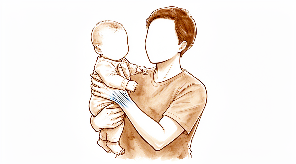
For patients: a plain-language version of this topic is available. See the patient guide.
Overview
De Quervain's tenosynovitis involves the first dorsal compartment, where release is an effective treatment for tendinopathy [5]. Non-operative management remains the preferred initial strategy, with corticosteroid injection being the only nonsurgical treatment that can potentially modify the disease course [17]. This approach, often combined with short-duration immobilization or hand therapy, is primary and effective [18, 19]. Combined orthosis and injection approaches are more effective than either intervention alone [23], and repeat injections remain a viable clinical option despite decreasing success rates with multiple attempts [33]. Patients with lower physical function scores or higher pain interference have significantly increased odds of eventually undergoing surgical release [13]. Furthermore, negative perceptions of consequences and worse pain catastrophizing are associated with worse baseline pain and reduced function in patients awaiting decompression [28]. Addressing misconceptions regarding symptom duration and consequences allows patients to make informed decisions aligned with their values [16].
Surgical indications include specific clinical scenarios such as triggering due to de Quervain tenosynovitis, which is rare but typically requires surgical release [8]. In cases where constriction involves only the extensor pollicis brevis in a separate compartment, exploration of both compartments is advised [2]. Post-traumatic de Quervain's syndrome is uncommon and often overlooked, but once diagnosed, it is typically successfully treated non-operatively [12]. Radiographs of the wrist are necessary to rule out other lesions causing wrist pain in patients not responding to conservative management [11]. Additionally, first dorsal compartment release during volar approach for distal radius fracture fixation reduces symptoms in patients with pre-existing De Quervain disease compared with the no release group during short-term follow-up [1].
Surgical techniques include simple release and Z-plasty, both of which are effective methods [10]. Tendoscopic and endoscopic releases provide earlier symptom relief, recovery, and scar satisfaction with fewer superficial radial nerve complications compared to open release, while maintaining equivalent long-term outcomes [4, 15]. The presence of a septum does not significantly affect clinical outcomes or complications following endoscopic release [9]. Reconstruction procedures on the first compartment are satisfactory for treating instability of the EPB and APL tendons after primary release [14], and extensor retinaculum reconstruction using the wide-awake approach can be broadly applied without specialized equipment [7]. Dissatisfaction after release can result from incomplete release, tendon subluxation, nerve injury, or recovery duration [5]. The scientific literature on the management of de Quervain tendinopathy is sparse and limited largely to uncontrolled cohorts with low-quality randomized trials [6].
Anatomy & Pathophysiology
First Dorsal Compartment Anatomy
The abductor pollicis longus and extensor pollicis brevis tendons are visible between the radial styloid proximally and the base of the first metacarpal distally [66]. Compartmental configuration varies significantly; in 72% of wrists, dye injected into the first dorsal compartment dispersed into a single compartment containing both tendons, while in 28% a separate septum existed for the extensor pollicis brevis tendon [93]. A common septum is found between the abductor pollicis longus and extensor pollicis brevis in 80% of patients requiring surgical release for de Quervain tenosynovitis [46]. Variations in the insertion of the abductor pollicis longus tendon are more common and occur in greater number per forearm than generally realized [52]. In cases where constriction involves only the extensor pollicis brevis in a separate compartment, exploration of both compartments is advised [2].
Ultrasonography consistently provided a reliable evaluation of the pertinent first extensor compartment anatomy and improved the accuracy of needle placement for first extensor compartment injection in a cadaver model [31]. The presence of an intracompartmental septum detected by ultrasound is associated with the failure of ultrasound-guided steroid injection in de Quervain’s syndrome [22].
Nerve Anatomy
Anatomic knowledge of the course of the superficial radial nerve and its branches is important during open release for avoiding nerve injury [36]. Surgical release of the first dorsal compartment requires care to protect the radial sensory nerve and identify all accessory compartments [50].
Pathophysiology & Etiology
The cause of De Quervain's tenosynovitis does not correlate with manual work or trauma based on cadaver studies, authors' clinical cases, and systematic review [34]. Conversely, De Quervain tenosynovitis results from anatomical factors associated with some mechanical stressors, with women more affected than men due to manual work influencing first compartment dynamics [77]. Testosterone replacement therapy is associated with an increased likelihood of de Quervain tenosynovitis and an increased likelihood of requiring surgical release for the condition [3]. Operative treatment for distal radius fractures confers a slightly greater risk of developing DeQuervain tenosynovitis within 1 year of injury or surgery [51]. Post-traumatic De Quervain's syndrome can occur following an injury causing radial-sided wrist pain that persists, with a clear temporal link from the accident to initial and continuing symptoms [30]. Tenosynovial swelling from rheumatoid arthritis may contribute to de Quervain disease [73].
Diagnostic Considerations
The wrist hyperflexion and abduction of the thumb test is a more precise tool for the diagnosis of de Quervain's disease than the Eichhoff's test [45]. In a large population analysis, the epidemiology of stenosing tenosynovitis of the first extensor compartment has been described [20].
Classification
Anatomical Variants: Septation of the first extensor compartment is more common in patients with de Quervain disease than in the general population [42]. When the extensor pollicis brevis tendon resides in a subcompartment, it is likely to do so particularly in patients with de Quervain's disease [21]. The presence of an intracompartmental septum does not significantly affect clinical outcomes or complications following endoscopic release for de Quervain's syndrome [9].
Other Considerations: Post-traumatic de Quervain's syndrome is very uncommon and often overlooked initially due to its rarity [12]. A patient apparently presenting with de Quervain's disease was classified as derangement syndrome using Mechanical Diagnosis and Therapy [39].
Clinical Presentation
Diagnostic Testing: The wrist hyperflexion and abduction of the thumb (WHAT) test is a more precise tool for the diagnosis of de Quervain's disease than the Eichhoff's test [45]. This test could be adopted to guide clinical diagnosis in the early stages of de Quervain's tendinopathy [45]. A staged version of the Finkelstein test is reliable, easy, and reproducible for diagnosing de Quervain's tendonitis [29]. This staged approach causes minimal discomfort compared to traditional descriptions [29].
Anatomical Variations: Septation of the first extensor compartment suggests an anatomical variation that may play an etiological role in de Quervain disease [42]. A common septum is found between the APL and EPB in 80% of patients requiring surgical release for de Quervain tenosynovitis [46].
Psychosocial Factors: More negative perceptions of the consequences of de Quervain's tenosynovitis are associated with worse pain and reduced function at baseline in patients awaiting surgical decompression [28]. Worse pain catastrophizing is associated with worse pain and reduced function at baseline in patients awaiting surgical decompression of de Quervain's tenosynovitis [28].
Systemic Associations: Testosterone replacement therapy is associated with an increased likelihood of de Quervain tenosynovitis [3]. It is also associated with an increased likelihood of requiring surgical release for de Quervain tenosynovitis [3]. Growth hormone abuse is associated with a more recalcitrant form of de Quervain tenosynovitis that does not respond well to nonsurgical treatment [48]. This abuse leads to an increased likelihood of surgical decompression for de Quervain tenosynovitis [48].
Post-Traumatic Course: Once diagnosed, post-traumatic de Quervain's syndrome is typically successfully treated non-operatively [12].
Investigations
Clinical Examination
A staged version of the Finkelstein test is reliable, easy, and reproducible for diagnosing de Quervain's tendonitis while causing minimal discomfort compared to traditional descriptions [29]. When the extensor pollicis brevis tendon is involved in thumb interphalangeal joint extension, particularly in patients with de Quervain's disease, it is likely to reside in a subcompartment of the first dorsal compartment [21].
Imaging
MRI: MRI is the modality of choice for imaging radiographically occult fractures of the hand and wrist [60]. High-resolution MRI aids in the evaluation of ligament injuries of the wrist, with a static magnetic field strength of at least 1.5 T using a dedicated wrist coil recommended for analyzing ligament insertions [69]. MRI has an expanding role in the evaluation of pathologic conditions of the elbow and wrist, requiring high-resolution images best obtained with surface coil technique and high field system [71]. MRI is useful in detecting additional marrow abnormalities in osteonecrosis, as seen in the lunate in Kienböck disease or in the scaphoid after fracture [71]. MRI provides earlier detection of synovitis and erosive bone changes associated with rheumatoid arthritis than do radiographs [71]. Tenosynovitis and tendon injuries in the wrist and hand can be assessed using MRI [71].
CT: CT scanning enables the 3D analysis of carpal dysfunction [69].
Dynamic fluoroscopy: Dynamic fluoroscopy shows abnormal motion between the scaphoid and lunate and changes in the kinematics of the midcarpal joint [69].
Arthroscopy: Arthroscopy is considered by many to be the diagnostic intervention of choice for determining the degree of injury to the wrist and can assess the condition of the cartilage [69].
Other Considerations
Patient-Reported Outcomes and Psychological Factors: A healthy inner narrative about de Quervain can alleviate symptoms and limitations, and surgery should be a choice based on values rather than misconceptions or cognitive biases [35].
Associated Conditions and Etiology: Testosterone replacement therapy is associated with an increased likelihood of both trigger finger and de Quervain tenosynovitis, and an increased likelihood of requiring surgical release for both conditions [3]. Growth hormone abuse is associated with a more recalcitrant form of de Quervain tenosynovitis that does not respond well to nonsurgical treatment, thus leading to increased likelihood of surgical decompression [48]. The cause of De Quervain's tenosynovitis does not correlate with manual work or trauma from cadaver studies, authors' clinical cases, and systematic review [34]. In analysis of a large population, the epidemiology of stenosing tenosynovitis of the first extensor compartment has been described [20].
Treatment
Non-Operative
Corticosteroid injection is the only available nonsurgical treatment that can potentially modify the course of de Quervain's tenosynovitis and is therefore the preferred initial treatment [17]. The combined use of corticosteroid injection and hand therapy intervention decreases pain and symptomology as measured through provocative testing, though the strength of the evidence is limited [19]. A single cortisone injection was effective in alleviating symptoms in 82% of patients, with over half remaining symptom-free for at least 12 months [32]. Injection of steroids and immobilization in a splint resulted in a satisfactory outcome in 62 per cent of wrists at a mean of eighteen months [27]. Post-traumatic de Quervain's syndrome is typically successfully treated non-operatively once diagnosed [12].
Operative
Indications: Patients who scored lower than 40 for physical function or higher than 60 for pain interference had significantly increased odds of eventually undergoing surgical release [13].
Surgical Approach / Technique: First dorsal compartment release during a volar approach for distal radius fracture fixation demonstrated a significantly greater reduction in de Quervain disease symptoms in the release group compared with the no release group during short-term follow-up [1]. When the extensor pollicis brevis tendon resides in a subcompartment of the first dorsal compartment, it is likely to be associated with de Quervain's disease [21].
Revision: Reconstruction procedures on the first compartment seemed to be satisfactory in treating instability of the EPB and APL tendons after primary surgical release for de Quervain's disease [14].
Complications
Operative Complications
Nerve injury: Endoscopic release for de Quervain's tenosynovitis is associated with fewer superficial radial nerve complications compared with open release [15]. Dissatisfaction following first dorsal compartment release can result from nerve injury, among other factors [5].
Tendon instability: Tendon subluxation or instability of the extensor pollicis brevis and abductor pollicis longus tendons following primary surgical release can be treated with reconstruction procedures on the first compartment [14]. Dissatisfaction following first dorsal compartment release can also result from tendon subluxation [5].
Incomplete release: Dissatisfaction following first dorsal compartment release can result from incomplete release [5]. In cases where constriction involves only the extensor pollicis brevis in a separate compartment, exploration of both compartments is advised to address symptoms [2].
Recovery duration: Dissatisfaction following first dorsal compartment release can result from recovery duration [5].
Non-Operative Complications and Risks
Steroid injection response: Patients respond to extra-articular steroid injections with gradual improvement over the course of the first week [37].
Recovery
Light activity (weeks): The evidence provided does not specify a typical week range for light activities such as desk work, driving, or light ADLs. However, one patient experienced striking relief within 24 hours after operation, regaining the ability to flex and extend the thumb [49].
Full activity (months): The evidence provided does not specify a month range for the return to manual work, sport, or full range of motion and strength.
Complete recovery / outcome plateau (months): The evidence provided does not specify a month range for when pain, strength, and final functional outcomes stabilise.
Rehabilitation protocol: The evidence provided does not detail specific PT phasing, immobilisation duration, weight-bearing/ROM progression, or sling/brace removal timing.
Functional milestones: The evidence provided does not report validated PROM trajectories or specific outcome-measure benchmarks.
Other Considerations: Tendoscopic release for de Quervain's disease provides earlier symptom relief and earlier recovery compared with traditional open release technique [4]. Endoscopic release for de Quervain's tenosynovitis provides earlier improvement after surgery compared with open release [15]. Tendoscopic release for de Quervain's disease is associated with fewer complications compared with traditional open release technique [4]. Tendoscopic release for de Quervain's disease results in a more desirable scar compared with traditional open release technique [4]. Endoscopic release for de Quervain's tenosynovitis results in greater scar satisfaction compared with open release [15]. Tendoscopic release for de Quervain's disease provides equivalent successful long-term outcome compared with traditional open release technique [4]. Dissatisfaction after first dorsal compartment release can result from recovery duration [5].
Key Evidence
- [L1] The current results demonstrated a significantly greater reduction in de Quervain disease symptoms in the release group compared with the no release group during the short-term follow-up. [1] (10.1016/j.jhsg.2024.03.009)
- [L4] In cases with symptoms of de Quervain's syndrome where the constriction involves only the extensor pollicis brevis in a separate compartment, exploration of both compartments is advised. [2] (10.2106/00004623-194931040-00019)
- [L2] TRT is associated with an increased likelihood of both trigger finger and de Quervain tenosynovitis, and an increased likelihood of requiring surgical release for both conditions. [3] (10.1016/j.jhsa.2024.01.018)
- [L3] The results of this study suggest that tendoscopic technique for de Quervain's disease could provide earlier symptom relief and earlier recovery with fewer complications and more desirable scar, as well as equivalent successful long-term outcome, when compared with traditional open release technique. [4] (10.1186/s13018-019-1393-5)
- [L4] Release of the first dorsal compartment is an effective treatment of de Quervain tendinopathy, though dissatisfaction can result from incomplete release, tendon subluxation, nerve injury, or recovery duration. [5] (10.1016/j.jhsa.2015.09.003)
- [L4] The scientific literature on the surgical and nonsurgical management of de Quervain tendinopathy is sparse and limited largely to uncontrolled cohorts with low-quality randomized trials. [6] (10.1016/j.jhsa.2013.06.003)
- [L4] These procedures can be broadly applied without specialized equipment for optimizing function in de Quervain tenosynovitis. [7] (10.1016/j.jhsa.2017.07.024)
- [L4] Triggering due to de Quervain tenosynovitis is a rare condition where surgical release is required in most cases. [8] (10.1016/j.jhsg.2022.04.004)
- [L4] The presence of a septum does not significantly affect clinical outcomes or complications following endoscopic release for de Quervain's syndrome. [9] (10.1177/17531934231214137)
- [L4] Both simple release and Z-plasty were effective surgical methods for de Quervain's disease. [10] (10.1177/1753193418818341)
- [L4] Radiographs of the wrist are necessary to rule out any other lesion resulting in wrist pain in patients not responding to conservative management of de Quervain's disease. [11] (10.1007/s11552-010-9258-8)
- [L4] Post-traumatic de Quervain's syndrome is very uncommon and often overlooked initially due to its rarity, but once diagnosed is typically successfully treated non-operatively. [12] (10.1177/1753193416646722)
- [L4] Patients who scored lower than 40 for physical function or higher than 60 for pain interference had significantly increased odds of eventually undergoing surgical release for de Quervain tenosynovitis. [13] (10.1016/j.jhsa.2023.07.005)
- [L4] The reconstruction procedures on the first compartment seemed to be satisfactory in treating instability of the EPB and APL tendons after primary surgical release for de Quervain's disease. [14] (10.1055/s-0037-1603686)
- [L1] Endoscopic release for de Quervain's tenosynovitis seems to provide earlier improvement after surgery, with fewer superficial radial nerve complications and greater scar satisfaction, when compared with open release. [15] (10.1302/0301-620x.95b7.31486)
- [L3] Addressing misconceptions about de Quervain's tenosynovitis in terms of the consequences for patients and how long their symptoms will last should allow patients to make informed decisions about the treatment that best matches their values. [16] (10.1097/corr.0000000000001577)
- [L4] According to the limited evidence available, injection of corticosteroids is the only available nonsurgical treatment that can potentially modify the course of de Quervain's tenosynovitis and is therefore the preferred initial treatment. [17] (10.1016/j.jhsa.2008.12.030)
- [L1] Corticosteroid injection with a short duration of immobilization remains the primary and effective treatment for de Quervain tenosynovitis. [18] (10.1016/j.jhsa.2024.03.003)
- [L1] The paper supports the combined use of corticosteroid injection and hand therapy intervention to decrease pain and symptomology as measured through provocative testing in patients with de Quervain's, though the strength of the evidence is limited. [19] (10.1016/j.jht.2015.12.004)
- [L2] In analysis of a large population, we have described the epidemiology of stenosing tenosynovitis of the first extensor compartment. [20] (10.1016/j.jhsa.2008.08.020)
- [L4] When it does, particularly in patients with de Quervain's disease, it is likely to reside in a subcompartment of the first dorsal compartment. [21] (10.1016/j.jhsa.2008.12.015)
- [L3] [22] (10.1177/1753193415611414)
- [L1] Combined orthosis/corticosteroid injection approaches are more effective than either intervention alone in the treatment of de Quervain's disease. [23] (10.1016/j.jht.2015.10.004)
- [L3] More negative perceptions of the consequences of de Quervain's tenosynovitis and worse pain catastrophizing are associated with worse pain and reduced function at baseline in patients awaiting surgical decompression of de Quervain's tenosynovitis. [28] (10.1097/corr.0000000000000992)
- [L4] The authors describe a staged version of the Finkelstein test that is reliable, easy, and reproducible for diagnosing de Quervain's tendonitis while causing minimal discomfort compared to traditional descriptions. [29] (10.1016/j.jhsa.2010.05.022)
- [Letter] [30] (10.1177/1753193417726668)
- [L5] Ultrasonography consistently provided a reliable evaluation of the pertinent first extensor compartment anatomy and, in this cadaver model, improved the accuracy of needle placement for first extensor compartment injection. [31] (10.5435/jaaos-d-15-00753)
- [L4] A single cortisone injection was effective in alleviating symptoms of de Quervain tendinopathy in 82% of patients, with over half remaining symptom-free for at least 12 months. [32] (10.1016/j.jhsa.2014.12.027)
- [L2] Although the success rate for the treatment of De Quervain's tenosynovitis decreases with multiple injections, repeat injections have a high rate of success and are a viable clinical option. [33] (10.1016/j.jhsa.2021.04.018)
- [Paper] The cause of De Quervain's tenosynovitis does not correlate with manual work or trauma from cadaver studies, authors' clinical cases, and systematic review. [34] (10.1055/s-0039-1683984)
- [L5] A healthy inner narrative about de Quervain can alleviate symptoms and limitations, and surgery should be a choice based on values rather than misconceptions or cognitive biases. [35] (10.1097/corr.0000000000001009)
- [L5] Anatomic knowledge of the course of the superficial radial nerve and its branches is important during open release for avoiding nerve injury. [36] (10.1016/j.jhsa.2013.12.004)
- [L1] Patients respond to extra-articular steroid injections with gradual improvement over the course of the first week. [37] (10.1016/j.jhsa.2007.08.002)
- [L4] A patient apparently presenting with de Quervain's disease was classified as derangement syndrome using MDT. [39] (10.1016/j.jht.2009.03.002)
- [L4] Septation of the first extensor compartment is more common in patients with de Quervain disease than in the general population, suggesting this anatomical variation may play an etiological role. [42] (10.2106/00004623-198668060-00016)
- [L2] The wrist hyperflexion and abduction of the thumb test is a more precise tool for the diagnosis of de Quervain's disease than the Eichhoff's test and could be adopted to guide clinical diagnosis in the early stages of de Quervain's tendinopathy. [45] (10.1177/1753193412475043)
- [L3] Our results suggest that growth hormone abuse is associated with a more recalcitrant form of de Quervain tenosynovitis that does not respond well to nonsurgical treatment, thus leading to increased likelihood of surgical decompression. [48] (10.1177/0363546509337993)
- [L5] In resistant cases, surgical release of the first dorsal compartment is done, taking care to protect the radial sensory nerve and identify all accessory compartments. [50] (10.5435/00124635-200712000-00009)
- [Paper] Operative treatment for distal radius fractures confers a slightly greater risk of developing DeQuervain tenosynovitis within 1 year of injury or surgery. [51] (10.1177/15589447251366456)
- [L4] The occurrence of variations in the insertion of the abductor pollicis longus tendon is more common and in greater number per forearm than has been generally realized. [52] (10.2106/00004623-195133020-00007)
- [L4] [77] (10.1177/0363546504268134)
- [L5] In 72% of wrists, dye dispersed into one compartment containing both tendons, but in 28% a separate septum existed for the EPB tendon. [93] (10.1177/1753193411409126)
See Also
References
[1] First Dorsal Compartment Release During Volar Approach for Distal Radius Fracture Fixation Reduces Symptoms in Patients With Pre-Existing De Quervain Disease. Journal of Hand Surgery Global Online. 2024. DOI: 10.1016/j.jhsg.2024.03.009
[2] AN UNUSUAL FORM OF DE QUERVAINʼS SYNDROME. The Journal of Bone & Joint Surgery. 1949. DOI: 10.2106/00004623-194931040-00019
[3] Testosterone Replacement Therapy and Associated Rates of Trigger Finger, de Quervain Tenosynovitis, and Their Subsequent Management. The Journal of Hand Surgery. 2024. DOI: 10.1016/j.jhsa.2024.01.018
[4] Tendoscopic versus open release for de Quervain’s disease: earlier recovery with 7.21 year follow-up. Journal of Orthopaedic Surgery and Research. 2019. DOI: 10.1186/s13018-019-1393-5
[5] Dissatisfaction After First Dorsal Compartment Release for de Quervain Tendinopathy. The Journal of Hand Surgery. 2016. DOI: 10.1016/j.jhsa.2015.09.003
[6] Current Treatment of de Quervain Tendinopathy. The Journal of Hand Surgery. 2013. DOI: 10.1016/j.jhsa.2013.06.003
[7] Extensor Retinaculum Reconstruction Using the Wide-Awake Approach. The Journal of Hand Surgery. 2017. DOI: 10.1016/j.jhsa.2017.07.024
[8] Triggering Thumb Is Not Always a Trigger Thumb. Journal of Hand Surgery Global Online. 2022. DOI: 10.1016/j.jhsg.2022.04.004
[9] Prediction of an intracompartmental septum and its effect on outcomes of endoscopic release for de Quervain’s syndrome. Journal of Hand Surgery (European Volume). 2023. DOI: 10.1177/17531934231214137
[10] Comparison between simple release and Z-plasty of retinaculum for de Quervain’s disease: a retrospective study. Journal of Hand Surgery (European Volume). 2019. DOI: 10.1177/1753193418818341
[11] Does Radial Styloid Abnormality in de Quervain's Disease Affect the Outcome of Management?. HAND. 2010. DOI: 10.1007/s11552-010-9258-8
[12] Post-traumatic de Quervain’s syndrome: a rare condition, often diagnosed late. Journal of Hand Surgery (European Volume). 2016. DOI: 10.1177/1753193416646722
[13] Association of Patient-Reported Outcomes Measurement Information System Measures With Injection and Surgical Treatment Response in Patients With De Quervain Tenosynovitis. The Journal of Hand Surgery. 2023. DOI: 10.1016/j.jhsa.2023.07.005
[14] Pulley Reconstruction for Symptomatic Instability of the Tendons of the First Extensor Compartment Following de Quervain's Release. Journal of Wrist Surgery. 2017. DOI: 10.1055/s-0037-1603686
[15] Endoscopicversusopen release in patients with de Quervain’s tenosynovitis. The Bone & Joint Journal. 2013. DOI: 10.1302/0301-620x.95b7.31486
[16] Are Patient Expectations and Illness Perception Associated with Patient-reported Outcomes from Surgical Decompression in de Quervain’s Tenosynovitis?. Clinical Orthopaedics & Related Research. 2020. DOI: 10.1097/corr.0000000000001577
[17] Nonsurgical Treatment for de Quervain's Tenosynovitis. The Journal of Hand Surgery. 2009. DOI: 10.1016/j.jhsa.2008.12.030
[18] Advancements in de Quervain Tenosynovitis Management: A Comprehensive Network Meta-Analysis. The Journal of Hand Surgery. 2024. DOI: 10.1016/j.jhsa.2024.03.003
[19] Clinical Relevance Commentary on: Hand therapy versus corticosteroid injections in the treatment of de Quervain's disease: A systematic review and meta-analysis. Journal of Hand Therapy. 2016. DOI: 10.1016/j.jht.2015.12.004
[20] Incidence of de Quervain's Tenosynovitis in a Young, Active Population. The Journal of Hand Surgery. 2009. DOI: 10.1016/j.jhsa.2008.08.020
[21] Thumb Interphalangeal Joint Extension By the Extensor Pollicis Brevis: Association With a Subcompartment and de Quervain's Disease. The Journal of Hand Surgery. 2009. DOI: 10.1016/j.jhsa.2008.12.015
[22] Presence of an intracompartmental septum detected by ultrasound is associated with the failure of ultrasound-guided steroid injection in de Quervain’s syndrome. Journal of Hand Surgery (European Volume). 2015. DOI: 10.1177/1753193415611414
[23] Hand therapy versus corticosteroid injections in the treatment of de Quervain's disease: A systematic review and meta-analysis. Journal of Hand Therapy. 2016. DOI: 10.1016/j.jht.2015.10.004
[27] Treatment of de Quervain tenosynovitis. A prospective study of the results of injection of steroids and immobilization in a splint.. The Journal of bone and joint surgery. American volume. 1991.
[28] Which Psychological Variables Are Associated With Pain and Function Before Surgery for de Quervain’s Tenosynovitis? A Cross-sectional Study. Clinical Orthopaedics & Related Research. 2019. DOI: 10.1097/corr.0000000000000992
[29] Staged Description of the Finkelstein Test. The Journal of Hand Surgery. 2010. DOI: 10.1016/j.jhsa.2010.05.022
[30] Letter and reply. Journal of Hand Surgery (European Volume). 2017. DOI: 10.1177/1753193417726668
[31] Ultrasonography-guided de Quervain Injection. Journal of the American Academy of Orthopaedic Surgeons. 2016. DOI: 10.5435/jaaos-d-15-00753
[32] de Quervain Tendinopathy: Survivorship and Prognostic Indicators of Recurrence Following A Single Corticosteroid Injection. The Journal of Hand Surgery. 2015. DOI: 10.1016/j.jhsa.2014.12.027
[33] De Quervain Tenosynovitis: An Evaluation of the Epidemiology and Utility of Multiple Injections Using a National Database. The Journal of Hand Surgery. 2022. DOI: 10.1016/j.jhsa.2021.04.018
[34] De Quervain's Tenosynovitis. Journal of Wrist Surgery. 2019. DOI: 10.1055/s-0039-1683984
[35] CORR Insights®: Which Psychological Variables Are Associated With Pain and Function Before Surgery for de Quervain’s Tenosynovitis? A Cross-sectional Study. Clinical Orthopaedics & Related Research. 2019. DOI: 10.1097/corr.0000000000001009
[36] The Relationship of the Superficial Radial Nerve and Its Branch to the Thumb to the First Extensor Compartment. The Journal of Hand Surgery. 2014. DOI: 10.1016/j.jhsa.2013.12.004
[37] Extra-Articular Steroid Injection: Early Patient Response and the Incidence of Flare Reaction. The Journal of Hand Surgery. 2007. DOI: 10.1016/j.jhsa.2007.08.002
[39] Application of Mechanical Diagnosis and Therapy to a Patient Diagnosed with de Quervain's Disease: A Case Study. Journal of Hand Therapy. 2009. DOI: 10.1016/j.jht.2009.03.002
[42] Anatomical variations in the first extensor compartment of the wrist. A clinical and anatomical study.. The Journal of Bone & Joint Surgery. 1986. DOI: 10.2106/00004623-198668060-00016
[45] The wrist hyperflexion and abduction of the thumb (WHAT) test: a more specific and sensitive test to diagnose de Quervain tenosynovitis than the Eichhoff’s Test. Journal of Hand Surgery (European Volume). 2013. DOI: 10.1177/1753193412475043
[46] Aaos Comprehensive Orthopaedic Review 3. Tendinopathy of the Hand and Wrist* > VII. Extensor Carpi Ulnaris Tenosynovitis.
[48] Abuse of Growth Hormone Increases the Risk of Persistent de Quervain Tenosynovitis. The American Journal of Sports Medicine. 2009. DOI: 10.1177/0363546509337993
[49] 10_1097_00000658_193804000_00015. 1938.
[50] de Quervain Tenosynovitis of the Wrist. Journal of the American Academy of Orthopaedic Surgeons. 2007. DOI: 10.5435/00124635-200712000-00009
[51] Incidence of DeQuervain Tenosynovitis After Distal Radius Fractures. HAND. 2025. DOI: 10.1177/15589447251366456
[52] ANATOMICAL AND CLINICAL STUDY OF THE VARIATIONS IN THE INSERTIONS OF THE ABDUCTOR POLLICIS LONGUS TENDON, ASSOCIATED WITH STENOSING TENDOVAGINITIS. The Journal of Bone & Joint Surgery. 1951. DOI: 10.2106/00004623-195133020-00007
[60] Orthopaedic Knowledge Update 13 Ebook Without Multimedia. Anatomy, Evaluation, Clinical Examination, and Imaging > Imaging: Advances in Imaging of the Hand and Upper Extremity > Magnetic Resonance Imaging.
[66] Exam Of The Hand Wrist 2Ed. 2.3 EXAMINATION OF THE MUSCULOTENDINOUS APPARATUS.
[69] Green S Operative Hand Surgery. Advanced Imaging.
[71] Campbell S Operative Orthopaedics 4 Volume Set. WRIST AND ELBOW.
[73] Campbell S Operative Orthopaedics 4 Volume Set. THUMB CARPODNETACARPAL ARTHRODESIS WITH KIRSCHNER WIRE OR BLADE-PLATE FIXATION > SYNOVITIS OF THE WRIST.
[77] De Quervain Disease in Volleyball Players. The American Journal of Sports Medicine. 2005. DOI: 10.1177/0363546504268134
[93] Accuracy of intrasheath injection techniques for de Quervain’s disease: a cadaveric study. Journal of Hand Surgery (European Volume). 2012. DOI: 10.1177/1753193411409126