Patients › Wrist
De Quervain's Tenosynovitis
Patient-facing topic on De Quervain's tenosynovitis (first dorsal compartment) — diagnosis, conservative management, and indications for de Quervain's release.
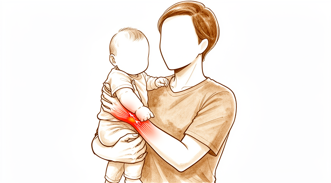
What you're feeling
De Quervain's tenosynovitis causes pain on the thumb side of your wrist, near the base of the thumb. The area can also feel tender and slightly swollen. The problem sits in a narrow tunnel that two thumb tendons slide through, and when that tunnel is irritated, the tendons catch as they move.
Certain movements make the pain worse. Bending your wrist towards the little-finger side, gripping firmly while bending the wrist, or pinching your thumb and index finger together can all set it off. You might notice it when lifting a kettle, turning a door handle, picking up your baby, or using scissors. Some people find the pain flares on waking or after a stretch of activity.
The cause is not clearly linked to your job or to a specific injury. It can appear alongside other wrist problems, but often it develops on its own.
Your surgeon can usually diagnose this by examining your wrist and hearing where the pain sits. Scans are not needed when the picture is clear. Imaging is only used if there is something else to check, such as an old wrist fracture or arthritis at the base of the thumb.
Most people who understand this condition start with simple treatment first, and most find their symptoms settle within one year. A steroid injection into the tender tunnel is the usual first step, and it works within two injections for 73.4% of people. Splinting alone is less effective than an injection.
If your pain is severe, or it stops you doing the things you need to do, surgery to release the tight tunnel may be discussed. This is called a release of the first extensor compartment, and it is the standard treatment when injections have not helped enough.
What's actually happening
The two tendons that move your thumb out and away from your hand run through a narrow tunnel on the thumb side of your wrist. Think of the tendons as smooth cords and the tunnel as a pulley strap that holds them close to the bone. In this condition, the tunnel becomes tight and thickened, so the cords lose their easy glide.
The friction between tendon and tunnel causes swelling inside that space. Over time, the tendon sheath itself changes: it becomes dense and fibrous, and the tissue takes on a gel-like quality. The tendons can also develop sticky bands that glue them to the tunnel wall. The result is a cramped, narrowed channel, and every thumb movement pulls the swollen cords through it. That pulling irritates pain sensors in the strap, which is why the pinch and grip movements you read about above hurt.
The tunnel can also be naturally narrow in some people. An extra wall inside it, splitting the space into two compartments, is present in about a third of the population, and some people have extra tendon slips sharing the same tight space. These variations are found on both wrists in some people, and they make crowding more likely.
This condition is often called tenosynovitis, which sounds like inflammation of the tendon lining. In fact, the tissue changes are closer to wear and degeneration than to classic inflammation. The swelling and thickening are real, but they come from the tendon sheath breaking down under load rather than from a simple flare of inflammation.
The pain is not a sign that you have damaged your wrist through overuse. No clear link has been established between hand use at work or a specific injury and this condition. It is more common in women, and it can appear after other wrist problems such as a wrist fracture treated with surgery.
What we can do about it
Dr Kieran Hirpara, an upper-limb surgeon at Mater Private Hospital Rockhampton, starts with the least invasive options that suit your condition. Patients are generally referred to our clinic by their GP; if a physiotherapist has suggested you see us, you will still need a referral from your GP in order to be eligible for the Medicare rebate. At your first visit we take a history, examine your wrist and arrange imaging only if something else needs checking. Because this problem is long-standing rather than a fresh injury, we usually begin with non-operative care.
The first steps are things you can do yourself. Resting the thumb and wrist helps, and a thumb spica splint holds the thumb and wrist still so the irritated tunnel gets a break. A splint can settle the pain while you wear it, but it does not keep working once the joints are moving freely again. Hand therapy can be added to an injection, and this combination has been shown to reduce pain on testing [2]. There are also treatments where a medicated cream is pushed into the skin by a small electric current, or where sound waves are pulsed into the tender area; these may improve how the wrist works and ease pain.
The main medical treatment is a cortisone injection into the tunnel. Cortisone is a strong anti-inflammatory medicine that calms the swelling around the tendons. A single injection relieved symptoms in 82% of patients, and more than half of those stayed symptom-free for at least 12 months. If you have diabetes, one injection is less likely to work for you, but repeat injections do not lose their effect. Combining the injection with a splint works better than an injection on its own. Other injections, such as hyaluronic acid or PRP, are not part of the treatment we offer for this condition.
If non-operative care has not given you enough relief, we will talk about surgery. The operation releases the tight tunnel so the tendons can glide freely again, and it is considered when injections and splinting have not settled your symptoms. We will go through what the operation involves, what recovery looks like and what the results are before you decide anything together with us.
What to expect
For most people, this condition settles with time. Most patients find their symptoms resolve within one year. Many people manage well without surgery: only about a third of people with this condition end up needing an operation within two years, and when surgery is needed, it usually happens within the first year.
Treatment aims to shorten that journey. A cortisone injection with a short period of splinting is the mainstay of early care, and it works for most people. If the first injection helps but the pain creeps back, a second injection is still a reasonable option. The success rate does drop with repeat injections, but they remain a useful tool.
If you have diabetes, a single injection is less likely to settle your symptoms than it is for other people. The good news is that further injections do not lose their effect in people with diabetes, so repeating the treatment stays worthwhile.
Surgery is reserved for the smaller group whose pain does not settle with injections and splinting. When that happens, releasing the tight tunnel gives lasting relief for the tendons, and the operation carries a low rate of complications.
How you think about the condition matters too. People who expect the worst tend to report more pain and less function, so a clear picture of what lies ahead can help you choose the treatment that fits your life. Worrying that you have damaged your wrist through overuse is not supported by the evidence, and knowing that can take some of the fear out of the flare-ups.
There is no single guaranteed timeline. Some wrists respond quickly to an injection, others need a repeat dose, and a few go on to surgery. What tends to happen when this condition is managed well is that the pain eases and the tendons glide freely again. Left alone, most cases still settle within a year, but you may spend months managing the discomfort in the meantime.
When to see someone
See your GP if the pain on the thumb side of your wrist keeps coming back, or if it stops you doing your usual tasks despite rest and a splint. Ask for a specialist review if injections have not settled things, or if the pain is severe enough to limit how you use your hand day to day. Some people find their symptoms ease within a year on their own, but waiting is not the only option, and treatment can shorten that stretch. If you notice new numbness, tingling or unusual sensations in the thumb or wrist that do not match the usual pattern, mention this at your appointment, as other wrist problems can sometimes mimic this condition.
In more depth
Advanced reading: the deeper science (optional)
This section goes further than you need for your own treatment decisions. De Quervain's tenosynovitis is worth the extra reading because of a small anatomical variant that accounts for much of the dissatisfaction after an operation that is otherwise reliable, and because the best non-operative result comes from combining two treatments rather than choosing between them.
The combination beats either part
The first dorsal compartment holds two tendons in a tunnel at the thumb side of the wrist. Treatment aims to settle inflammation in that tunnel and reduce the load passing through it.
A network meta-analysis of 823 patients concluded that corticosteroid injection with a short duration of immobilisation remains the primary and effective treatment, with extracorporeal shockwave therapy a secondary option [1]. Looking at the components directly, combined orthosis and corticosteroid injection approaches are more effective than either intervention alone [2].
That is a more specific instruction than "try a splint, then an injection if it fails". The evidence supports doing both together, with the splint worn for a defined short period after the injection.
A second injection is still worth having
Where symptoms return, the reflex is to conclude injections have failed and to move to surgery. The data across a large cohort say otherwise: although the success rate decreases with multiple injections, repeat injections have a high rate of success and are a viable clinical option [3].
Declining success with repetition is expected. But a lower success rate is not the same as futility, and a second injection remains a reasonable step rather than a delaying tactic.
The variant that explains most surgical disappointment
Release of the first dorsal compartment is effective, and where it disappoints there is usually a specific reason. Dissatisfaction can result from incomplete release, tendon subluxation, nerve injury, or simply the duration of recovery, and an unidentified and unreleased extensor pollicis brevis subsheath is a discrete source of dissatisfaction [4].
This deserves unpacking because it is the single most useful fact in this section. In a substantial proportion of people the compartment is not one tunnel but two, with the extensor pollicis brevis tendon running in its own separate sheath alongside the other. A release that opens the main compartment and stops leaves that second sheath intact, and the tendon inside it still compressed. The wrist is opened, the operation is completed as described, and the symptoms persist.
It is also why the two other listed causes matter: releasing too widely allows the tendons to subluxate out of the groove with wrist movement, producing a painful snap, and the superficial branch of the radial nerve crosses immediately over the operative field. The compartment must be opened completely but not excessively, with a nerve in the way, which is why an apparently minor operation warrants the same care as a larger one.
Not everything at the radial wrist is de Quervain's
Pain in this region has a differential worth knowing, because the treatments differ: arthritis at the base of the thumb, intersection syndrome a few centimetres further up the forearm, and radial nerve irritation can all produce pain in overlapping territory. A localised tenderness directly over the compartment, with pain reproduced by ulnar deviation of the wrist with the thumb tucked in the palm, is what points to the tendon sheath rather than to its neighbours.
References for the advanced reading
- Chong HH, Pradhan A, Dhingra M, Liong W, Hau MY, Shah R. Advancements in de Quervain tenosynovitis management: a comprehensive network meta-analysis of randomized controlled trials. J Hand Surg Am. 2024;49(6):557-69.
- Cavaleri R, Schabrun SM, Te M, Chipchase LS. Hand therapy versus corticosteroid injections in the treatment of de Quervain's disease: a systematic review and meta-analysis. J Hand Ther. 2016;29(1):3-11.
- Hassan K, Sohn A, Shi L, Lee M, Wolf JM. De Quervain tenosynovitis: an evaluation of the epidemiology and utility of multiple injections using a national database. J Hand Surg Am. 2022;47(3):284.e1-284.e6.
- Rogozinski B, Lourie GM. Dissatisfaction after first dorsal compartment release for de Quervain tendinopathy. J Hand Surg Am. 2016;41(1):117-9.
Evidence & references
This is the clinical evidence summary written for health professionals. It is technical, and it lists the research this page was built from. You do not need to read it to understand your treatment or to make a decision about it.
Overview
Anatomy
- Anatomical variations in the first extensor compartment are clinically relevant to the pathophysiology and treatment of De Quervain's tenosynovitis [1].
Non-Operative Management
- Corticosteroid injection is the only available nonsurgical treatment that can potentially modify the course of de Quervain's tenosynovitis [2].
- Corticosteroid injection with a short duration of immobilization is the primary and effective treatment for de Quervain tenosynovitis [14].
- The success rate for treating De Quervain's tenosynovitis decreases with multiple injections [25].
- Repeat injections for De Quervain's tenosynovitis have a high rate of success and are a viable clinical option [25].
- Patients presenting with de Quervain's tenosynovitis may favor initial nonsurgical management [8].
Operative Management
- Surgical release of De Quervain's tenosynovitis remains the gold standard treatment [6].
- Longitudinal incision for surgical release offers the advantage of easy identification of the compartment [6].
- Longitudinal incision for surgical release offers the advantage of more complete releases of tendon sheath and peritendinous adhesions [6].
- Longitudinal incision for surgical release offers the advantage of less risk of palmar subluxation of tendons [6].
- Release of the first extensor compartment for refractory de Quervain's disease results in good clinical outcomes with minimal morbidity [12].
- Endoscopic release of the extensor compartment is an effective and safe procedure for patients with de Quervain's disease who are unresponsive to nonoperative treatments [28].
- Endoscopic release of the extensor compartment is associated with a slight increase in operation time compared to open release [28].
- Endoscopic release for de Quervain's tenosynovitis provides earlier improvement after surgery compared with open release [30].
- Endoscopic release for de Quervain's tenosynovitis is associated with fewer superficial radial nerve complications compared with open release [30].
- Endoscopic release for de Quervain's tenosynovitis is associated with greater scar satisfaction compared with open release [30].
- Pulley reconstruction should be considered part of the surgical treatment for de Quervain tenosynovitis [4].
- Pulley reconstruction for de Quervain tenosynovitis provides satisfactory medium-term results [4].
Patient Factors and Outcomes
- Patients who scored lower than 40 for physical function had significantly increased odds of eventually undergoing surgical release for de Quervain tenosynovitis [23].
- Patients who scored higher than 60 for pain interference had significantly increased odds of eventually undergoing surgical release for de Quervain tenosynovitis [23].
- Addressing misconceptions about the consequences of de Quervain's tenosynovitis and symptom duration allows patients to make informed decisions about treatment [5].
Anatomy & Pathophysiology
Anatomical Variations
- The EPB tendon is the most dorsal tendon in the first dorsal compartment and can be identified by its distally oriented muscle fibers [15].
- The APL tendon has no muscular fibers in the area of the first dorsal compartment [15].
- Separate compartments for the EPB and APL tendons are occasionally found, requiring identification of a septum [15].
- If the EPB tendon is absent, the floor of the compartment may show a Y-shaped tendinous insertion of the brachioradialis tendon [15].
- The superficial branch of the radial nerve is expected to be encountered in more than 50% of patients undergoing surgery for De Quervain's tenosynovitis [58].
- The radial artery courses dorsally just beyond the radial styloid, requiring caution during septum resection [15].
Pathological Mechanisms
- De Quervain's syndrome is defined as inflammation of the APL and EPB tendons in the first dorsal compartment caused by friction within a tight osteoligamentous tunnel [3].
- Friction between the APL and EPB tendons leads to compromised blood flow and nutrition, resulting in adhesion development and tendon stenosis [3].
- De Quervain's syndrome may involve myxoid degeneration, a process where connective tissues are replaced by a gelatinous or mucoid substance [3].
- De Quervain's syndrome is associated with trauma, extensor carpi ulnaris involvement, ligament instability, and repetitive strain [3].
- Tenovaginitis in the first extensor compartment is characterized by thickening of the fibrous sheath [11].
- De Quervain's disease results from anatomical factors associated with mechanical stressors [43].
- Women are more affected by De Quervain's disease than men due to manual work influencing first compartment dynamics [43].
- Training intensity is a major factor in determining De Quervain's disease in volleyball players [43].
- Wrist position influences the excursion of the extensor pollicis brevis tendon [44].
- Stenosing tenosynovitis causes a significant decrease in maximum velocity during slow fist tasks [48].
Classification
Anatomical Variations and Pathophysiology
- De Quervain's syndrome is defined as a condition originating when the abductor pollicis longus (APL) and extensor pollicis brevis (EPB) tendons in the first dorsal compartment become inflamed through friction within a tight osteoligamentous tunnel [3].
- The condition is characterized by compromised blood flow and nutrition, leading to adhesions and tendon stenosis [3].
- De Quervain's syndrome may represent a myxoid degeneration, defined as a degenerative process where connective tissues are replaced by a gelatinous or mucoid substance [3].
- Terminology for the condition varies, including tendinosis (degeneration without inflammation), tendinopathy (injury to the tendon), and tenosynovitis (inflammation or thickening of the fibrous sheath wall) [3].
- De Quervain's syndrome may not be an isolated pathology and has been associated with trauma, extensor carpi ulnaris involvement, ligament instability, and repetitive strain [3].
- The prevalence of a septated first dorsal compartment is considerably higher in patients with De Quervain tenosynovitis than previously reported [42].
- An intracompartmental septum between the APL and EPB tendons is present in a mean of 43% of wrists in cadaveric studies, with a range of 20% to 75% [17].
- The presence of an intracompartmental septum detected by ultrasound is associated with the failure of ultrasound-guided steroid injection [17].
- In a prospective study, 22 of 30 wrists (73%) that failed nonsurgical treatment and underwent operative release were found to have a separate compartment for the extensor pollicis brevis [32].
- The prevalence of a separate extensor pollicis brevis compartment in patients failing nonsurgical treatment is significantly higher than in the general population [32].
- In cases where constriction involves only the extensor pollicis brevis in a separate compartment, exploration of both compartments is advised [18].
- Radial styloid abnormalities do not affect the outcome of management for de Quervain's disease [10].
Epidemiology and Risk Factors
- The unadjusted incidence rate of de Quervain's tenosynovitis in a young, active population was 0.94 per 1000 person-years [31].
- The unadjusted incidence rate for female patients was 2.81 per 1000 person-years, compared with 0.62 per 1000 person-years for male patients [31].
- The adjusted incidence rate ratio for females, with males as the referent category, was 4.45 (95% CI 4.28, 4.62) [31].
- The highest incidence rate was observed in the ≥40-year-old group at 1.37 per 1000 person-years [31].
- The adjusted incidence rate ratio for the ≥40-year-old group, compared to the <20-year-old group, was 3.65 (95% CI 3.26, 4.09) [31].
- Non-white race is a risk factor for de Quervain's tenosynovitis, with an adjusted incidence rate ratio of 1.31 (95% CI 1.21, 1.42) for black race and 1.17 (95% CI 1.05, 1.25) for other races compared to white race [31].
- De Quervain's syndrome affects approximately 1.3% of women and 0.5% of men in a population of adults of working age [17].
Diagnostic and Clinical Considerations
- Routine wrist radiography prior to surgeon evaluation is wasteful as patients do not benefit from the resulting radiologic data [7].
- Addressing misconceptions about the consequences of de Quervain's tenosynovitis and symptom duration allows patients to make informed treatment decisions [5].
- Patients with a physical function score lower than 40 or a pain interference score higher than 60 on PROMIS measures had significantly increased odds of undergoing surgical release [23].
Clinical Presentation
Anatomy and Pathophysiology
- De Quervain's syndrome is defined as a condition where the abductor pollicis longus (APL) and extensor pollicis brevis (EPB) tendons in the first dorsal compartment become inflamed through friction, leading to pain [3].
- In De Quervain's syndrome, blood flow and nutrition become compromised, causing adhesions that lead to tendon stenosis [3].
- The condition is referred to by various terminologies including tendinosis, tendinopathy, and tenovaginitis/tenosynovitis [3].
- Fritz de Quervain described pain over the first dorsal compartment as a tenosynovitis following repetitive activity in 1895 [3].
- Theodor Kocher described de Quervain's as a "fibrous tendovaginitis" around the same time, noting that his patients lacked a history of repetitive strain [3].
- The pain in de Quervain's disease is attributed to friction of the EPL and APB against the pulley of zone 7 of the first extensor compartment [35].
- This friction results in initial tendinopathy followed by reactive thickening of the pulley [35].
- A septum can be present between the EPB and APL tendons within the first dorsal compartment [35].
- The EPB tendon is very small in bulk, while the APL has a variable number of tendons in its final portion at insertion [35].
- De Quervain's syndrome may not be an isolated pathology and may be associated with trauma, extensor carpi ulnaris involvement, ligament instability, or repetitive strain [3].
Clinical Symptoms and Signs
- Patients with de Quervain's tenosynovitis mostly complain of soreness and tenderness on the radial side of the distal radius [35].
- Symptoms are exacerbated by ulnar deviation of the thumb [35].
- Symptoms are exacerbated by a strong grasp combined with flexion and radial deviation of the wrist [35].
- Symptoms are exacerbated by firm pinching together of the index finger and thumb [35].
- Physical examination reveals tenderness and swelling directly over the first dorsal compartment [35].
- Palpation findings include feeling for moving nodularity, tendon rub, or popping directly over the tendon [38].
Diagnostic Testing
- Finkelstein's test is described as the standard test to confirm the diagnosis of de Quervain's tenovaginitis [35].
- The WHAT test (wrist hyperflexion and abduction of the thumb) is a more specific and sensitive test to diagnose de Quervain tenosynovitis than Eichhoff’s Test [35].
- Finkelstein's test is superior to Eichhoff's test in the investigation of de Quervain's disease [38].
- The clinical diagnosis of de Quervain's tenosynovitis involves pain and tenderness over the radial styloid and either pain at the radial styloid reproduced by resisted thumb extension or a positive Finkelstein's test result [26].
- Using the asymptomatic thumb as a standard measure to identify the symptomatic thumb's impairments associated with de Quervain's disease may be invalid [39].
Imaging
- Plain radiographic findings do not routinely predict the need for surgery or alter treatment courses in the initial diagnosis of De Quervain's tenosynovitis [37].
- In a study of 200 patients, 69.1% had at least one positive radiographic finding, with carpometacartic joint arthritis being the most common finding seen in 30.9% of cases [37].
- No radiographic findings altered the course of treatment in patients with isolated De Quervain's tenosynovitis [37].
Patient Factors and Perception
- More negative perceptions of the consequences of de Quervain's tenosynovitis are associated with worse pain and reduced function at baseline in patients awaiting surgical decompression [21].
- Worse pain catastrophizing is associated with worse pain and reduced function at baseline in patients awaiting surgical decompression of de Quervain's tenosynovitis [21].
Investigations
Imaging
- Ultrasound is a useful imaging technique for diagnosing de Quervain's disease and provides important information about anatomic variations within the first extensor compartment [40].
- Ultrasound is a worthwhile preoperative investigation in case of de Quervain's disease [41].
- Styloid abnormalities do not affect the outcome of management for de Quervain's disease [10].
Anatomical Variations
- Anatomic variations in the first extensor compartment are clinically relevant in the pathophysiology and treatment of De Quervain's tenosynovitis [1].
- The presence of a septum does not significantly affect clinical outcomes or complications following endoscopic release for de Quervain's syndrome [20].
- When the extensor pollicis brevis tendon is associated with thumb interphalangeal joint extension, it is likely to reside in a subcompartment of the first dorsal compartment, particularly in patients with de Quervain's disease [46].
Treatment
Non-Operative
- Injection of corticosteroids is the only available nonsurgical treatment that can potentially modify the course of de Quervain's tenosynovitis and is the preferred initial treatment [2].
- One or two local injections of 1 ml triamcinolonacetonide 10 mg/ml provided by general practitioners lead to short-term improvement in participants with de Quervain's tenosynovitis compared to placebo [19].
- Corticosteroid injections are a useful treatment for de Quervain's tenosynovitis, leading to treatment success 73.4% of the time within 2 injections [13].
- Pain and clinical outcomes significantly improved after corticosteroid injection in de Quervain's disease [29].
- Although the success rate for the treatment of De Quervain's tenosynovitis decreases with multiple injections, repeat injections have a high rate of success and are a viable clinical option [25].
- Patients with diabetes mellitus have a decreased probability of success following a single corticosteroid injection for de Quervain tenosynovitis in comparison to nondiabetic patients [27].
- The effectiveness of each additional corticosteroid injection does not appear to diminish in patients with diabetes mellitus [27].
- The presence of an intracompartmental septum detected by ultrasound is associated with the failure of ultrasound-guided steroid injection in de Quervain’s syndrome [17].
- Providers should remain cognizant that patients presenting with de Quervain's tenosynovitis may favor initial nonsurgical management [8].
- Addressing misconceptions about de Quervain's tenosynovitis in terms of the consequences for patients and how long their symptoms will last should allow patients to make informed decisions about the treatment that best matches their values [5].
- The practice of obtaining routine wrist radiography prior to surgeon evaluation is wasteful as patients do not benefit from the resulting radiologic data [7].
Operative
- Longitudinal incision offers the advantage of easy identification of the compartment, more complete releases of tendon sheath and peritendinous adhesions, and less risk of palmar subluxation of tendons [6].
- The release of the first extensor compartment for refractory de Quervain's disease resulted in good clinical outcomes with minimal morbidity [12].
- Endoscopic release of the extensor compartment is an effective and safe procedure in patients with de Quervain's disease who are unresponsive to nonoperative treatments [28].
- Endoscopic release of the first extensor compartment is associated with a slight increase in operation time compared to open release [28].
- Pulley reconstruction should be considered part of the surgical treatment for de Quervain tenosynovitis and gives satisfactory medium-term results [4].
- Anatomic variations are frequently encountered in recalcitrant cases of de Quervain tenosynovitis [50].
- Examples of anatomic variations encountered in recalcitrant cases include multiple slips of the abductor pollicis longus and the extensor pollicis brevis in its own separate compartment [50].
- Outcomes for surgical treatment of de Quervain tenosynovitis are generally excellent [50].
- Complications of surgical treatment for de Quervain tenosynovitis include iatrogenic injury to the superficial branch of the radial nerve, tendon subluxation, complex regional pain syndrome, and recurrence due to incomplete release [50].
- The dorsal retinaculum is released during surgical treatment to prevent volar tendon subluxation [50].
- Surgical decompression should be considered if corticosteroid injection fails [17].
- The volarly based retinacular flap must remain over the released tendons to prevent volar tendon subluxation during surgical release [15].
- The first dorsal compartment is opened on its dorsoulnar side during surgical release [15].
- If the abductor pollicis longus and extensor pollicis brevis tendons cannot be easily retracted from the radial styloid, the surgeon should look for additional “aberrant” tendons and separate compartments [15].
- Releasing the extensor pollicis brevis tendon first and then the abductor pollicis longus usually reveals the presence or absence of a septum [15].
- If there is no extensor pollicis brevis present, the surgeon should inspect the floor of the compartment for the footprint of the brachioradialis tendon [15].
- The brachioradialis tendon has a Y-shaped tendinous insertion, and if this is clearly seen, release of this compartment is assured [15].
- Resection of a pronounced septum is warranted during surgical release, ensuring not to injure the underlying radial artery coursing dorsally just beyond the radial styloid [15].
Complications
Anatomical Variations and Surgical Risks
- The longitudinal incision technique creates a longer area in which skin scar may make cutaneous nerves more subject to scar adherence [15].
- The radial artery courses dorsally just beyond the radial styloid and must be avoided during septum resection [15].
- The volarly based retinacular flap must remain over the released tendons to prevent volar tendon subluxation [15].
- A longitudinal incision offers a less risk of palmar subluxation of tendons compared to other approaches [6].
Diagnostic and Anatomical Complications
- Finkelstein's test is a descriptive error that can produce a false positive [9].
- Extensor pollicis longus tenosynovitis can mimic de Quervain's disease because of its course through the first extensor compartment [9].
- The prevalence of a separate compartment for the extensor pollicis brevis is significantly higher in patients with unsatisfactory non-operative outcomes than in the general population [32].
- An intracompartmental septum is present in a mean 43% of wrists in cadaveric studies, with a range of 20 to 75% across different series [17].
Treatment-Related Complications and Outcomes
- Patients with diabetes mellitus have a decreased probability of success following a single corticosteroid injection for de Quervain tenosynovitis compared to nondiabetic patients [27].
Recovery
Non-Operative
- Corticosteroid injection with a short duration of immobilization remains the primary and effective treatment for de Quervain tenosynovitis [14].
- According to the limited evidence available, injection of corticosteroids is the only available nonsurgical treatment that can potentially modify the course of de Quervain's tenosynovitis and is therefore the preferred initial treatment [2].
- Corticosteroid injections lead to treatment success 73.4% of the time within 2 injections [13].
- Most informed patients initially elect symptomatic treatment for de Quervain's disease, and most experience symptom resolution within one year [33].
Operative
- Endoscopic release for de Quervain's tenosynovitis seems to provide earlier improvement after surgery, with fewer superficial radial nerve complications and greater scar satisfaction, when compared with open release [30].
- Pulley reconstruction should be considered part of the surgical treatment for de Quervain tenosynovitis, as the technique gives satisfactory medium-term results [4].
Patient Factors and Prognosis
- Patients who scored lower than 40 for physical function or higher than 60 for pain interference had significantly increased odds of eventually undergoing surgical release for de Quervain tenosynovitis [23].
- More negative perceptions of the consequences of de Quervain's tenosynovitis and worse pain catastrophizing are associated with worse pain and reduced function at baseline in patients awaiting surgical decompression [21].
Key Evidence
- [L4] These variations are clinically relevant in the pathophysiology and treatment of De Quervain's tenosynovitis. [1] (10.1016/j.bjps.2016.08.020)
- [L4] According to the limited evidence available, injection of corticosteroids is the only available nonsurgical treatment that can potentially modify the course of de Quervain's tenosynovitis and is therefore the preferred initial treatment. [2] (10.1016/j.jhsa.2008.12.030)
- [L4] [3] (10.1177/1758998315599796)
- [L4] The authors believe the technique gives satisfactory medium-term results and should be considered part of the surgical treatment for de Quervain tenosynovitis. [4] (10.1055/s-0035-1556862)
- [L3] Addressing misconceptions about de Quervain's tenosynovitis in terms of the consequences for patients and how long their symptoms will last should allow patients to make informed decisions about the treatment that best matches their values. [5] (10.1097/corr.0000000000001577)
- [L4] Surgical release of De Quervain's tenosynovitis remains the gold standard treatment, and longitudinal incision offers advantage of easy identification of compartment, more complete releases of tendon sheath and peritendinous adhesions and less risk of palmar subluxation of tendons. [6] (10.1007/s12306-018-0585-1)
- [L3] The practice of obtaining routine wrist radiography prior to surgeon evaluation is wasteful as patients do not benefit from the resulting radiologic data. [7] (10.1055/s-0037-1606124)
- [L4] Providers should remain cognizant that patients presenting with de Quervain's tenosynovitis may favor initial nonsurgical management. [8] (10.1016/j.jhsg.2024.01.009)
- [L4] [9] (10.1016/j.jhsa.2014.09.024)
- [L4] Though considered as a manifestation of de Quervain's disease by some authors, styloid abnormalities do not affect the outcome of management as proved in this study. [10] (10.1007/s11552-010-9258-8)
- [L4] [11] (10.1054/jhsb.1999.0277)
- [L4] The release of the first extensor compartment for refractory de Quervain's disease resulted in good clinical outcomes with minimal morbidity. [12] (10.4055/cios.2014.6.4.405)
- [L3] This study indicates that corticosteroid injections are a useful treatment for de Quervain's tenosynovitis, leading to treatment success 73.4% of the time within 2 injections. [13] (10.1177/1558944716681976)
- [L1] Corticosteroid injection with a short duration of immobilization remains the primary and effective treatment for de Quervain tenosynovitis. [14] (10.1016/j.jhsa.2024.03.003)
- [L3] [17] (10.1177/1753193415611414)
- [L4] In cases with symptoms of de Quervain's syndrome where the constriction involves only the extensor pollicis brevis in a separate compartment, exploration of both compartments is advised. [18] (10.2106/00004623-194931040-00019)
- [L1] One or two local injections of 1 ml triamcinolonacetonide 10 mg/ml provided by general practitioners leads to improvement in the short term in participants with de Quervain's tenosynovitis when compared to placebo. [19] (10.1186/1471-2474-10-131)
- [L4] The presence of a septum does not significantly affect clinical outcomes or complications following endoscopic release for de Quervain's syndrome. [20] (10.1177/17531934231214137)
- [L3] More negative perceptions of the consequences of de Quervain's tenosynovitis and worse pain catastrophizing are associated with worse pain and reduced function at baseline in patients awaiting surgical decompression of de Quervain's tenosynovitis. [21] (10.1097/corr.0000000000000992)
- [L4] Patients who scored lower than 40 for physical function or higher than 60 for pain interference had significantly increased odds of eventually undergoing surgical release for de Quervain tenosynovitis. [23] (10.1016/j.jhsa.2023.07.005)
- [L2] Although the success rate for the treatment of De Quervain's tenosynovitis decreases with multiple injections, repeat injections have a high rate of success and are a viable clinical option. [25] (10.1016/j.jhsa.2021.04.018)
- [L1] [26] (10.1002/14651858.cd005616.pub2)
- [L4] Patients with diabetes mellitus have a decreased probability of success following a single corticosteroid injection for de Quervain tenosynovitis in comparison to nondiabetic patients, but the effectiveness of each additional injection does not appear to diminish. [27] (10.1016/j.jhsa.2022.02.018)
- [L4] Endoscopic release of the extensor compartment seems to be an effective and safe procedure in patients with de Quervain's disease who are unresponsive to nonoperative treatments, despite a little increase in operation time. [28] (10.1016/j.bjps.2011.05.015)
- [L1] Pain and clinical outcomes significantly improved after corticosteroid injection in de Quervain's disease. [29] (10.1016/j.otsr.2019.11.015)
- [L1] Endoscopic release for de Quervain's tenosynovitis seems to provide earlier improvement after surgery, with fewer superficial radial nerve complications and greater scar satisfaction, when compared with open release. [30] (10.1302/0301-620x.95b7.31486)
- [L2] [31] (10.1016/j.jhsa.2008.08.020)
- [L4] Most informed patients initially elect symptomatic treatment for de Quervain's disease, and most experience symptom resolution within one year. [33] (10.1007/s12593-009-0018-3)
- [L2] [35] (10.1177/1753193412475043)
- [L4] [37] (10.1055/s-0040-1716522)
- [L3] [38] (10.1055/s-0038-1626690)
- [L3] This finding may question the validity of using the asymptomatic thumb as a standard measure to identify the symptomatic thumb's impairments associated with de Quervain's disease. [39] (10.1197/j.jht.2008.03.004)
- [L5] Ultrasound is a useful imaging technique for diagnosing de Quervain's disease and provides important information about anatomic variations within the first extensor compartment. [40] (10.1136/bcr-2021-242173)
- [L4] Ultrasound is a worthwhile preoperative investigation in case of de Quervain's disease. [41] (10.1007/s12593-009-0001-z)
- [L3] The prevalence of a septated first dorsal compartment is considerably higher than previously reported, most notably in patients with De Quervain tenosynovitis. [42] (10.1177/1558944718810864)
- [L4] [43] (10.1177/0363546504268134)
- [L4] In vivo EPB tendon excursion measures have been quantified, and wrist position has been found to have an influence on excursion. [44] (10.1016/j.jht.2017.12.004)
- [L4] When it does, particularly in patients with de Quervain's disease, it is likely to reside in a subcompartment of the first dorsal compartment. [46] (10.1016/j.jhsa.2008.12.015)
- [L3] Those subjects demonstrate a significant decrease in maximum velocity in slow fist tasks, highlighting the need for comprehensive assessment to ascertain the full extent of functional limitations that can occur in the setting of hand pathology. [48] (10.1177/1558944717729218)
- [L2] The anatomical findings are consistent with previous studies, with an expectation to encounter the superficial branch of the radial nerve in more than 50% of patients. [58] (10.1055/s-0039-1688700)
References
[1] Surgical anatomy of the first extensor compartment: A systematic review and comparison of normal cadavers vs. De Quervain syndrome patients. Journal of Plastic, Reconstructive & Aesthetic Surgery. 2017. DOI: 10.1016/j.bjps.2016.08.020
[2] Nonsurgical Treatment for de Quervain's Tenosynovitis. The Journal of Hand Surgery. 2009. DOI: 10.1016/j.jhsa.2008.12.030
[3] De Quervain’s syndrome: It may not be an isolated pathology. Hand Therapy. 2015. DOI: 10.1177/1758998315599796
[4] Pulley Reconstruction As Part of the Surgical Treatment for de Quervain Disease: Surgical Technique with Medium-Term Results. Journal of Wrist Surgery. 2015. DOI: 10.1055/s-0035-1556862
[5] Are Patient Expectations and Illness Perception Associated with Patient-reported Outcomes from Surgical Decompression in de Quervain’s Tenosynovitis?. Clinical Orthopaedics & Related Research. 2020. DOI: 10.1097/corr.0000000000001577
[6] Functional outcome of De Quervain’s tenosynovitis with longitudinal incision in surgically treated patients. MUSCULOSKELETAL SURGERY. 2019. DOI: 10.1007/s12306-018-0585-1
[7] Deferring Routine Wrist Radiography Does Not Affect Management of de Quervain Tendinopathy Patients. Journal of Wrist Surgery. 2017. DOI: 10.1055/s-0037-1606124
[8] De Quervain’s Tenosynovitis: As Seen from the Perspective of the Patient. Journal of Hand Surgery Global Online. 2024. DOI: 10.1016/j.jhsg.2024.01.009
[9] Longitudinal Split Tear of the Extensor Pollicis Brevis Tendon: Report of 2 Cases. The Journal of Hand Surgery. 2015. DOI: 10.1016/j.jhsa.2014.09.024
[10] Does Radial Styloid Abnormality in de Quervain's Disease Affect the Outcome of Management?. HAND. 2010. DOI: 10.1007/s11552-010-9258-8
[11] De Quervain’s Disease. Journal of Hand Surgery. 2000. DOI: 10.1054/jhsb.1999.0277
[12] Surgical Release of the First Extensor Compartment for Refractory de Quervain's Tenosynovitis: Surgical Findings and Functional Evaluation Using DASH Scores. Clinics in Orthopedic Surgery. 2014. DOI: 10.4055/cios.2014.6.4.405
[13] Effectiveness of Corticosteroid Injections for Treatment of de Quervain’s Tenosynovitis. HAND. 2016. DOI: 10.1177/1558944716681976
[14] Advancements in de Quervain Tenosynovitis Management: A Comprehensive Network Meta-Analysis. The Journal of Hand Surgery. 2024. DOI: 10.1016/j.jhsa.2024.03.003
[15] Campbell S Operative Orthopaedics 4 Volume Set. EXCISION OF NECROTIC MUSCLES COMBINED WITH NEUROLYSIS OF MEDIAN AND ULNAR NERVES FOR SEVERE CONTRACTURE > SURGICAL TREATMENT OF DE QUERVAIN DISEASE.
[17] Presence of an intracompartmental septum detected by ultrasound is associated with the failure of ultrasound-guided steroid injection in de Quervain’s syndrome. Journal of Hand Surgery (European Volume). 2015. DOI: 10.1177/1753193415611414
[18] AN UNUSUAL FORM OF DE QUERVAINʼS SYNDROME. The Journal of Bone & Joint Surgery. 1949. DOI: 10.2106/00004623-194931040-00019
[19] Randomised controlled trial of local corticosteroid injections for de Quervain's tenosynovitis in general practice. BMC Musculoskeletal Disorders. 2009. DOI: 10.1186/1471-2474-10-131
[20] Prediction of an intracompartmental septum and its effect on outcomes of endoscopic release for de Quervain’s syndrome. Journal of Hand Surgery (European Volume). 2023. DOI: 10.1177/17531934231214137
[21] Which Psychological Variables Are Associated With Pain and Function Before Surgery for de Quervain’s Tenosynovitis? A Cross-sectional Study. Clinical Orthopaedics & Related Research. 2019. DOI: 10.1097/corr.0000000000000992
[23] Association of Patient-Reported Outcomes Measurement Information System Measures With Injection and Surgical Treatment Response in Patients With De Quervain Tenosynovitis. The Journal of Hand Surgery. 2023. DOI: 10.1016/j.jhsa.2023.07.005
[25] De Quervain Tenosynovitis: An Evaluation of the Epidemiology and Utility of Multiple Injections Using a National Database. The Journal of Hand Surgery. 2022. DOI: 10.1016/j.jhsa.2021.04.018
[26] Corticosteroid injection for de Quervain's tenosynovitis. Cochrane Database of Systematic Reviews. 2009. DOI: 10.1002/14651858.cd005616.pub2
[27] Effectiveness of Corticosteroid Injections in Diabetic Patients With De Quervain Tenosynovitis. The Journal of Hand Surgery. 2022. DOI: 10.1016/j.jhsa.2022.02.018
[28] Does endoscopic release of the first extensor compartment have benefits over open release in de Quervain’s disease?. Journal of Plastic, Reconstructive & Aesthetic Surgery. 2011. DOI: 10.1016/j.bjps.2011.05.015
[29] Prospective randomized comparison of ultrasonography-guided and blind corticosteroid injection for de Quervain's disease. Orthopaedics & Traumatology: Surgery & Research. 2020. DOI: 10.1016/j.otsr.2019.11.015
[30] Endoscopicversusopen release in patients with de Quervain’s tenosynovitis. The Bone & Joint Journal. 2013. DOI: 10.1302/0301-620x.95b7.31486
[31] Incidence of de Quervain's Tenosynovitis in a Young, Active Population. The Journal of Hand Surgery. 2009. DOI: 10.1016/j.jhsa.2008.08.020
[32] Treatment of de Quervain tenosynovitis. A prospective study of the results of injection of steroids and immobilization in a splint.. The Journal of bone and joint surgery. American volume. 1991.
[33] Patient-centered care of de Quervain’s disease. Journal of Hand and Microsurgery. 2009. DOI: 10.1007/s12593-009-0018-3
[35] The wrist hyperflexion and abduction of the thumb (WHAT) test: a more specific and sensitive test to diagnose de Quervain tenosynovitis than the Eichhoff’s Test. Journal of Hand Surgery (European Volume). 2013. DOI: 10.1177/1753193412475043
[37] Are Plain X-Rays Necessary in the Diagnosis of De Quervain's Tenosynovitis?. Journal of Wrist Surgery. 2020. DOI: 10.1055/s-0040-1716522
[38] Finkelstein's Test Is Superior to Eichhoff's Test in the Investigation of de Quervain's Disease. Journal of Hand and Microsurgery. 2018. DOI: 10.1055/s-0038-1626690
[39] Bilateral_Thu_mb's_Active_Range_of_Motion_and_Strength_in_de_Quervain's_Disease_S0894113008000483. Journal of Hand Therapy. 2008. DOI: 10.1197/j.jht.2008.03.004
[40] Type II de Quervain’s disease: depicting subcompartmentalisation with ultrasound. BMJ Case Reports. 2021. DOI: 10.1136/bcr-2021-242173
[41] Preoperative ultrasound in de Quervain’s disease: an investigation worth doing. Journal of Hand and Microsurgery. 2009. DOI: 10.1007/s12593-009-0001-z
[42] Prevalence of a Septated First Dorsal Compartment Among Patients With and Without De Quervain Tenosynovitis: An In Vivo Anatomical Study. HAND. 2018. DOI: 10.1177/1558944718810864
[43] De Quervain Disease in Volleyball Players. The American Journal of Sports Medicine. 2005. DOI: 10.1177/0363546504268134
[44] Ultrasound assessment of extensor pollicis brevis tendon excursion in different wrist positions in healthy people. Journal of Hand Therapy. 2019. DOI: 10.1016/j.jht.2017.12.004
[46] Thumb Interphalangeal Joint Extension By the Extensor Pollicis Brevis: Association With a Subcompartment and de Quervain's Disease. The Journal of Hand Surgery. 2009. DOI: 10.1016/j.jhsa.2008.12.015
[48] Dynamic Functional Assessment of Hand Motion Using an Animation Glove: The Effect of Stenosing Tenosynovitis. HAND. 2017. DOI: 10.1177/1558944717729218
[50] Miller S Review Of Orthopaedics. 2. Flexor tendon injury > 5. de Quervain tenosynovitis (Fig. 7.35).
[58] A Prospective Evaluation of the Anatomy of the First Dorsal Compartment in Patients Requiring Surgery for De Quervain's Tenosynovitis. Journal of Wrist Surgery. 2019. DOI: 10.1055/s-0039-1688700