Clinicians › Hand
Hand Infections
Paronychia, deep-space and flexor sheath infections — recognition, antibiotics, drainage.
For patients: a plain-language version of this topic is available. See the patient guide.
Overview
Hand infections present a considerable surgical challenge despite advances in medical technology, as they are associated with high complication rates that are often difficult to manage [3, 7]. Delayed diagnosis can result in amputation or death, making early identification and treatment essential for optimal outcomes [1]. Although modern antibiotics have reduced some risks, infections involving a variety of organisms continue to be a source of morbidity and possible long-term disability [6]. Understanding the different types of hand infections, which are often unique to the specific anatomy of the hand, is critical to decreasing overall morbidity [2]. With prompt and appropriate care, most patients with soft tissue hand infections can achieve full resolution [9].
Prompt diagnosis, surgical debridement, and antibiotic coverage remain the standard of care, though challenges persist in immunosuppressed patients and those with intravenous drug use [14]. Aggressive treatment is emphasized for immunosuppressed patients to prevent limb- and life-threatening complications [25]. The rising incidence of virulent and MRSA infections is a key consideration, with the proportion of surgically treated hand infections due to CA-MRSA increasing over the last decade [25, 34]. Empiric antibiotic selection for acute infections should be based on infection severity, patient comorbidities, and local MRSA prevalence [21].
Deep infection after hand surgery is uncommon but can lead to significant morbidity [15]. Prophylactic antibiotics are indicated for procedures lasting longer than 2 hours, contaminated wounds, and open fractures, while their use in small elective soft tissue procedures is debated and often not necessary [15]. Routine preoperative antibiotics are not supported for clean hand surgery [25], a finding supported by a prospective multicenter trial showing no significant difference in infection rates in elective hand surgery with or without preoperative antibiotics [63]. For specific presentations, outpatient management of diabetic hand infections provides a safe alternative to inpatient admission in selected patients [66], and initial bedside debridement of dorsal hand infections is at least as effective as formal operating room debridement, decreasing hospital days without increasing complications [67].
Anatomy & Pathophysiology
General Principles & Epidemiology
Hand and upper-extremity infections are usually a clinical diagnosis, though imaging and laboratory evaluation aid in the diagnostic process [8]. Misdiagnosis of mimicking conditions often occurs, while treatment for true hand infections is available and successful [16]. A rising incidence of finger infections presenting to emergency departments has been demonstrated, with 40- to 59-year-old patients most at risk [24]. Hand involvement is an independent risk factor for hospital admission for patients presenting to the emergency department with cellulitis [32]. Known risk factors for surgical hand infections include prolonged hospitalization, chronic illness, intravenous drug abuse, and prior hand infection [32]. An immunocompromised state, intravenous drug abuse, diabetes mellitus, and steroid use all predispose to hand infections [29]. The most common site of hand infections is subcutaneous tissue, and the most common mechanism is trauma [29]. Physicians should bear in mind the unique environments and characteristics of the pediatric hand, including the frequency of fingers in mouths, open growth plates, and typically more robust circulation with fewer systemic comorbidities [57].
Bony & Soft Tissue Anatomy
The skeleton of the hand and wrist consists of 27 bones, of which 19 are long bones [52]. The skeleton is divided into five rays, each ray making up a polyarticulated chain comprising the metacarpals and phalanges [52]. The hand is capable of conforming to the shape of objects to be grasped or studied through a structure consisting of 19 bones, 17 articulations, and 19 muscles situated entirely within the hand, and about the same number of tendons activated by the forearm muscles [35]. The extrinsic extensors run through six different fibroosseous retinacular compartments at the wrist level [51]. The first (most radial) compartment contains the abductor pollicis longus and the extensor pollicis brevis [51]. The second extensor compartment contains the extensor carpi radialis longus and the extensor carpi radialis brevis [51]. The third compartment contains the extensor pollicis longus, which runs longitudinally down the forearm through the third compartment and turns abruptly radialward about Lister tubercle [51]. The fourth extensor compartment contains the extensor indicis proprius lying deep to the four tendons of the extensor digitorum communis [51]. The fifth compartment contains the extensor digiti quinti [51]. The extensor carpi ulnaris tendon runs through the sixth compartment and inserts at the base of the little finger metacarpal [51].
The extrinsic finger flexors are the flexor digitorum profundus and the flexor digitorum superficialis [51]. The flexor digitorum profundus inserts on the proximal volar aspect of the distal phalanx, flexing the distal interphalangeal joint as well as the proximal interphalangeal and metacarpophalangeal joints [51]. The flexor digitorum superficialis acts as a flexor of the proximal interphalangeal and metacarpophalangeal joints [51]. There are seven interosseous muscles, four dorsal and three volar [77]. The dorsal interossei are abductors and lie to the radial side of the index and middle fingers and the ulnar side of the middle and ring fingers [77]. The volar interossei are adductors and lie to the ulnar side of the index finger and the radial side of the ring and little fingers [77]. The middle finger has two dorsal interossei (abductors) and no volar interossei (adductors) because the central axis of the hand lies within it [77].
The dorsal skin of the hand is thin, possesses a normal pilosebaceous system, and has loose connections with deeper planes allowing free gliding [83]. Flexion of the fingers produces a significant lengthening of the dorsal skin, with an average increase of 3 cm in the middle finger as it goes from extension to full flexion [83]. The palmar integument may be subdivided into two separate zones by the oppositional crease of the thumb [76]. The skin of the radial portion of the palm covers the thenar eminence and is the mobile portion [76]. The skin of the ulnar and distal portion covers the hypothenar eminence where the skin has poor mobility [76]. The central triangular part of the palm has skin that is fixed and poorly vascularized, covering almost directly the superficial palmar aponeurosis [76]. The digital neurovascular structures are surrounded by a diffuse network of thin transverse oblique fibers, with fibers dorsal to the bundle called Cleland ligament and those palmar to the bundle called Grayson ligament [84].
The tenosynovial sheath of the flexor pollicis longus is continuous with the radial bursa, and the tenosynovial sheath to the little finger is continuous with the ulnar digital bursa [81]. In some patients, the radial and ulnar bursae communicate, allowing a so-called horseshoe abscess to spread between the thumb and little finger if infection occurs in the flexor tendon sheath of either one of these digits [81]. The fibroosseous tunnel, or digital flexor sheath, extends distally to the proximal aspect of the distal phalanx and consists of annular pulleys providing mechanical stability and cruciate pulleys providing flexibility [81]. The A2 and A4 pulleys are the most essential in maintaining the mechanical advantage of the flexor tendons [81]. The tenosynovium that lines the fibroosseous tunnel supplies both nutrition and lubrication to the poorly vascularized flexor tendons [81]. Within the flexor tendon sheath, tendon vascularity is supplied via the vincula system: the vinculum longus and brevis [81]. The pulp of the fingertip consists of multiple small compartments of subcutaneous fat separated by fibrous septae between the distal phalanx and dermis [65].
Pathophysiology & Microbiology
Invading organisms employ a range of pathogenic molecules and strategies known as virulence factors, including adhesins, invasins, and antiphagocytic factors such as Streptococcal M protein [131]. Bacteria may damage the host by production of secreted products including toxins, enzymes, and exopolysaccharides [131]. Examples of bacterial toxins include alpha Hemolysin, Panton-Valentine Leukocidin (PVL), Phenol soluble Modulins (PSMs), Epidermal Cell Differentiation Inhibitor (EDIN), Exfoliative Toxins (ETs), Staphylococcal Enterotoxins (SEs), and Toxic-Shock Syndrome Toxin 1 (TSST-1) [131]. Some secreted products can enter host cells and manipulate their machinery through intracellular changes in metabolic regulatory networks governed by protein sensors and non-coding regulatory ribonucleic acid molecules [131]. More sophisticated forms of bacterial damage occur through immuno-evasion, immunosuppression, or reprogramming of cell death pathways [131].
Pyogenic flexor tenosynovitis is typically caused by a puncture wound, although hematogenous seeding is possible [125]. Staphylococcus aureus was found in 40% to 75% of positive cultures for pyogenic flexor tenosynovitis in several series [125]. Methicillin-resistant S. aureus (MRSA) has been found in up to 29% of pyogenic flexor tenosynovitis cases in several series [125]. Other commonly isolated bacteria for pyogenic flexor tenosynovitis include S. epidermidis, β-hemolytic Streptococcus species, and Pseudomonas aeruginosa [125]. Mixed flora infections were found in 36 of 61 patients (59%) with pyogenic flexor tenosynovitis, with 26% of cultures demonstrating mixtures of anaerobic and aerobic organisms [125]. Rare causes of pyogenic flexor tenosynovitis include Eikenella corrodens from a human bite or Pasteurella multocida from an animal bite [125]. With persistent tenosynovial infection, pressures within the flexor sheath can exceed 30 mm Hg, rendering the tendons ischemic in the presence of infection [33]. Delay in treatment of flexor tenosynovitis may lead to damage to the flexor tendon, with resulting adhesion, loss of excursion, finger stiffness, and impaired function [33].
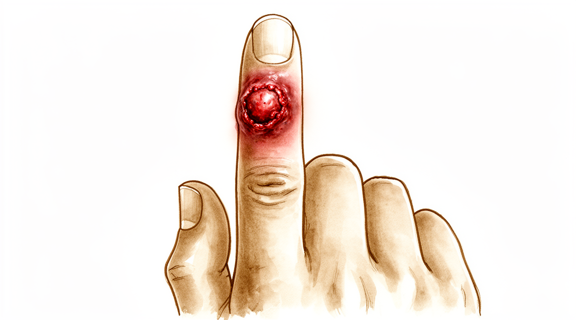
As a felon abscess forms, swelling and pressure within the pulp compartments increase, often causing significant discomfort [65]. The resulting local vascular compromise in a felon promotes further necrosis and spread of the infection, occasionally resulting in infections of the distal phalanx, distal interphalangeal (DIP) joint, or flexor tendon sheath [65]. Chronic paronychia is caused by Candida in most cases, with C. albicans responsible for the majority (70% to 97%) of cases [68]. Chronic paronychia is characterized by a relatively well-localized area of skin inflammation proximal to the cuticle of a fingernail, where the skin becomes pink, warm, indurated, glistening, and tense [68]. The cuticle in chronic paronychia is chronically rounded and detached from the nail plate, allowing free passage of moisture and water under the eponychium [68]. Unless the disease process of chronic paronychia is stopped, secondary thickening, ridging, and discoloration of the nail can occur, eventually resulting in nail loss [68].
Actinomycosis is most commonly caused by Actinomyces israelii, a normal inhabitant of the oral cavity [91]. In the hand, a closed-fist injury or a bite is the most common cause of actinomycosis, designated as "punch actinomycosis" when oral flora is implanted over the metacarpophalangeal joint or metacarpal head on impact [91]. Once actinomycosis is established locally, it spreads contiguously in a slow but progressive manner, ignoring tissue planes [91]. Contiguous invasion of subjacent bones occurs in actinomycosis, with the bones of the hand and wrist showing cystic, sclerotic, or lytic changes [91]. Mycetoma is a characteristic clinical triad of tumification, draining sinuses, and granules in the discharging pus in the distal aspect of a limb [92]. Mycetoma infection begins in the skin and subcutaneous tissues as a nodule or nodules and tends to follow fascial planes as it spreads proximally and mediolaterally [92]. With increasing depth, mycetoma progressively infects and destroys all connective tissues and eventually bone [92].
Sporotrichosis is the most common cutaneous and lymphatic (lymphocutaneous) fungal infection in North America [94]. The infection in sporotrichosis is usually nonopportunistic and affects healthy subjects, with 80% of infections occurring in the upper extremity [94]. Sporotrichosis is characterized by linear marching nodules on the forearm and arm, which represent spread through local lymphatic channels [94]. Mycobacterial tenosynovitis is the most common form of TB of the hand, with mycobacteria having more predilection for tenosynovium than joints, bone, subcutaneous tissue, or nerve [96]. Flexor tendons of the fingers, and carpal tunnel with involvement of palm, wrist, and forearm are affected more often than extensor tendons in mycobacterial tenosynovitis [96]. Nontuberculous mycobacterial (NTM) tenosynovitis is more common than TB mycobacterial tenosynovitis [96]. More than half of the reported hand NTM tenosynovial infections are due to M. marinum [96]. The second most common infecting organism for NTM tenosynovitis is M. kansasii, and the ratio of M. kansasii and M. marinum is 1:5 [96]. The third most frequent infecting organism for NTM tenosynovitis is M. avium-intracellulare complex [96]. Inflammatory signs such as erythema, warmth, and tenderness are absent in mycobacterial tenosynovitis, presenting as a "cold abscess" [96]. The most common presentation of all mycobacterial tenosynovitis is digital flexor tenosynovitis [96]. Tubercular finger tenosynovitis may appear as a "sausage finger" [96].
Blastomycosis is a rare fungal infection caused by Blastomyces dermatitidis, reported mostly in North America’s Ohio and Mississippi River valleys [98]. Primary cutaneous blastomycosis can be caused by direct implantation into the skin or secondarily by dissemination of the fungus from the lungs [98]. Clinical findings of blastomycosis are nonspecific and multifarious, appearing as a plaque, ulcer, or nodules on a finger, hand, or forearm [98]. Contiguous spread to bone, joint, or flexor tendon of a finger can occur in blastomycosis [98]. Osteoarticular lesions occur in 60% of patients who have systemic blastomycosis and may include septic arthritis and osteomyelitis in the hand and elbow [98].
The continued emergence of antibiotic-resistant bacteria and the development of only a few new classes of antibiotics over the past 50 years have made the treatment of acute hand infections problematic [29]. Hand stiffness, contractures, and even amputation can result from missed diagnoses or delayed treatment of acute hand infections [29]. Community-acquired MRSA (CA-MRSA) hand infections increased rapidly in prevalence over the past two decades, but may be decreasing as polymicrobial infections become more frequent [32]. Anywhere from 30% to 70% of surgical hand infections are MRSA related [32]. Multidrug-resistant strains of MRSA are being increasingly reported with a higher incidence of resistance to clindamycin and levofloxacin [32]. Risk factors for multidrug-resistant MRSA pathogens at an urban center included young age, intravenous drug use, and nosocomial infection [32]. Infections in immunosuppressed patients were more likely to involve deeper structures such as joints, bone, tendons, and muscle [32]. Patients with immunosuppression are at a higher risk for atypical infections [32].
Chronic infections of the hand and upper extremity can be caused by a variety of agents: viruses, bacteria, mycobacteria, fungi, Prototheca, protozoa, parasites, and insects [31]. An infection may be superficial and affect the skin or nails, or it may affect subcutaneous tissue, or it may be deep and affect the nerves, tendons (tenosynovium), joints (synovium), bone, and (rarely) muscles [31]. Chronic lesions of the hand, both superficial and deep, have a nonspecific presentation and early biopsy and cultures facilitate diagnosis [31]. An infection that does not respond to antibiotics, incision, drainage, or debridement is suspect for a chronic infection [31]. The most common chronic hand infection traditionally has been tuberculosis (TB) and Hansen disease [31]. Nontuberculous mycobacterial (NTM) infections of the hand are now more common than Mycobacterium tuberculosis infections [31]. Tenosynovial infections of both TB and NTM types are far more common than joint and bone infections [31]. In North America, the most common fungal infection is sporotrichosis [31]. Hansen disease is the most common chronic infection affecting the hand in developing countries and infects peripheral nerves [31]. Chronic hand infections caused by protozoa, Prototheca, parasites, and insects are sporadically encountered in Africa, Asia, and South America but are rare in North America [31].
The increasing incidence of HIV infection and the implantation of prosthetic devices are important in the increasing incidence of local and systemic candidiasis [43]. The most common species recovered from specimens sent for tissue culture for candidiasis include C. albicans, C. tropicalis, C. glabrata, and C. parapsilosis [43]. Flexor and extensor tenosynovitis of the hand has been reported in newborns and in AIDS patients with Candida [43].
Classification
Hand infections are grouped by microorganism into bacterial (mycobacterial and others), fungal, and viral types [19]. Atypical hand infections are caused by organisms such as Mycobacterium, fungi, and viruses [27]. These atypical infections exist within a wide spectrum of presentations, ranging from cutaneous lesions to deep infections such as tenosynovitis and osteomyelitis [27]. They can manifest as either acute infections with obvious swelling, erythema, and pain, or more indolent, chronic infections [27]. Chronic hand infections are uncommon and require a high index of suspicion for early diagnosis [19].
Necrotizing Soft Tissue Infections: Necrotizing soft tissue infections (NSTIs) can be classified based on anatomic location, depth of involvement, or microbial pathogen [61]. Type II necrotizing soft tissue infections are monomicrobial and comprise approximately 20% to 30% of cases [61]. These are caused by group A beta-hemolytic streptococci (GAS) either alone or in association with Staphylococcus aureus [61]. Type IV fungal necrotizing soft tissue infections are rare and primarily affect immunocompromised patients [61]. They often have a high mortality rate and aggressive clinical course with rapid extension of involved areas [61].
Subcutaneous Fungal Infections: There are three major subcutaneous fungal infections of the hand: chronic paronychia, sporotrichosis, and phaeomycotic cysts [68]. Chronic paronychia is usually initially misdiagnosed as a bacterial infection [68]. A phaeomycotic cyst is a deep dermal or subcutaneous infection that results from the traumatic implantation of a dematiaceous (pigmented) fungus such as Exophiala or Phialophora [68].
Mycetoma: Mycetoma of the hand and upper extremity has been graded into five stages [118]. Stage I is characterized by a small, firm, painless subcutaneous nodule or nodules under the skin (nodular stage) [118]. Stage II is characterized by nodules becoming abscesses and draining granules through sinuses to the surface of the skin or to the surface of an ulcerated nodule (sinusoidal stage) [118]. Stage III is characterized by progression to osteomyelitis (skeletal stage) [118]. Stage IV is characterized by limb deformity occurring over the course of a year or more (skeletal stage) [118]. Stage V is characterized by lesions metastasizing to the axilla and the chest wall (metastatic stage) [118].
Michon Classification: The Michon classification for severity of flexor tenosynovitis includes Stage I, characterized by increased fluid in the sheath, primarily serous exudate [33]. Stage II is characterized by cloudy/purulent fluid and granulomatous synovium [33]. Stage III is characterized by septic necrosis of tendon, pulleys, or tendon sheath [33].
Other Considerations: A new classification system for open fractures of the hand divides patients into three groups based on a risk score to predict infection requiring re-debridement [95]. The 1993 Revised Classification System for HIV Infection categorizes patients based on CD4+ T-cell count and clinical conditions [114]. In this system, a CD4+ T-cell count of <200/μL corresponds to category 3 (A3, B3, or C3) [114].
Clinical Presentation
Hand infections encompass a diverse array of entities with the potential for serious morbidity [13]. The incidence of finger infections presenting to emergency departments is rising, with 40- to 59-year-old patients most at risk [24]. Diagnosis is usually clinical, though imaging and laboratory evaluation aid in the process [8]. A careful history and physical examination determine the location of the infection, extent of spread, presence of swelling, lymphangitis, lymphadenitis, and joint involvement [101]. Fluctuance can be difficult to identify in the hand [101]. The specificity of all inflammation markers (WBC, C-reactive protein, ESR) is inadequate for diagnosis [101]. Misdiagnosis of conditions that mimic hand infections often occurs [16]. Conditions that can be confused with hand infections include gout, acute calcium deposition, pseudogout, pyogenic granuloma, insect bites, pyoderma gangrenosum, foreign bodies, factitious lesions, herpetic gangrenes, metastatic lesions, silicone synovitis, granuloma annulare, rheumatoid arthritis, nonspecific tenosynovitis, reactions to intravenous medications (e.g., chemotherapeutic agents), and Sweet syndrome [101].
Acute Infections
Prompt diagnosis and treatment are important because hand stiffness, contractures, and even amputation can result from missed diagnoses or delayed treatment [29]. Community-acquired MRSA has become the most common cause of culture-positive hand infections in the United States [101]. In a retrospective review of 110 patients, the overall prevalence of MRSA infection was 34%, and patients with MRSA hand infections experienced a substantial delay in receiving appropriate antibiotics compared to those with non-MRSA infections [22]. A study of 815 culture-positive hand infections over a 10-year period found clindamycin resistance to MRSA rising from 4% to 31% and levofloxacin resistance rising from 12% to 56% [101]. Renal disease and diabetes increase the risk of failed outpatient management of cellulitic hand infections [53]. For invasive Group A Streptococcus hand infections, which are often limb- and life-threatening, prompt diagnosis and early debridement are of the utmost importance to improve outcomes [60].
Flexor Tenosynovitis
The diagnosis of infectious flexor tenosynovitis (FTS) has historically been made based on physical exam using Kanavel's signs [58]. The specificity of Kanavel's signs has come into question, although a higher number of Kanavel signs were present in the FTS group (2.9) compared to the finger cellulitis group (0.5) [58]. Radiographic soft tissue swelling does not distinguish FTS from other causes [58]. Magnetic resonance imaging (MRI) access and time spent obtaining MRI may delay diagnosis, with a negative impact on patient outcomes [58]. Ultrasound has been briefly studied as a diagnostic tool with high sensitivity, but mediocre specificity, and comes with inherent user variability and limitations with access and equipment [58]. Prompt diagnosis and early treatment with broad-spectrum intravenous antibiotics and surgical irrigation and debridement are essential to avoid complications and preserve hand function in pyogenic flexor tenosynovitis [17]. Early diagnosis and prompt treatment are required to preserve the digit and prevent morbidity and loss of hand function in septic flexor tenosynovitis [30]. Severe infection can still lead to impaired function or amputation despite timely and thorough treatment of pyogenic flexor tenosynovitis [17, 23]. Continuous postoperative irrigation is no longer used due to concerns regarding swelling and hindrance of finger motion [55].
Septic Arthritis
Diagnosis of septic arthritis of the wrist is made based mainly on a thorough patient history, physical examination, and joint aspiration, as no serum laboratory values have been shown to consistently confirm wrist joint infection [39]. Finger joint infections usually result from the spread of infection in adjacent structures, direct penetration of the joint, and less commonly, hematogenous spread [93]. Involved joints in finger joint infections are usually swollen, tender, and warm, and the finger is usually held in slight flexion [93]. Careful inspection and palpation may reveal a fluctuant joint effusion, and active and passive motions are usually quite painful [93]. Fluid obtained from a septic joint is usually turbid, opaque, or grossly purulent [93]. The joint fluid WBC is usually greater than 50,000/mm³ [93]. Lowering the cell count threshold to 17,500 increases the sensitivity of the diagnosis of septic arthritis to 83% [93]. The polymorphonuclear count in septic joint fluid is usually greater than 75% [93]. The synovial fluid glucose in septic arthritis is 40 mg or less [93]. S. aureus is usually the organism isolated from septic hand and wrist joints [93].
Chronic and Atypical Infections
Chronic hand infections are grouped by microorganism into bacterial (mycobacterial and others), fungal, and viral types [19]. Chronic infections of the hand and upper extremity are rare and are primarily a problem of diagnosis [31]. Early suspicion, biopsy, and diagnosis of a chronic infection is the mainstay of all ensuing treatment principles [31]. An infection that does not respond to antibiotics, incision, drainage, or debridement is suspect [31]. "Culture a tumor and biopsy an infection" is a useful adage when an unusual lesion is encountered [31]. Atypical hand infections are difficult to recognize and treat due to their indolent nature and nonspecific symptoms [11]. Atypical infections of the hand are caused by organisms such as Mycobacterium, fungi, and viruses, and often do not respond to conventional management [27]. Having a high clinical suspicion for atypical hand infections is vital because diagnosis often requires special tests and/or cultures [27]. Obtaining a detailed medical, work, and travel history is extremely important for diagnosing atypical hand infections [27]. An indolent clinical course, late diagnosis, and delayed treatment are common in atypical hand infections [27]. Atypical hand infections are commonly misdiagnosed or diagnosed in a delayed fashion [27]. Fungal infections of the hand are most commonly cutaneous infections involving the skin and nails [56]. Hand surgeons should consider mycobacterial infection in patients receiving TNF inhibitors with atypical presentations of tenosynovitis or postoperative infections [38]. Acute Nocardia infection of the hand should be considered in children with suppurative infections that do not respond to initial treatment [89].
Immunocompromised Patients
Hand infections in patients with HIV, diabetes, and those on immunosuppressive treatment present with specific clinical pictures and possible complications [59]. Blue nails and clubbing are both a sign of HIV infection [72]. Red fingers syndrome has been reported in patients with HIV and hepatitis C infection [72]. Fingers of patients with AIDS may be red with painless erythema and periungual telangiectasia [72]. Nails in patients with AIDS may be blue with painless clubbing [72]. Approximately 10% of patients who presented with hand infections in metropolitan hospital emergency departments had HIV infection [72]. Of intravenous drug users who presented with hand and upper extremity infections, 80% had HIV infection [72]. In a review of 74 HIV-seropositive patients treated for upper extremity infections, 26 infections (29%) required more than one operation and 11 (12%) resulted in amputation [72]. Patients with AIDS were significantly more likely to present with spontaneous infection than those who were HIV seropositive [72]. In a study of 28 patients, 5 had necrotizing fasciitis; all needed more than one debridement, and one required forearm amputation [72]. TB remains the main infection in patients with AIDS [72]. Hansen disease is the most common chronic infection affecting the hand in developing countries [31]. Hansen disease infects peripheral nerves, and the level of suspicion should be high when peripheral neuropathy of the ulnar nerve, with or without nerve enlargement, is seen in an immigrant [31].
Investigations
Aspiration: Diagnosis of septic arthritis of the wrist relies primarily on a thorough patient history, physical examination, and joint aspiration [39]. No serum laboratory values have been shown to consistently confirm wrist joint infection [39].
Laboratory: Early identification of atypical hand infections requires appropriate laboratory testing [11]. Diagnosis of these conditions often requires special tests and/or cultures [27]. Obtaining a detailed medical, work, and travel history is extremely important for the diagnosis of atypical hand infections [27].
CT: Contrast-enhanced computed tomography (CT) has been evaluated to increase the successful diagnosis of infectious flexor tenosynovitis (FTS) [58]. MRI, CT, and 3-phase bone scans may add to the radiographic diagnosis made on plain radiographs for hand osteomyelitis, but all have poor sensitivity and specificity [108].
MRI: MRI may add to the radiographic diagnosis made on plain radiographs for hand osteomyelitis, but it has poor sensitivity and specificity [108].
Bone scan: 3-phase bone scans may add to the radiographic diagnosis made on plain radiographs for hand osteomyelitis, but they have poor sensitivity and specificity [108].
Other Considerations: Deep tissue cultures are the most accurate method of diagnosis for osteomyelitis of the hand [108]. The presence of an indolent course suggests a fungal or mycobacterial cause in hand osteomyelitis [108]. Tissue biopsy is recommended for the diagnosis of nonmarinum, nontuberculous mycobacterial infections of the upper extremity [75]. Incisional biopsy is indicated for patients with lytic destructive lesions of the bones of the hand [115]. Biopsy must be strongly considered before the initiation of treatment for presumed deep infection because misdiagnosis of mimicking conditions often occurs [115]. The specificity of Kanavel's signs for the diagnosis of infectious flexor tenosynovitis has come into question [58]. Ultrasound has been briefly studied as a diagnostic tool for flexor tenosynovitis with high sensitivity, but mediocre specificity [58]. Ultrasound for flexor tenosynovitis diagnosis comes with inherent user variability and limitations with access and equipment [58].
Treatment
General Principles
Prompt diagnosis, surgical debridement, and antibiotic coverage constitute the standard of care for hand infections [14]. Missed diagnoses or delayed treatment can result in hand stiffness, contractures, and even amputation [29]. Specimens of infected tissue must be sent for aerobic and anaerobic cultures [29]. If chronic hand infection is suspected, fungal and atypical cultures should also be obtained [29]. Patients with immunosuppression are at higher risk for atypical infections and require rapid, aggressive treatment due to high potential for increased morbidity [32]. Glucocorticoids are the most frequent immunosuppressive medication among patients with upper extremity infections [32]. Known risk factors for hand infections include prolonged hospitalization, chronic illness, IV drug abuse, and prior hand infection [32]. At an urban center, risk factors for multidrug-resistant pathogens included young age, intravenous drug use, and nosocomial infection [32].
Antibiotic Management
Empiric Therapy: A systematic review recommends empiric coverage for CA-MRSA if local prevalence rates exceed 10% to 15% [32]. Early treatment is empiric and often covers the most common pathogen, Staphylococcus aureus [22]. The incidence of MRSA infection in a county emergency department study was found to be 72% [32]. A prospective randomized trial demonstrated increased cost and mean hospital stay for patients treated with cefazolin compared with vancomycin for MRSA hand infections [32]. With the use of a formal hand infection treatment algorithm, there was no statistical difference in appropriate antibiotic delivery time and length of stay between CA-MRSA and non-MRSA hand infections [69].
Targeted Therapy: The best form of antibiotic treatment is based on culture sensitivities [22]. Vancomycin is effective for infections caused by gram-positive bacteria [33]. Ciprofloxacin is most effective for gram-negative organisms, including Pseudomonas [33]. Fluconazole and AmB are the drugs of choice for use in patients with deep candidiasis [43]. For periprosthetic fungal infection, radical synovectomy, tenosynovectomy, and removal of the implant are recommended in addition to a chemotherapy combination of amphotericin B and 5-fluorouracil [43].
Prophylaxis: For low-risk, traumatic injuries with clean wounds and no devitalized tissue, no antibiotic prophylaxis is indicated [99]. For injuries in immunocompromised patients, gram-positive cocci coverage is indicated for antibiotic prophylaxis [99]. For wounds with devitalized tissue, gram-positive cocci coverage is indicated if the wound, tendon, or joint space is contaminated [99]. For animal and human bites other than superficial abrasions, a first-generation cephalosporin is indicated for antibiotic prophylaxis [99]. In patients with bites that may contain Pasteurella multocida or Eikenella corrodens, penicillin or amoxicillin-clavulanate potassium should be considered [99]. In immunocompromised patients with bites, erythromycin or amoxicillin-clavulanate should be considered [99]. In patients with sepsis and petechial rash, intravenous ciprofloxacin and clindamycin should be considered [99]. Patients with sepsis or petechial rash should be hospitalized [99]. For puncture wounds, antibiotic prophylaxis is a case-by-case decision [99].
Route of Administration: The mainstay of treatment for serious hand infections remains surgical debridement and antibiotics [22]. The use of IV antibiotics reflects a traditional belief that parenteral therapy is superior to oral therapy [116]. A recent multicenter, randomized, controlled noninferiority study of 1,050 patients concluded that oral antibiotic therapy was noninferior to IV antibiotic therapy when used during the first 6 weeks for complex orthopedic infections including osteomyelitis, prosthetic joint infection, and fixation-device infection [116]. Findings from a study of complex orthopedic infections suggest that oral antibiotics may also be feasible for complex hand infections [116].
Surgical Management
Osteomyelitis: Management principles for osteomyelitis include good surgical debridement and culture-guided antimicrobial therapy [4]. Antibiotic therapy for osteomyelitis typically lasts 4–6 weeks [4]. The optimal length and mode of administration of antibiotics for hand osteomyelitis remain under study [4]. Management of osteomyelitis of the hand and wrist should consist of a combined surgical and medical approach to achieve the most favorable outcome [44].
Mycobacterial and Atypical Infections: Initial treatment for mycobacterial infections involves a combination of long-term antibiotics and surgical debridement to cure the infection [10]. Reconstructive procedures aid in restoring hand function lost secondary to the disease process in mycobacterial infections [10]. Early identification through appropriate laboratory testing and surgical treatment paired with medical management is imperative for eradication of the causative organism in atypical hand infections [11]. Biopsy and cultures should be considered as a part of a diagnostic workup for any atypical lesion [31]. Consultation with an infectious disease specialist, microbiology personnel, and a pathologist improves the accuracy of a diagnosis when organisms are scarce, slow to grow, and require special media and temperature to grow [31]. Pharmacologic treatment of a chronic hand infection requires close monitoring for serious side effects and drug resistance [31]. Recurrence of the infection due to drug resistance may occur because of poor patient compliance or poor prescription practices [31].
Pyogenic Flexor Tenosynovitis: Early diagnosis and prompt treatment are required to preserve the digit and prevent morbidity and loss of hand function in pyogenic flexor tenosynovitis [30]. When early tenosynovitis is suspected, immediate treatment with antibiotics and splinting may abort the spread of infection if the patient’s symptoms have been present for less than 48 hours [33]. If nonsurgical treatment is selected for early tenosynovitis, patients should be followed closely with a low threshold for hospital admission [33]. Good results have been reported in patients with pyogenic flexor tenosynovitis treated with surgical drainage, followed by outpatient management with intravenous antibiotics, wound care, and rehabilitation [33]. If gross pus is obtained from the aspiration of the digital flexor sheath, surgical drainage usually is indicated [33]. Needle aspiration of the flexor sheath through cellulitic tissue creates the risk of inoculating the uninfected sheath with bacteria [33]. Delay in treatment may lead to damage to the flexor tendon, with resulting adhesion, loss of excursion, finger stiffness, and impaired function [33]. The prognosis for function is poor if an infection in the flexor sheath produces pus that must be drained [33]. An open or closed irrigation technique can be used for drainage of flexor tenosynovitis [33]. If an open technique is used for flexor tenosynovitis, healing and rehabilitation are prolonged and full motion may not be regained [33]. The use of a continuous postoperative irrigation catheter has not been shown to improve outcomes for flexor tenosynovitis, but rather increases postoperative pain and adds difficulty to postoperative care [33]. The Michon classification recommends minimally invasive drainage and catheter irrigation for stage I or II flexor tenosynovitis [33]. The Michon classification recommends extensile open debridement and possible amputation for stage III flexor tenosynovitis characterized by septic necrosis of tendon, pulleys, or tendon sheath [33]. The Michon classification has not been validated [33]. Patients presenting with subcutaneous purulence had amputation rates of 8% for pyogenic flexor tenosynovitis [33]. Patients presenting with ischemic changes had amputation rates of 59% for pyogenic flexor tenosynovitis [33]. Despite timely and thorough treatment, severe infection can nevertheless lead to impaired function or even amputation of the affected digit in pyogenic flexor tenosynovitis [23].
Abscess and Dorsal Infections: For patients with fluctuance concerning for abscess, ultrasonography may be of value as a diagnostic test [32]. Ultrasonography was shown to have a 78.4% positive predictive value of identifying an abscess and a negative predictive value of 90% to rule out an abscess in a series of 179 patients [32]. Early antibiotic administration has not been shown to greatly reduce bacterial culture growth from hand abscess so long as decompression is performed within 24 hours [32]. After surgical decompression, debridement, and irrigation of the abscess, packing is often used, at least initially, to allow for continued drainage [32]. No difference has been shown between different soaks and daily dressing changes in clearing the infection postoperatively [32]. An initial debridement of dorsal hand infections at the bedside is at least as effective as formal debridement in the operating room [67]. Bedside debridement of dorsal hand infections decreases the number of formal debridements and hospital days without increasing complications [67].
Felon and Paronychia: Surgical drainage is the mainstay of treatment for felon [65]. Drainage for felon should be accomplished without violating the flexor sheath or DIP joint [65]. A midaxial incision along the non-pressure-bearing side of the digit or a longitudinal incision over the volar pulp skin is preferred for felon drainage [65]. The wound for felon is left open, and warm soapy soaks are initiated, to allow drainage [65]. When the paronychia has progressed to abscess formation, drainage, with or without removal of the involved portion of the nail plate from the fold, is required [65]. When drainage is pursued for paronychia, cultures should be taken whenever possible [65]. Antibiotics as well as warm soapy soaks should be recommended following drainage of paronychia [65]. Nail plate removal and eponychial marsupialization is the recommended treatment for chronic paronychia [65]. The “Swiss roll” technique has also been described to treat chronic paronychia [65].
Herpetic Whitlow: Surgical drainage or debridement of herpetic whitlow lesions is contraindicated [65]. Bacterial superinfection, viral encephalitis, and death have been reported following surgical drainage or debridement of herpetic whitlow [65]. When administered early, oral acyclovir may lessen symptom severity in herpetic whitlow [65]. In children with herpetic whitlow, a 10-day course of a penicillinase-resistant oral antibiotic is required if cultures from blistering dactylitis reveal growth [65].
Adjuncts and Rehabilitation: A very high wire survival rate (99.5%) and a very low rate of pin site infection (4.5%) were confirmed with no cases of bony infection requiring further surgery when a specific protocol was adopted [26]. The recommendation to leave wires exposed in adult hand and wrist surgery is supported when the specific protocol for reducing pin site infection is adopted [26]. Complex infections often require surgery and a prolonged course of IV antibiotics administered in an outpatient setting [116]. Rehabilitation after treatment of a hand infection is essential to prevent stiffness [116]. Splinting in the early stages with the hand in intrinsic plus position, followed by early mobilization of all digits, will help maximize function [116].
Complications
Mortality and Limb Threat: Delayed diagnosis of hand infections can result in amputation or death [1]. Despite modern antibiotics, hand infections continue to be a source of morbidity and possible long-term disability [6]. Atypical hand infections may present as rapidly disseminating processes that are life-threatening and limb-threatening [27]. Fungal infections following organ transplantation can have an associated mortality as high as 76% [104]. In necrotizing myofasciitis caused by histoplasmosis, the infection can result in fatality [104]. A case of cutaneous histoplasmosis in a renal transplant patient required above-elbow amputation to save the patient's life after multiple debridements and antifungal medications [104]. Another case of cutaneous histoplasmosis in a renal transplant patient with hand and forearm necrotizing myofasciitis required below-elbow amputation [104].
Immunocompromised and Systemic Risk: Infections in immunosuppressed patients are more likely to involve deeper structures such as joints, bone, tendons, and muscle [32]. Patients with systemic conditions such as HIV, diabetes, or those on immunosuppressive therapy are at increased risk for hand infections associated with severe complications [102]. Diabetics are at increased risk for hand infections associated with severe complications, including necrotizing fasciitis [102]. Immunocompromised patients are at a higher risk for atypical infections [32]. The potential for increased morbidity is high in immunosuppressed patients with hand infections [32]. In a matched cohort study, the most frequent immunosuppressive medication among patients with upper extremity infections was glucocorticoids [32].
Flexor Tenosynovitis: In flexor tenosynovitis, persistent infection can cause pressures within the flexor sheath to exceed 30 mm Hg, rendering tendons ischemic [33]. Delay in treatment of flexor tenosynovitis may lead to damage to the flexor tendon, resulting in adhesion, loss of excursion, finger stiffness, and impaired function [33]. The prognosis for function is poor if a flexor tenosynovitis infection produces pus that must be drained [33]. If an open technique is used for flexor tenosynovitis drainage, healing and rehabilitation are prolonged and full motion may not be regained [33]. Patients presenting with subcutaneous purulence in flexor tenosynovitis had amputation rates of 8% [33]. Patients presenting with ischemic changes in flexor tenosynovitis had amputation rates of 59% [33]. In the Michon classification for flexor tenosynovitis, stage III is characterized by septic necrosis of the tendon, pulleys, or tendon sheath [33]. The treatment recommendation for stage III flexor tenosynovitis includes extensile open debridement and possible amputation [33].
Microbiological Complications: Risk factors for multidrug-resistant MRSA strains at an urban center include young age, intravenous drug use, and nosocomial infection [32]. Patients with a history of intravenous drug use are 11 times more likely to have concurrent clindamycin resistance in MRSA hand abscesses [105]. Patients with nosocomial acquired MRSA are 5 times more likely to have concurrent clindamycin resistance in hand abscesses [105]. In a study of 110 patients with culture-positive hand infections, the overall prevalence of MRSA infection was 34% [22]. Since 1961, the rates of both community-acquired MRSA and health care–associated MRSA hand infections have increased rapidly [48]. Community-acquired MRSA hand infections increased rapidly in prevalence over the past two decades but may be decreasing as polymicrobial infections become more frequent [32]. A prospective randomized trial of patients with MRSA hand infections demonstrated increased cost and mean hospital stay for patients treated with cefazolin compared with vancomycin [32].
Amputation and Functional Loss: Ray amputation can narrow the palm span by 10%, with a resultant decrease in grip strength [111]. Loss of hand span can decrease hand width, which decreases forearm rotation, strength, and grip stability [111]. Patients who underwent primary ray resection lost an average of 28% of grip strength [111]. Patients who underwent primary ray resection lost an average of 26% of opposition [111]. Patients who underwent primary ray resection lost an average of 13% of key pinch strength [111]. In a series of 20 patients following ray amputation, grip strength decreased by 27% [111]. In a series of 20 patients following ray amputation, three-point pinch decreased by 22% [111]. Patients who had amputation of central digits had impaired manual dexterity compared to those with border digit amputations [111]. Cold intolerance was the most common persistent symptom in a series of 41 patients following ray amputation, affecting 35 patients [111]. Fifty-nine percent of patients in a series of 41 patients following ray amputation had suboptimal results owing to hyperesthesia in the thumb/long finger web space [111]. Hyperesthesia in the thumb/long finger web space interfered with hand function in 37% of patients [111]. In a series of 34 patients undergoing elective ray amputations, 21 complained of pain in the residual limb before transmetacarpal amputation [111]. In a series of 34 patients undergoing elective ray amputations, only 3 experienced complete relief of residual limb pain after the procedure [111]. Thumb amputation is associated with substantial disability because the thumb provides over 40% of hand function [103]. Amputations involving multiple digits may occur following trauma, infection, hypothermia, or vascular injuries [103]. The incidence of multiple-digit amputations is about 3 to 6 times greater in men than in women [103]. The incidence of multiple-digit amputations is 4.9 times greater in left-handed than in right-handed individuals [103].
Other Considerations: Misdiagnosis of conditions mimicking hand infections often occurs [16]. In a study of 652 patients with hand infections, 84 revision procedures were required in 72 patients [47]. In a study of 652 patients with hand infections, 112 patients (17%) did not attend the postoperative clinic appointment and were lost to follow-up [47]. In a study of 652 patients with hand infections, 134 of 485 tested patients (28%) were HIV positive [47]. In a study of 652 patients with hand infections, 22 of 75 patients with known CD4 counts (29%) had a CD4 count below 200 [47]. In a study of 652 patients with hand infections, two patients with a CD4 count below 200 had revision surgery [47]. In a study of 652 patients with hand infections, one patient with a CD4 count below 200 had an unspecified outcome mentioned in the truncated text [47]. In a study of 652 patients with hand infections, 167 patients (26%) were not tested for HIV [47]. In a study of 652 patients with hand infections, CD4 counts were not obtained in 59 of 134 HIV-positive patients [47]. In a study of 652 patients with hand infections, 53 of 75 patients with known CD4 counts (71%) had a CD4 count above 200 [47]. Outcomes such as stiffness, fibrosis, sepsis, and amputation can be avoided with prompt treatment of hand abscesses [105]. The study demonstrated a rising incidence of finger infections presenting to EDs, with 40- to 59-year-old patients most at risk [24]. In a study of 674 index procedures for hand infections, the different types included felon (331), web-space (187), paronychia (71), deep-space (61), superficial (55), flexor tenosynovitis (26), necrosis (15) and septic arthritis (12) [47]. In a series of 8 patients with soft tissue sarcomas of the hand who underwent partial hand resection, 6 were disease-free [103]. In a series of 8 patients with soft tissue sarcomas of the hand who underwent partial hand resection, the mean Musculoskeletal Tumor Society score was 26 (range, 19 to 29) [103]. Patients who had undergone double-ray amputations for soft tissue sarcomas had lower functional outcome scores [103]. In a series of double-ray amputations for high-grade soft tissue sarcomas, four of five patients were disease-free with a mean grip strength of 24% compared with the contralateral hand [103]. Patients with double-ray amputations maintained good key, tip, and tripod pinch when the deep motor branch of the ulnar nerve was preserved [103]. A 13-year-old boy with acute lymphocytic leukemia underwent small finger ray amputation for rapidly disseminating mucormycosis infection [103]. A 9-month-old HIV-positive Nigerian girl presented with blistering dactylitis of the right middle finger, and radiograph showed autoamputation of the terminal phalanx and osteomyelitis in the middle phalanx of the third finger [70]. Syphilitic dactylitis in the infant may resemble tuberculous spina ventosa [70]. Pathologic fractures of the metaphysis in congenital syphilis can masquerade as pseudoparalysis [70]. Tertiary syphilis lesions in the hand present as gummas, which are nonspecific, chronic granulomatous lesions that may involve tissues from skin to bone [70]. Cutaneous gumma can occur in syphilis, yaws, and TB and all appear similar [70]. The infection rate after open hand fracture remains relatively low [49]. A large study of clean, elective hand procedures based on 516,986 patients found no difference in the risk of postoperative infection in patients who received prophylactic antibiotics and those who did not [106]. The overall 30-day surgical site infection rate was 1.5% in the antibiotic prophylaxis group and 1.4% in the group not receiving antibiotics [106]. A database study that included 44,305 patients who had outpatient hand surgery procedures identified infections in fewer than 1% [106]. Predictive factors for surgical site infection after hand surgery were government-funded insurance and residence in a rural area [106]. Diabetes, obesity, and tobacco use were not associated with an increased risk of infection after hand surgery [106]. The study confirms a very high wire survival rate (99.5%) and a very low rate of pin site infection (4.5%) with no cases of bony infection requiring further surgery when a specific Ilizarov protocol is adopted [26]. The safety of performing elective hand surgery during the pandemic remains unclear [46]. Early identification and treatment of hand infections are essential to achieve optimal outcomes [1]. Treatment outcomes for infections of the hand have improved dramatically using established guidelines [5]. This article reviews the clinical spectrum and microbiology of the most common infections of the hand and discusses current concepts for their treatment to increase physician awareness of diagnosis and management [12]. Management principles for osteomyelitis of the hand include good surgical debridement and culture-guided antimicrobial therapy [4]. Antibiotic therapy for osteomyelitis of the hand typically lasts 4–6 weeks [4]. The optimal length and mode of administration of antibiotic therapy for osteomyelitis in the hand remain under study [4]. Specific presentations and treatments are emphasized for each type of chronic hand infection [19]. This review provides an update on the contemporary taxonomy of atypical organisms and focuses on the clinical aspects of disease diagnosis and treatment for atypical hand infections [20]. They exist within a wide spectrum of presentations, ranging from cutaneous lesions to deep infections such as tenosynovitis and osteomyelitis [27]. In addition to medical therapies, surgical debridement is often required to effectively treat atypical hand infections [27]. Most hand infections are caused by common Staphylococcus and Streptococcus bacterial species [27]. Infections caused by atypical organisms, such as Mycobacterium, viruses, and fungi are becoming more common, especially among immunocompromised patients [27]. Surgical debridement is often required to eliminate the offending organism or lower the disease burden in atypical hand infections [27]. Chronic infections of the hand and upper extremity are rare indeed and are primarily a problem of diagnosis [31]. They often are not considered in the differential diagnosis of hand lesions [31]. Many surgeons encounter their first case by surprise unless an unusual diagnosis
Recovery
Other Considerations: Recovery trajectories for hand infections are heavily influenced by the specific pathogen and patient comorbidities, with no standardized timeline for return to activity provided in the current evidence. Management principles for osteomyelitis include good surgical debridement and culture-guided antimicrobial therapy, with antibiotic therapy typically lasting 4–6 weeks [4]. The optimal length and mode of antibiotic administration for hand osteomyelitis remain under study [4]. Initial treatment for mycobacterial hand infections involves a combination of long-term antibiotics and surgical debridement to cure the infection [10]. Reconstructive procedures aid in restoring hand function lost secondary to the mycobacterial disease process [10].
Prophylactic antibiotics are indicated for hand surgery procedures lasting longer than 2 hours, contaminated wounds, and open fractures [15]. The use of prophylactic antibiotics in small elective soft tissue hand procedures is debated and often not necessary [15]. Despite timely and thorough treatment, severe pyogenic flexor tenosynovitis infection can lead to impaired function or even amputation of the affected digit [17, 23]. There is a rising incidence of virulent and MRSA infections [25]. Aggressive treatment is needed in immunosuppressed patients to prevent limb and life-threatening complications [25].
In a retrospective review of 110 patients, the overall prevalence of MRSA infection was 34% [22]. Empiric coverage for CA-MRSA should be provided if local prevalence rates exceed 10% to 15% [32]. Ultrasonography has a 78.4% positive predictive value of identifying an abscess and a negative predictive value of 90% to rule out an abscess [32]. No difference has been shown between different soaks and daily dressing changes in clearing the infection postoperatively after surgical decompression, débridement, and irrigation of the abscess [32].
Among intravenous drug users who presented with hand and upper extremity infections, 80% had HIV infection [72]. Among 14 AIDS patients with hand infections, almost one-third needed multiple debridements and resulted in amputation of a finger or hand [72]. Early amputation to maximize disease-free survival may be appropriate for patients with hand osteomyelitis and arterial calcification [130]. A very high wire survival rate (99.5%) and a very low rate of pin site infection (4.5%) were confirmed with no cases of bony infection requiring further surgery when a specific Ilizarov protocol was adopted [26].
Key Evidence
- [L5] Early identification and treatment of hand infections are essential to achieve optimal outcomes, as delayed diagnosis can result in amputation or death. [1] (10.1016/j.jhsa.2018.05.027)
- [L5] The morbidity of hand infections can be decreased with understanding of the different types of hand infections often unique to the particular anatomy of the hand. [2] (10.5435/00124635-199607000-00006)
- [L5] Hand infections are associated with a high rate of complications that are often difficult to manage. [3] (10.1016/j.hcl.2020.03.010)
- [L5] Management principles include good surgical debridement and culture-guided antimicrobial therapy, with antibiotic therapy typically lasting 4–6 weeks, though the optimal length and mode of administration in the hand remain under study. [4] (10.1177/1753193415612373)
- [L5] Using these guidelines, the treatment outcomes for infections of the hand have improved dramatically. [5] (10.1016/s0749-0712(21)00413-3)
- [L4] Despite modern antibiotics, hand infections with a variety of organisms continue to be a source of morbidity and possible long-term disability. [6] (10.1016/j.ijid.2005.06.009)
- [L5] Infections of the hand and upper extremity pose a considerable challenge to the surgeon despite recent advances in medical technology. [7] (10.1016/s0749-0712(21)00470-4)
- [L5] Hand and upper-extremity infections are usually a clinical diagnosis, but imaging and laboratory evaluation aid in diagnosis. [8] (10.1016/j.hcl.2020.03.002)
- [L4] With prompt and appropriate care, most soft tissue hand infection patients can achieve full resolution of their infections. [9] (10.7717/peerj.513)
- [L5] Initial treatment involves a combination of long-term antibiotics and surgical debridement to cure the infection, with reconstructive procedures aiding in restoring hand function lost secondary to the disease process. [10] (10.1016/j.hcl.2020.03.013)
- [L4] Atypical hand infections are difficult to recognize and treat due to their indolent nature and nonspecific symptoms; early identification through appropriate laboratory testing and surgical treatment paired with medical management is imperative for eradication of the causative organism. [11] (10.1016/j.jhsa.2025.09.023)
- [L5] This article reviews the clinical spectrum and microbiology of the most common infections of the hand and discusses current concepts for their treatment to increase physician awareness of diagnosis and management. [12] (10.1302/2058-5241.4.180082)
- [L5] Hand infections include a diverse array of entities with potential for serious morbidity. [13] (10.1016/j.jhsa.2011.05.035)
- [L5] Prompt diagnosis, surgical debridement, and antibiotic coverage remain the standard of care for hand infections, though challenges persist with immunosuppressed patients and intravenous drug use. [14] (10.1016/j.hcl.2020.03.001)
- [Paper] Deep infection after hand surgery is uncommon but can lead to significant morbidity; prophylactic antibiotics are indicated for procedures lasting longer than 2 hours, contaminated wounds, and open fractures, while their use in small elective soft tissue procedures is debated and often not necessary. [15] (10.1016/j.hcl.2014.12.007)
- [L5] The authors emphasize that while treatment for true hand infections is available and successful, misdiagnosis of mimicking conditions often occurs. [16] (10.1016/s0749-0712(21)00414-5)
- [L5] Prompt diagnosis and early treatment with broad-spectrum intravenous antibiotics and surgical irrigation and debridement are essential to avoid complications and preserve hand function, though severe infection can still lead to impaired function or amputation. [17] (10.1016/j.jhsa.2019.04.011)
- [L4] The complications associated with mycobacterial hand infections can be significant. [18] (10.1177/1558944720940064)
- [L5] Chronic hand infections are uncommon and require a high index of suspicion for early diagnosis; they are grouped by microorganism into bacterial (mycobacterial and others), fungal, and viral types, with specific presentations and treatments emphasized for each. [19] (10.1016/j.jhsa.2014.04.003)
- [L5] This review provides an update on the contemporary taxonomy of atypical organisms and focuses on the clinical aspects of disease diagnosis and treatment for atypical hand infections. [20] (10.1016/s0749-0712(21)00422-4)
- [L5] The choice of empiric antibiotics for acute infections of the hand should be based on the severity of the infection, the comorbidities of the patient, and local prevalence of MRSA. [21] (10.1016/j.jhsa.2009.10.024)
- [L3] [22] (10.1097/blo.0b013e3180986729)
- [Paper] Despite timely and thorough treatment, severe infection can nevertheless lead to impaired function or even amputation of the affected digit. [23] (10.1055/s-0039-1700370)
- [L3] The study demonstrated a rising incidence of finger infections presenting to EDs, with 40- to 59-year-old patients most at risk. [24] (10.1177/1558944720915614)
- [L4] The study confirms a very high wire survival rate (99.5%) and a very low rate of pin site infection (4.5%) with no cases of bony infection requiring further surgery, supporting the recommendation to leave wires exposed in adult hand and wrist surgery when this specific protocol is adopted. [26] (10.1177/1753193421991318)
- [L5] [27] (10.1016/j.ocl.2016.12.013)
- [L5] [29] (10.1016/j.jhsa.2014.03.031)
- [L5] Early diagnosis and prompt treatment are required to preserve the digit and prevent morbidity and loss of hand function. [30] (10.1016/j.hcl.2020.03.005)
- [L3] The proportion of surgically treated hand infections due to CA-MRSA has increased during the last decade. [34] (10.1016/j.jhsa.2009.09.004)
- [L4] Hand surgeons should consider mycobacterial infection in patients receiving TNF inhibitors with atypical presentations of tenosynovitis or postoperative infections. [38] (10.1016/j.jhsa.2009.09.018)
- [L5] Diagnosis is made based mainly on a thorough patient history, physical examination, and joint aspiration, as no serum laboratory values have been shown to consistently confirm wrist joint infection. [39] (10.5435/jaaos-d-16-00414)
- [L5] Management of osteomyelitis of the hand and wrist should consist of a combined surgical and medical approach to achieve the most favorable outcome. [44] (10.1016/j.jhsa.2009.03.020)
- [L4] The safety of performing elective hand surgery during the pandemic remains unclear, and more studies with larger samples are needed to clarify this question. [46] (10.1177/15589447211064360)
- [L3] [47] (10.1177/1753193420977791)
- [L5] Since 1961, the rates of both community-acquired MRSA and health care–associated MRSA hand infections have increased rapidly. [48] (10.1016/j.hcl.2020.03.003)
- [L1] Overall, the infection rate after open hand fracture remains relatively low. [49] (10.1177/1558944716643294)
- [L3] [53] (10.1186/s13018-023-03911-5)
- [L5] Continuous postoperative irrigation is no longer used due to concerns regarding swelling and hindrance of finger motion. [55] (10.1016/j.jhsa.2010.11.033)
- [L5] Fungal infections of the hand are most commonly cutaneous infections involving the skin and nails and can be treated with topical or local therapy. [56] (10.1016/j.hcl.2020.03.009)
- [L5] Although many management principles are the same in pediatric and adult patients, physicians should bear in mind the unique environments and characteristics of the pediatric hand, including the frequency of fingers in mouths, open growth plates, and typically more robust circulation with fewer systemic comorbidities. [57] (10.1016/j.hcl.2020.03.012)
- [L3] [58] (10.1177/15589447221092058)
- [L5] The purpose of this article is to provide an outline of the most common and some of the more exotic organisms causing hand infections in patients with human immunodeficiency virus/acquired immunodeficiency syndrome, diabetes, and patients on immunosuppressive treatment, discussing presentation, clinical picture, evidence-based approaches in treatment, and possible complications. [59] (10.1016/j.jhsa.2018.07.001)
- [L4] Prompt diagnosis and early debridement are of the utmost importance to improve outcomes for these often limb- and life-threatening infections. [60] (10.1177/17531934241268983)
- [L4] [61] (10.5435/jaaos-d-17-00716)
- [L2] Our prospective multicenter trial showed no significant difference in infection rate in elective hand surgery whether antibiotics were administered preoperatively or not. [63] (10.1177/1558944719842238)
- [L4] Outpatient management of diabetic hand infections provides a safe alternative to inpatient admission in selected patients. [66] (10.1177/17531934231196026)
- [L3] An initial debridement of dorsal hand infections at the bedside is at least as effective as formal debridement in the operating room, decreasing the number of formal debridements and hospital days without increasing complications. [67] (10.1177/1558944719836234)
- [L4] With the use of a formal hand infection treatment algorithm, we did not identify a statistical difference in appropriate antibiotic delivery time and length of stay between ca-MRSA and non-MRSA hand infections. [69] (10.1016/j.jhsa.2008.11.021)
- [L4] Early consideration in differential diagnoses of chronic, painful swelling, nodular or inflammatory lesions, or septic arthritis is crucial, with tissue biopsy and early involvement with an infectious disease specialist recommended. [75] (10.1016/j.jhsa.2022.03.019)
- [L4] Acute Nocardia infection of the hand should be considered in children with suppurative infections that do not respond to initial treatment. [89] (10.1016/j.jhsa.2018.03.039)
- [L3] The new classification system divides patients into three groups based on a risk score to predict infection requiring re-debridement. [95] (10.1177/17531934231187553)
- [L5] [102] (10.1016/j.hcl.2020.03.008)
- [L3] [105] (10.1016/j.jhsa.2014.12.044)
- [L5] [116] (10.1016/j.jhsa.2024.09.001)
- [L5] [125] (10.5435/jaaos-20-06-373)
- [L4] Early amputation to maximize disease-free survival may be appropriate for patients with hand osteomyelitis and arterial calcification. [130] (10.1177/1753193420981871)
- [L5] [131] (10.1177/17531934231174819)
See Also
References
[1] Hand Infections. The Journal of Hand Surgery. 2019. DOI: 10.1016/j.jhsa.2018.05.027
[2] Hand Infections: Treatment Recommendations for Specific Types. Journal of the American Academy of Orthopaedic Surgeons. 1996. DOI: 10.5435/00124635-199607000-00006
[3] Complications of Hand Infections. Hand Clinics. 2020. DOI: 10.1016/j.hcl.2020.03.010
[4] Osteomyelitis of the hand. Journal of Hand Surgery (European Volume). 2015. DOI: 10.1177/1753193415612373
[5] THE HISTORY OF HAND INFECTIONS. Hand Clinics. 1998. DOI: 10.1016/s0749-0712(21)00413-3
[6] Epidemiology of bacterial hand infections. International Journal of Infectious Diseases. 2006. DOI: 10.1016/j.ijid.2005.06.009
[7] HAND INFECTIONS IN THE TRAUMA PATIENT. Hand Clinics. 1999. DOI: 10.1016/s0749-0712(21)00470-4
[8] Imaging and Laboratory Workup for Hand Infections. Hand Clinics. 2020. DOI: 10.1016/j.hcl.2020.03.002
[9] Hand infections: a retrospective analysis. PeerJ. 2014. DOI: 10.7717/peerj.513
[10] Mycobacterial Infections in the Hand and Wrist. Hand Clinics. 2020. DOI: 10.1016/j.hcl.2020.03.013
[11] Atypical Hand Infections. The Journal of Hand Surgery. 2026. DOI: 10.1016/j.jhsa.2025.09.023
[12] Infections of the hand: an overview. EFORT Open Reviews. 2019. DOI: 10.1302/2058-5241.4.180082
[13] Hand Infections. The Journal of Hand Surgery. 2011. DOI: 10.1016/j.jhsa.2011.05.035
[14] Hand Infections. Hand Clinics. 2020. DOI: 10.1016/j.hcl.2020.03.001
[15] Infection After Hand Surgery. Hand Clinics. 2015. DOI: 10.1016/j.hcl.2014.12.007
[16] MIMICKERS OF HAND INFECTIONS. Hand Clinics. 1998. DOI: 10.1016/s0749-0712(21)00414-5
[17] Pyogenic Flexor Tenosynovitis: Evaluation and Treatment Strategies. The Journal of Hand Surgery. 2019. DOI: 10.1016/j.jhsa.2019.04.011
[18] Mycobacterial Infections of the Hand. HAND. 2020. DOI: 10.1177/1558944720940064
[19] Chronic Hand Infections. The Journal of Hand Surgery. 2014. DOI: 10.1016/j.jhsa.2014.04.003
[20] ATYPICAL HAND INFECTIONS. Hand Clinics. 1998. DOI: 10.1016/s0749-0712(21)00422-4
[21] Empiric Antibiotics for Acute Infections of the Hand. The Journal of Hand Surgery. 2010. DOI: 10.1016/j.jhsa.2009.10.024
[22] Timeliness of Appropriate Antibiotics in Hand Infections. Clinical Orthopaedics & Related Research. 2007. DOI: 10.1097/blo.0b013e3180986729
[23] Pyogenic Flexor Tenosynovitis: Evaluation and Treatment Strategies. Journal of Hand and Microsurgery. 2019. DOI: 10.1055/s-0039-1700370
[24] A Nationwide Epidemiological Analysis of Finger Infections Presenting to Emergency Departments in the United States From 2012 to 2016. HAND. 2020. DOI: 10.1177/1558944720915614
[25] Chapter 30 Bone and Soft-Tissue Infections of the Hand and Wrist. 2020.
[26] Reducing the incidence of pin site infection in hand surgery with the use of a protocol from Ilizarov. Journal of Hand Surgery (European Volume). 2021. DOI: 10.1177/1753193421991318
[27] Atypical Hand Infections. Orthopedic Clinics of North America. 2017. DOI: 10.1016/j.ocl.2016.12.013
[29] Acute Hand Infections. The Journal of Hand Surgery. 2014. DOI: 10.1016/j.jhsa.2014.03.031
[30] Pyogenic Flexor Tenosynovitis: Evaluation and Management. Hand Clinics. 2020. DOI: 10.1016/j.hcl.2020.03.005
[31] Green S Operative Hand Surgery. GENERAL PRINCIPLES.
[32] Orthopaedic Knowledge Update 13 Ebook Without Multimedia. Bone and Soft-Tissue Infections of the Hand and Wrist > Soft-Tissue Infections > Hand Cellulitis and Abscesses.
[33] Campbell S Operative Orthopaedics 4 Volume Set. TUMORS AND TUMOROUS CONDITIONS OF THE HAND > TENOSYNOVITIS.
[34] Community-Acquired Methicillin-Resistant Staphylococcus aureus in Surgically Treated Hand Infections. The Journal of Hand Surgery. 2010. DOI: 10.1016/j.jhsa.2009.09.004
[35] Exam Of The Hand Wrist 2Ed. INTRODUCTION.
[38] Mycobacterial Hand Infections Occurring Postoperatively in Patients Treated With Tumor Necrosis Factor-α Inhibitors for Inflammatory Arthritis: Report of Three Cases. The Journal of Hand Surgery. 2010. DOI: 10.1016/j.jhsa.2009.09.018
[39] Septic Arthritis of the Wrist. Journal of the American Academy of Orthopaedic Surgeons. 2018. DOI: 10.5435/jaaos-d-16-00414
[43] Green S Operative Hand Surgery. Candidiasis.
[44] Current Recommendations in the Management of Osteomyelitis of the Hand and Wrist. The Journal of Hand Surgery. 2009. DOI: 10.1016/j.jhsa.2009.03.020
[46] Evaluation of the COVID-19 Infection Rate in the Perioperative Period of Elective Surgeries of the Hand and Microsurgery. HAND. 2021. DOI: 10.1177/15589447211064360
[47] Factors affecting suboptimal outcomes in hand infections. Journal of Hand Surgery (European Volume). 2020. DOI: 10.1177/1753193420977791
[48] Antibiotic Management and Antibiotic Resistance in Hand Infections. Hand Clinics. 2020. DOI: 10.1016/j.hcl.2020.03.003
[49] Timing of Debridement and Infection Rates in Open Fractures of the Hand. HAND. 2016. DOI: 10.1177/1558944716643294
[51] A Lange Medical Book Current Diagnosis Treatment In Orthopedics Fifth Edition. 9Hand Surgery > Image DISORDERS OF THE MUSCULATURE OF THE HAND.
[52] Exam Of The Hand Wrist 2Ed. 1.1 SKELETON OF THE HAND > The osseous skeleton.
[53] Renal disease and diabetes increase the risk of failed outpatient management of cellulitic hand infections: a retrospective cohort study. Journal of Orthopaedic Surgery and Research. 2023. DOI: 10.1186/s13018-023-03911-5
[55] Septic Flexor Tenosynovitis. The Journal of Hand Surgery. 2011. DOI: 10.1016/j.jhsa.2010.11.033
[56] Fungal Infections of the Hand. Hand Clinics. 2020. DOI: 10.1016/j.hcl.2020.03.009
[57] Pediatric Hand Infections. Hand Clinics. 2020. DOI: 10.1016/j.hcl.2020.03.012
[58] Contrast Enhanced Computed Tomography in the Diagnosis of Acute Pyogenic Flexor Tenosynovitis. HAND. 2022. DOI: 10.1177/15589447221092058
[59] Hand Infections in the Immunocompromised Patient: An Update. The Journal of Hand Surgery. 2019. DOI: 10.1016/j.jhsa.2018.07.001
[60] A surge in the incidence of invasive Group A Streptococcus hand infections: a single Hand Unit experience. Journal of Hand Surgery (European Volume). 2024. DOI: 10.1177/17531934241268983
[61] Necrotizing Infections of the Hand and Wrist: Diagnosis and Treatment Options. Journal of the American Academy of Orthopaedic Surgeons. 2020. DOI: 10.5435/jaaos-d-17-00716
[63] Prospective Analysis of Hand Infection Rates in Elective Soft Tissue Procedures of the Hand: The Role of Preoperative Antibiotics. HAND. 2019. DOI: 10.1177/1558944719842238
[65] Aaos Comprehensive Orthopaedic Review 3. Infections of the Hand* > I Fingertip Infections.
[66] The incidence and severity of diabetic hand infection presentations during the COVID-19 pandemic. Journal of Hand Surgery (European Volume). 2023. DOI: 10.1177/17531934231196026
[67] Initial Debridement of Dorsal Hand Abscesses in the Operating Room Does Not Improve Outcomes. HAND. 2019. DOI: 10.1177/1558944719836234
[68] Green S Operative Hand Surgery. Subcutaneous Fungal Infections.
[69] Community-Acquired Methicillin-Resistant Staphylococcus aureus Infections of the Hand: Prevalence and Timeliness of Treatment. The Journal of Hand Surgery. 2009. DOI: 10.1016/j.jhsa.2008.11.021
[70] Green S Operative Hand Surgery. Syphilis > Clinical Findings.
[72] Green S Operative Hand Surgery. Clinical Manifestations of AIDS Infection in the Hands.
[75] Nonmarinum, Nontuberculous Mycobacterial Infections of the Upper Extremity: A Multi-Institutional Descriptive Report. The Journal of Hand Surgery. 2023. DOI: 10.1016/j.jhsa.2022.03.019
[76] Exam Of The Hand Wrist 2Ed. Functional cutaneous units.
[77] Green S Operative Hand Surgery. Interosseous and Hypothenar Muscles.
[81] A Lange Medical Book Current Diagnosis Treatment In Orthopedics Fifth Edition. 9Hand Surgery > FLEXOR TENDON INJURY.
[83] Exam Of The Hand Wrist 2Ed. The dorsal skin.
[84] Green S Operative Hand Surgery. PERTINENT ANATOMY.
[89] An Acute Nocardia Infection in a Pediatric Hand. The Journal of Hand Surgery. 2019. DOI: 10.1016/j.jhsa.2018.03.039
[91] Green S Operative Hand Surgery. CHRONIC BACTERIAL INFECTIONS > Actinomycosis.
[92] Green S Operative Hand Surgery. Clinical Findings.
[93] Campbell S Operative Orthopaedics 4 Volume Set. TUMORS AND TUMOROUS CONDITIONS OF THE HAND > SEPTIC ARTHRITIS.
[94] Green S Operative Hand Surgery. Sporotrichosis > Rose thorn disease (rose gardener’s disease, cutaneous sporotrichosis).
[95] Open fractures of the hand: a new classification based on risk score to predict infection requiring re-debridement. Journal of Hand Surgery (European Volume). 2023. DOI: 10.1177/17531934231187553
[96] Green S Operative Hand Surgery. Deep Mycobacterial Infections > Mycobacterial Tenosynovitis.
[98] Green S Operative Hand Surgery. Blastomycosis.
[99] Campbell S Operative Orthopaedics 4 Volume Set. RESULTS OF SUTURE OF THE SCIATIC NERVE > Antibiotic Prophylaxis for Hand Injuries.
[101] Campbell S Operative Orthopaedics 4 Volume Set. TUMORS AND TUMOROUS CONDITIONS OF THE HAND > GENERAL APPROACH TO HAND INFECTIONS.
[102] Hand Infections Associated with Systemic Conditions. Hand Clinics. 2020. DOI: 10.1016/j.hcl.2020.03.008
[103] Green S Operative Hand Surgery. MULTIPLE-DIGIT AMPUTATIONS.
[104] Green S Operative Hand Surgery. Histoplasmosis.
[105] Risk Factors Associated With Clindamycin-Resistant, Methicillin-Resistant Staphylococcus aureus in Hand Abscesses. The Journal of Hand Surgery. 2015. DOI: 10.1016/j.jhsa.2014.12.044
[106] Campbell S Operative Orthopaedics 4 Volume Set. TUMORS AND TUMOROUS CONDITIONS OF THE HAND > FACTORS INFLUENCING HAND INFECTIONS.
[108] Orthopaedic Knowledge Update 13 Ebook Without Multimedia. Bone and Soft-Tissue Infections of the Hand and Wrist > Bone Infections.
[111] Green S Operative Hand Surgery. RAY AMPUTATIONS.
[114] Green S Operative Hand Surgery. Classification and Clinical Diagnosis > ETABLE 3.2 Grading for AIDS Based on Clinical Conditions and CD4+ Lymphocyte Count.
[115] Green S Operative Hand Surgery. BOX 59.1 Ganglions of the Hand and Wrist > Ewing's Sarcoma.
[116] Diagnosis and Management of Hand Infections. The Journal of Hand Surgery. 2024. DOI: 10.1016/j.jhsa.2024.09.001
[118] Green S Operative Hand Surgery. Staging.
[125] Flexor Tendon Sheath Infections of the Hand. Journal of the American Academy of Orthopaedic Surgeons. 2012. DOI: 10.5435/jaaos-20-06-373
[130] Hand osteomyelitis in arterial calcification, diabetes mellitus and end-stage renal failure: a comparison of 210 cases over 12 years. Journal of Hand Surgery (European Volume). 2021. DOI: 10.1177/1753193420981871
[131] Hand infection: a management approach based on a new understanding of combined bacterial and neutrophil mediated tissue damage. Journal of Hand Surgery (European Volume). 2023. DOI: 10.1177/17531934231174819