Clinicians › Wrist
Wrist Ganglia
Dorsal and volar wrist ganglia (scapholunate-origin and radial-artery-adjacent).
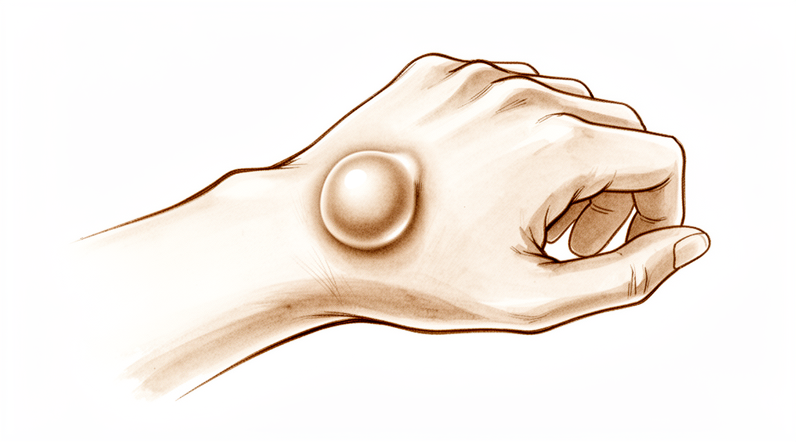
For patients: a plain-language version of this topic is available. See the patient guide.
Overview
The dorsal wrist ganglion is the prototype of all hand and wrist ganglions, accounting for 60% to 70% of cases, with the main cyst typically located directly over the scapholunate ligament [5]. The volar wrist ganglion is the second most common, representing 18% to 20% of cases and most frequently arising over the distal radial edge or scaphoid tubercle [18]. In pediatric populations, dorsal lesions predominate with a female predilection, whereas in children under 10 years, volar wrist ganglions are the primary presentation [2, 11]. Preoperative diagnosis is confirmed via transillumination or aspiration for dorsal lesions [5], while ultrasound helps confirm the articular origin of volar cysts [20]. Careful palpation with digital compression often reveals the extent and pedicle direction of dorsal ganglions [5]. Routine wrist radiography is not cost-effective due to a low prevalence of therapeutically significant findings [6].
Natural history data indicate that approximately 40% of wrist ganglion lesions decrease over the first 6 years after evaluation by a hand surgeon [44]. In children under 10, 69% to 79% of volar wrist ganglions display spontaneous regression within 12-18 months [11]. Pediatric hand ganglions resolve at a greater rate than wrist ganglions [3]. There is no consensus on the best management for pediatric wrist ganglia, as no single treatment modality confers a particular advantage or disadvantage [1]. Observation and/or splinting will likely be helpful in the resolution of a majority of pediatric hand and wrist ganglions [10]. Surgical excision is indicated for pediatric ganglions that are symptomatic, do not resolve after approximately 2 months of observation and/or splinting, or recur [10].
Open surgical excision offers a significantly lower chance of recurrence compared with aspiration [15]. Surgical intervention for wrist ganglions has about a 10% recurrence rate [44], and excision of primary wrist ganglia significantly reduces symptoms with low recurrence rates and high patient satisfaction [9]. Most ganglions recur after aspiration [44], though performing at least 1 aspiration before surgical excision improves the cost-effectiveness of dorsal wrist ganglion treatment [49]. Arthroscopic resection is a reasonable alternative to open excision for radiocarpal volar ganglia, offering decreased postoperative morbidity and a high rate of good results [21]. For dorsal lesions, arthroscopy is a good alternative to open surgery, though it is a difficult procedure requiring adequate experience [37]. Outcomes, recurrence, and complication rates after 4 years of follow-up support the use of arthroscopy for dorsal wrist ganglions [16].
Anatomy & Pathophysiology
Epidemiology & Demographics
Volar wrist ganglions represent the second most common ganglion of the hand and wrist, accounting for 18% to 20% of cases [18]. They have been identified as the site of 13% to 30% of ganglion cysts about the hand and wrist [121]. In the military population, the incidence of dorsal wrist ganglia was higher compared with the civilian population [19]. Specifically, volar wrist ganglions comprised 17% of all ganglions diagnosed in the military population and 25% of ganglions diagnosed in the civilian population [121]. The incidence of volar wrist ganglions was 3.28 per 10,000 person-years in the civilian population [121]. In the military population, the incidence of volar wrist ganglions was 3.73 per 10,000 person-years for male personnel and 7.98 per 10,000 person-years for female personnel [121]. Patients with wrist hyperlaxity have a predisposition to developing ganglions [65]. An association between wrist ganglions and ligamentous hyperlaxity does not prove causation, but the possibility of the same underlying pathological entity causing both can be envisioned [28].
Dorsal Wrist Ganglion Anatomy
Dorsal ganglions typically appear between the extensor pollicis longus and extensor digitorum communis tendons [5]. They may occur anywhere else between the extensor tendons and can be connected to the scapholunate ligament through an elongated pedicle [5]. A small, mucin-filled duct is invariably seen piercing the transverse fibers of the scapholunate ligament, connecting the underlying scapholunate joint with the main cyst [5]. Smaller, occult dorsal ganglions are easily overlooked and can often only be palpated with the involved wrist in marked volar flexion [59]. Dorsal ganglions occasionally occur in association with an underlying scapholunate diastasis [59]. Arthroscopic assessment of 16 wrists with painful dorsal ganglia revealed abnormalities in the scapholunate joint in 10 of the wrists [55]. Only one of 16 wrist radiographs in a study of painful dorsal ganglia showed radiological scapholunate widening [55]. A mild chronic "sprain" of the scapholunate ligament is present in most cases of dorsal wrist ganglion, which does not give rise to the well-recognised radiological and clinical features associated with scapholunate instability [55].
Volar Wrist Ganglion Anatomy
The majority of volar ganglions occur either directly over the distal edge of the radius or slightly more distally over the scaphoid tubercle [18]. Volar wrist ganglions are typically found arising from the radioscaphoid joint (65%), scaphotrapezial joint (34%), or metacarpotrapezial joint (1%) [121]. The volar wrist ganglion arising from the radiocarpal joint occurs under the volar wrist crease between the flexor carpi radialis and abductor pollicis longus tendons [18]. The main cyst of a volar wrist ganglion may be intertwined with bifurcating branches of the radial artery [18]. Volar ganglions may be multiloculated, extending under the thenar muscles, along the flexor carpi radialis tendon, into the carpal canal, and under the first extensor compartment [18]. The radial artery is frequently intimately attached to the wall of a volar ganglion and may be completely encircled by it [18]. Operation-related complications after arthroscopic volar wrist ganglionectomy are associated with the anatomical location of the ganglion being distal to the bifurcation of the radial artery and concurrently penetrated up to the superficial fascia layer [39].
Pathogenesis & Etiology
Ganglions are defined as cystic swellings closely connected to joints or tendon sheaths which contain mucinous material [47]. The etiology of ganglions is unknown [47]. Hypotheses advanced for the pathogenesis of ganglions include retention cyst, herniation of tendon or capsular synovia, bursal transformation, neoplasia, and mucinous degeneration of fibrous tissue [47]. Carp and Stout reported that a ganglion does not initially connect with the joint and that communication occurs secondary to degeneration of the capsule which then ruptures [47]. Injections of contrast material into ganglions usually fail to show a communication into the joint, but injections into the wrist have demonstrated communications into the ganglions [47]. In 40 of 59 patients in the study by Andrén and Eiken, contrast medium passed from the joint into the ganglion through a tortuous narrowed duct [47]. The articular (synovial) theory has gained worldwide acceptance as a scientifically proven explanation for the formation of intraneural ganglions [40]. Intraneural ganglions are rare, with the ulnar nerve being the most commonly involved nerve in the upper extremity [40].
Differential Diagnosis & Associated Conditions
Dorsal wrist ganglions can be confused with carpal bosses, which are osteoarthritic spurs or prominences that develop at the base of the second or third carpometacarpal joints [25]. A small ganglion is associated with a carpal boss in 30% of cases [25]. Carpal bosses are more common in women, in the right hand, and between the third and fourth decades [25]. Chronic tenosynovitis of the extensor tendons can be confused with a dorsal wrist ganglion but is distinguished by the diffuse nature of the swelling and the puckering seen with digital extension (tuck sign) [59]. Dorsal prominence of the proximal pole of the scaphoid secondary to intercarpal instability may be confused with a painful occult ganglion [59]. Intraosseous carpal bone cysts are a rare cause of chronic wrist pain that can progress to pathological fracture and tendon compromise [42]. Ganglions do arise on or within extensor tendons, typically over the metacarpals, and are distinguished by their proximal motion with the fingers in extension [95]. Volar wrist ganglia have been associated with triangular fibrocartilage lesions, particularly in patients with a positive ulnocarpal stress test [43].
Classification
Anatomical and Histological Subtypes: Dorsal wrist ganglions may occur anywhere between the extensor tendons, connected to the scapholunate ligament through an elongated pedicle [5]. While ganglions have been reported in other carpal joints, they are rare [5]. Cystic soft tissue tumours of the dorsal aspect of the wrist present as two distinct histological subtypes [113]. Both histological tissue types can coexist at recurrence, with equal recurrence rates observed in both initial synovial and ganglion groups [113].
Intraneural Ganglions: Intraneural ganglions of the hand and wrist are rare, with the ulnar nerve being the most commonly involved nerve in the upper extremity [40]. Most patients with intraneural ganglions are aged 30 to 50 years, though occurrence in children has also been reported [40].
Other Considerations: Pediatric Presentation: In pediatric populations, ganglions most commonly affect the dorsal wrist and demonstrate a female predilection [2]. Surgical Implication: Failure to identify and excise the pedicle attachment to the scapholunate ligament increases the likelihood of recurrence [5].
Clinical Presentation
Epidemiology and Demographics
Wrist ganglions in pediatric populations most commonly affect the dorsal wrist and demonstrate a female predilection [2]. In the adult population, female gender is a significant predictor of ipsilateral wrist ganglion cyst formation following wrist arthroscopy [84]. An association between wrist ganglions and ligamentous hyperlaxity has been identified, though it does not prove causation [28].
Physical Examination and Clinical Features
Dorsal Ganglions: The main cyst of a dorsal wrist ganglion is usually located directly over the scapholunate ligament and is easily seen and diagnosed [5]. A typical dorsal ganglion appears between the extensor pollicis longus and extensor digitorum communis tendons [5].
Volar Ganglions: The majority of volar wrist ganglions occur either directly over the distal edge of the radius or slightly more distally over the scaphoid tubercle [18]. Volar ganglions arising from the radiocarpal joint occur under the volar wrist crease between the flexor carpi radialis and abductor pollicis longus tendons [18]. Although volar ganglions may appear small clinically, they can be surprisingly extensive at surgery, with multiloculated cysts extending under the thenar muscles, along the flexor carpi radialis tendon, into the carpal canal, and under the first extensor compartment [18]. Volar wrist ganglia can present as trigger finger pathology at the wrist [12]. An occult volar wrist ganglion may remain asymptomatic and present as an incidentaloma during radial artery forearm free flap harvest [24].
Other Presentations: A carpal boss presents as a firm, bony, nonmobile, tender mass visible and palpable at the base of the carpometacarpal joints, especially when the wrist is flexed [25]. Intraneural ganglions of the hand and wrist most commonly involve the ulnar nerve, though almost any other nerve in the vicinity of a joint can be involved [40]. Most patients with intraneural ganglions present with a painless mass, while some may present with symptoms of nerve irritation or entrapment neuropathy [40].
Diagnostic Evaluation
Transillumination or aspiration confirms the diagnosis of a wrist ganglion preoperatively [5]. Sonography can localize occult ganglia and differentiate nerves and arteries [64]. Preoperative ultrasound scanning helps confirm the articular origin and nature of a cystic mass in volar wrist ganglions [20]. MRI is an excellent diagnostic modality to evaluate rapidly growing upper extremity masses and distinguish ganglions from malignant processes [54].
In patients with the clinical diagnosis of wrist ganglion cyst, quality of care would not be compromised by abandoning the practice of routine submission of surgical specimens for pathological examination after excision [36, 50]. Pathological examination is recommended only when the clear gelatinous fluid typical of a ganglion cyst is not encountered at surgery [50]. The prevalence of a concordant pathological diagnosis in surgically excised wrist ganglions was 98.6% [50].
Pediatric Presentation and Natural History
In a child with a wrist ganglion, if the cyst ultimately resolved, it usually did so within 18 months [32]. Surgical excision is indicated for pediatric wrist ganglia that are symptomatic, do not resolve after approximately 2 months of observation and/or splinting, or recur [10].
Patient Factors and Comorbidities
Female patients who have preoperative pain around dorsal wrist ganglia were the most likely to have residual pain after surgery [4]. It is important to ascertain the precise concerns of patients with ganglia and not assume that they attend the hand clinic with symptoms which justify surgical excision [34].
Investigations
Clinical Assessment: Careful preoperative palpation of the cyst with digital compression often reveals its extent and the direction of the pedicle [5]. Transillumination or aspiration confirms the diagnosis preoperatively [5]. Palpation for areas of maximal tenderness is one of the most useful tools in the diagnosis of wrist pathology, especially in patients with chronic dysfunctions [53]. The natural inclination to study radiographs or special imaging studies and reports prior to a thorough history and physical examination should be avoided, as this introduces cognitive bias, which can affect one’s thinking and decision making [53].
Plain radiography: Routinely performing wrist radiography is not cost-effective in the evaluation and treatment decision-making process for patients with a wrist ganglion due to a low prevalence of therapeutically significant findings [6]. However, review of the patient's preoperative radiographs to rule out an interosseous component is wise [5]. Dorsal wrist ganglions can be confused with carpal bosses, which are osteoarthritic spurs or prominences that develop at the base of the second or third carpometacarpal joints (or both) [25]. Radiologically, the mass is best visualized with the hand in 30 to 40 degrees of supination and 20 to 30 degrees of ulnar deviation (“carpal boss view”) [25]. A small ganglion is associated with a carpal boss in 30% of cases, thus adding to confusion of it with the more common dorsal wrist ganglion [25].
Ultrasound and Sonography: Sonography has some applications in investigating disorders of the hand and can allow dynamic study of tendons and show tenosynovitis [64]. Some foreign bodies and tumors, such as occult ganglia, can be localized by sonography [64]. Preoperative ultrasound scanning helps to confirm the articular origin and nature of the cystic mass for volar wrist ganglions [20]. Sonography-guided wrist arthroscopy provides several advantages for surgeons, including visualization of the ganglions and ganglion stalk, as well as of the arthroscopic shaver and adjacent structures such as nerves, vessels, and tendons to perform surgery safely [114].
MRI: MRI is the most sensitive technique currently in use for showing the soft tissues, the vascularity of both bones and soft tissues, and in assessing the extension of bony tumors into soft tissues [104]. A dedicated wrist coil provides enhanced resolution of wrist structures [79]. CNNs can detect ganglion cysts in wrist MRI [103]. A high rate of false-positive findings on MR images of normal subjects has been reported [79].
Arthrography: A wrist arthrogram can be performed immediately prior to the arthroscopic intervention or as a separate investigative procedure for volar wrist ganglions [20]. The relative position of the contrast-filling stalk can aid the identification of the true stalk during the actual arthroscopic procedure and hence facilitate subsequent decompression [20]. The use of an intraoperative arthrogram of a ganglion as arising from the radiocarpal joint may preclude the use of the technique if there is an absence of demonstrable contrast filling the stalk with communication from the radiocarpal joint [20]. Ganglia arising from the midcarpal joint or STT joint will not be revealed by arthrogram of the radiocarpal joint, unless one of the interosseous ligaments at the proximal carpal row is ruptured and creates a free communication between the radiocarpal and the midcarpal joint [20].
CT: CT scanning is not as useful in the wrist as that in other parts of the skeleton [104]. A lipoma or a ganglion can be clearly differentiated from dense tumors using a CT scan, though it is difficult to be sure whether the tumor is benign or malignant [104].
Treatment
Non-Operative
Conservative management is the initial approach for many patients, as approximately 40% of wrist ganglion lesions decrease in size over the first 6 years after evaluation [44]. The spontaneous resolution rate for wrist ganglions is reported to be between 28% and 58% [86]. Aspiration, with or without cortisone injection, achieves an average success rate of 35% to 50% [86]. In pediatric populations, observation and/or splinting is likely to be helpful in the resolution of a majority of hand and wrist ganglions [10]. Performing at least one aspiration before surgical excision improves the cost-effectiveness of dorsal wrist ganglion treatment [49]. For occult dorsal carpal ganglions, conservative treatment with immobilization and steroid injections directly into the dorsal capsule is recommended as initial management [59]. Patients should be assessed to ascertain their precise concerns, as they may not attend the hand clinic with symptoms that justify surgical excision [34].
Operative
Indications: Surgical intervention is considered when conservative measures fail or symptoms persist. Excision of the posterior interosseous nerve at the level of the radiocarpal joint may help alleviate pain and add to postoperative comfort in patients with occult dorsal ganglions [59]. Arthroscopic dorsal wrist ganglionotomy is best indicated in a symptomatic patient with a dorsal wrist ganglion arising from the scapholunate interval [96]. The best indication for arthroscopic volar wrist ganglionotomy is a sessile type of volar wrist ganglion arising from the radiocarpal joint and centered on the proximal wrist crease [20]. Mobile ganglia with long pedicles are poor candidates for arthroscopic surgery [20].
Surgical Approach / Technique: The dorsal wrist ganglion accounts for 60% to 70% of all hand and wrist ganglions [5]. Most dorsal ganglions can be approached through a transverse incision over the proximal carpal row [5]. The diagnosis of ganglion cyst should be made before commitment to a transverse incision because this incision is not readily incorporated into a limb-sparing incision if a malignant soft tissue tumor is subsequently diagnosed [5]. The main cyst and its pedicle are mobilized down to the underlying joint capsule, which is opened along the border of the radius and scaphoid's proximal pole [5]. A small, mucin-filled duct is invariably seen piercing the transopen fibers of the scapholunate ligament, connecting the underlying scapholunate joint with the main cyst [5]. The ganglion and its capsular attachments are tangentially excised off the scapholunate ligament [5]. Synovial and capsular attachments along the distal margin of the scapholunate ligament are excised to give an unobstructed view of the head and neck of the capitate [5]. The key to successful open treatment is excision of the ganglion stalk based at the scapholunate ligament and its associated dorsal-capsular attachments [86]. Open ganglionectomy generally creates a defect in the capsule of 1 to 1.5 cm, which is left open [86].
For volar ganglions, the main cyst may be intertwined with bifurcating branches of the radial artery, making delicate dissection imperative [18]. The Allen test should be performed routinely to assess the patency of the radial and ulnar arteries before volar ganglion excision [18]. Longitudinal incisions allow for optimal visualization during volar ganglion excision [18]. The pedicle of a volar ganglion is traced to the volar joint capsule, usually the scaphotrapezial or radiocarpal ligament [18]. Ganglion attachments are excised approximately 3 ± 4 mm from the joint capsule during volar ganglion surgery [18]. Capsular closure is unnecessary after volar ganglion excision and only delays early mobilization [18]. Wrist motion should begin within the first 2 weeks after volar ganglion surgery [18]. Curved incisions appear to consistently provide more attractive scars on the volar aspect of the wrist, especially near the volar wrist creases [18].
Arthroscopic resection of dorsal wrist ganglions with routine midcarpal joint exploration appeared to reduce recurrence at 1 year without negatively impacting patient outcomes [8]. Outcomes, recurrence, and complication rates after 4 years of follow-up support the use of arthroscopy as a treatment for dorsal wrist ganglion [16]. The proposed classification of ganglia helps minimize the area of resection required during arthroscopic treatment [33]. Advancements in wrist arthroscopy have lowered the recurrence rates of dorsal wrist ganglions [94]. Recurrence rates following arthroscopic excision of dorsal ganglions have been reported from 0% to 17% [100]. In a study of 18 patients undergoing arthroscopic excision of dorsal ganglions, the stalk could be identified in 61% of cases and the reported number of recurrences at an average of 16 months was 0 [100]. In a study of 30 patients, the ability to identify the stalk during arthroscopic excision was 79%, with 2 patients having a recurrence at final follow-up [100]. In a study of 45 patients, the reported recurrence rate was 0% and the stalk was identified in only 16% of cases [100].
For arthroscopic dorsal wrist ganglionotomy, the author prefers to perform the procedure under PSLA to minimize anesthetic risk [96]. A 1.9 mm arthroscope is preferred for arthroscopic dorsal wrist ganglionotomy to improve scar appearance [96]. Two portals are made at 1-2 and 6R for arthroscopic dorsal wrist ganglionotomy [96]. Lidocaine is infiltrated around the stalk of the ganglion near the 3-4 portal to minimize pain during the resection process [96]. Placing the surgical scar on the outer aspect of the wrist in contrast to the more central location of the usual 3-4 portal can produce a more pleasant esthetic outcome [96]. A clear arthroscopic view is maintained by establishing outflow at the 6U portal using an 18-gauge needle [96]. The arthroscope is advanced toward the SL joint interval dorsally, where there is always localized synovial hypertrophy [96]. A 2.0 mm shaver is introduced from the 1-2 portal to remove the synovial folding and display the dorsal aspect of the SL ligament [96]. The arthroscope is directed distal and dorsally toward the junction between the SL ligament and the dorsal capsular reflection of the radiocarpal joint where the ganglion stalk may arise [96]. Pressing the ganglion externally can lead to bulging motion at the interval of the capsule [96]. Most ganglia will rupture after 1 to 3 minutes of shaving during arthroscopic dorsal wrist ganglionotomy [96]. A sizable capsular resection of 5 to 10 mm in diameter should be made in the dorsal capsule to ensure resection of the potential one to 10 mm valve diameter at the stalk [96]. Care is taken to avoid damaging intracapsular dorsal wrist ligaments and extensor tendons during shaving [96]. All wounds are closed with Steri-Strips only, with no suture required [96]. Bulky dressing and bandaging is applied for 1 to 2 days to reduce swelling, followed by light dressing to allow full mobilization of the wrist and fingers [96]. The patient is encouraged to perform self-massaging of the original ganglion site for 3 weeks to prevent pseudorecurrence formation due to regurgitation of joint fluid into the empty cyst remnant [96].
For arthroscopic volar ganglionotomy, preoperative ultrasound scanning helps to confirm the articular origin and nature of the cystic mass [20]. An intraoperative arthrogram can identify the stalk of a ganglion arising from the radiocarpal joint, with absence of demonstrable contrast filling the stalk precluding the use of the technique [20]. Ganglia arising from the interval between the RSC and LRL ligaments show up at the more lateral aspect of the distal radius during arthrogram [20]. Ganglia arising from the interval between the LRL and SRL ligaments show up at a more central position of the distal radius during arthrogram [20]. Ganglia arising from the midcarpal joint or STT joint will not be revealed by arthrogram of the radiocarpal joint unless an interosseous ligament at the proximal carpal row is ruptured [20]. The author prefers to perform arthroscopic volar ganglionotomy under PSLA without the use of a tourniquet to monitor possible iatrogenic damage to the radial artery [20]. Abnormal synovitis at the interligamentous sulcus is a hallmark of a volar ganglion from the radiocarpal joint [46]. Pressing the ganglion externally should demonstrate bulging of the origin of the ganglion as viewed intraarticularly [46]. The ligament interval with abnormal synovitis should be shaved, but not the ligament itself, to abolish the abnormal check-valve of the ganglion [46]. The cyst wall should not be shaved during arthroscopic volar ganglionotomy as the radial artery is at risk [46]. Overaggressive shaving may cause injury to the volar radiocarpal ligaments [46]. Attempted shaving of the cyst wall may cause radial artery damage and bleeding [46]. No stitch is required for wound closure to optimize the scar appearance after arthroscopic volar ganglionotomy [46]. Compressive bandaging for 2 days is used to reduce saline swelling after arthroscopic volar ganglionotomy [46]. Patients should be informed about the possibility of pseudorecurrence phenomenon of up to 3 weeks postoperative and instructed to do daily regular massage at the ganglionotomy site [46]. Arthroscopic drainage of a ganglion from the STT joint is feasible with the addition of new portals [20]. Decompression of a ganglion arising from the STT interval is generally more difficult compared with the standard volar ganglion [46]. A small arthroscope of 1.9 mm and small shaver of 2 mm are mandatory for decompression of a ganglion arising from the STT interval [46].
Complications: Surgical intervention for wrist ganglions has an approximate recurrence rate of 10% [44]. Surgical recurrence rates for ganglions range from 4% to 40% [62]. Complications of ganglion surgery include infection, nerve injury, and wrist stiffness [62]. Unexpected branches of the radial sensory or lateral antebrachial cutaneous nerves may be injured during volar ganglion excision, leading to troublesome neuromas [18]. Injuries to the radial artery during volar ganglion excision can be repaired microscopically [18]. Some authors recommend leaving a portion of the cyst wall attached to the radial artery to avoid arterial injury [18]. Nerve injury during wrist arthroscopy is related to portal placement or suture of the TFCC and typically affects the dorsal sensory branch of the radial or ulnar nerve [29]. ECU tendinitis may be related to portal placement or the suture knot after TFCC repair [29]. Metacarpophalangeal joint pain is typically caused by overdistraction and is transient [29]. Wrist stiffness is an uncommon complication of wrist arthroscopy of uncertain etiology [29]. Inadequate instruction to the patient on massaging the ganglionotomy site at the postoperative period may lead to persistence of pseudorecurrence of the ganglion [46].
Other Considerations: The prevalence of a concordant pathological diagnosis in 429 consecutive adult patients undergoing surgical excision of a wrist ganglion was 98.6% [50]. Worse hand function following dorsal wrist ganglion excision is associated with recurrence following prior surgery, worse baseline hand function, and lower treatment credibility [61]. The superficial branch of the radial nerve averages 16 mm (5 to 22 mm) from the 3-4 portal [29]. The dorsal sensory branch of the ulnar nerve averages 8 mm (0 to 14 mm) from the 6R portal [29]. The 1-2 portal carries a high risk of injury to the superficial branch of the radial nerve [29]. The 6U portal carries a high risk of injury to the dorsal sensory branch of the ulnar nerve [29]. Arthroscopy of the wrist is typically safe, with minor and transient complications [29]. A retrospective review reports a low recurrence rate of 5.3% for pediatric wrist ganglion cysts [91]. In a cohort of 33,884 pediatric patients with a ganglion cyst, the mean age was 13.1 ± 4.3 years and the cohort was predominantly female (66%) [92]. Most pediatric patients with a ganglion cyst received no diagnostic imaging (90%) [92]. Conservative management was maintained in 91% of pediatric patients who did not receive any intervention [92]. Approximately 12% of pediatric patients with a ganglion cyst proceeded to surgical excision [92]. Among pediatric patients that proceeded to surgery, 4.1% underwent surgery for a recurrent ganglion after their initial excision with an average time to second surgery of 1.6 ± 1.3 years [92].
Complications
Recurrence and Natural History: Surgical intervention for wrist ganglions carries an approximately 10% recurrence rate [44]. Failure to identify the pedicle and excise its attachment to the scapholunate ligament increases the likelihood of recurrence [5]. Approximately 40% of wrist ganglion lesions decrease over the first 6 years after evaluation by a hand surgeon [44].
Nerve Injury: Unexpected branches of the radial sensory or lateral antebrachial cutaneous nerves may be injured during volar wrist ganglion excision, leading to troublesome neuromas [18]. Extensions of the routine incision into the carpal canal must avoid injury to the palmar cutaneous branch of the median nerve during volar wrist ganglion excision [18]. Neuromas caught in dorsal scars are particularly painful because they are stimulated not only by direct touch but also by stretching of the surrounding skin, nerve, and scar when the wrist and fingers are flexed [108]. During wrist arthroscopy, the 1-2 portal carries a high risk of injury to the superficial branch of the radial nerve [29], while the 6U portal carries a high risk of injury to the dorsal sensory branch of the ulnar nerve [29].
Vascular Injury: The radial artery is frequently intimately attached to the wall of a volar ganglion and may even be completely encircled by the ganglion [18]. Injuries to the radial artery during volar wrist ganglion excision can be repaired microscopically [18]. Some authors recommend leaving a portion of the cyst wall attached to the radial artery to avoid arterial injury during volar wrist ganglion excision [18]. Operation-related complications after arthroscopic volar wrist ganglionectomy are associated with its anatomical location, specifically when distal to the bifurcation of the radial artery and concurrently penetrated up to the superficial fascia layer [39]. Attempted shaving of the cyst wall during arthroscopic volar wrist ganglionotomy may cause radial artery damage and bleeding [46]. Overaggressive shaving during arthroscopic volar wrist ganglionotomy may cause injury to the volar radiocarpal ligaments [46].
Other Operative Complications: A synovial fistula is a rare complication of recurrent dorsal wrist ganglion excision, potentially caused by extensive capsular excision and steroid injection [60]. Wrist stiffness is less common than with dorsal ganglions after volar excision, but it can occur if early motion is not encouraged [18]. Unpleasant scars are not an uncommon problem after volar wrist ganglion excision, and curved incisions appear to consistently provide more attractive scars, especially near the volar wrist creases [18]. Infection is an uncommon complication of wrist arthroscopy [29]. ECU tendinitis may be related to portal placement or the suture knot after TFCC repair during wrist arthroscopy [29]. Tendon injury may result from improper portal placement during wrist arthroscopy [29]. Metacarpophalangeal joint pain is typically caused by overdistraction and is transient during wrist arthroscopy [29]. Wrist stiffness is an uncommon complication of uncertain etiology following wrist arthroscopy [29].
Other Considerations: The diagnosis of ganglion cyst should be made before commitment to a transverse incision because this type of incision is not readily incorporated into a limb-sparing incision in the event of a subsequent diagnosis of a malignant soft tissue tumor [5]. Inadequate instruction to the patient on massaging the ganglionotomy site at the postoperative period may lead to persistence of pseudorecurrence of the ganglion after arthroscopic volar wrist ganglionotomy [46].
Recovery
Pediatric
Postoperative Care and Complications
Rehabilitation protocol: Wrist motion should begin within the first 2 weeks after volar wrist ganglion excision [18]. Early motion is a critical component of the postoperative regimen, as stiffness of the wrist is less common after volar ganglion excision than after dorsal ganglion excision, but it can occur if early motion is not encouraged [18].
Key Evidence
- [L4] There is no consensus within the literature regarding the best management of pediatric wrist ganglia, and no single treatment modality confers a particular advantage or disadvantage over another. [1] (10.1177/1558944720966716)
- [L2] Ganglions in pediatric populations, which most commonly affect the dorsal wrist, demonstrate a female predilection. [2] (10.1016/j.jhsa.2021.02.026)
- [L4] Pediatric ganglions of the hand have a greater rate of resolution than ganglions of the wrist. [3] (10.1016/j.jhsa.2023.07.002)
- [L4] Female patients who have preoperative pain around dorsal wrist ganglia were the most likely to have residual pain after surgery. [4] (10.1016/j.arthro.2013.04.002)
- [L4] The study concludes that routinely performing wrist radiography is not cost-effective in the evaluation and treatment decision-making process for patients with a wrist ganglion due to a low prevalence of therapeutically significant findings. [6] (10.1007/s11552-007-9032-8)
- [Paper] This method is safer and more reliable for treating volar wrist ganglia. [7] (10.1016/j.eats.2011.12.007)
- [L3] Routine midcarpal joint exploration during arthroscopic excision of dorsal wrist ganglions appeared to reduce recurrence at 1 year without negatively impacting patient outcomes. [8] (10.1177/17531934251405730)
- [L4] Surgical excision of primary wrist ganglia significantly reduces patient symptoms with low recurrence rates and high patient satisfaction. [9] (10.1177/1753193411434376)
- [L4] While observation and/or splinting will likely be helpful in resolution of a majority of pediatric hand and wrist ganglions, surgical excision is indicated for those that are symptomatic, do not resolve after approximately 2 months of observation and/or splinting, or recur. [10] (10.1007/s11552-008-9122-2)
- [L4] In children aged <10 years, ganglions mainly occur on the volar wrist and can be treated expectantly, with 69% to 79% displaying spontaneous regression within a span of 12-18 months. [11] (10.1016/j.jhsa.2021.12.015)
- [L4] This is the first reported case of triggering pathology at the wrist to be treated with interventional radiological measures rather than open surgery and demonstrates the efficacy of the technique. [12] (10.1177/1753193412453699)
- [L3] This study suggests that open excision of dorsal wrist ganglia leads to a lower recurrence rate than does arthroscopic excision. [13] (10.1177/15589447211003184)
- [L4] Patients whose occupation or activities require forceful wrist extension should be counseled on the considerable risk of residual pain and functional limitations that may occur after open dorsal wrist ganglion excision. [14] (10.1016/j.jhsa.2015.05.030)
- [L1] Open surgical excision offers significantly lower chance of recurrence compared with aspiration in the treatment of wrist ganglions. [15] (10.1016/j.jhsa.2014.12.014)
- [L4] The outcomes, recurrence, and complications rates after 4 years of follow-up presented in this study support the use of arthroscopy as a treatment for dorsal wrist ganglion. [16] (10.1177/1558944717743601)
- [L3] The incidence of dorsal wrist ganglia was higher in the military compared with the civilian population. [19] (10.1016/j.jhsg.2020.08.001)
- [L1] The arthroscopic resection of radiocarpal volar ganglia is a reasonable alternative to open excision with decreased postoperative morbidity, easy technique, and high rate of good results. [21] (10.1080/02844310802210897)
- [L4] Arthroscopic ganglionectomy through an intrafocal cystic portal is a safe and efficacious option for the treatment of painful wrist ganglia. [23] (10.1016/j.arthro.2009.08.021)
- [L5] Occult volar wrist ganglion may remain asymptomatic and present as an incidentaloma during FRAFF harvest. [24] (10.1007/s12593-012-0079-6)
- [L4] The results confirm that high patient satisfaction can be achieved for arthroscopic treatment of occult dorsal wrist ganglia. [27] (10.1007/s00402-016-2539-0)
- [L3] Although an association between wrist ganglions and ligamentous hyperlaxity does not prove causation, the possibility of the same underlying pathological entity causing both can be envisioned. [28] (10.1016/j.jhsa.2013.08.109)
- [L4] In a child with a wrist ganglion, if the cyst ultimately resolved, it usually did so within 18 months. [32] (10.1016/j.jhsa.2019.10.032)
- [L4] The proposed classification of ganglia helps minimize the area of resection required. [33] (10.1054/jhsb.2001.0620)
- [L4] It is important to ascertain the precise concerns of patients with ganglia and not assume that they attend the hand clinic with symptoms which justify surgical excision. [34] (10.1054/jhsb.2000.0504)
- [L4] In patients with the clinical diagnosis of wrist ganglion cyst, the quality of care would not be compromised by abandoning the practice of routine submission of surgical specimens for pathological examination after excision of the ganglion cyst. [36] (10.1016/s0363-5023(10)60107-4)
- [L4] Arthroscopic treatment of a dorsal wrist ganglion is a good alternative to open surgery, though it is a difficult procedure requiring adequate experience. [37] (10.1054/jhsb.1999.0290)
- [L2] The systematic review and survey of Canadian hand surgeons reveal that hand surgeons are divided regarding the need to immobilize the wrist after dorsal wrist ganglion excision. [38] (10.1177/15589447211014631)
- [L3] The operation-related complications after arthroscopic volar wrist ganglionectomy are associated with its anatomical location: distal to the bifurcation of the radial artery and concurrently penetrated up to the superficial fascia layer. [39] (10.1186/s12891-025-08766-x)
- [L4] [40] (10.1016/j.jhsa.2015.05.025)
- [L4] Intraosseous carpal bone cysts are a rare cause of chronic wrist pain that can progress to pathological fracture and tendon compromise; once identified, they require careful clinical and radiographic assessment with surgical intervention indicated for symptomatic cases. [42] (10.1007/s11552-015-9750-2)
- [L4] Therefore, arthroscopy should be contemplated as the primary treatment option for patients with painful ganglions of the wrist if they are in a radiopalmar location with a positive ulnocarpal stress test and for patients with recurrent radiopalmar ganglions. [43] (10.1016/j.jhsa.2012.04.042)
- [L5] Current best evidence suggests that about 40% of lesions decrease over the first 6 years after evaluation by a hand surgeon, that most ganglions recur after aspiration, and that surgical intervention has about a 10% recurrence rate, leaves scars, and has some risk for adverse events. [44] (10.1016/j.jhsa.2010.11.048)
- [L4] [47] (10.2106/00004623-197254070-00009)
- [L2] As patient preferences may preclude routinely performing 2 aspirations, performing at least 1 aspiration before surgical excision improves the cost-effectiveness of dorsal wrist ganglions treatment. [49] (10.1016/j.jhsa.2022.09.002)
- [L3] [50] (10.1016/j.jhsa.2010.03.021)
- [L4] MRI is an excellent diagnostic modality to evaluate rapidly growing upper extremity masses and distinguish ganglions from malignant processes. [54] (10.1007/s11552-007-9083-x)
- [L4] [55] (10.1080/028443101750523267)
- [Case_report] A synovial fistula is a rare complication of recurrent dorsal wrist ganglion excision, potentially caused by extensive capsular excision and steroid injection. [60] (10.1016/j.jhsa.2012.02.015)
- [L2] Worse hand function was associated with recurrence following prior surgery, worse baseline hand function and lower treatment credibility. [61] (10.1177/17531934231153029)
- [L5] Surgical recurrence rates range from 4% to 40%, and complications include infection, nerve injury, and wrist stiffness. [62] (10.1016/j.hcl.2004.03.015)
- [L3] Patients with wrist hyperlaxity have a predisposition to developing ganglions, a finding corroborated by independent investigations using similar prospective cohort designs. [65] (10.1016/j.jhsa.2013.11.025)
- [L3] Wrist arthroscopy is associated with a 1.24% incidence of ipsilateral wrist ganglion cyst formation, with female gender being a significant predictor of development. [84] (10.1177/1558944720939203)
- [L4] [86] (10.1016/s0749-0712(21)00020-2)
- [L4] This retrospective review reports a low recurrence rate of 5.3% for pediatric wrist ganglion cysts, indicating potential merit in the surgeon's operative and postoperative techniques. [91] (10.1177/1558944717751195)
- [L4] [92] (10.1177/15589447251314142)
- [L4] [100] (10.1016/j.hcl.2013.08.020)
- [L4] CNNs can detect ganglion cysts in wrist MRI. [103] (10.1186/s12891-025-09011-1)
- [L4] The study demonstrated two histologically distinct tissue types at primary surgery and the coexistence of both tissue types at recurrence, with equal recurrence rates in both initial synovial and ganglion groups. [113] (10.1177/17531934241251721)
- [L4] Sonography-guided wrist arthroscopy provides several advantages for surgeons, including visualization of the ganglions and ganglion stalk, as well as of the arthroscopic shaver and adjacent structures such as nerves, vessels, and tendons to perform surgery safely. [114] (10.1016/j.jhsa.2012.04.012)
- [L4] [121] (10.5435/jaaos-d-22-00105)
See Also
References
[1] Wrist Ganglion Cysts in Children: An Update and Review of the Literature. HAND. 2020. DOI: 10.1177/1558944720966716
[2] Clinical Presentation and Characteristics of Hand and Wrist Ganglion Cysts in Children. The Journal of Hand Surgery. 2021. DOI: 10.1016/j.jhsa.2021.02.026
[3] Natural History of Pediatric Hand and Wrist Ganglion Cysts: Longitudinal Follow-Up of a Prospective, Dual-Center Cohort. The Journal of Hand Surgery. 2023. DOI: 10.1016/j.jhsa.2023.07.002
[4] Arthroscopic Excision of Dorsal Wrist Ganglion: Factors Related to Recurrence and Postoperative Residual Pain. Arthroscopy. 2013. DOI: 10.1016/j.arthro.2013.04.002
[5] Green S Operative Hand Surgery. BOX 59.1 Ganglions of the Hand and Wrist > Operative Treatment > Dorsal Wrist Ganglion.
[6] The Use of Routine Wrist Radiography is Not Useful in the Evaluation of Patients with a Ganglion Cyst of the Wrist. HAND. 2007. DOI: 10.1007/s11552-007-9032-8
[7] Sonography‐Assisted Arthroscopic Resection of Volar Wrist Ganglia: A New Technique. Arthroscopy Techniques. 2012. DOI: 10.1016/j.eats.2011.12.007
[8] Arthroscopic resection of dorsal wrist ganglions with or without midcarpal exploration. Journal of Hand Surgery (European Volume). 2025. DOI: 10.1177/17531934251405730
[9] Patient outcomes following wrist ganglion excision surgery. Journal of Hand Surgery (European Volume). 2012. DOI: 10.1177/1753193411434376
[10] Pediatric Ganglion Cysts of the Hand and Wrist: An Epidemiologic Analysis. HAND. 2008. DOI: 10.1007/s11552-008-9122-2
[11] Pediatric Ganglions of the Hand and Wrist: A Review of Current Literature. The Journal of Hand Surgery. 2022. DOI: 10.1016/j.jhsa.2021.12.015
[12] Volar wrist ganglion presenting as trigger finger. Journal of Hand Surgery (European Volume). 2012. DOI: 10.1177/1753193412453699
[13] Recurrence Rates of Dorsal Wrist Ganglion Cysts After Arthroscopic Versus Open Surgical Excision: A Retrospective Comparison. HAND. 2021. DOI: 10.1177/15589447211003184
[14] Outcomes of Open Dorsal Wrist Ganglion Excision in Active-Duty Military Personnel. The Journal of Hand Surgery. 2015. DOI: 10.1016/j.jhsa.2015.05.030
[15] Wrist Ganglion Treatment: Systematic Review and Meta-Analysis. The Journal of Hand Surgery. 2015. DOI: 10.1016/j.jhsa.2014.12.014
[16] Arthroscopic Resection of Dorsal Wrist Ganglion: Results and Rate of Recurrence Over a Minimum Follow-up of 4 Years. HAND. 2017. DOI: 10.1177/1558944717743601
[18] Green S Operative Hand Surgery. BOX 59.1 Ganglions of the Hand and Wrist > Operative Treatment > Volar Wrist Ganglion.
[19] Epidemiology of Symptomatic Dorsal Wrist Ganglia in Active Duty Military and Civilian Populations. Journal of Hand Surgery Global Online. 2020. DOI: 10.1016/j.jhsg.2020.08.001
[20] Green S Operative Hand Surgery. AUTHOR'S PREFERRED METHOD OF TREATMENT: ARTHROSCOPIC PARTIAL WRIST FUSION > Volar Wrist Ganglion.
[21] Articular ganglia of the volar aspect of the wrist: Arthroscopic resection compared with open excision. A prospective randomised study. Scandinavian Journal of Plastic and Reconstructive Surgery and Hand Surgery. 2008. DOI: 10.1080/02844310802210897
[23] Arthroscopic Ganglionectomy Through an Intrafocal Cystic Portal for Wrist Ganglia. Arthroscopy. 2010. DOI: 10.1016/j.arthro.2009.08.021
[24] Incidentaloma at Radial Artery Forearm Free Flap Harvest: An Occult Volar Wrist Ganglion. Journal of Hand and Microsurgery. 2013. DOI: 10.1007/s12593-012-0079-6
[25] Green S Operative Hand Surgery. BOX 59.1 Ganglions of the Hand and Wrist > Carpmetacarpal Boss.
[27] Arthroscopic resection of occult dorsal wrist ganglia. Archives of Orthopaedic and Trauma Surgery. 2016. DOI: 10.1007/s00402-016-2539-0
[28] Ligamentous Hyperlaxity and Dorsal Wrist Ganglions. The Journal of Hand Surgery. 2013. DOI: 10.1016/j.jhsa.2013.08.109
[29] Aaos Comprehensive Orthopaedic Review 3. Wrist Arthroscopy > V. Complications.
[32] Wrist Ganglia in Children: Nonsurgical Versus Surgical Treatment. The Journal of Hand Surgery. 2020. DOI: 10.1016/j.jhsa.2019.10.032
[33] Arthroscopic Diagnosis and Treatment of Dorsal Wrist Ganglion. Journal of Hand Surgery. 2001. DOI: 10.1054/jhsb.2001.0620
[34] Ganglia: The Patient’s Perception. Journal of Hand Surgery. 2000. DOI: 10.1054/jhsb.2000.0504
[36] Necessity of Routine Pathological Examination following Surgical Excision of Wrist Ganglions. The Journal of Hand Surgery. 2010. DOI: 10.1016/s0363-5023(10)60107-4
[37] Arthroscopic Resection of Dorsal Wrist Ganglia and Treatment of Recurrences. Journal of Hand Surgery. 2000. DOI: 10.1054/jhsb.1999.0290
[38] Immobilization of the Wrist After Dorsal Wrist Ganglion Excision: A Systematic Review and Survey of Current Practice. HAND. 2021. DOI: 10.1177/15589447211014631
[39] Anatomical location of volar wrist ganglion in preoperative MRI is a risk factor for operation-related complications after arthroscopic ganglionectomy. BMC Musculoskeletal Disorders. 2025. DOI: 10.1186/s12891-025-08766-x
[40] Intraneural Ganglions of the Hand and Wrist. The Journal of Hand Surgery. 2015. DOI: 10.1016/j.jhsa.2015.05.025
[42] Intraosseous Ganglion Cysts of the Carpus: Current Practice. HAND. 2015. DOI: 10.1007/s11552-015-9750-2
[43] Ganglions of the Wrist and Associated Triangular Fibrocartilage Lesions: A Prospective Study in Arthroscopically-treated Patients. The Journal of Hand Surgery. 2012. DOI: 10.1016/j.jhsa.2012.04.042
[44] Wrist Ganglions. The Journal of Hand Surgery. 2011. DOI: 10.1016/j.jhsa.2010.11.048
[46] Green S Operative Hand Surgery. AUTHOR'S PREFERRED METHOD OF TREATMENT: ARTHROSCOPIC PARTIAL WRIST FUSION > Arthroscopic Volar Wrist Ganglionotomy.
[47] Ganglions of the Wrist and Hand. The Journal of Bone & Joint Surgery. 1972. DOI: 10.2106/00004623-197254070-00009
[49] Minimizing Costs for Dorsal Wrist Ganglion Treatment: A Cost-Minimization Analysis. The Journal of Hand Surgery. 2023. DOI: 10.1016/j.jhsa.2022.09.002
[50] Necessity of Routine Pathological Examination After Surgical Excision of Wrist Ganglions. The Journal of Hand Surgery. 2010. DOI: 10.1016/j.jhsa.2010.03.021
[53] Green S Operative Hand Surgery. Diagnosis and Treatment > Assessment of the Symptomatic Wrist.
[54] Compression Neuropathy of the Radial Nerve Due to Ganglion Cysts. HAND. 2008. DOI: 10.1007/s11552-007-9083-x
[55] Arthroscopic findings in patients with painful wrist ganglia. Scandinavian Journal of Plastic and Reconstructive Surgery and Hand Surgery. 2001. DOI: 10.1080/028443101750523267
[59] Green S Operative Hand Surgery. BOX 59.1 Ganglions of the Hand and Wrist > Operative Treatment > Occult Dorsal Carpal Ganglion.
[60] Synovial Fistula as a Complication of Recurrent Dorsal Wrist Ganglion Excision: Case Report. The Journal of Hand Surgery. 2012. DOI: 10.1016/j.jhsa.2012.02.015
[61] Factors associated with self-reported pain and hand function following dorsal wrist ganglion excision. Journal of Hand Surgery (European Volume). 2023. DOI: 10.1177/17531934231153029
[62] Ganglion cysts and other tumor related conditions of the hand and wrist. Hand Clinics. 2004. DOI: 10.1016/j.hcl.2004.03.015
[64] Exam Of The Hand Wrist 2Ed. Special techniques for imaging the hand and wrist.
[65] Increased Prevalence of Ganglion Formation Among Patients With Wrist Hyperlaxity. The Journal of Hand Surgery. 2014. DOI: 10.1016/j.jhsa.2013.11.025
[79] Campbell S Operative Orthopaedics 4 Volume Set. NERVE INJURIES AT THE LEVEL OF THE HAND AND WRIST > RADIOGRAPHIC TECHNIQUES.
[84] Prevalence of Ganglion Cyst Formation After Wrist Arthroscopy: A Retrospective Longitudinal Analysis of 2420 Patients. HAND. 2020. DOI: 10.1177/1558944720939203
[86] ARTHROSCOPIC RESECTION OF DORSAL GANGLION OF THE WRIST. Hand Clinics. 1995. DOI: 10.1016/s0749-0712(21)00020-2
[91] Pediatric Ganglion Cysts: A Retrospective Review. HAND. 2018. DOI: 10.1177/1558944717751195
[92] Incidence of Surgery for Pediatric Wrist Ganglion Cysts. HAND. 2025. DOI: 10.1177/15589447251314142
[94] Orthopaedic Knowledge Update Sports Medicine 6. Hand and Wrist Injuries > Summary.
[95] Green S Operative Hand Surgery. BOX 59.1 Ganglions of the Hand and Wrist > Carpmetacarpal Boss > Ganglions of Extensor Tendons.
[96] Green S Operative Hand Surgery. AUTHOR'S PREFERRED METHOD OF TREATMENT: ARTHROSCOPIC PARTIAL WRIST FUSION > Arthroscopic Wrist Synovectomy > Dorsal Wrist Ganglion.
[100] Arthroscopic Excision of Ganglion Cysts. Hand Clinics. 2014. DOI: 10.1016/j.hcl.2013.08.020
[103] Automated detection of wrist ganglia in MRI using convolutional neural networks. BMC Musculoskeletal Disorders. 2025. DOI: 10.1186/s12891-025-09011-1
[104] Exam Of The Hand Wrist 2Ed. CT scans.
[108] Campbell S Operative Orthopaedics 4 Volume Set. NERVE INJURIES AT THE LEVEL OF THE HAND AND WRIST > SUPERFICIAL RADIAL NERVE.
[113] Cystic soft tissue tumours of the dorsal aspect of the wrist have two distinct histological subtypes. Journal of Hand Surgery (European Volume). 2024. DOI: 10.1177/17531934241251721
[114] Sonography-guided Arthroscopy for Wrist Ganglion. The Journal of Hand Surgery. 2012. DOI: 10.1016/j.jhsa.2012.04.012
[121] Ganglions in the Hand and Wrist: Advances in 2 Decades. Journal of the American Academy of Orthopaedic Surgeons. 2023. DOI: 10.5435/jaaos-d-22-00105