Patients › Elbow
肘关节骨关节炎
Osteoarthritis of the elbow — primary and post-traumatic, conservative and surgical options.
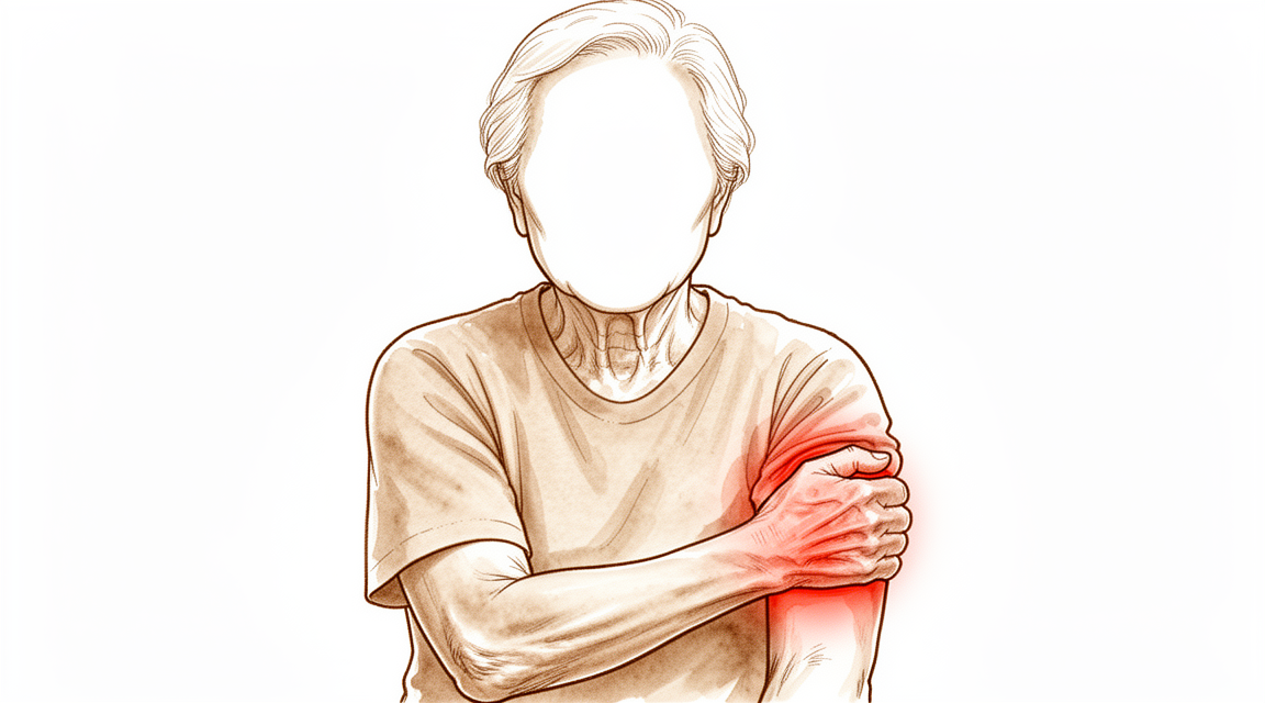
您正在感受到的症状
肘关节炎通常表现为活动最末端的僵硬和疼痛。您可能会发现无法将手臂完全伸直,或者无法完全弯曲。疼痛往往出现在这些极限位置,而不是活动的中段。许多人还会注意到关节出现卡顿、弹响或交锁,有时伴有疼痛,这是因为骨赘以及软骨或骨的小游离碎片卡在了关节内。
需要把手臂伸直或完全弯曲的日常事务会变得困难。伸手去够高处的架子、用手撑着从椅子上站起来,或者把叉子送到嘴边,都可能变得不便。旋转前臂的动作,比如转动钥匙或螺丝刀,在早期通常仍然没有问题,到后期才会变得僵硬。这对您的限制有多大,在很大程度上取决于您的工作和日常需要。重体力劳动、提举重物或投掷动作对关节造成的负担远大于伏案工作。
夜间疼痛并非肘关节炎的典型表现。如果您的肘部在夜间使您痛醒,这提示另有原因,值得请医生检查。如果疼痛没有缓解、在数周内不断加重,或使您无法工作或使用手臂,应请您的全科医生评估,您也可以要求我们安排专科医生评估。
有些肘关节炎患者还会出现尺神经受压,尺神经是走行于肘部内侧后方的神经。这可能导致无名指和小指刺痛或麻木,或手部无力。如果您注意到这些症状,请在下次复诊时提出,以便我们评估这条神经。
根本问题是关节面的磨损,加上肘部边缘长出的额外骨质。这些额外的骨质,连同逐渐收紧的关节包膜和游离碎片,在活动范围的末端从物理上阻挡了肘部。有趣的是,关节内的间隙往往仍然保留,因此在影像检查上,关节看起来可能比您的症状所提示的要好。
实际发生了什么
在您的肘部内,有三个小关节协同工作。其中一个让手臂弯曲和伸直。另外两个让前臂旋转,使您可以把手掌转向上或转向下。每个关节面都覆盖着一层光滑而润滑的软骨,有点像好锅上的不粘涂层。当这层涂层完好时,关节面之间几乎没有摩擦地相互滑动。
在肘关节炎中,这层涂层会在一些地方磨薄,身体的反应是在关节边缘长出额外的骨质。这些称为骨赘的骨性突起会在肘部的前方和后方堆积。关节包膜,即包绕关节的一层组织鞘套,也会收紧和缩短。软骨或骨的小碎片可能脱落,作为游离体在关节内漂浮。
这些变化共同解释了您的感受。骨赘和紧绷的包膜就像活动末端的物理挡块,使肘部无法完全伸直或弯曲。游离体卡在活动的关节面之间时,就会引起卡顿、弹响或交锁。在早期,疼痛出现在活动末端的极限位置,因为阻挡正发生在那里。随着更多的软骨涂层被磨掉,疼痛也可能蔓延到活动的中段。
有两点使肘关节炎与髋关节或膝关节的关节炎有所不同。第一,关节内的间隙往往保持较宽,因此X光片看起来可能比您的实际感受要好。第二,磨损往往对关节的某一部分影响更重,即前臂一根骨头的顶端与其上方上臂骨末端相接的部位。您推或提重物时,这个部位承受很大的负荷,这就是为什么重体力劳动、举重和投掷动作与这种疾病有关。
它也可能继发于旧伤。骨折或关节的其他损伤可能使关节面变得略微不平整,因此之后会出现不均匀的磨损。
我们如何处理该问题
Mater Private Hospital Rockhampton 的上肢外科医生 Kieran Hirpara 医生会从适合您病情的最微创方案入手。患者通常由全科医生转诊至我们的诊所;如果理疗师建议您就诊,您仍需获得全科医生的转诊才能符合 Medicare 报销资格。在您的首次就诊时,我们会采集病史,检查您的肘部,并在需要时安排X光检查。X光片可以显示关节前方和后方的骨赘。CT扫描能提供更详细的图像,如果最终需要手术,它有助于我们规划手术。
对于这样一个长期存在的问题,我们通常先尝试非手术治疗。改变工作和提举重物的方式可以减轻关节的负担,休息可以缓解急性发作。手部治疗的目的是保持您现有的活动范围,并增强肘部周围的力量。抗炎药片是一类常用的缓解疼痛和肿胀的药物,可以帮助改善症状。在讨论手术之前,我们会先充分尝试这些方案。
如果这些措施未能带来足够的改善,手术可能值得考虑。通常在疼痛无法缓解、僵硬使您无法完成日常事务,或者肘部出现疼痛性的交锁或卡顿时,才会建议手术。主要的手术是关节清理,称为清创术。它会切除阻碍活动的骨赘和游离碎片,并松解紧绷的关节包膜,同时保留您自身的关节面。根据需要处理的部位,这项手术可以通过小的锁孔切口完成,也可以通过开放切口完成。我们会与您详细讨论哪种方式适合您的肘部,并共同决定治疗方案。
预期情况
肘关节炎是一种长期疾病。它通常不会自行消失,但往往呈现一定的规律:有比较平稳的时期,在肘部劳累过度时则会急性发作。在早期,疼痛出现在活动末端,关节内的间隙往往仍然保留。随着磨损进展,疼痛可能蔓延到活动的中段,僵硬也可能加重。
如果不治疗,骨赘和紧绷的关节包膜会持续阻挡活动末端,因此僵硬往往会随时间推移逐渐加重。在早期处理中,非手术治疗仍然是第一步,并且可以在早期阶段带来缓解。改变工作和提举重物的方式、手部治疗以及抗炎药片,可以使症状缓解到足以让您继续正常生活的程度。如果这些措施不再起效,这就提示手术可能会有帮助。
针对这种疾病的手术目的是去除机械性阻挡,而不是置换关节。清除骨赘和游离碎片并松解紧绷的包膜,可以改善大多数人的活动范围和功能,并发症发生率和再手术率都很低。这种改善是切实的,但并非治愈:它不能使软骨再生,也不能阻止关节炎进展。有些人在关节清理后的数年里,会发现活动范围又逐渐减少,而X光片上的变化往往会在十年左右的时间里不断进展。对于磨损性关节炎,很少会进行全肘关节置换,而且通常不用于65岁以下或活动量大的人,因为人工关节在高负荷下会磨损。
如果您的肘部变得发烫、发红、肿胀和疼痛,尤其是伴有发热时,请当天前往急诊科。不需要全科医生的转诊。如果您的症状没有缓解、在数周内不断加重、使您在夜间痛醒,或使您无法工作或使用手臂,请去看您的全科医生,或要求我们安排专科医生评估。
何时就医
大多数肘关节炎是缓慢发展的,因此您有时间采取措施。如果僵硬和疼痛没有缓解、在数周内不断加重,或使您无法工作或使用手臂,请去看您的全科医生,或要求我们安排专科医生评估。反复出现的交锁或卡顿也值得评估,因为关节内的游离碎片可能阻碍活动。如果无名指和小指出现刺痛或麻木,也应告知医生,因为这提示尺神经受到了挤压。
有一组症状需要当天就医。如果您的肘部变得发烫、发红、肿胀和疼痛,尤其是伴有发热时,请前往急诊科。不需要全科医生的转诊。关节内感染可能会迅速损伤关节,因此这种情况不能等到常规预约再处理。
深入探讨
Advanced reading: the deeper science (optional)
本节内容超出了您做出自身治疗决策所需的深度。肘关节骨关节炎值得额外阅读,因为看似能一劳永逸解决问题的手术——即像髋关节和膝关节那样常规进行的关节置换术——在肘关节处的表现截然不同,而这一单一事实决定了整个治疗阶梯的走向。
为何肘关节置换并非首选方案
全肘关节置换术是有效的,但其对负荷的耐受性不及髋关节或膝关节置换术。一项纳入 2,118 例类风湿关节炎患者的汇总研究显示,尽管肘关节置换术持续提供令人满意的效果,但其 假体失败率和并发症率显著高于髋关节和膝关节置换术 [1]。
其后果是,患者需遵守永久性的提重限制,而非仅在恢复期采取的临时预防措施。由于肘关节位于长力臂的末端,手部施加的适度重量会在假体处产生巨大的力,这些力会随时间推移导致假体松动。
因此,肘关节的治疗阶梯与下肢相反。在髋关节,关节置换术是晚期关节炎的标准手术。而在肘关节,该手术主要保留给年龄较大、功能需求较低的患者,以及炎症性关节炎而非磨损性关节炎患者。
病因与严重程度同等重要
两名肘关节磨损程度相同的患者,其预后可能因关节磨损的原因不同而存在差异。在 679 名患者中,关节炎的病因影响了特定假体失效模式方面的结局,且类风湿关节炎患者的功能结局优于因创伤后情况接受置换手术的患者 [2]。
这一点值得强调,因为它与大多数人的预期相反,即由单次损伤导致的关节损伤应比由系统性疾病导致的关节损伤预后更好。其解释在于负荷需求:创伤后关节炎往往发生在生活体力要求更高的较年轻人群中,而假体所承受的力超出了其设计范围。
清创术是承担主要工作的手术
对于原发性磨损性关节炎,主要治疗手段并非关节置换,而是清创术:清除在关节活动末端阻碍关节运动的骨赘和游离体,松解紧张的关节囊,同时保留关节面。
证据是一致的。在 1,097 例患者中,清创术取得了良好的中期功能效果,且使用关节镜技术并未增加并发症 [3]。在 871 例患者中,无论是开放手术还是关节镜下的骨关节囊清创术,均可靠地改善了屈曲、伸展及功能评分,且并发症发生率较低 [4];一项针对 586 例患者的荟萃分析发现,清创术对原发性肘关节骨关节炎的致残症状有效,且并发症发生率可接受 [5]。
请注意清创术被要求完成的任务。它并不重塑关节面,也不能阻止关节炎的进展。它移除的是活动末端的机械性阻挡,因此它有助于主要主诉为肘关节无法完全伸直或弯曲、且在活动极限处出现疼痛性卡顿的患者;而对于在整个活动弧范围内均有疼痛的患者,其帮助则较小。
开放手术或关节镜手术并非决定性问题
与多种肘部手术一样,技术选择引发的争议往往超出了证据所能支持的范围。上述综述发现,两种方法均安全且有效;一项针对 639 例患者的叙述性综述得出结论,无法确定哪种手术方式更优 [6]。
实际的决定因素是需要触及的部位。关节镜能很好地处理前室和后室的操作;但对于需要广泛关节囊松解的僵硬肘关节,或解剖结构扭曲且必须直视并保护神经的病例,开放手术可能更安全。
参考文献
[1] Chou TA, Ma H, Wang J, Tsai S, Chen C, Wu P, et al. 类风湿关节炎患者全肘关节置换术:系统评价与荟萃分析。Bone Joint J. 2020;102-B(8):967-80. https://doi.org/10.1302/0301-620X.102B8.BJJ-2019-1465.R1
[2] Wang J, Ma H, Chou TA, Tsai S, Chen C, Wu P, et al. 类风湿关节炎与创伤后病变全肘关节置换术后的结局:系统评价与荟萃分析。Bone Joint J. 2019;101-B(12):1489-97. https://doi.org/10.1302/0301-620X.101B12.BJJ-2019-0799.R1
[3] White CHR, Ravi V, Watson J, Badhrinarayanan S, Phadnis J. 关节镜与开放清创术治疗关节炎性肘关节的系统评价。Arthroscopy. 2020;37(2):747-58. https://doi.org/10.1016/j.arthro.2020.09.005
[4] Guerrero EM, Bullock GS, Helmkamp JK, Madrid A, Ledbetter L, Richard MJ, et al. 关节镜与开放骨囊清创术治疗原发性肘关节骨关节炎的临床影响:系统评价。J Shoulder Elbow Surg. 2020;29(4):689-98. https://doi.org/10.1016/j.jse.2019.12.003
[5] de Klerk HH, Welsink CL, Spaans AJ, Verweij LPE, van den Bekerom MPJ. 原发性肘关节骨关节炎的关节镜与开放清创术:系统评价与荟萃分析。EFORT Open Rev. 2020;5(12):874-82. https://doi.org/10.1302/2058-5241.5.190095
[6] Poonit K, Zhou X, Zhao B, Sun C, Yao C, Zhang F, et al. 开放或关节镜清创术治疗肘关节骨关节炎:叙述性综述。BMC Musculoskelet Disord. 2018;19(1). https://doi.org/10.1186/s12891-018-2318-x
Evidence & references
This is the clinical evidence summary written for health professionals. It is technical, and it lists the research this page was built from. You do not need to read it to understand your treatment or to make a decision about it.
Overview
Epidemiology and Demographics
- Primary osteoarthritis of the elbow is a relatively rare occurrence, seen in only 1% to 2% of patients with elbow arthrosis [3].
- Symptomatic primary osteoarthritis of the elbow affects 2% of the population [6].
- Primary osteoarthritis of the elbow is confined almost exclusively to men [3].
- Men are affected more often than women with a 4:1 ratio [6].
- The average age of presentation for primary elbow osteoarthritis is 50 years, with a range of 20 to 70 years [6].
- Hand dominance and strenuous manual labor are associated with primary osteoarthritis of the elbow [6].
- Secondary causes of elbow osteoarthritis include trauma, osteochondritis dissecans, and synovial osteochondromatosis [6].
Pathoanatomy
- Osteoarthritis of the elbow is characterized by osteophyte formation, capsular contracture, and loose bodies, often with relative preservation of the joint space [6].
- Periarticular hypertrophic osteophytes act as a mechanical block at the end ranges of flexion and extension [6].
- Advanced elbow osteoarthritis rarely presents with joint space narrowing [6].
- Osteoarthritis typically involves the radiocapitellar joint articular cartilage preferentially, with relative preservation of the ulnohumeral articular surfaces [6].
Clinical Presentation and Evaluation
- Patients typically present with loss of terminal extension and flexion and painful catching, clicking, or locking of the elbow [6].
- Pain in elbow osteoarthritis is typically noted at the end ranges of motion and not through the midrange [6].
- Night pain is not typical of elbow osteoarthritis; if present, an inflammatory cause of the arthritis should be considered [6].
- Ulnar neuropathy is present in up to 50% of patients with elbow osteoarthritis [6].
- Standard AP and lateral radiographs typically show osteophyte formation at the coronoid process (anterior and medial), coronoid fossa, radial fossa, radial head, olecranon tip, and olecranon fossa [6].
- Radiographs typically show preserved joint spaces at the ulnohumeral joint and mildly narrowed joint spaces at the radiocapitellar joint [6].
- Radiographs typically underestimate the number of loose bodies present in the elbow [6].
- CT may be useful for surgical planning, allowing a detailed assessment of osteophytes and the presence of loose bodies [6].
Treatment Principles
- Nonoperative treatment remains the first step in the early management of elbow osteoarthritis [4].
- The goal of treatment is to obtain a low level of pain with sufficient motion range to ensure good function, while preserving future surgical options and delaying elbow arthroplasty to the extent possible [1].
- The appropriate treatment for elbow arthritis depends on the etiology, severity, patient age, and functional demands [8].
- Treatment of elbow arthritis must be individualized based on etiology, severity, patient age, and functional demands [14].
- Nonsurgical management may provide relief in early stages of elbow arthritis [14].
Nonoperative Management
- Rest, NSAIDs, corticosteroid injections, and activity modification are the mainstays of nonsurgical treatment for elbow osteoarthritis [6].
Surgical Indications and Options
- Surgical indications for elbow osteoarthritis include failure to respond to nonsurgical interventions, loss of motion that interferes with activities of daily living, and painful locking or catching of the elbow [6].
- Joint-sparing procedures such as débridement, excision of osteophytes, capsular release, and removal of loose bodies are preferred surgical options [6].
- Total elbow arthroplasty is rarely indicated for elbow osteoarthritis and is not indicated for patients younger than 65 years or physically active patients because of concerns about implant longevity [6].
- Surgical options must be tailored to cartilage integrity and bone structure, with total elbow arthroplasty generally avoided in young, active patients due to poor durability [35].
- Total elbow arthroplasty is a treatment option for elbows severely affected by rheumatoid arthritis, with recent reports showing decreased prevalence of complications, maintenance of excellent pain relief, and functional restoration [32].
- Total elbow arthroplasty remains associated with substantial complication and reoperation rates [43].
Arthroscopic Procedures
- Elbow arthroscopic osteocapsular arthroplasty is a safe, efficacious treatment for patients with mild to moderate osteoarthritis [2].
- Arthroscopic treatment of elbow osteoarthritis provides good short-term outcomes in primary elbow osteoarthritis and is associated with a low complication rate [7].
- Arthroscopic debridement for primary degenerative osteoarthritis results in statistically significant and clinically relevant improvement in elbow range of motion and clinical outcomes with low complication and reoperation rates [15].
- Arthroscopic treatment of elbow osteoarthritis significantly improved 6-month clinical results for functional scores, pain, strength, and range of motion [30].
- Arthroscopic osteocapsular arthroplasty can be recommended for its favorable overall treatment outcomes for elbow osteoarthritis [29].
- Arthroscopic osteocapsular arthroplasty is comparable to the open procedure in managing primary osteoarthritis of the elbow [73].
- The open procedure shows better outcomes than arthroscopic osteocapsular arthroplasty in the improvement of flexion limitation [73].
- Contraindications for arthroscopic elbow procedures include severe contracture and periarticular heterotopic ossification [6].
- Relative contraindications for arthroscopic elbow procedures include prior ulnar nerve transposition and prior extensive open procedures [6].
- Osteocapsular arthroplasty refers to the arthroscopic technique for elbow joint débridement, involving capsular release, loose body removal, and excision of osteophytes [6].
- Miniopen ulnar nerve decompression is a safe and effective procedure that can be indicated for ulnar neuropathy in the setting of elbow osteoarthritis [117].
Open Procedures
- The Outerbridge-Kashiwagi arthroplasty is the classic open procedure, in which the olecranon fossa is trephinated and osteophytes are removed [6].
- Limitations of the Outerbridge-Kashiwagi procedure include incomplete anterior release and incomplete osteophyte removal anteriorly [6].
- The Outerbridge-Kashiwagi procedure is an effective and safe way of treating both posttraumatic arthritis and osteoarthritis of the elbow [13].
- Both open elbow debridement and the Outerbridge-Kashiwagi procedure had excellent survivorship until conversion to total elbow arthroplasty and are viable options in the treatment of primary elbow osteoarthritis and post-traumatic cases [22].
- A medial or lateral column approach can be used for open débridement, loose body removal, osteophyte resection, and capsulectomy depending upon the location of the pathology and concomitant procedures to be performed [6].
- Capsulectomy and debridement for primary osteoarthritis of the elbow through a medial trans-flexor approach is associated with a low rate of complications and is safe and effective [5].
Outcomes and Survivorship
- Patients with either posttraumatic or primary degenerative osteoarthritis can expect satisfactory elbow function and an improvement in pain with little chance of reoperation at the midterm of the follow-up duration [11].
- Open and arthroscopic debridement procedures seem to be safe and effective in the treatment of elbow osteoarthritis [61].
Complications
- Deep infections in the elbow are more common than other joints treated arthroscopically, with a rate of 0.8% to 2.2% [6].
- Transient nerve palsies complicate 1% to 3% of cases, with radial and ulnar nerves being the most common [6].
- Other complications of elbow osteoarthritis surgery include stiffness (heterotopic ossification), hematoma formation, and synovial ganglion formation [6].
Anatomy & Pathophysiology
Epidemiology & Demographics
- Primary osteoarthritis most commonly affects middle-aged males (approximately 50 years old), manual laborers, throwing athletes, and heavy weight lifters [70].
Etiology & Secondary Causes
- Posttraumatic arthritis is commonly seen after elbow injuries such as distal humerus fractures, radial head fractures, proximal ulna fractures, or elbow fracture-dislocations [12].
- Arthritis can develop in response to the initial cartilage insult or secondary to residual articular incongruities from injuries [12].
- Postinjury malunions, nonunions, or residual instability can alter kinematics and load across the elbow, predisposing the elbow to degenerative changes [12].
- Posttraumatic arthritis of the elbow is particularly challenging to treat due to associated bone loss and its frequent occurrence in younger populations with higher functional demands [12].
- Unlike primary osteoarthritis, rheumatoid arthritis frequently involves the elbow joint, can be bilateral, and often involves other joints [70].
- Rheumatoid arthritis causes an intense inflammatory synovitis that leads to progressive joint destruction, involving the ulnohumeral joint early in the disease process [70].
Pathoanatomy & Wear Patterns
- Rarely, advanced disease presents with joint space narrowing [6].
- Cadaveric studies have demonstrated that radiocapitellar degeneration is more common with increasing age [18].
- The posteromedial aspect of the radial head appears to be consistently involved with reciprocal changes seen on the capitellum in primary elbow osteoarthritis [18].
- Osteophytic change occurs predominantly in the ulnohumeral compartment of the elbow, whereas joint space narrowing more frequently affects the radiocapitellar articulation [53].
- Ulnohumeral osteophytes were found in 95% of patients with symptomatic osteoarthritis, while radiohumeral osteophytes were found in 59% [78].
- The primary pathology of elbow osteoarthritis is loss of articular cartilage with resulting osteophyte formation on the olecranon process, coronoid process, and their respective fossae [70].
- Secondary resultant changes in elbow osteoarthritis involve osteophyte formation along the margin of the radial head and formation of loose bodies that may result in mechanical block or crepitance [70].
- Three-dimensional computational models identified unique regions of bony impingement in elbow osteoarthritis, such as between the radial head and a posterior capitellar osteophyte in extension [54].
Clinical Presentation
- Pain is typically noted at the end ranges of motion and not through the midrange [6].
- Night pain is not typical; if present, an inflammatory cause of the arthritis should be considered [6].
- Forearm rotation is relatively preserved until later in the disease process [6].
- Most patients initially complain of pain at terminal limits of motion as a result of capsular stretch and osteophyte impingement at the extremes of motion [70].
- Later in the disease process, pain through the mid arc of motion develops as the extent of cartilage loss progresses [70].
Imaging Findings
- Radiographs typically show osteophyte formation at the coronoid process (anterior and medial), coronoid fossa, radial fossa, radial head, olecranon tip, and olecranon fossa [6].
- Joint spaces at the ulnohumeral joint are usually preserved, and those at the radiocapitellar joint are mildly narrowed [6].
- Loose bodies may be evident on radiographs, which typically underestimate the number present [6].
- The ulnohumeral articular joint space is preferentially preserved with osteoarthritis [71].
- Osteophytes are typically seen on the coronoid and olecranon tips, with loss of the concavity of the radial head, coronoid, and olecranon fossa [71].
- Ulnohumeral joint space loss suggests inflammatory or posttraumatic arthritis rather than primary osteoarthritis [71].
Classification
Radiographic Classification Systems
- The Broberg and Morrey classification system is based on osteophyte formation and joint space narrowing [18].
- The Hasting and Retting classification system focuses on radiocapitellar wear and subluxation [18].
- The Broberg and Morrey and Hasting and Retting classification systems are both X-ray based [18].
- Kwak et al. described a CT-based method of quantifying elbow arthritis [18].
- The CT-based classification demonstrated high correlation with the visual analog scale and the Mayo Elbow Performance Score [18].
- The CT-based classification demonstrated moderate correlation with range of motion [18].
- A new radiographic classification system was developed based on the lack of radiographic evidence for morphologic predisposition in primary elbow osteoarthritis [27].
- The extent of degenerative joint disease of the radiocapitellar joint had a negative effect on postoperative outcome [27].
- The developed radiographic classification system appears valid in predicting postoperative outcome [27].
- A bony landmarks classification system effectively delineated osteophyte distribution in elbow patients [58].
Reliability and Validity
- The Broberg and Morrey and Hasting and Retting classification systems demonstrated substantial intraobserver and interobserver reliability for evaluating radiographic severity of post-traumatic arthritis and primary osteoarthritis of the elbow [39].
- The CT-based staging system was highly reproducible and clinically feasible compared with previous plain radiograph-based staging systems for elbow osteoarthritis [28].
- The 4 grades of the Broberg and Morrey classification system have only fair interobserver reliability [118].
- Interobserver reliability for the Broberg and Morrey classification system is influenced by subspecialty and experience [118].
- Surgeons with more than 10 years of experience had greater agreement in arthrosis ratings than surgeons with less experience [118].
- Surgeons who treated more than 10 elbow fractures per year had better agreement in arthrosis ratings than those treating fewer fractures [118].
- Simplified binary rating systems for elbow arthrosis resulted in moderate agreement among observers [118].
- Interobserver agreement for radiographic assessment and classification in a cohort study of arthroscopic management yielded k values ranging from 0.81 to 0.85 [44].
Clinical Presentation
Epidemiology and Demographics
- The prevalence of primary elbow osteoarthritis in Japanese subjects aged 50-89 years was 25.2%, with most cases being asymptomatic [17].
- The prevalence of elbow osteoarthritis was 55.0% in respondents aged 40 years or older, with a symptomatic prevalence of 22.6% [25].
- Men are affected more often than women with primary elbow osteoarthritis at a 4:1 ratio [6].
- Older age, male sex, and a history of elbow trauma are identified as significant risk factors for elbow osteoarthritis [25].
Pathoanatomy
- Advanced disease rarely presents with joint space narrowing [6].
- Primary osteoarthritis of the elbow is unique due to relative preservation of articular cartilage and maintenance of joint space with hypertrophic osteophyte formation [21].
- Radiocapitellar degeneration is more common with increasing age [18].
- The posteromedial aspect of the radial head appears to be consistently involved in primary elbow osteoarthritis, with reciprocal changes seen on the capitellum [18].
History and Symptoms
- Night pain is not typical for elbow osteoarthritis; if present, an inflammatory cause of the arthritis should be considered [6].
- Most patients initially complain of pain at terminal limits of motion as a result of capsular stretch and osteophyte impingement at the extremes of motion, with or without mechanical symptoms [70].
- The degree of disability caused by osteoarthritis depends on the patient’s vocation and physical disability [6].
Physical Examination
- Inspection should check for prior surgical incisions and joint effusion at the lateral soft spot [6].
- Pain is usually felt at the end ranges of flexion and extension rather than throughout the arc during range of motion assessment [6].
Imaging
- Standard AP and lateral radiographs should be obtained for evaluation of elbow osteoarthritis [6].
- Joint spaces at the ulnohumeral joint are usually preserved on radiographs [6].
- Joint spaces at the radiocapitellar joint are mildly narrowed on radiographs [6].
- CT may be useful for surgical planning and allows a detailed assessment of osteophytes and the presence of loose bodies [6].
- CT scans with 3D reconstructions may be useful for evaluating the extent and location of disease and for surgical planning [101].
Investigations
History and Physical Examination
- Patients with elbow osteoarthritis typically present with loss of terminal extension and flexion, along with painful catching, clicking, or locking of the elbow [6].
- Pain in elbow osteoarthritis is typically noted at the end ranges of motion rather than through the midrange [6].
- The presence of night pain in a patient with elbow arthritis suggests an inflammatory cause rather than typical osteoarthritis [6].
- Physical examination for elbow osteoarthritis includes inspection for prior surgical incisions and joint effusion at the lateral soft spot [6].
- Forearm rotation is relatively preserved until later in the disease process of elbow osteoarthritis [6].
- The degree of disability caused by elbow osteoarthritis depends on the patient’s vocation and physical disability [6].
Imaging
- Standard AP and lateral radiographs are the initial imaging modality for evaluating elbow osteoarthritis [6].
- Radiographs of the elbow in osteoarthritis typically show osteophyte formation at the coronoid process, coronoid fossa, radial fossa, radial head, olecranon tip, and olecranon fossa [6].
- In elbow osteoarthritis, joint spaces at the ulnohumeral joint are usually preserved, while those at the radiocapitellar joint are mildly narrowed [6].
- CT imaging may be useful for surgical planning by allowing a detailed assessment of osteophytes and the presence of loose bodies [6].
- CT has greater sensitivity than radiographs for the detection of osteophytes and loose bodies in primary elbow osteoarthritis [139].
- A CT-based staging system for elbow osteoarthritis is highly reproducible and clinically feasible compared with plain radiograph-based staging systems [28].
- On imaging assessment for elbow arthritis, signs of impingement such as osteophytes and filling of fossae are more common than cartilage lesions like joint narrowing [145].
- Three-dimensional computational models can identify the locations and volumes of bony impingement in patients with elbow osteoarthritis [54].
- Pre-operative simulation results can be used as an index to determine which osteophytes to remove during arthroscopic surgery for elbow osteoarthritis [50].
- A bony landmarks classification system effectively delineates osteophyte distribution in patients with elbow osteoarthritis [58].
- The Broberg and Morrey classification for elbow arthritis is based on osteophyte formation and joint space narrowing [18].
- The Hasting and Retting classification system for elbow arthritis focuses on radiocapitellar wear and subluxation [18].
- Both the Broberg and Morrey and Hasting and Retting classification systems demonstrated substantial intraobserver and interobserver reliability for evaluating radiographic severity of elbow arthritis [39].
- A CT-based method of quantifying elbow arthritis described by Kwak et al. demonstrates high correlation with visual analog scale and the Mayo Elbow Performance Score, and moderate correlation with range of motion [18].
Treatment
Nonoperative Management
- Nonsurgical treatments can be effective for reducing symptoms but have limited effectiveness for improving range of motion limitations and pain related to impinging osteophytes [66].
Surgical Indications
- Surgical treatment for elbow arthritis is based on disease etiology, severity of degeneration, and patient age [10].
- Failure to respond to nonsurgical interventions is an indication for surgical treatment of elbow osteoarthritis [6].
- Loss of motion that interferes with activities of daily living is an indication for surgical treatment of elbow osteoarthritis [6].
- Painful locking or catching of the elbow is an indication for surgical treatment of elbow osteoarthritis [6].
- The goal of treatment for post-traumatic osteoarthritis of the elbow is to obtain a low level of pain with sufficient motion range to ensure good function, while preserving future surgical options and delaying elbow arthroplasty to the extent possible [1].
Joint-Sparing Procedures
- Joint-sparing procedures such as débridement, excision of osteophytes, capsular release, and removal of loose bodies are preferred for elbow osteoarthritis [6].
- Osteocapsular debridement is an effective surgical treatment option for patients with symptomatic primary elbow osteoarthritis who have failed conservative management [46].
- Surgical debridement is an effective treatment for the disabling symptoms of primary elbow OA with an acceptable complication rate [63].
- Elbow arthroscopic debridement for primary degenerative osteoarthritis results in statistically significant and clinically relevant improvement in elbow range of motion and clinical outcomes with low complication and reoperation rates [15].
- Arthroscopic debridement for elbow osteoarthritis provides satisfactory pain relief, improvement of elbow motion, and good functional outcome [64].
- Arthroscopic debridement for the elbow osteoarthritis provided satisfactory pain relief, improvement of elbow motion, and good functional outcome [68].
- Elbow AOA is a safe, efficacious treatment for patients with mild to moderate osteoarthritis [2].
- This minimally invasive technique provides good short-term outcomes in primary elbow osteoarthritis and is associated with a low complication rate [7].
- The study recommends arthroscopic debridement in the surgical management of patients with osteoarthritis of the elbow [9].
- This procedure is a valuable adjunct in the management of the arthritic elbow, serving as an intermediate step between nonoperative management and elbow replacement surgery [36].
- Both open elbow debridement and the OK procedure had excellent survivorship until conversion to total elbow arthroplasty and are viable options in the treatment of primary elbow osteoarthritis and post-traumatic cases [22].
- The OK procedure is an effective and safe way of treating both posttraumatic arthritis and osteoarthritis of the elbow [13].
- The modified Outerbridge-Kashiwagi procedure combining mini-open and arthroscopic techniques achieved short-term clinical success in pain relief and function restoration in patients with elbow osteoarthritis [105].
- Most patients had an uneventful postoperative course and have shown a painless elbow joint, with satisfactory functional recovery at short-term follow-up after open debridement and radiocapitellar replacement [34].
- The medial approach is effective for the treatment of advanced primary osteoarthritis of the elbow, especially in patients with ulnar nerve symptoms as well as medial osteophytes [19].
- Arthroscopic osteocapsular arthroplasty is comparable to the OPEN procedure in managing primary osteoarthritis of the elbow; however, the OPEN procedure shows the better outcome in improvement of flexion limitation [73].
- Open and arthroscopic debridement procedures seem to be safe and effective in the treatment of elbow OA [61].
- In subjects with slight or moderate OA and preserved elbow joint congruence, arthroscopy provides good-to-satisfactory results [44].
- The extent of degenerative joint disease of the radiocapitellar joint had a negative effect on postoperative outcome, and the developed classification system appears valid in predicting postoperative outcome [27].
- A reproducible triceps tendon-guided arthroscopic olecranon debridement technique can restore adequate joint space, mobility, and function to the patient with degenerative elbow osteoarthritis [23].
Arthroplasty and Salvage Procedures
- Total elbow arthroplasty is rarely indicated for elbow osteoarthritis, and it is not indicated for patients younger than 65 years or physically active patients because of concerns about implant longevity [6].
- Total elbow arthroplasty is best reserved for low demand, elderly patients who will be able to comply with the 5-lb weightlifting restriction imposed postoperatively to protect the implants from bearing wear, hardware loosening, or failure [100].
- Open or arthroscopic débridement may be effective in the treatment of early arthritis, whereas interposition arthroplasty or total elbow arthroplasty is best reserved for more advanced cases [100].
- Total elbow arthroplasty has become a reliable treatment option for elbows severely affected by rheumatoid arthritis, with recent reports showing decreased prevalence of complications, maintenance of excellent pain relief, and functional restoration [32].
- The study observed favorable midterm results with the capitellocondylar total elbow replacement, with good pain relief and improved function of the elbow [52].
- Although total elbow arthroplasty has become an accepted treatment for a variety of degenerative and traumatic elbow conditions, concerns regarding its durability in young and/or high-demand patients make elbow arthrodesis an acceptable alternative in this population [98].
- Elbow arthrodesis is reserved for patients with painful arthritis who are not candidates for total elbow arthroplasty, especially individuals who place high demands on the upper extremities, such as manual laborers [98].
- For unilateral arthrodesis of the elbow, a position of 90 to 100 degrees of flexion is desirable to provide the most powerful grip strength [98].
- Bilateral elbow arthrodesis rarely is indicated because of resultant functional limitations [98].
- If bilateral elbow arthrodesis is indicated, one elbow should be placed in 110 to 120 degrees of flexion to permit the patient to reach the mouth, and the other should be placed in 45 to 65 degrees to aid in personal hygiene [98].
- Interposition arthroplasty is considered for intrinsic contractures in young patients (20 to 50 years) with articular cartilage destruction in whom the anatomic architecture of the distal humerus and proximal ulna are relatively preserved [69].
Procedural Considerations and Contraindications
- Contraindications for arthroscopic procedures for elbow osteoarthritis include severe contracture and periarticular heterotopic ossification [6].
- Relative contraindications for arthroscopic procedures for elbow osteoarthritis include prior ulnar nerve transposition and prior extensive open procedures [6].
- Regardless of the type of procedure used, ulnar nerve transposition and release of the posterior bundle of the medial collateral ligament (MCL) should be considered for patients who have less than 90° to 100° of elbow flexion [6].
- Ulnar nerve decompression/transposition and release of the posterior bundle of the MCL should be considered for patients who have less than 90° to 100° of elbow flexion [66].
- During arthroscopic surgery, joint distention moves the capsule away from bone, but the distance between the neurovascular structures and the capsule remain unchanged; therefore, the nerves remain at risk with capsular work [6].
- Neurovascular structures at risk during portal placement, débridement, and capsular release include median nerve (anteromedial), the ulnar nerve (posteromedial), and the radial nerve (lies adjacent to the anterolateral capsule) [6].
- The brachialis muscle protects the median nerve and brachial artery during capsular procedures [6].
- The olecranon osteophytosis extends medially and laterally and not just at the tip, requiring resection to be extended along the medial and lateral aspects of the olecranon to allow maximal extension and prevent impingement [6].
- Coronoid osteophytosis extends medially and not just at the tip, requiring resection to be extended medially if necessary to maximize the restoration of flexion range of motion [6].
- Elbow arthroscopy is technically demanding, and several neurovascular structures that are at risk during the procedure include the radial, ulnar, and median nerves [66].
- Unlike other joints, arthroscopic débridement in the elbow can be very effective at reducing symptoms and improving range of motion [66].
Complications
- Deep infections in the elbow are more common than in other joints treated arthroscopically, with a rate of 0.8% to 2.2% [6].
- Infection can manifest as superficial minor wound complications or deep infection [6].
- Infection is associated with intraoperative corticosteroid injections [6].
- Transient nerve palsies complicate 1% to 3% of cases [6].
- Radial and ulnar nerves are the most commonly affected in transient nerve palsies [6].
- Heterotopic ossification is a complication associated with stiffness [6].
- Hematoma formation is a recognized complication [6].
- Synovial ganglion formation is a recognized complication [6].
- During arthroscopic surgery, joint distention moves the capsule away from bone, but the distance between neurovascular structures and the capsule remains unchanged, keeping nerves at risk during capsular work [6].
- The median nerve is at risk during portal placement, débridement, and capsular release due to its anteromedial location [6].
- The ulnar nerve is at risk during portal placement, débridement, and capsular release due to its posteromedial location [6].
- The radial nerve is at risk during portal placement, débridement, and capsular release as it lies adjacent to the anterolateral capsule [6].
- Total elbow arthroplasty is associated with substantial complication and reoperation rates [43].
- Total elbow replacement is associated with a high complication rate and may be warranted only for seriously disabled patients [113].
- Total elbow replacement for patients with rheumatoid arthritis has no more perioperative complications than for those who do not have rheumatoid arthritis [123].
Recovery
Nonoperative Management
- Rest, NSAIDs, corticosteroid injections, and activity modification are the mainstays of nonsurgical treatment [6].
Surgical Indications and Goals
- Surgical indications include failure to respond to nonsurgical interventions, loss of motion that interferes with activities of daily living, and painful locking or catching of the elbow [6].
Arthroscopic Outcomes
- Patients with primary or posttraumatic elbow osteoarthritis who underwent arthroscopic debridement can expect satisfactory elbow function and an improvement in pain with little chance of reoperation at the midterm of the follow-up duration [11].
- Clinical outcomes for patients with primary elbow osteoarthritis who underwent arthroscopic osteocapsular arthroplasty improved from preoperative assessment to short- and medium-term follow-up [31].
- Range of motion decreased between short- and medium-term follow-up after arthroscopic osteocapsular arthroplasty for primary elbow osteoarthritis [31].
Open Surgical Outcomes
- Most patients undergoing open debridement and radiocapitellar replacement for primary or post-traumatic arthritis of the elbow had an uneventful postoperative course and showed a painless elbow joint with satisfactory functional recovery at short-term follow-up [34].
- A tendency of advancing roentgenological osteoarthritic changes was recognized in most patients 10 years after the Outerbridge-Kashiwagi method [80].
Total Elbow Arthroplasty Outcomes
- Total elbow arthroplasty is rarely indicated and is not indicated for patients younger than 65 years or physically active patients because of concerns about implant longevity [6].
- The main objective of elbow joint replacement in arthritic diseases is to decrease pain, increase joint stability, and improve overall range of motion [51].
- In most cases, elbow function was maintained in the long-term without loosening of the implant following Kudo type-5 total elbow arthroplasty for rheumatoid arthritis [20].
- The capitellocondylar total elbow replacement provided significant long-term relief of pain and improvement in functional status and range of motion in patients with rheumatoid arthritis, with improvements not deteriorating over time [120].
- The mean Mayo elbow performance score significantly improved from 48 points preoperatively to 83 points at final follow-up for GSB III total elbow arthroplasty in patients with rheumatoid arthritis [75].
- In the course of total elbow arthroplasty, satisfactory supportive range of motion was restored to patients' elbow joints [76].
- Long-term follow-up showed acceptable results of the type-3 Kudo total elbow arthroplasty in patients with rheumatoid arthritis [72].
Complications
Key Evidence
- [L4] The goal of treatment is to obtain a low level of pain with sufficient motion range to ensure good function, while preserving future surgical options and delaying elbow arthroplasty to the extent possible. [1] (10.1016/j.otsr.2013.11.004)
- [L4] Elbow AOA is a safe, efficacious treatment for patients with mild to moderate osteoarthritis. [2] (10.1016/j.jhsa.2015.11.018)
- [L5] Primary osteoarthritis of the elbow is a relatively rare occurrence, seen in only 1% to 2% of patients with elbow arthrosis, and is confined almost exclusively to men. [3] (10.1016/s0894-1130(00)80037-x)
- [L5] Nonoperative treatment remains the first step in the early management of elbow osteoarthritis. [4] (10.2106/jbjs.e.00568)
- [L4] This approach is associated with a low rate of complications and is safe and effective for the treatment of primary osteoarthritis of the elbow. [5] (10.1016/j.jhsa.2011.07.018)
- [L4] This minimally invasive technique provides good short-term outcomes in primary elbow osteoarthritis and is associated with a low complication rate. [7] (10.1016/j.otsr.2019.09.003)
- [L5] The appropriate treatment for elbow arthritis depends on the etiology, severity, patient age, and functional demands. [8] (10.1016/j.jhsa.2009.02.019)
- [L4] The study recommends this technique in the surgical management of patients with osteoarthritis of the elbow. [9] (10.1302/0301-620x.96b2.30714)
- [L5] Surgical treatment for elbow arthritis is based on disease etiology, severity of degeneration, and patient age. [10] (10.1016/j.jhsa.2007.12.022)
- [L3] Patients with either pathology can expect satisfactory elbow function and an improvement in pain with little chance of reoperation at the midterm of the follow-up duration. [11] (10.1016/j.jseint.2021.07.018)
- [L5] [12] (10.1016/j.jhsa.2022.12.014)
- [L4] The OK procedure is an effective and safe way of treating both posttraumatic arthritis and osteoarthritis of the elbow. [13] (10.1016/j.jse.2015.11.052)
- [L5] Treatment of elbow arthritis must be individualized based on etiology, severity, patient age, and functional demands; nonsurgical management may provide relief in early stages, while surgical options range from arthroscopic debridement for pain at motion extremes to total elbow arthroplasty for pain throughout the arc of motion. [14] (10.1016/j.jhsa.2012.12.037)
- [L1] Elbow arthroscopic debridement for primary degenerative osteoarthritis results in statistically significant and clinically relevant improvement in elbow range of motion and clinical outcomes with low complication and reoperation rates. [15] (10.1016/j.arthro.2017.08.247)
- [L3] The prevalence of primary elbow osteoarthritis in Japanese subjects aged 50-89 years was 25.2%, with most cases being asymptomatic. [17] (10.1016/j.jse.2021.07.015)
- [L3] [18] (10.1177/17585732251327183)
- [L4] The medial approach is effective for the treatment of advanced primary osteoarthritis of the elbow, especially in patients with ulnar nerve symptoms as well as medial osteophytes. [19] (10.2106/jbjs.d.02684)
- [L3] In most cases, elbow function was maintained in the long-term without loosening of the implant. [20] (10.1302/0301-620x.99b6.bjj-2016-1033.r2)
- [L4] Primary osteoarthritis of the elbow is unique due to relative preservation of articular cartilage and maintenance of joint space with hypertrophic osteophyte formation. [21] (10.5435/00124635-200802000-00005)
- [L4] Both open elbow debridement and the OK procedure had excellent survivorship until conversion to total elbow arthroplasty and are viable options in the treatment of primary elbow osteoarthritis and post-traumatic cases. [22] (10.1016/j.jse.2022.01.138)
- [L5] In doing so, we present a reproducible technique that can restore adequate joint space, mobility, and function to the patient with degenerative elbow osteoarthritis. [23] (10.1016/j.eats.2024.103332)
- [L3] The prevalence of elbow OA was 55.0% in respondents aged 40 years or older, with a symptomatic prevalence of 22.6%; older age, male sex, and a history of elbow trauma were identified as significant risk factors. [25] (10.1016/j.jse.2018.02.049)
- [L4] The extent of degenerative joint disease of the radiocapitellar joint had a negative effect on postoperative outcome, and the developed classification system appears valid in predicting postoperative outcome. [27] (10.1016/j.jse.2007.03.014)
- [L4] CT-based staging system was highly reproducible and clinically feasible, compared with previous plain radiograph-based staging systems, for elbow osteoarthritis. [28] (10.1016/j.joca.2019.03.004)
- [L4] Arthroscopic osteocapsular arthroplasty can be recommended for its favorable overall treatment outcomes for elbow osteoarthritis. [29] (10.1016/j.jse.2019.09.036)
- [L3] Arthroscopic treatment of elbow osteoarthritis significantly improved 6-month clinical results for functional scores, pain, strength and range of motion. [30] (10.1016/j.otsr.2019.09.002)
- [L4] Serial assessment of patients with primary elbow OA who underwent arthroscopic OCA showed that the clinical outcomes improved from preoperative assessment to short- and medium-term follow-up, although ROM decreased between short- and medium-term follow-up. [31] (10.1177/23259671231162398)
- [L4] Total elbow arthroplasty has become a reliable treatment option for elbows severely affected by rheumatoid arthritis, with recent reports showing decreased prevalence of complications, maintenance of excellent pain relief, and functional restoration. [32] (10.2106/00004623-199805000-00008)
- [L4] Most patients had an uneventful postoperative course and have shown a painless elbow joint, with satisfactory functional recovery at short-term follow-up. [34] (10.1016/j.jse.2011.08.071)
- [L4] Surgical options must be tailored to cartilage integrity and bone structure, with total elbow arthroplasty generally avoided in young, active patients due to poor durability. [35] (10.1016/j.jhsg.2025.100736)
- [L4] This procedure is a valuable adjunct in the management of the arthritic elbow, serving as an intermediate step between nonoperative management and elbow replacement surgery. [36] (10.1016/s1058-2746(99)90131-3)
- [L3] Both the BM and HR classification systems demonstrated substantial intraobserver and interobserver reliability for evaluating radiographic severity of post-traumatic arthritis and primary osteoarthritis of the elbow. [39] (10.1016/j.jse.2014.10.015)
- [L4] Total elbow arthroplasty remains associated with substantial complication and reoperation rates. [43] (10.1016/j.jhsg.2026.100981)
- [L4] [44] (10.1007/s12306-015-0365-0)
- [L2] Osteocapsular debridement is an effective surgical treatment option for patients with symptomatic primary elbow osteoarthritis who have failed conservative management. [46] (10.1016/j.jse.2020.01.060)
- [L4] These results can be used as an index to determine the osteophytes to be removed during arthroscopic surgery for elbow osteoarthritis. [50] (10.1016/j.jseint.2026.101667)
- [L4] The main objective of elbow joint replacement in arthritic diseases is to decrease pain, increase joint stability and improve overall range of motion. [51] (10.1007/s12306-010-0070-y)
- [L4] The study observed favorable midterm results with the capitellocondylar total elbow replacement, with good pain relief and improved function of the elbow. [52] (10.1016/j.jse.2004.10.010)
- [L3] Osteophytic change occurs predominantly in the ulnohumeral compartment of the elbow, whereas joint space narrowing more frequently affects the radiocapitellar articulation. [53] (10.1016/j.jse.2006.08.005)
- [L4] Three-dimensional computational models identified the locations and volumes of bony impingement in patients with osteoarthritis of the elbow and highlighted unique regions of impingement, such as between the radial head and a posterior capitellar osteophyte in extension. [54] (10.1016/j.jhsa.2013.03.035)
- [L3] The bony landmarks classification system effectively delineated osteophyte distribution in elbow patients. [58] (10.1186/s13018-025-06145-9)
- [L5] However, from the data we obtained the open and arthroscopic debridement procedures seem to be safe and effective in the treatment of elbow OA. [61] (10.1186/s12891-018-2318-x)
- [L1] Surgical debridement is an effective treatment for the disabling symptoms of primary elbow OA with an acceptable complication rate. [63] (10.1302/2058-5241.5.190095)
- [L4] Arthroscopic debridement for elbow osteoarthritis provides satisfactory pain relief, improvement of elbow motion, and good functional outcome. [64] (10.1016/j.jse.2014.01.009)
- [L4] Arthroscopic debridement for the elbow osteoarthritis provided satisfactory pain relief, improvement of elbow motion, and good functional outcome. [68] (10.1016/s0363-5023(11)60056-7)
- [L3] This long-term follow-up study showed acceptable results of the type-3 Kudo total elbow arthroplasty in patients with rheumatoid arthritis. [72] (10.2106/00004623-200110000-00008)
- [L3] Arthroscopic osteocapsular arthroplasty is comparable to the OPEN procedure in managing primary osteoarthritis of the elbow; however, the OPEN procedure shows the better outcome in improvement of flexion limitation. [73] (10.1016/j.arthro.2018.11.057)
- [L4] The mean Mayo elbow performance score significantly improved from 48 points preoperatively to 83 points at final follow-up. [75] (10.1007/s10165-011-0509-5)
- [L4] In the course of total elbow arthroplasty, satisfactory supportive range of motion was restored to patients' elbow joints. [76] (10.1136/annrheumdis-2014-eular.2916)
- [L4] [78] (10.1016/j.jse.2008.03.012)
- [L4] However, a tendency of advancing roentgenological osteoarthritic changes was recognized in most patients 10 years after OKM. [80] (10.1007/bf01234111)
- [L4] The modified Outerbridge-Kashiwagi procedure combining mini-open and arthroscopic techniques achieved short-term clinical success in pain relief and function restoration in patients with elbow osteoarthritis. [105] (10.1016/j.jse.2020.05.007)
- [L3] Total elbow replacement is associated with a high complication rate and therefore may be warranted only for seriously disabled patients. [113] (10.2106/jbjs.d.02734)
- [L4] Miniopen ulnar nerve decompression was safe and effective procedure and can be indicated for ulnar neuropathy in the setting of elbow osteoarthritis. [117] (10.1016/j.arthro.2020.12.055)
- [L3] [118] (10.1016/j.jhsa.2011.12.043)
- [L3] The capitellocondylar total elbow replacement provided significant long-term relief of pain and improvement in functional status and range of motion in patients with rheumatoid arthritis, with improvements not deteriorating over time. [120] (10.2106/00004623-199304000-00004)
- [L3] Total elbow replacement for patients with RA has no more complications perioperatively than for those who do not have RA. [123] (10.1016/j.jse.2008.06.012)
- [L1] CT has greater sensitivity than radiographs for the detection of osteophytes and loose bodies in primary elbow osteoarthritis. [139] (10.1016/j.jse.2021.04.001)
- [L3] On the imaging assessment, signs of impingement (osteophytes and filling of fossae) are more common than cartilage lesions (joint narrowing). [145] (10.1016/j.otsr.2019.09.012)
References
[1] Post-traumatic osteoarthritis of the elbow. Orthopaedics & Traumatology: Surgery & Research. 2014. DOI: 10.1016/j.otsr.2013.11.004
[2] Outcomes of Elbow Arthroscopic Osteocapsular Arthroplasty. The Journal of Hand Surgery. 2016. DOI: 10.1016/j.jhsa.2015.11.018
[3] Osteoarthritis and traumatic arthritis of the elbow. Journal of Hand Therapy. 2000. DOI: 10.1016/s0894-1130(00)80037-x
[4] Management of Elbow Osteoarthritis. The Journal of Bone & Joint Surgery. 2006. DOI: 10.2106/jbjs.e.00568
[5] Capsulectomy and Debridement for Primary Osteoarthritis of the Elbow Through a Medial Trans-Flexor Approach. The Journal of Hand Surgery. 2011. DOI: 10.1016/j.jhsa.2011.07.018
[6] Aaos Comprehensive Orthopaedic Review 3. Arthritis and Arthroplasty of the Elbow > I. Osteoarthritis.
[7] Arthroscopic treatment of elbow osteoarthritis. Orthopaedics & Traumatology: Surgery & Research. 2019. DOI: 10.1016/j.otsr.2019.09.003
[8] Elbow Arthritis: Current Concepts. The Journal of Hand Surgery. 2009. DOI: 10.1016/j.jhsa.2009.02.019
[9] Arthroscopic debridement in the treatment of patients with osteoarthritis of the elbow, based on computer simulation. The Bone & Joint Journal. 2014. DOI: 10.1302/0301-620x.96b2.30714
[10] Surgical Options for the Arthritic Elbow. The Journal of Hand Surgery. 2008. DOI: 10.1016/j.jhsa.2007.12.022
[11] Midterm outcomes and survivorship of arthroscopic elbow debridement: a comparison of posttraumatic versus primary degenerative osteoarthritis. JSES International. 2022. DOI: 10.1016/j.jseint.2021.07.018
[12] Elbow Arthritis. The Journal of Hand Surgery. 2023. DOI: 10.1016/j.jhsa.2022.12.014
[13] The outerbridge-kashiwagi procedure for primary degenerative arthritis of the elbow vs. post traumatic arthritis. Journal of Shoulder and Elbow Surgery. 2016. DOI: 10.1016/j.jse.2015.11.052
[14] Elbow Arthritis: Current Concepts. The Journal of Hand Surgery. 2013. DOI: 10.1016/j.jhsa.2012.12.037
[15] Arthroscopic Debridement for Primary Degenerative Osteoarthritis of the Elbow Leads to Significant Improvement in Range of Motion and Clinical Outcomes: A Systematic Review. Arthroscopy. 2017. DOI: 10.1016/j.arthro.2017.08.247
[17] Prevalence and associated factors of primary elbow osteoarthritis in the Japanese general elderly population: a Japanese cohort survey randomly sampled from a basic resident registry. Journal of Shoulder and Elbow Surgery. 2022. DOI: 10.1016/j.jse.2021.07.015
[18] Sex-related differences in wear patterns in primary elbow osteoarthritis. Shoulder & Elbow. 2025. DOI: 10.1177/17585732251327183
[19] Débridement Arthroplasty for Primary Osteoarthritis of the Elbow. Journal of Bone and Joint Surgery. 2005. DOI: 10.2106/jbjs.d.02684
[20] Kudo type-5 total elbow arthroplasty for patients with rheumatoid arthritis. The Bone & Joint Journal. 2017. DOI: 10.1302/0301-620x.99b6.bjj-2016-1033.r2
[21] Primary Osteoarthritis of the Elbow: Current Treatment Options. Journal of the American Academy of Orthopaedic Surgeons. 2008. DOI: 10.5435/00124635-200802000-00005
[22] Long-term survivorship of open débridement and débridement arthroplasty for elbow arthritis: a retrospective chart review. Journal of Shoulder and Elbow Surgery. 2022. DOI: 10.1016/j.jse.2022.01.138
[23] Triceps Tendon‐Guided Arthroscopic Olecranon Debridement for Posterior Impingement Caused by Degenerative Elbow Osteoarthritis: A Technical Note. Arthroscopy Techniques. 2025. DOI: 10.1016/j.eats.2024.103332
[25] The prevalence of elbow osteoarthritis in Japanese middle-aged and elderly populations: the relationship between risk factors and function. Journal of Shoulder and Elbow Surgery. 2018. DOI: 10.1016/j.jse.2018.02.049
[27] Primary osteoarthritis of the elbow: Lack of radiographic evidence for morphologic predisposition, results of operative debridement at intermediate follow-up, and basis for a new radiographic classification system. Journal of Shoulder and Elbow Surgery. 2008. DOI: 10.1016/j.jse.2007.03.014
[28] Intraobserver and interobserver reliability of the computed tomography-based radiographic classification of primary elbow osteoarthritis: comparison with plain radiograph-based classification and clinical assessment. Osteoarthritis and Cartilage. 2019. DOI: 10.1016/j.joca.2019.03.004
[29] Arthroscopic osteocapsular arthroplasty for advanced-stage primary osteoarthritis of the elbow using a computed tomography–based classification. Journal of Shoulder and Elbow Surgery. 2020. DOI: 10.1016/j.jse.2019.09.036
[30] Arthroscopic debridement for osteoarthritis of the elbow: Results and analysis of predictive factors. Orthopaedics & Traumatology: Surgery & Research. 2019. DOI: 10.1016/j.otsr.2019.09.002
[31] Serial Changes in Clinical Outcomes After Arthroscopic Osteocapsular Arthroplasty for Primary Elbow Osteoarthritis: A Medium-term Follow-up Study. Orthopaedic Journal of Sports Medicine. 2023. DOI: 10.1177/23259671231162398
[32] Total Elbow Arthroplasty in Patients Who Have Juvenile Rheumatoid Arthritis. The Journal of Bone & Joint Surgery*. 1998. DOI: 10.2106/00004623-199805000-00008
[34] Open debridement and radiocapitellar replacement in primary and post-traumatic arthritis of the elbow: a multicenter study. Journal of Shoulder and Elbow Surgery. 2012. DOI: 10.1016/j.jse.2011.08.071
[35] Management of Severe Elbow Arthritis in a Young Patient. Journal of Hand Surgery Global Online. 2025. DOI: 10.1016/j.jhsg.2025.100736
[36] Arthroscopic management of the arthritic elbow: Indications, technique, and results. Journal of Shoulder and Elbow Surgery. 1999. DOI: 10.1016/s1058-2746(99)90131-3
[39] Reliability testing of two classification systems for osteoarthritis and post-traumatic arthritis of the elbow. Journal of Shoulder and Elbow Surgery. 2015. DOI: 10.1016/j.jse.2014.10.015
[43] Total Elbow Arthroplasty Complication, Reoperation, and Revision Rates: A Comparison Between Arthroplasty for Arthritis Versus Fracture. Journal of Hand Surgery Global Online. 2026. DOI: 10.1016/j.jhsg.2026.100981
[44] Arthroscopic joint debridement and capsular release in primary and post-traumatic elbow osteoarthritis: a retrospective blinded cohort study with minimum 24-month follow-up. MUSCULOSKELETAL SURGERY. 2015. DOI: 10.1007/s12306-015-0365-0
[46] The Clinical Impact of Different Approaches to Osteocapsular Debridement for Primary Osteoarthritis of the Elbow: A Systematic Review. Journal of Shoulder and Elbow Surgery. 2020. DOI: 10.1016/j.jse.2020.01.060
[50] Predicting impinging osteophytes based on pre-operative simulation results to guide arthroscopic débridement for elbow osteoarthritis. JSES International. 2026. DOI: 10.1016/j.jseint.2026.101667
[51] Linked semi-constrained total elbow prosthesis in chronic arthritis: results of 18 cases. MUSCULOSKELETAL SURGERY. 2010. DOI: 10.1007/s12306-010-0070-y
[52] Capitellocondylar total elbow replacement in late-stage rheumatoid arthritis. Journal of Shoulder and Elbow Surgery. 2005. DOI: 10.1016/j.jse.2004.10.010
[53] Radiographic changes at the elbow in primary osteoarthritis: A comparison with normal aging of the elbow joint. Journal of Shoulder and Elbow Surgery. 2007. DOI: 10.1016/j.jse.2006.08.005
[54] Identifying the Location and Volume of Bony Impingement in Elbow Osteoarthritis by 3-Dimensional Computational Modeling. The Journal of Hand Surgery. 2013. DOI: 10.1016/j.jhsa.2013.03.035
[58] Bony landmarks guided mapping of the osteophytes of the elbow osteoarthritis patients: a three dimensional computed tomograph based study. Journal of Orthopaedic Surgery and Research. 2025. DOI: 10.1186/s13018-025-06145-9
[61] Treatment of osteoarthritis of the elbow with open or arthroscopic debridement: a narrative review. BMC Musculoskeletal Disorders. 2018. DOI: 10.1186/s12891-018-2318-x
[63] Arthroscopic and open debridement in primary elbow osteoarthritis: a systematic review and meta-analysis. EFORT Open Reviews. 2020. DOI: 10.1302/2058-5241.5.190095
[64] Arthroscopic débridement for primary osteoarthritis of the elbow: analysis of preoperative factors affecting outcome. Journal of Shoulder and Elbow Surgery. 2014. DOI: 10.1016/j.jse.2014.01.009
[66] Aaos Comprehensive Orthopaedic Review 3. Arthritis and Arthroplasty of the Elbow > Osteoarthritis.
[68] Factors Affecting the Results of Arthroscopic Debridement for the Elbow Osteoarthritis. The Journal of Hand Surgery. 2011. DOI: 10.1016/s0363-5023(11)60056-7
[69] Aaos Comprehensive Orthopaedic Review 3. Elbow Stiffness* > VI. Treatment.
[70] Orthopaedic Knowledge Update 13 Ebook Without Multimedia. Elbow Degenerative Conditions and Nerve Disorders > Etiologies.
[71] Aaos Comprehensive Orthopaedic Review 3. Imaging of the Shoulder and Elbow > II. Elbow.
[72] Kudo Total Elbow Arthroplasty in Patients with Rheumatoid Arthritis. The Journal of Bone and Joint Surgery-American Volume. 2001. DOI: 10.2106/00004623-200110000-00008
[73] Clinical Outcome of Osteocapsular Arthroplasty for Primary Osteoarthritis of the Elbow: Comparison of Arthroscopic and Open Procedure. Arthroscopy: The Journal of Arthroscopic & Related Surgery. 2019. DOI: 10.1016/j.arthro.2018.11.057
[75] Clinical and radiological results of GSB III total elbow arthroplasty in patients with rheumatoid arthritis. Modern Rheumatology. 2011. DOI: 10.1007/s10165-011-0509-5
[76] AB0306 Does Total Elbow Arthroplasty Improve Disease Activity Scores and HAQ in Patients with Rheumatoid Arthritis Treated with Biologics?. Annals of the Rheumatic Diseases. 2014. DOI: 10.1136/annrheumdis-2014-eular.2916
[78] Pattern of osteophyte distribution in primary osteoarthritis of the elbow. Journal of Shoulder and Elbow Surgery. 2008. DOI: 10.1016/j.jse.2008.03.012
[80] Outerbridge-Kashiwagi's method for arthroplasty of osteoarthritis of the elbow — 44 elbows followed for 8–16 years. Journal of Orthopaedic Science. 1996. DOI: 10.1007/bf01234111
[98] Campbell S Operative Orthopaedics 4 Volume Set. ARTHROSCOPIC SHOULDER ARTHRODESIS FOR BRACHIAL PLEXUS INJURY > ELBOW ARTHRODESIS.
[100] Orthopaedic Knowledge Update 13 Ebook Without Multimedia. Elbow Degenerative Conditions and Nerve Disorders > Summary.
[101] Orthopaedic Knowledge Update 13 Ebook Without Multimedia. Elbow Degenerative Conditions and Nerve Disorders > Evaluation.
[105] Clinical outcomes of a combined arthroscopic and mini-open Outerbridge-Kashiwagi procedure for elbow osteoarthritis. Journal of Shoulder and Elbow Surgery. 2020. DOI: 10.1016/j.jse.2020.05.007
[113] Primary Souter-Strathclyde Total Elbow Prosthesis in Rheumatoid Arthritis. Journal of Bone and Joint Surgery. 2005. DOI: 10.2106/jbjs.d.02734
[117] Arthroscopic Osteocapsular Arthroplasty with Mini‐Open Ulnar Nerve Decompression for Elbow Arthritis. Arthroscopy. 2021. DOI: 10.1016/j.arthro.2020.12.055
[118] Radiographic Arthrosis After Elbow Trauma: Interobserver Reliability. The Journal of Hand Surgery. 2012. DOI: 10.1016/j.jhsa.2011.12.043
[120] Capitellocondylar total elbow replacement in rheumatoid arthritis. Long-term results.. The Journal of Bone & Joint Surgery. 1993. DOI: 10.2106/00004623-199304000-00004
[123] Comparison of perioperative complications in patients with and without rheumatoid arthritis who receive total elbow replacement. Journal of Shoulder and Elbow Surgery. 2009. DOI: 10.1016/j.jse.2008.06.012
[139] Effectiveness of radiographs and computed tomography in evaluating primary elbow osteoarthritis. Journal of Shoulder and Elbow Surgery. 2021. DOI: 10.1016/j.jse.2021.04.001
[145] Can preoperative imaging predict the outcomes after arthroscopic release for elbow arthritis?. Orthopaedics & Traumatology: Surgery & Research. 2019. DOI: 10.1016/j.otsr.2019.09.012