Patients › Hand
黏液囊肿
Mucous cysts – common bumps near finger joints, often linked to arthritis, and treatment options.
您的症状
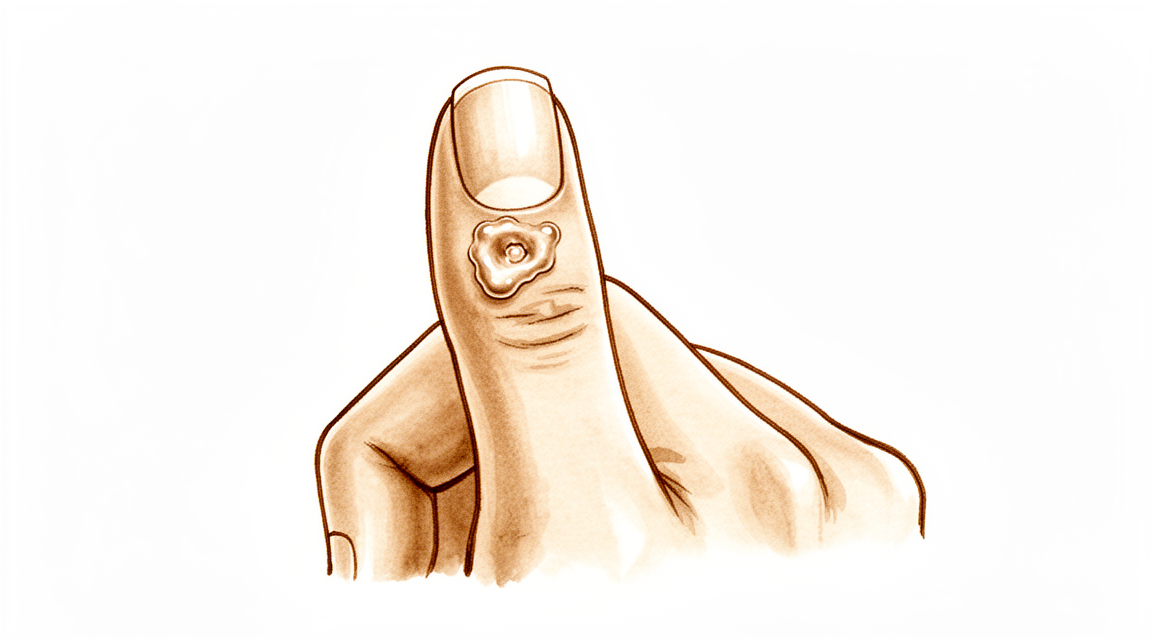
黏液囊肿是出现在手指背面、通常正好位于指甲后方的一个小而坚硬的肿块。它内部充满浓稠、透明的液体。肿块本身通常不痛,但它可能会压迫附近的结构并引起不适。
您可能会注意到指甲本身发生变化。囊肿紧邻甲床,因此在增大时可能会压迫指甲。这常常导致指甲上出现一条纵贯全长的凹槽或嵴。囊肿表面的皮肤也可能变薄并变得发亮。在某些情况下,皮肤会破溃,囊肿会通过一个小通道渗出液体。
肿块往往正好位于手指在指甲前最后一个关节弯曲的地方。这会让一些日常事务变得不便。把手伸进紧窄的口袋、拧瓶盖或捡起小硬币时可能会感到不适。打字或写字可能会压到肿块,使其疼痛。
这种囊肿与同一关节的退行性关节炎有关。一个称为骨赘的小骨刺会刺激关节,并产生充满囊肿的液体。因此,在您大量使用手部之后,关节可能会酸痛,此时肿块也可能有更明显的压痛。
如果囊肿表面的皮肤破开并有液体渗出,请保持该部位清洁并及时就医。从皮肤通向手指关节的通道可能让感染进入,而关节感染需要立即治疗。如果您的手指变得发热、发红、肿胀和疼痛,尤其是伴有发烧时,请当天前往急诊科。无需全科医生转诊。
不要自行刺破或挤压囊肿。这可能会让感染进入关节。
如果肿块在增大、指甲畸形让您困扰,或症状在数周内没有缓解,请就诊您的全科医生或要求专科医生评估。
实际发生了什么
您手指的最后一个关节就像一个小铰链。与任何关节一样,它内衬一层柔软的套膜,会产生少量液体,使关节面保持顺畅滑动。发生黏液囊肿时,该关节已经出现了退行性关节炎。关节内衬受到刺激,开始产生超过关节所需的液体。
与此同时,关节炎常常在关节边缘形成一个小骨刺。这个骨刺从内部压迫并磨穿关节套膜。液体随后通过磨损处渗出,并积聚在手指背面的皮肤下。您能看到和摸到的肿块就是这些被困住的液体,它位于关节的一侧、正好在指甲后方。
囊肿本身是一个薄壁的囊袋,而不是一种增生物。它的囊壁由支撑组织的纤维构成,没有自己真正的内衬。由于囊壁很薄,而其上方的皮肤往往更薄,轻微的碰撞就可能使其破裂。发生这种情况时,里面透明的胶冻状液体就会渗出。
这解释了您刚刚读到的症状。肿块紧邻甲床,因此在指甲生长时,囊肿的压力可能会在指甲上压出凹槽。关节酸痛是因为关节炎同时存在,而囊肿正是关节炎的一个征象。关节边缘的骨刺使液体不断流出,因此只要骨刺存在,即使囊肿渗漏或被抽吸之后,它也往往会重新充盈。
这也是为什么治疗的重点在于关节,而不仅仅是肿块。去除骨刺可以从源头上阻止渗漏,囊肿在不再得到液体补充后就会消退。
我们能做什么
Mater Private Hospital Rockhampton 的上肢外科医生 Kieran Hirpara 医生会从适合您病情的最微创方案入手。患者通常由全科医生转诊至我们的诊所;如果理疗师建议您就诊,您仍需获得全科医生的转诊,才有资格享受 Medicare 报销。在门诊就诊时,我们会采集病史、检查您的手指,并在必要时安排影像学检查。大多数黏液囊肿与关节的退行性关节炎有关,因此我们通常首先尝试非手术治疗,在非手术治疗未能带来足够改善时再考虑手术。
第一步通常只是观察等待。有些囊肿会一直保持很小,几乎不造成困扰。保护手指会有帮助:避免反复碰撞肿块,并在进行会压迫肿块的活动时多加小心。如果关节在大量使用后酸痛,减少该活动可以让情况缓解。手部治疗可以帮助改善僵硬,并让手指保持舒适地活动。在决定任何进一步措施之前,请先在几周内给予这些简单措施充分的尝试。
止痛方法很简单。按照说明服用药房购买的普通止痛药,可以缓解关节酸痛。抗炎药可以缓解囊肿背后的关节炎。这些药物治疗的是不适,而不是肿块本身。
我们不提供的一种方案是用针抽吸囊肿。这是一种方便的门诊操作,但之后大约有一半的情况囊肿会复发。此外还有将感染带入关节的风险,这也是我们建议您不要在家中自行刺破或挤压囊肿的原因。
当囊肿不断重新充盈、指甲畸形让您困扰,或囊肿表面的皮肤已经变薄或正在破溃时,会考虑手术。手术治疗的是关节,而不仅仅是肿块。关节边缘的骨刺会被切除,从而从源头上阻止液体渗漏,囊肿在不再得到液体补充后就会消退。在某些情况下,囊肿也会与一小块局部皮瓣一起被切除,这适用于已使其表面皮肤变薄的囊肿。将骨刺与囊肿的蒂部一并切除,囊肿复发的几率最低。我们会与您讨论哪种方法适合您的手指,并共同做出决定。
预期情况
若不予处理,黏液囊肿往往会不断复发,而不是消失。只要关节边缘的骨刺存在,它就会持续为囊肿补充液体。有些囊肿会长期保持很小,几乎不造成困扰。另一些则会缓慢增大,使指甲凹槽进一步加深,或使肿块表面的皮肤变薄直至渗漏。
如果不治疗,主要风险就是您已经读到的那些:在囊肿压迫期间指甲会一直有嵴,以及皮肤破溃并形成通向关节的通道。这个通道值得认真对待。如果您的手指变得发热、发红、肿胀和疼痛,尤其是伴有发烧时,请当天前往急诊科。无需全科医生转诊。
针对关节进行治疗后,前景很明确。去除骨刺可以从源头上阻止液体渗漏,囊肿在不再得到液体补充后就会消退。骨刺被切除后,囊肿极少复发。如果同时切除囊肿本身,复发率也保持在较低水平:经手术治疗的囊肿中,每 100 个约有 2 个复发;使用局部皮瓣闭合该区域时,每 100 个约有 1.4 个复发。接受这种治疗的人通常对瘢痕的外观感到满意,并且愿意再次接受这种治疗。
恢复过程不算繁重,但也不能完全忽视。手指常常会在几周内感到僵硬和压痛,抓握或捏取等日常动作需要一段时间才能恢复正常。术后的手部治疗由 Extend Rehabilitation 的 Ruby Doolan 负责。Ruby 是一名手部治疗师:她会指导您的锻炼,并为您制作所需的任何支具。大多数人在数周至几个月内即可恢复正常使用手部。
一旦甲床不再受压,指甲凹槽通常也会改善。对此需要耐心:变形的那段指甲必须长出来,这需要几个月时间。
如果症状在数周内没有缓解,或正在加重,请就诊您的全科医生或要求专科医生评估。
何时就医
大多数黏液囊肿并不紧急,许多通过简单护理就能缓解。少数征象确实需要采取行动。
如果您的手指变得发热、发红、肿胀和疼痛,尤其是伴有发烧时,请当天前往急诊科。这提示关节感染,需要立即治疗。如果囊肿表面的皮肤已经破开并有液体渗出,也应如此,因为这个开口可能让感染进入。
如果肿块不断增大、指甲凹槽让您困扰,或囊肿表面的皮肤已经变薄,请就诊您的全科医生或要求专科医生评估。出现这些征象时,值得讨论治疗。如果症状在数周内没有缓解,或正在加重,您的全科医生可以为您安排转诊。
深入探讨
Advanced reading: the deeper science (optional)
本节内容超出了您自身治疗决策所需的范围。黏液囊肿值得额外阅读,原因在于一个虽小但证据充分的手术见解:囊肿本身并非问题所在,且效果最佳的手术操作未必会将其切除。
骨刺是病因,而非囊肿
黏液囊肿起源于退行性变的指尖关节。骨性突起(骨赘)刺激并穿透关节囊,关节液经此缺损流出并在皮下积聚。囊肿是该过程的可见末端,而非其起源。
这一认识具有直接的手术意义,在一项系列研究中,仅切除骨赘而不切除囊肿在大多数病例中实现了完全消退,并被描述为一种提供微创方法的良好治疗选择 [1]。
切除骨刺即关闭了“水龙头”。囊肿不再被填充,便会消退。这与腕部腱鞘囊肿的规律相同,即决定复发的因素是蒂部而非囊体,这也解释了为何单纯抽吸或穿刺黏液囊肿如此可靠地失败。
在囊肿切除部位,疗效同样良好
另一种方法是在切除囊肿的同时,利用局部皮瓣闭合缺损。该方法同样可靠:在69例患者中,采用局部推进皮瓣进行手术切除的复发率为1.4%,患者对瘢痕的满意度较高,且愿意再次接受该手术 [2]。
两种方法均有效,且均针对基础关节进行处理。实际差异在于涉及皮肤的范围:长期存在的囊肿会使覆盖皮肤变薄,有时甚至出现渗液,在这种情况下,无论对骨骼采取何种处理,变薄的皮肤都需要切除并替换。
指甲为何变形,以及能否恢复
沿指甲纵向延伸的沟槽或脊状隆起是常见的伴随症状,且往往比肿块本身更令人担忧。其成因是机械性的:囊肿恰好位于甲母质(即甲床中负责生成指甲的部分)正上方,并对其施加压力,导致指甲在生成过程中出现缺陷。
关键在于,这种影响是压迫而非破坏。一旦囊肿压力解除,指甲通常能正常生长,但需要数月时间才能使变形部分从指尖长出。因此,指甲变形是治疗囊肿的理由,而非其永久性后果。
谨慎对待穿刺的原因
黏液囊肿与关节直接相通。无论是有意穿刺,还是因囊肿较大导致其表面皮肤破溃,都会形成一条从外界通向指间关节的通道,而小关节的化脓性关节炎远比囊肿本身更为严重。
这正是反对自行引流看似仅为液体积聚水疱的囊肿的实用理由,也是为何对自发性排液的囊肿需紧急处理而非仅观察的原因。
参考文献
[1] Lee H, Kim P, Jeon I, Kyung H, Ra I, Kim T. Osteophyte excision without cyst excision for a mucous cyst of the finger. J Hand Surg Eur Vol. 2013;39(3):258-61. https://doi.org/10.1177/1753193413478549
[2] Johnson SM, Treon K, Thomas S, Cox QGN. A reliable surgical treatment for digital mucous cysts. J Hand Surg Eur Vol. 2013;39(8):856-60. https://doi.org/10.1177/1753193413508540
Evidence & references
This is the clinical evidence summary written for health professionals. It is technical, and it lists the research this page was built from. You do not need to read it to understand your treatment or to make a decision about it.
Overview
- The scientific data regarding mucous cysts consist almost entirely of retrospective studies, and much of what is done or recommended is based on expert opinion [3].
- Surgical treatment of mucous cysts is not a totally benign procedure, and complications do occur contrary to literature reporting glowing results [5].
- In-office excision is a safe and effective option for treating digital mucous cysts [6].
- Complications are rare and occur only in cysts associated with fistula, justifying their early surgical treatment [7].
- Surgery provided definitive treatment with no major long-term problems [10].
- Surgical excision was the primary treatment, with no reported recurrences [11].
- Incomplete excisions will invariably result in recurrence [12].
- Surgical excision with a local advancement skin flap is a reliable treatment for digital mucous cysts, demonstrating a low recurrence rate of 1.4% and high patient satisfaction regarding the scar and willingness to undergo the procedure again [13].
- The 2% recurrence rate (2 of 113) obtained in this series illustrates the advantage of this procedure [16].
- Osteophyte excision without cyst excision may be a good treatment choice for mucous cyst of the finger, providing a less invasive method with complete resolution in most cases [20].
- The final recurrence rate of less than 1.5% may be acceptable and comparable with other techniques [21].
- Excision with local flap cover offers benefit in terms of a reduced recurrence rate and is particularly valuable where the cyst has caused marked thinning of the skin [30].
- Excision with local flap cover would seem to offer benefit in terms of a reduced recurrence rate, and is particularly valuable where the cyst has caused marked thinning of the skin [34].
- Excision of the cyst and complete removal of the marginal osteophyte eradicates mucous cysts with extremely rare recurrence [1].
- A total dorsal capsulectomy alone was a simple treatment for mucous cysts and did not lead to any recurrence [8].
Anatomy & Pathophysiology
Clinical Presentation and Location
- Mucous cysts are small, firm, cystic masses that appear just distal to the distal interphalangeal joint [23].
- The lesion is always located to one side of the midline [23].
- Mucous cysts are rarely greater than fifteen millimeters in diameter [23].
- The skin over the mucous cyst is thinned out and occasionally may be ulcerated [23].
- Pressure on the mucous cyst does not usually result in a decrease in its size [23].
- Longitudinal grooving of the nail may be noted, occasionally occurring prior to the appearance of the cyst [23].
- Mucous cysts are usually small and located to one side as they emerge from the joint beside the extensor mechanism [28, 29].
- Occasionally, mucous cysts may spread across the whole of the dorsum of the digit between the distal interphalangeal joint and nail fold [28, 29].
- The overlying skin of a mucous cyst is frequently thin, and minimal trauma may result in rupture [28, 29].
Histology and Ultrastructure
- The histological appearance of a mucous cyst is essentially identical to that of ganglions, synovial cysts, and similar lesions appearing on other areas of the extremities [23].
- The mucous cyst is surrounded by an undemarcated fibrous capsule [23].
- The basic structure of the mucous cyst is myxomatous with interspersed fibroblasts [23].
- Areas of myxomatous degeneration tend to coalesce to form a multiloculated cyst [23].
- An epithelial lining has not been reported for mucous cysts [23].
- Digital mucous cysts and wrist ganglia show no anatomic difference at the light and scanning electron microscopic levels [19].
- The wall of a digital mucous cyst consists of multidirectional strata of collagen fibers with rare fibroblast-like cells on the inner surface [19].
- There are no signs of myxomatous degeneration in the wall of a digital mucous cyst [19].
- The surface of the digital mucous cyst wall is generally flat, with some areas showing localized elevations [19].
- Multifunctional mesenchymal cells are located beneath the lining of the ganglion and digital mucous cyst [19].
- These multifunctional mesenchymal cells contain abundant rough endoplasmic reticulum, smooth-walled vacuoles, and Golgi complexes [19].
- The ganglion fluid may be produced by these multifunctional cells and reaches the ganglion cavity by flowing through the porous collagen matrix of the ganglion wall [19].
- A digital mucous cyst has a large globular dome-shaped interconnecting cavity having a wall of collagen fibers covered by a fenestrated pseudomembrane [19].
- A porous network of collagen fibers is present in the wall of the digital mucous cyst on cross section [19].
Etiology and Pathogenesis
- Degenerative arthritis of the distal interphalangeal joint is a frequent associated finding with mucous cysts [23].
- An incidence of 78 percent for degenerative arthritis of the distal interphalangeal joint has been reported in association with mucous cysts [23].
- The etiology indicated by evidence is that the lesion arises from the joint capsule [23].
- Mucous cysts are often associated with degenerative changes at the distal interphalangeal joints [28, 29].
- Theories on the pathogenesis of mucous cysts include myxoid degeneration, repeated local trauma, vascular insufficiency, and synovial herniation [28, 29].
- Studies suggest that mucous cysts arise due to excessive hyaluronic acid production from a degenerate distal interphalangeal joint [28, 29].
Classification
- The lesion is a small, firm, cystic mass which appears just distal to the distal interphalangeal joint [23].
- The lesion is always located to one side of the mid-line [23].
- The lesion is rarely greater than fifteen millimeters in diameter [23].
- The skin over the lesion is thinned out and occasionally may be ulcerated [23].
- Pressure on the lesion does not usually result in a decrease in its size [23].
- Degenerative arthritis of the distal interphalangeal joint is a frequent associated finding [23].
- An incidence of 78 per cent has been reported for degenerative arthritis of the distal interphalangeal joint associated with mucous cysts [23].
- The histological appearance is essentially identical to that of ganglions, synovial cysts, and similar lesions appearing on other areas of the extremities [23].
- The lesion is surrounded by an undemarcated fibrous capsule [23].
- The basic structure is myxomatous with interspersed fibroblasts [23].
- An epithelial lining has not been reported [23].
- In spite of their different clinical settings, digital mucous cysts and wrist ganglia show no anatomic difference at the light and scanning electron microscopic levels [19].
- The wall of the digital mucous cyst consisted of multidirectional strata of collagen fibers with rare fibroblast-like cells on the inner surface [19].
- There were no signs of myxomatous degeneration in the wall of the digital mucous cyst [19].
- The surface of the digital mucous cyst was generally flat, with some areas showing localized elevations [19].
- Multifunctional mesenchymal cells beneath the lining of the ganglion are postulated to produce collagen fibers, elastic fibers, and the interfibrillary mucopolysaccharide matrix [19].
- The porous nature of the ganglion wall was confirmed in the ultrastructural study of digital mucous cysts [19].
- A previous ultrastructural study of digital mucous cysts demonstrated a large globular dome-shaped interconnecting cavity having a wall of collagen fibers covered by a fenestrated pseudomembrane [19].
- The current study confirmed the anatomic organization of a large globular dome-shaped interconnecting cavity having a wall of collagen fibers covered by a fenestrated pseudomembrane [19].
- A porous network of collagen fibers in the wall was revealed on cross section [19].
Clinical Presentation
- The mucous cyst presents as a small, firm, cystic mass located just distal to the distal interphalangeal joint [23].
- The lesion is easily distinguished from a Heberden's node by its placement distal to the distal interphalangeal joint [23].
- The histological appearance of the mucous cyst is essentially identical to that of ganglions, synovial cysts, and similar lesions appearing on other areas of the extremities [23].
- The basic structure of the lesion is myxomatous with interspersed fibroblasts [23].
- An epithelial lining has not been reported for the mucous cyst [23].
- The wall of the digital mucous cyst consists of a porous network of collagen fibers [19].
- The fluid within the digital mucous cyst is produced by underlying mesenchymal cells [19].
Investigations
- Ultrasound is a powerful modality for evaluation of pathologic conditions in the hand and wrist, providing a cost-effective and expedient alternative and/or adjunct to MRI [33].
- Ultrasound is best used when there is a specific clinical question regarding a well-localized abnormality [33].
- Ultra-sonography provides a new ability to study the complex anatomical structures of the hand, with the technique being quick and simple to perform [38].
- A careful physical examination is essential to direct care and future testing if indicated [35].
- Diagnostic tests such as imaging and serum laboratory studies are useful in this determination but can be expensive, time consuming, and often nonspecific [35].
Treatment
Operative Techniques
- Total dorsal capsulectomy alone is a simple treatment for mucous cysts that did not lead to any recurrence [8].
- Osteophyte-sparing treatment of mucous cysts has a final recurrence rate of less than 1.5% [21].
- Surgical excision was the primary treatment in a series with no reported recurrences [11].
- Surgery provided definitive treatment for mucous cysts with no major long-term problems [10].
- The 2% recurrence rate (2 of 113) obtained in a series of 191 excised cysts illustrates the advantage of the procedure [4, 16].
- The authors of a study on osteophyte excision without cyst excision state that their principle is not to do more damage to the friable skin because the skin is not the culprit of the cyst [15].
Outcomes and Complications
- Complications of surgical treatments include infection, recurrence, nail deformity, swelling, stiffness and pain [28].
- This report provides additional basis for prophylactic extirpation of mucous cyst of the finger [2].
- This case emphasizes the utility of a pathohistological analysis in cases where doubts arise about the initial diagnosis of a benign tumorous lesion [14].
Non-Operative and Adjunctive Considerations
- Aspiration with injection of steroid is a convenient technique that can be performed in an outpatient setting but is followed by a recurrence rate of 50% [28].
- Surgical treatment has a higher cure rate than aspiration with injection of steroid [28].
- Radical treatment with complete excision of the stalk and underlying osteophytes seems to reduce recurrence rates [28].
Complications
- Complications are rare and occur only in cysts associated with fistula [7].
- Aspiration with injection of steroid is followed by a recurrence rate of 50% [28].
- The 2% recurrence rate (2 of 113) was obtained in a series of surgically treated mucous cysts [16].
- Surgical excision with a local advancement skin flap demonstrated a low recurrence rate of 1.4% [13].
- A total dorsal capsulectomy alone did not lead to any recurrence [8].
- Eccrine porocarcinomas have a substantial risk of metastasis, high risk of local recurrence, and are potentially fatal [24].
Recovery
- Surgical excision with a local advancement skin flap demonstrated a low recurrence rate of 1.4% and high patient satisfaction regarding the scar and willingness to undergo the procedure again [13].
Key Evidence
- [L4] Excision of the cyst and complete removal of the marginal osteophyte eradicates mucous cysts with extremely rare recurrence. [1] (10.2106/00004623-197355030-00013)
- [L4] This report provides additional basis for prophylactic extirpation of mucous cyst of the finger. [2] (10.1016/s0363-5023(84)80152-5)
- [L4] The scientific data regarding mucous cysts consist almost entirely of retrospective studies, and much of what is done or recommended is based on expert opinion. [3] (10.1016/j.jhsa.2010.01.029)
- [L5] Of the 191 mucous cysts excised, 113 had at least 6 months of follow-up time. [4] (10.1016/j.jhsa.2010.07.028)
- [L4] Surgical treatment of mucous cysts is not a totally benign procedure, and complications do occur contrary to literature reporting glowing results. [5] (10.1016/s0266-7681(97)80067-8)
- [Paper] In-office excision is a safe and effective option for treating digital mucous cysts. [6] (10.1177/15589447251350168)
- [L4] Complications are rare and occur only in cysts associated with fistula, justifying their early surgical treatment. [7] (10.1016/j.main.2015.06.001)
- [L4] A total dorsal capsulectomy alone was a simple treatment for mucous cysts and did not lead to any recurrence. [8] (10.1016/j.jhsa.2014.03.004)
- [L4] Surgery provided definitive treatment with no major long-term problems. [10] (10.1053/jhsu.2003.50088)
- [L5] Incomplete excisions will invariably result in recurrence. [12] (10.1016/0363-5023(93)90302-j)
- [L4] Surgical excision with a local advancement skin flap is a reliable treatment for digital mucous cysts, demonstrating a low recurrence rate of 1.4% and high patient satisfaction regarding the scar and willingness to undergo the procedure again. [13] (10.1177/1753193413508540)
- [L5] This case emphasizes the utility of a pathohistological analysis in cases where doubts arise about the initial diagnosis of a benign tumorous lesion. [14] (10.1007/s00402-008-0794-4)
- [L5] The authors state that their principle is not to do more damage to the friable skin because the skin is not the culprit of the cyst. [15] (10.1177/1753193414546990)
- [L4] The 2% recurrence rate (2 of 113) obtained in this series illustrates the advantage of this procedure. [16] (10.1016/0363-5023(94)90071-x)
- [L5] [19] (10.1016/s0363-5023(88)80143-6)
- [L4] Osteophyte excision without cyst excision may be a good treatment choice for mucous cyst of the finger, providing a less invasive method with complete resolution in most cases. [20] (10.1177/1753193413478549)
- [L4] The final recurrence rate of less than 1.5% may be acceptable and comparable with other techniques. [21] (10.5435/jaaosglobal-d-21-00164)
- [L4] [23] (10.2106/00004623-197254070-00008)
- [L4] Prompt recognition and appropriate treatment are critical because eccrine porocarcinomas have a substantial risk of metastasis, high risk of local recurrence, and are potentially fatal. [24] (10.1016/j.jhsa.2016.07.112)
- [L4] [28] (10.1177/1753193408103498)
- [L4] [29] (10.1177/1753193409103498)
- [L4] Excision with local flap cover offers benefit in terms of a reduced recurrence rate and is particularly valuable where the cyst has caused marked thinning of the skin. [30] (10.1016/0266-7681(90)90064-b)
- [L5] Ultrasound is a powerful modality for evaluation of pathologic conditions in the hand and wrist, providing a cost-effective and expedient alternative and/or adjunct to MRI, best used when there is a specific clinical question regarding a well-localized abnormality. [33] (10.1016/j.jhsa.2009.02.010)
- [L4] Excision with local flap cover would seem to offer benefit in terms of a reduced recurrence rate, and is particularly valuable where the cyst has caused marked thinning of the skin. [34] (10.1016/0266-7681_90_90064-b)
- [L4] Ultra-sonography provides a new ability to study the complex anatomical structures of the hand, with the technique being quick and simple to perform. [38] (10.1016/0266-7681(90)90133-o)
References
[1] Marginal Osteophyte Excision in Treatment of Mucous Cysts. The Journal of Bone & Joint Surgery. 1973. DOI: 10.2106/00004623-197355030-00013
[2] Infected mucous cyst of the finger. The Journal of Hand Surgery. 1984. DOI: 10.1016/s0363-5023(84)80152-5
[3] Mucous Cysts. The Journal of Hand Surgery. 2010. DOI: 10.1016/j.jhsa.2010.01.029
[4] Mucous Cysts. The Journal of Hand Surgery. 2010. DOI: 10.1016/j.jhsa.2010.07.028
[5] Complications Following Mucous Cyst Excision. Journal of Hand Surgery. 1997. DOI: 10.1016/s0266-7681(97)80067-8
[6] Safety and Cost-Effectiveness of In-Office Digital Mucous Cyst Excisions. HAND. 2025. DOI: 10.1177/15589447251350168
[7] Surgical treatment of mucous cysts by subcutaneous excision and osteophyte resection: Results in 68 cases at a mean 6.63 years’ follow-up. Chirurgie de la Main. 2015. DOI: 10.1016/j.main.2015.06.001
[8] Total Dorsal Capsulectomy for the Treatment of Mucous Cysts. The Journal of Hand Surgery. 2014. DOI: 10.1016/j.jhsa.2014.03.004
[10] Treatment of mucous cysts of the fingers: Review of 134 cases with minimum 2-year follow-up evaluation. The Journal of Hand Surgery. 2003. DOI: 10.1053/jhsu.2003.50088
[11] 10.1177-15589447261433068. n.d..
[12] Juvenile hyaline fibromatosis of the hand in an adult. The Journal of Hand Surgery. 1993. DOI: 10.1016/0363-5023(93)90302-j
[13] A reliable surgical treatment for digital mucous cysts. Journal of Hand Surgery (European Volume). 2013. DOI: 10.1177/1753193413508540
[14] Malignant Natural-Killer cell neoplasm presenting as a mucous cyst on the distal interphalangeal joint of the finger. Archives of Orthopaedic and Trauma Surgery. 2008. DOI: 10.1007/s00402-008-0794-4
[15] Re: Lee, H.-J., Kim, P.-T., Jeon, I.-H., Kyung, H.-S., Ra, I.-H. and Kim, T.-K. Osteophyte excision without cyst excision for a mucous cyst of the finger. J Hand Surg Eur. 2014, 39: 258–61. Journal of Hand Surgery (European Volume). 2014. DOI: 10.1177/1753193414546990
[16] Outcome of surgically treated mucous cysts of the hand. The Journal of Hand Surgery. 1994. DOI: 10.1016/0363-5023(94)90071-x
[19] A surface ultrastructure study of ganglia and digital mucous cysts. The Journal of Hand Surgery. 1988. DOI: 10.1016/s0363-5023(88)80143-6
[20] Osteophyte excision without cyst excision for a mucous cyst of the finger. Journal of Hand Surgery (European Volume). 2013. DOI: 10.1177/1753193413478549
[21] Osteophyte-Sparing Treatment of Mucous Cysts: Case Analysis and Surgical Technique. JAAOS: Global Research and Reviews. 2021. DOI: 10.5435/jaaosglobal-d-21-00164
[23] Etiology and Treatment of the So-Called Mucous Cyst of the Finger. The Journal of Bone & Joint Surgery. 1972. DOI: 10.2106/00004623-197254070-00008
[24] Eccrine Porocarcinoma Presenting as a Hand Cyst. The Journal of Hand Surgery. 2016. DOI: 10.1016/j.jhsa.2016.07.112
[28] Use of Wolfe Graft for the Treatment of Mucous Cysts. Journal of Hand Surgery (European Volume). 2009. DOI: 10.1177/1753193408103498
[29] Use of Wolfe Graft for the Treatment of Mucous Cysts. Journal of Hand Surgery (European Volume). 2009. DOI: 10.1177/1753193409103498
[30] Mucous cyst of the distal interphalangeal joint: Treatment by simple excision or excision and rotation flap. The Journal of Hand Surgery: Journal of the British Society for Surgery of the Hand. 1990. DOI: 10.1016/0266-7681(90)90064-b
[33] Ultrasound of the Hand and Wrist. The Journal of Hand Surgery. 2009. DOI: 10.1016/j.jhsa.2009.02.010
[34] Mucous Cyst of the Distal Interphalangeal Joint: Treatment by Simple Excision or Excision and Rotation Flap. Journal of Hand Surgery. 1990. DOI: 10.1016/0266-7681_90_90064-b
[35] Orthopaedic Knowledge Update 13 Ebook Without Multimedia. Anatomy, Evaluation, Clinical Examination, and Imaging > Evaluation and Clinical Examination: Current Concepts.
[38] Diagnostic medical ultrasound in the management of hand injuries. The Journal of Hand Surgery: Journal of the British Society for Surgery of the Hand. 1990. DOI: 10.1016/0266-7681(90)90133-o