Patients › Wrist
舟骨骨折
Scaphoid fractures — recognition, the high non-union risk, casting and percutaneous/open fixation.
您的感受
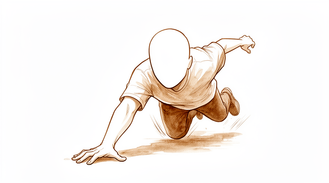
舟骨骨折通常发生在手掌撑地跌倒时,或在运动中。舟骨是手腕拇指侧的一块小骨头,就在拇指根部下方。这种骨折常由站立高度的低处跌倒引起,但在男性中,更多是由较强的撞击(例如运动或车祸)造成。有些人在受伤那一刻只感到一阵锐痛,除此之外并无其他感觉。
受伤后,手腕拇指侧会立即疼痛。该侧可能出现肿胀和瘀青。拇指根部正下方的部位,即一个被称为"解剖学鼻烟壶"的小凹陷处,按压时常有压痛。您的手腕可能感到僵硬,完全弯曲手腕、用手腕撑身体,或在极端姿势下让手腕受力时,都可能引起疼痛。将拇指和食指捏在一起,或沿拇指的长轴方向推压拇指,也可能引起疼痛。
这些体征可能出乎意料地轻微。该凹陷处的肿胀可能很轻,有些人的手腕仍能活动自如,以至于以为只是扭伤。这正是舟骨骨折容易被漏诊的原因之一。多达30%至40%的舟骨骨折在首次X光片上无法显示,有些要在再次检查手腕并重新拍片后才能确诊,最常见的是在受伤后10至14天。
在最初的几天到几周内,疼痛通常在早期最为严重。活动手腕、抓握,或用这只手从椅子上撑起身都会引起疼痛。最初几天常有夜间隐痛。随着愈合开始,疼痛通常会逐渐缓解,但拇指下方凹陷处的压痛可能持续到第二周。
如果您的症状没有缓解、在数周内逐渐加重、在夜间把您弄醒,或使您无法工作或无法使用这只手,请去看您的全科医生或要求专科评估。如果您联系不上诊所,请前往离您最近的急诊科。
实际发生了什么
舟骨是手腕拇指侧的一块小型弯曲骨骼。它位于腕关节内部,连接构成手腕的两排小骨。可以把它想象成火车前后两部分之间的连接器:当它完好时,两部分会顺畅地一起弯曲和伸直。当它断裂时,手腕的两部分就可能开始彼此不同步地活动。
大多数骨折发生在骨骼中部,称为腰部。当您跌倒时手腕被迫向后弯,这里承受最大的弯曲力。约70%至80%的舟骨骨折发生在腰部。其余的发生在更靠近前臂或更靠近拇指的位置。
这块骨头的血液供应很特殊。血液从靠近拇指的远端流入,然后沿着骨头向后流动。骨折可能切断靠近前臂那一块骨头的血液供应。没有血液,那一块骨头就难以愈合。这就是为什么骨折的位置如此重要,也是为什么一些骨折即使在X光片上看起来相似,治疗方式也会不同。
骨骼通过重新连接而愈合,就像手臂骨折愈合一样。新骨会在骨折处形成,直到两块骨头重新合为一体。但要做到这一点,骨折块必须保持固定不动,并有充足的血液供应。如果骨折移位,或者在您使用手腕时两块骨头相互错动,骨头就可能无法连接。如果骨头不能连接,手腕可能会在多年中逐渐磨损,因为两排腕骨持续不同步地活动。
简而言之,这就是力学上的问题:一个依赖这个连接器的关节,而连接器断了。
我们如何处理
Mater Private Hospital Rockhampton 的上肢外科医生 Kieran Hirpara 医生会根据您的具体伤情匹配治疗方案。有些舟骨骨折无需手术即可良好愈合,而另一些则需要在受伤后尽快手术,因此及时评估非常重要。我们会观察骨折的位置、骨折块是否移位,以及您的手在工作和运动中需要做什么。患者通常由全科医生(GP)转诊至我们的诊所;如果物理治疗师建议您就诊,您仍需获得全科医生的转诊,才有资格享受 Medicare 报销。
如果骨折稳定或几乎没有移位,石膏或夹板通常是合适的选择。它可以在骨骼愈合期间将其固定不动,我们会在此过程中通过复查影像来检查愈合情况。在受伤后 6 个月内发现的骨折,即使诊断较晚,仍可单靠石膏固定愈合。骨骼愈合后,手部治疗师会分阶段指导您恢复活动。证据中有一点需要注意:在第一个月内服用抗炎止痛药片,与骨骼不愈合的几率升高有关,因此我们会与您讨论不会影响愈合的止痛方法。
对于某些损伤,手术从一开始就是推荐的治疗,而不是退而求其次的选择。靠近前臂一端的骨折在石膏固定中常常无法愈合,手术治疗能让它们的愈合可靠得多。骨折块已移位或骨骼不稳定的骨折,也会采用手术治疗,以便骨折块在愈合期间保持在正确位置。一枚小螺钉从内部将骨头固定住,这让一些人可以更早停止佩戴石膏,更快重返工作,平均早约 7 周。对另一些人来说,这确实需要共同做出选择:稳定的骨折可以在石膏中愈合,但这意味着要多戴几周石膏,重返工作也较慢,而手术本身也有小的风险。我们会与您一起权衡这些利弊。
无论您选择哪种方式,最初几周的情况都相似。止痛药让您在骨骼开始愈合时保持舒适。保护手腕意味着要严格遵守固定计划,因为骨折块之间的活动正是阻碍愈合的原因。在愈合足够牢固后,Extend Rehabilitation 的 Ruby Doolan 会在合适的阶段开始为您进行手部治疗。她会指导您的锻炼,并为您制作所需的任何夹板。如果您的症状没有缓解、在数周内逐渐加重、在夜间把您弄醒,或使您无法工作或无法使用这只手,请去看您的全科医生或要求专科评估。
预期情况
愈合情况取决于骨折的位置以及开始治疗的早晚。受伤后 4 周内接受治疗的骨折,比更晚治疗的骨折愈合得更可靠;受伤后超过 31 天才接受治疗的骨折,不愈合的几率更高。在石膏中,骨骼被固定不动,新骨在骨折处形成。如果做手术,一枚小螺钉从内部将骨折块固定在一起,一些人可以更早停止佩戴石膏,并平均早约 7 周重返工作。对于稳定且几乎没有移位的骨折,两种方式都能让手腕功能良好,而且尚无证据表明哪一种在长期上更有优势。
无论哪种方式,最初几周的情况都差不多。您的手腕受到保护,疼痛逐渐缓解,您要避免让这只手受力。随着骨骼愈合,手部治疗师会分阶段指导您恢复活动和握力。大多数人先恢复日常活动,再恢复繁重工作。运动放在最后,要等到确认愈合并且力量恢复之后。如果骨骼愈合,几乎所有手腕都能取得良好的结果,即使骨骼愈合后形状略有改变。
主要可能出现的问题是愈合缓慢或不愈合,称为骨不连。在接受手术治疗的骨折中,超过 10% 会发生这种情况;如果治疗开始较晚,或骨折位于靠近前臂的一端,则更容易发生。如果骨骼不愈合,手腕可能会在多年中逐渐发展为磨损性关节炎。在未愈合骨折从未接受治疗的人群中,受伤五年或以上者有 97% 已发展为关节炎。已愈合的骨折也可能再次骨折:有一例记录在愈合后 7 个月内再次骨折。手腕固定期间常会出现僵硬,一旦骨骼稳固,通过治疗通常会改善。
如果您的症状没有缓解、在数周内逐渐加重、在夜间把您弄醒,或使您无法工作或无法使用这只手,请去看您的全科医生或要求专科评估。如果您联系不上诊所,请前往离您最近的急诊科。
何时就医
如果您的手腕明显变形、受伤处有开放性伤口,或手指变得苍白、冰冷、发白或发青,请立即寻求紧急救治。受伤后手部突然失去感觉或活动能力,也需要在当天前往急诊科。
对于大多数舟骨损伤,情况要不明显得多,而这正是陷阱所在。手腕可能仍能活动,肿胀也可能很轻。如果您的疼痛没有缓解,或者随着愈合进展,肿胀、活动或握力没有逐周改善,请去看您的全科医生或要求专科评估。症状在数周内逐渐加重、在夜间把您弄醒,或使您无法工作或无法使用这只手,也值得认真检查。如果您联系不上诊所,请前往离您最近的急诊科。
深入探讨
Advanced reading: the deeper science (optional)
本节内容超出了您自行做出治疗决策所需的深度。舟骨骨折值得额外阅读,因为骨折在骨骼上的具体位置比几乎所有其他特征都更为重要,且支持手术治疗的论点并非大多数人通常所认为的那样。
骨折在骨上的位置使风险增加七倍
舟骨的大部分血液供应来自进入其远端附近的血管,因此血液沿骨向近端极逆向流动。腰部骨折会中断该骨折线远端所有区域的血液供应。
这一后果已被量化。综合已发表的系列研究,34%的急性舟骨近端骨折在非手术治疗下会进展为骨不连,且与采用相同治疗方式的更远端骨折相比,此类骨折发生骨不连的相对风险为7.5 [1]。
这就是为什么在影像片上看似相似的两处骨折可能带来完全不同的治疗建议。远端骨折使用石膏固定有很高的愈合几率。而采用相同方式治疗的近端极骨折,约有三分之一的病例无法愈合。
手术降低了骨不连发生率,但并未改善最终结局
对于常见的腰部骨折,将手术治疗与石膏固定治疗进行对比,所得结果值得仔细解读。手术组的骨不连发生率降低了三倍,功能恢复更快,握力和活动范围在短期内表现更优,但并发症更多。两组在疼痛、压痛、费用或功能结局方面无显著差异 [2]。
因此,手术以手术并发症为代价,换取了骨愈合的可靠性和恢复速度的提升,且两组最终趋向于相同的结局。这使得该决策取决于特定个体对更快、更确定路径的价值评估:体力劳动者或运动员对这几个月的权衡,与能够耐受石膏固定的患者截然不同。
诊断是避免大多数危害的关键
由于漏诊的舟骨骨折是导致骨不愈合的骨折类型,诊断路径至关重要。解剖学鼻烟壶区压痛是最具敏感性的临床检查,且联合检查可提高骨折的后验概率,这可用于限制不必要的制动、就诊和影像学检查 [3]。
最后这一分句尤为实用。联合体格检查发现不仅关乎捕捉骨折,还能识别出哪些人可以安全地停止佩戴石膏。因此,即使手腕感觉好转,进行结构化的再评估也值得参加。
对于已形成的骨不连,更复杂的移骨并非更优的移骨
当骨折未能愈合时,标准治疗是植骨术,而带血管蒂的移骨(即携带自身血供的移骨)在直觉上更优。
证据并不支持这一观点。现有证据表明,在舟骨骨不连中,带血管蒂植骨并未比非带血管蒂植骨产生显著更优的结果,尽管作者指出潜在的纳入偏倚降低了确定性 [4]。愈合率数据相近:带血管蒂移骨为 84%,非带血管蒂移骨为 80%,各报告之间存在较大差异,可归因于患者、骨折、治疗及研究设计等因素 [5]。
鉴于带血管蒂植骨是耗时更长且技术要求更高的手术,这种近乎持平的结果值得知晓。此处的纳入偏倚具有特定方向,带血管蒂移骨往往用于更困难的病例,因此该比较可能低估了其效果。但这并不支持将其常规视为更优选择。
参考文献
[1] Eastley N, Singh H, Dias JJ, Taub N. 近端舟骨骨折后的愈合率;现有证据的荟萃分析与综述。J Hand Surg Eur Vol. 2012;38(8):888-97. https://doi.org/10.1177/1753193412451424
[2] Symes TH, Stothard J. 舟骨急性骨折治疗的系统综述。J Hand Surg Eur Vol. 2011;36(9):802-10. https://doi.org/10.1177/1753193411412151
[3] Mallee WH, Henny EP, van Dijk CN, Kamminga SP, van Enst WA, Kloen P. 舟骨骨折的临床诊断评估:系统综述与荟萃分析。J Hand Surg Am. 2014;39(9):1683-1691.e2. https://doi.org/10.1016/j.jhsa.2014.06.004
[4] Duncumb JW, Robinson PG, Williamson TR, Murray IR, Campbell D, Molyneux SG, et al. 舟骨骨不连手术的植骨术:系统综述与荟萃分析。Bone Joint J. 2022;104-B(5):549-58. https://doi.org/10.1302/0301-620X.104B5.BJJ-2021-1114.R1
[5] Ferguson DO, Shanbhag V, Hedley H, Reichert I, Lipscombe S, Davis TRC. 舟骨骨折骨不连:使用骨移植进行外科治疗的系统综述。J Hand Surg Eur Vol. 2015;41(5):492-500. https://doi.org/10.1177/1753193415604778
Evidence & references
This is the clinical evidence summary written for health professionals. It is technical, and it lists the research this page was built from. You do not need to read it to understand your treatment or to make a decision about it.
Overview
Epidemiology and Natural History
- Nondisplaced scaphoid fractures heal in the vast majority of cases with strict immobilization [18].
- Displaced scaphoid fractures have a propensity for nonunion due to displacement and rotation [18].
- Pediatric scaphoid fractures have excellent outcomes [1].
Diagnostic and Evidence Status
- Even some well-established and widely used principles of scaphoid fracture management are supported by an insufficient amount of evidence, with many decisions based on small case series [2].
- The management of scaphoid fractures remains a source of controversy with no established gold standard for immobilization, acute surgical repair, or nonunion management despite decades of study [40].
- Currently, there is insufficient evidence to support the most effective treatment for acute scaphoid fractures [66].
- Scaphoid fracture and nonunion management continues to be an area of expanding evidence with opportunities to improve knowledge and familiarization with current evidence-based data [33].
- The definition of instability of scaphoid fractures and the indications for conservative treatment must be considered carefully [28].
Nonoperative Treatment
- Nondisplaced scaphoid fractures can be effectively treated nonoperatively with union rates approaching or exceeding those of operative intervention [62].
- This study did not demonstrate a true long-term benefit of internal fixation, compared with nonoperative treatment, for acute nondisplaced or minimally displaced scaphoid fractures [11].
- Cast immobilization for scaphoid fractures presenting 21 days or more after injury is a reasonable option [38].
Operative Treatment
- Operative intervention is recommended for displaced fractures [62].
- Early internal fixation is increasingly favored even for nondisplaced fractures [4].
- Internal fixation of scaphoid fractures is indicated in certain acute situations and in chronic nonunion cases [29].
- Appropriately performed acute percutaneous internal fixation is now a standard treatment option for a selected group of patients with acute scaphoid fracture [31].
- For all indications, the scaphoid staple has a high union rate and a low complication rate [12].
- In patients with subacute scaphoid fractures, volar percutaneous fixation performed with appropriate indications achieved excellent outcomes [76].
- The authors prefer to treat nondisplaced acute scaphoid fractures in the athlete on an individualized basis [71].
Nonunion and Malunion
- Despite improvements in diagnosis and surgical techniques, nonunion rates remain high [4].
- The clinical outcomes of malunited scaphoids after reconstruction for scaphoid fractures nonunion did not differ significantly from well-united scaphoids at a minimum 5-year follow-up [10].
- Virtually all scaphoid fractures which unite have a good outcome, regardless of malunion [13].
- The rationale, indications, contraindications, technique and results of bone grafting scaphoid nonunions with grafts harvested from the medial femoral condyle are presented [69].
- Patients with recent scaphoid fractures that failed treatment may also be treated with distal scaphoid resection [70].
- The procedure is considered pertinent for certain recent scaphoid nonunions [202].
Postoperative Care
- The optimal protocol for postoperative immobilization following operative treatment of scaphoid fractures remains controversial [81].
Anatomy & Pathophysiology
Bony Anatomy
- The scaphoid is a small, irregular S-shaped tubular bone located in the proximal carpal row on the radial aspect of the wrist [99].
- The scaphoid lies entirely within the wrist joint at a 45-degree plane to the longitudinal and horizontal axis of the wrist [99].
- The scaphoid articulates with the trapezium/trapezoid on its distal surface, the radius on its proximal/lateral surface, and the capitate and lunate on its medial surface [99].
- The proximal articular surface of the scaphoid is convex and articulates with the radius [99].
- The capitate head articulates with a sulcus on the scaphoid located across the radial articular surface, providing a socket-like fit [99].
- The scaphoid gently pronates and flexes distally such that the distal pole sits ulnarly angulated relative to the proximal pole [99].
- The distal articular surface of the scaphoid has two distinct facets for the trapezium and trapezoid, forming the STT joint [99].
- Over 80% of the scaphoid surface is covered with articular cartilage [99].
- The scaphoid is divided into three regions: proximal pole, waist, and distal pole (tubercle) [139].
- The proximal pole of the scaphoid articulates with the scaphoid fossa of the distal radius and the lunate [139].
- The scaphoid is oriented in the carpus with an intrascaphoid angle averaging 40 ± 3 degrees in the coronal plane and 32 ± 5 degrees in the sagittal plane [139].
- The scaphoid is the only carpal bone that bridges the proximal and distal carpal rows and acts as a tie-rod [139].
- The scaphoid acts as a midcarpal joint “bridge” linking and synchronizing the motions of the proximal and distal carpal rows as part of the key intercalated segment [99].
- Motion of the scaphoid includes rotation proximally and gliding distally, while providing stability to the midcarpal joint [99].
- The scaphoid is ridged across its nonarticular dorsoradial surface, along which the critical dorsal ridge vessels traverse [99].
- The dorsal ridge of the scaphoid is the insertion point for both the dorsal component of the scapholunate and intercarpal ligaments [99].
- There are no tendon attachments to the scaphoid [99].
Ligamentous Anatomy
- The ligamentous attachments of the scaphoid are predominantly found on the nonarticular dorsoradial surface [99].
- The short intrinsic ligaments provide stability to the scaphoid through attachments to the other carpal bones, in particular the lunate, and merge with the extrinsic ligaments and capsule of the wrist [99].
- The radioscapocapitate ligament does not attach to the bone itself but crosses the waist, acting as a sling across it allowing it to rotate [99].
- The radioscaphocapitate (RSC) ligament originates from the volar radial aspect of the radius, crosses the volar concavity of the scaphoid waist, and proceeds ulnarly toward the capitate [139].
- The RSC ligament acts as a fulcrum around which the scaphoid rotates [139].
- The scaphoid can fracture around the RSC ligament fulcrum at the waist [139].
- The scaphocapitate ligament originates from the distal scaphoid and inserts into the border between the trapezoid facet and the capitate facet [139].
- The scaphocapitate ligament inserts into the volar waist of the capitate distal to the RSC ligament [139].
- The scaphocapitate ligament, along with the scaphotrapezial ligament, functions as a primary restraint of the distal pole [139].
- The scapholunate interosseous ligament (SLIL) is a stout ligament connecting the scaphoid to the lunate and is the primary stabilizer [139].
- The dorsal aspect of the SLIL is composed of transverse collagen fibers, whereas the palmar ligament is composed of oblique collagen fibers inserting to the volar capsular ligaments [139].
- The dorsal portion of the SLIL is twice as strong as the palmar portion [139].
- Only 20 to 30 degrees of motion is possible at an intact scapholunate interval [139].
- The dorsal region of the SLIL resists palmar-dorsal translation and gap, whereas the volar portion resists rotation [139].
Vascular Anatomy
- The blood supply of the scaphoid is predominantly retrograde [139].
- 70% to 80% of the intraosseous and proximal pole vascular supply of the scaphoid is from branches of the radial artery entering distally through the dorsal ridge [139].
- Volar branches from the radial artery or superficial palmar arch enter in the region of the tubercle and provide blood supply to 20% to 30% of the bone in the region of the distal pole [139].
- The proximal pole also receives blood supply from the radioscapholunate ligament (ligament of Testut) and direct scapholunate branches from the palmar and dorsal transverse carpal arches [139].
- Venous drainage from the proximal pole of the scaphoid is via the dorsal ridge into the venae comitantes of the radial artery [139].
- The waist of the scaphoid has minimal or no perforating vasculature [99].
- No vessels perforate the proximal dorsal cartilaginous area or through the scapholunate ligament [99].
- Proximal fractures are associated with at least temporary disruption of the interosseous blood supply to the proximal pole [99].
- Only 67% of scaphoid bones have arterial foramina throughout their length, including the distal, middle, and proximal thirds [107].
- 13% of scaphoid bones have blood supply predominantly in the distal third [107].
- 20% of scaphoid bones have most of the arterial foramina in the waist area with no more than a single foramen near the proximal third [107].
- One third of scaphoid fractures occurring in the proximal third may be without adequate blood supply [107].
- The prevalence of osteonecrosis can be 35% in fractures at the proximal pole level [107].
- Vessels enter the scaphoid from the radial artery laterovolarly, dorsally, and distally [107].
- The laterovolar and dorsal systems share in the blood supply to the proximal two thirds of the scaphoid [107].
- 70% to 80% of the interosseous circulation is provided through branches of the radial artery entering through the dorsal ridge [107].
- 20% to 30% of the bone in the distal tuberosity region receives its blood supply from volar branches of the radial artery [107].
Mechanism of Injury
- Acute scaphoid fractures account for 2% to 3% of all fractures, approximately 10% of all hand fractures, and between 60% and 80% of all carpal fractures [74].
- The incidence of scaphoid fractures is quoted in the literature with a range from 1.5 to 121 fractures per 100,000 persons per year [74].
- The mean age for scaphoid fractures in the literature ranges from 25 to 35 years [74].
- Males are significantly younger at the time of injury compared to females [74].
- A male predominance is seen with a male to female ratio of approximately 2.5:1 [74].
- Scaphoid fractures usually occur after a fall on to the outstretched hand or during sports [74].
- Sports injuries are associated with a true scaphoid fracture [74].
- Low-energy falls from standing height occur more frequently in females, while males are more likely to sustain fractures after high-energy injuries such as sports or motor-vehicle collisions [74].
- Fractures of the scaphoid are increasingly documented after punching or assault-related injuries [74].
- The usual mechanism of injury is forced hyperextension of the wrist [17].
- Patients classically present with wrist pain following a fall onto the outstretched hand, with almost 90% recalling a hyperextension injury [30].
- Hyperextension past 95 degrees is the usual position of injury for scaphoid fractures [116].
- Other mechanisms such as axial loading and hyperflexion of the wrist have been postulated to produce scaphoid fractures [116].
- With the hyperextension mechanism, a fracture of the scaphoid usually begins at the volar waist with a tensile failure [116].
- Forces propagate to the dorsal surface with compression loading until failure occurs in hyperextension injuries [116].
- Fracture of the scaphoid occurs when the wrist is dorsiflexed to at least 95 degrees and radially deviated to at least 10 degrees [123].
- In this position, the proximal pole of the scaphoid is held firmly between the radius, capitate, radioscaphocapitate ligament, and the palmar capsule [123].
- With the wrist radially deviated, the radioscaphocapitate ligament is relaxed and unable to relieve the increasing force being applied to the radiopalmar aspect of the scaphoid [123].
- When axial loading and/or dorsal compression of the scaphoid occurs in this position, the scaphoid fractures most frequently through the waist [123].
- The waist is subject to maximal bending movement and has a characteristically lower trabecular volume [123].
- Wrist deviation may predict the location of the fracture, with the line of the midcarpal joint crossing the proximal pole in radial deviation and the distal pole in ulnar deviation [123].
- Fractures of the waist are usually the result of shear forces across the scaphoid [123].
- Tubercle fractures appear to be caused by either compression or avulsion [123].
- The size of a proximal pole fracture is dependent on the level of the proximal extent of the joint facet with the capitate [123].
- Smaller proximal pole fractures can be caused by an avulsion of the attachment of the scapholunate ligament [123].
- The scaphoid usually fractures on tension at the radial-palmar side during a fall on the outstretched palm [107].
- The proximal pole locks in the scaphoid fossa of the radius, and the distal pole moves excessively dorsal during injury [107].
- 60% to 80% of scaphoid fractures occur at the scaphoid waist or midportion [107].
- 17% of patients with scaphoid fractures have other fractures of the carpus and forearm [107].
- Associated injuries include transscaphoid perilunar dislocations, fractures of the trapezium, Bennett fractures, fractures of the radial head, dislocations of the lunate, and fractures at the distal end of the radius [107].
- In a cadaveric study, wrists placed in extreme dorsiflexion and ulnar deviation produced fractures through the scaphoid waist as the scaphoid impinged on the dorsal rim of the radius [116].
- Proximal scaphoid fractures resulted from dorsal subluxation during forced hyperextension in cadaveric studies [116].
- Carpal dislocations and scapholunate ligament tears were reproduced with wrist extension and ulnar deviation, combined with intercarpal supination [116].
Fracture Patterns and Epidemiology
- Scaphoid fractures account for almost 75% of all carpal fractures [17].
- Scaphoid fractures are rare in children and in the elderly [17].
- Fractures occur in three anatomical locations: distal tubercle, waist, and proximal pole [17].
- Waist fractures represent 70% of scaphoid fractures [14].
- Proximal pole fractures represent 20% of scaphoid fractures [14].
- Distal pole fractures represent 10% of scaphoid fractures [14].
- Fractures tend to occur at the waist partly because the RSC ligament acts as a fulcrum over which the scaphoid waist fractures [14].
- Some fractures, especially distal oblique and waist fractures, are unstable, which predisposes to non-union or malunion [17].
- The estimated incidence rate for the U.S. population is 1.47 fractures per 100,000 person-years [90].
- Scaphoid fractures made up 2.36% of wrist fractures overall in the U.S. population [90].
- 66.4% of scaphoid fractures occurred in males [90].
- The incidence rate ratio for gender, using females as the referent group, was 2.04 [90].
- Scaphoid fracture occurrence showed a peak incidence in the second and third decades [90].
- The incidence rate of scaphoid injury for those aged 10 to 19 years was 3.38 per 100,000 [90].
- The incidence rate of scaphoid injury for those aged 20 to 29 years was 2.34 per 100,000 person-years [90].
- Scaphoid fractures are the most common carpal injury in the pediatric population [77].
- Scaphoid fractures account for approximately 3% of hand and carpal fractures and 0.34% of all fractures in children [77].
- The majority of pediatric scaphoid fractures now occur at the waist, similar to adult patterns [77].
- In a review of 1000 consecutive hand injuries, only 18% involved fractures of the carpus, with the scaphoid being the most commonly injured carpal bone at 58% [131].
Pathophysiology and Kinematics
- The scaphoid is critical to the coordination of normal carpal kinematics, and its fracture has significant biomechanical consequences to the wrist [98].
- With an unstable displaced scaphoid fracture, the kinematics of the wrist is altered [123].
- Joint compressive forces, trapezium–scaphoid shear stress, and capitolunate rotation moments act upon the scaphoid, leading to a dissociation of the proximal and distal carpal rows [123].
- This dissociation permits the natural tendency of the two carpal rows to fail by collapsing, assuming a lunate-extended posture [123].
- The scaphoid will assume an anteverted position, the lunate and triquetrum may subluxate forward and rotate dorsally, and the capitate and hamate subluxate dorsally and proximally, producing the dorsal intercalated segment instability (DISI) deformity [123].
- The collapse pattern seen with chronic scaphoid nonunion is known as scaphoid nonunion advanced collapse (SNAC) and appears as a DISI deformity [123].
- The proximal and distal fracture fragments can collapse giving a characteristic flexed or “humpback” position on radiographs with an intrascaphoid angle of greater than approximately 30 degrees [123].
- After a simulated scaphoid waist fracture, the proximal and distal segments of the scaphoid moved independently [216].
- The distal scaphoid assumed a relatively flexed stance and displayed increased motion after osteotomy [216].
- The proximal scaphoid fragment and lunate assumed a relatively extended stance and displayed less motion after osteotomy [216].
- These kinematic abnormalities produced significant interfragmentary motion that would be expected to complicate normal fracture healing [216].
- The spontaneous collapse of the two scaphoid fragments produced a dorsal angulation or “humpback" deformity that simulated the clinical situation of displaced scaphoid nonunions [216].
- The scaphoid serves an important role maintaining normal alignment of the carpal bones and producing normal wrist motion [216].
- Scaphoid nonunions have a dramatic impact on carpal kinematics, partially uncoupling the proximal and distal carpal rows [102].
- Problem fractures and non-unions of the scaphoid are associated with major alterations in wrist kinematics and a higher incidence of premature carpal collapse and degenerative arthritis [61].
- Untreated displaced fractures of the waist will usually angulate as the volar bone is reabsorbed, yielding a “humpback” flexion deformity of the scaphoid [116].
- The resultant radial column shortening and the extension of the proximal scaphoid pole releases the lunate to rotate into DISI under the influence of the attached triquetrum [116].
- Untreated scaphoid nonunion will predictably progress to arthritic change, termed scaphoid nonunion advanced collapse (SNAC) [116].
- Arthritic change arises at the radial styloid articulation with the distal scaphoid pole (stage I) and is followed by degeneration
Classification
General Classification Schemes and Reliability
- Three common classifications used for scaphoid fractures are the Mayo classification, Russe classification, and Herbert classification [224].
- The Mayo and Russe classifications are based on anatomic planes of the scaphoid [224].
- The Herbert classification defines stable and unstable fractures [224].
- Some series have shown limited prognostic value and poor inter- and intraobserver reliability of scaphoid fracture classification schemes [224].
- The Herbert classification may be particularly helpful in determining treatment options [224].
- Scaphoid nonunions are not easy to categorize and have been described by anatomic location or with clinically specific terms such as stable, fibrous, sclerotic, unstable, humpback, synovial, cystic, pseudarthrosis, or avascular [212].
- There is a need for a validated prognostic classification system for scaphoid nonunions that can allow comparisons between outcome studies [120].
- Several classification schemes have been proposed for scaphoid nonunion, generally based on factors such as time since injury, mobility of the fragments, cystic or flexion deformity, and degenerative change [214].
Herbert Classification
- In the Herbert classification, Type A fractures are acute stable fractures [213].
- In the Herbert classification, Type A1 fractures are fractures of the tubercle [213].
- In the Herbert classification, Type A2 fractures are undisputed "crack" fractures of the waist [213].
- In the Herbert classification, Type B fractures are acute unstable fractures [213].
- In the Herbert classification, Type B1 fractures are oblique fractures of the distal third [213].
- In the Herbert classification, Type B2 fractures are displaced or mobile fractures of the waist [213].
- In the Herbert classification, Type B3 fractures are proximal pole fractures [213].
- In the Herbert classification, Type B4 fractures are fracture dislocations of the carpus [213].
- In the Herbert classification, Type B5 fractures are comminuted fractures [213].
- In the Herbert classification, Type C fractures are delayed union [213].
- In the Herbert classification, Type D fractures are established non-union [213].
- In the Herbert classification, Type D1 fractures are fibrous non-union [213].
- In the Herbert classification, Type D2 fractures are sclerotic non-union (pseudoarthrosis) [213].
- Type A Herbert classification fractures include fractures of the tubercle (A1) and an incomplete fracture of the waist (A2) [224].
- Type B Herbert classification fractures include subtypes B1 (oblique fractures of the distal third), B2 (displaced or mobile fractures of the waist), B3 (proximal pole fractures), B4 (fracture dislocations), and B5 (comminuted fractures) [224].
- Type C Herbert classification fractures show delayed union after more than 6 weeks of plaster immobilization [224].
- Type D Herbert classification fractures are established nonunions, either fibrous (D1) or sclerotic (D2) [224].
Schernberg Classification
- The Schernberg classification classifies scaphoid fractures as being through the proximal pole (type 1), proximal part of the body (type 2), distal part of the body (type 3), tuberosity (type 4) or distal segment (type 5) [211].
- The Schernberg classification includes partial fractures of the tuberosity as type 6 [211].
Slade and Geissler Classification
- The Slade and Geissler classification of scaphoid fracture nonunion includes Type 1 as delayed presentation for 4–12 weeks [214].
- The Slade and Geissler classification of scaphoid fracture nonunion includes Type 2 as fibrous union with minimal fracture line [214].
- Slade and Dodds described a classification of scaphoid nonunion into six grades [222].
- In the Slade and Dodds classification, grades I, II, and III correspond to delayed presentation, fibrous nonunion, and minimal sclerosis [222].
- In the Slade and Dodds classification, grades IV and V correspond to bone loss without significant flexion deformity [222].
- In the Slade and Dodds classification, grade VI corresponds to more advanced resorption [222].
Pediatric Classification
- D’Arienzo proposed a three-part classification system for pediatric scaphoid fractures based on the age of the child and the presumed degree of ossification [217].
- Type 1 pediatric scaphoid fractures occur in children younger than age 8 years [217].
- Type 1 pediatric scaphoid fractures may be purely chondral or may involve part of the ossific nucleus [217].
- Type 2 pediatric scaphoid fractures are osteochondral fractures and occur in patients aged 8 to 11 years [217].
- Type 3 pediatric scaphoid fractures occur in adolescents aged ≥12 years [217].
- In Type 3 pediatric scaphoid fractures, the scaphoid is almost completely ossified [217].
- Pediatric scaphoid fractures may be classified according to anatomic location: tuberosity, transverse distal pole, avulsion distal pole, waist, and proximal pole [217].
- In children, fractures of the distal third of the scaphoid are the most common [217].
Nonunion Classification Characteristics
- A revised classification of scaphoid nonunions focuses on the width of the devitalized scaphoid zone and circumstances that complicate the healing process when additional structural or biologic enhancements are needed [212].
- Scaphoid nonunions can be divided roughly into two groups: early nonunions without substantial bone resorption, and older nonunions with substantial bone resorption [212].
- Complicating factors in treating scaphoid nonunions include perfusion, deformity, and instability (bony or ligamentous) [212].
- Type I scaphoid nonunions are defined as delayed or fibrous union with no deformity [214].
- Scaphoid nonunions with minimal fracture sclerosis (<1 mm) require only rigid fixation to heal if there is adequate perfusion [214].
- Scaphoid nonunions with minimal bone resorption of the anterior cortical bone and minimal fracture sclerosis (<2 mm confirmed by CT scan) still have the potential for healing in the early stages with screw fixation alone [214].
Clinical Presentation
Epidemiology and Mechanism
- Scaphoid fractures account for 2% to 3% of all fractures, approximately 10% of all hand fractures, and between 60% and 80% of all carpal fractures [74].
- The annual incidence of true radiographically confirmed acute scaphoid fractures is approximately 29 per 100,000 persons per year [74].
- The mean age for scaphoid fracture in the literature ranges from 25 to 35 years [74].
- There is a male predominance with a male-to-female ratio of approximately 2.5:1 [74].
- Scaphoid fractures usually occur after a fall onto the outstretched hand or during sports [74].
- Males are more likely to sustain scaphoid fractures after high-energy injuries such as sports or motor-vehicle collisions, while females more frequently sustain them after low-energy falls from standing height [74].
- Scaphoid fractures are increasingly documented after punching or assault-related injuries [74].
- Almost 90% of patients recall a hyperextension injury [30].
- In the pediatric population, scaphoid fractures account for approximately 3% of hand and carpal fractures and 0.34% of all fractures in children [77].
- Pediatric scaphoid fracture patterns are shifting toward the waist, similar to adults, due to increased participation in high-energy extreme sports and increasing BMIs [77].
Symptoms and Signs
- Patients classically present with pain on the radial side of the wrist [14].
- There may be swelling on the radial side of the wrist [14].
- There is usually a history of trauma, such as falling on an outstretched hand, collision of the wrist against a person or heavy obstacle, or a direct blow against an object [14].
- Limited range of motion and pain when applying extended wrist loading or positioning the wrist in extreme positions of flexion or extension may be present [14].
- In the acute phase, pain, swelling, ecchymosis, and tenderness around the region of the scaphoid may be present [30].
- Slight fullness in the anatomical snuffbox may be present [17].
- Precisely localized tenderness in the anatomical snuffbox is an important diagnostic sign [17].
- Swelling in the snuffbox is more often present with fractures than without after 2 weeks [47].
- Wrists with chronic injury may have swelling in the dorsoradial wrist [14].
- Pain on longitudinal compression of the thumb (scaphoid axial compression test) is a sign of scaphoid fracture [14].
- The main complaint is radial-sided wrist pain with localized tenderness over the scaphoid in the region of the anatomical snuffbox [30].
Physical Examination Findings
- "Snuffbox tenderness" applies predominantly to waist fractures, which represent 70% of scaphoid fractures [14].
- Proximal pole fractures are the second most common type, accounting for 20% of scaphoid fractures [14].
- Distal pole fractures are the least common, accounting for 10% of scaphoid fractures [14].
- Fractures tend to occur at the waist partly because the radioscaphocapitate (RSC) ligament acts as a fulcrum over which the scaphoid waist fractures [14].
- The distal pole of the scaphoid should be palpated at the scaphoid tubercle on the palmar aspect of the wrist [14].
- With radial deviation of the wrist, the prominence of the distal pole moves palmarly toward the examiner’s thumb [14].
- The proximal pole is palpated dorsally in line with the second ray just distal to the dorsal radius lip [14].
- The scapholunate ligament is in line between the second and third rays just distal to the dorsal radius lip and corresponds to the 3-4 wrist arthroscopy portal [14].
- The proximal pole is located just radial to the scapholunate ligament/3-4 portal area [14].
- Anatomical snuffbox tenderness has a sensitivity of 87–100% and a specificity of 3–98% [30].
- Axial compression of the thumb has a sensitivity of 48–100% and a specificity of 22–97% [30].
- Scaphoid tubercle tenderness has a sensitivity of 82–100% and a specificity of 17–57% [30].
- Pain on ulnar deviation has a sensitivity of 67–100% and a specificity of 17–60% [30].
- Pain on radial deviation has a sensitivity of 67–90% and a specificity of 31–42% [30].
- Reduced range of movement of the thumb has a sensitivity of 65–66% and a specificity of 38–59% [30].
- Thumb–index finger pinch has a sensitivity of 75–79% and a specificity of 44–76% [30].
- No single clinical sign has been found to be adequately sensitive or specific for scaphoid fracture [30].
- Anatomical snuffbox tenderness is oversensitive and has poor specificity [30].
- In a study of 246 patients with suspected scaphoid fracture, anatomical snuffbox tenderness had a sensitivity of 90% and a specificity of 40% [30].
- In the same study of 246 patients, scaphoid tubercle tenderness had a sensitivity of 87% and a specificity of 57% [30].
- A combination of anatomical snuffbox tenderness, scaphoid tubercle tenderness, and anatomical snuffbox pain on longitudinal compression of the thumb generated a sensitivity of 100% and a specificity of 74% [30].
- The combination of anatomical snuffbox tenderness, scaphoid tubercle tenderness, and scaphoid axial compression test has a sensitivity of 87% to 100% and a specificity of 74% [14].
- Clinical signs are inadequate indicators of scaphoid fracture when used alone and should be combined to achieve a more accurate clinical diagnosis [42].
- The sensitivity of the scaphoid compression test is 70.5% and the specificity is 21.8% [119].
- Pain on thumb–index finger pinch and anatomical snuffbox pain on pronation of the forearm are most suggestive of a true scaphoid fracture [30].
- The best predictors of fracture within 72 hours of injury are the absence of pain on ulnar deviation of the wrist and pain on thumb–index finger pinch [30].
- Scaphoid tubercle tenderness is most predictive at week 2 [30].
- A clinical scaphoid score (CSS) of 4 or higher requires an MRI, using a scoring system of 3 points for anatomical snuffbox tenderness with the wrist in ulnar deviation, 2 points for tenderness over the scaphoid tubercle, and 1 point for pain upon longitudinal compression of the thumb [30].
- Most scaphoid fractures are missed due to failure to consider the possibility of the injury and search for clinical signs [45].
Diagnostic Challenges and Missed Fractures
- Up to 30% to 40% of scaphoid fractures are not identified on initial assessment and investigation with standard four-view radiographs [30].
- Patients with a suspected occult scaphoid fracture are reevaluated after 1 to 2 weeks of immobilization in a forearm cast or splint [80].
- An examination by a specialist after the injury has become less painful allows for a more accurate physical examination and substantially increases the sensitivity of detecting a scaphoid fracture [80].
- The combination of conventional radiographs and two clinical examinations does not provide adequate diagnostic certainty for scaphoid fractures, as a true fracture was identified in only about 40% of patients [6].
- The combination of conventional radiographs and clinical reassessment does not increase the accuracy of these diagnostic tests compared with conventional radiographs alone [19].
- The incidence of fractures of the scaphoid in patients diagnosed initially as a clinical fracture of the scaphoid is very low [15].
- Even at 2 weeks, it is very difficult to separate clinically those patients with proven fractures from those in whom there was clinical suspicion of a fracture which could not be substantiated on either scintigraphy or radiography [22].
- Some patients with fractures were almost completely asymptomatic, whereas some who did not have a fracture had signs and symptoms very suggestive of a fracture [22].
- Radiography cannot be used as the gold standard for diagnosing a scaphoid fracture [22].
- Fractures not visible on original radiographs show up on repeat radiographs at 2 weeks in only 2% of cases [22].
- In a retrospective review of 108 patients with a diagnosis of "clinical" fracture of the scaphoid, no fracture was demonstrated on radiographs after a period of immobilization [22].
- Dorsal avulsion fractures of the scaphoid are indistinguishable from X-ray negative scaphoid fractures because both present with similar clinical signs and isotope bone scans without a specific view [21].
- If initial radiographs are negative, a fracture of the trapezium is more likely to be present than one of the scaphoid, despite high levels of clinical suspicion for scaphoid injuries [48].
- The high rates of delayed presentation and incomplete evaluation and treatment suggest a strong need for better patient and doctor education on the subject of scaphoid injuries and nonunions [32].
- Pediatric scaphoid fractures often present late because pain and swelling can be subtle in the anatomic snuffbox [77].
Investigations
Clinical Examination
- Patients with scaphoid fractures typically present with pain on the radial side of the wrist, swelling, and a history of trauma such as falling on an outstretched hand [14].
- "Snuffbox tenderness" is predominantly associated with waist fractures, which represent 70% of scaphoid fractures [14].
- Proximal pole fractures account for 20% of scaphoid fractures, while distal pole fractures account for 10% [14].
- The full physical examination of the scaphoid should include palpation of the waist, distal pole, and proximal pole [14].
- A combination of anatomic snuffbox tenderness, scaphoid tubercle tenderness, and the scaphoid axial compression test has a sensitivity of 87% to 100% and a specificity of 74% for scaphoid fracture [14].
- Anatomical snuffbox tenderness has a sensitivity range of 87–100% and a specificity range of 3–98% [30].
- Axial compression of the thumb has a sensitivity range of 48–100% and a specificity range of 22–97% [30].
- Scaphoid tubercle tenderness has a sensitivity range of 82–100% and a specificity range of 17–57% [30].
- A combination of anatomic snuffbox tenderness, scaphoid tubercle tenderness, and anatomic snuffbox pain on longitudinal compression of the thumb generated a sensitivity of 100% and a specificity of 74% in the first 24 hours after injury [30].
- Pain on thumb–index finger pinch and anatomic snuffbox pain on pronation of the forearm were the most suggestive clinical signs of a true scaphoid fracture [30].
- The absence of pain on ulnar deviation of the wrist and pain on thumb–index finger pinch were the best predictors of fracture within 72 hours of injury [30].
- Scaphoid tubercle tenderness was the most predictive clinical sign at week 2 [30].
- A clinical scaphoid score (CSS) of 4 or higher, based on anatomic snuffbox tenderness with ulnar deviation, scaphoid tubercle tenderness, and pain upon longitudinal compression of the thumb, indicates the need for MRI [30].
- Clinical examination along with early MRI scan should form the basis of diagnosing a suspected scaphoid fracture [50].
Radiography
- Standard radiographic views for scaphoid fractures include posteroanterior, lateral, oblique, and scaphoid views [59].
- A true scaphoid pisiform capitate (SPC) lateral radiograph allows for a true assessment of carpal alignment [59].
- The scaphoid view is taken with the wrist in ulnar deviation to take the scaphoid out of its usual position of flexion and pronation [59].
- A clenched pencil view is useful for assessing associated dynamic scapholunate widening and SNAC/SLAC wrist changes [59].
- Radiographs may not show a fracture in the first few days after injury, but the break is usually much clearer two weeks later due to bone resorption and slight displacement [17].
- Up to 30% to 40% of scaphoid fractures are not identified on initial assessment with standard four-view radiographs [30].
- Radiographs are initially nondiagnostic in more than 30% of cases [124].
- The combination of conventional radiographs and two clinical examinations does not provide adequate diagnostic certainty, as a true fracture was identified in only about 40% of patients [6].
- Due to low agreement between observers and poor diagnostic performance, 6-week radiographs are not adequate for evaluating suspected scaphoid fractures [26].
- Plain radiography is approximately 50% sensitive for the detection of a scaphoid fracture [112].
- Dorsal avulsion fractures of the scaphoid are indistinguishable from X-ray negative scaphoid fractures without a specific view [21].
- There is no consensus regarding the imaging modality and measurements to use to define a scaphoid fracture as 'nondisplaced' [20].
Advanced Imaging
- If there is a strong clinical suspicion of a scaphoid fracture which cannot be confirmed by conventional radiology, bone scintigraphy is a valuable diagnostic tool [7].
- The best diagnostic strategy for clinically suspected scaphoid fractures consists of initial radiography followed by bone scintigraphy in patients with negative radiographs [16].
- The specificity of bone scanning for scaphoid fractures is 100%, as no false-negative scans occurred [23].
- Bone scintigraphy has a high sensitivity of 95%, but specificity varies between 60% and 95% [49].
- MRI is the definitive way to confirm or exclude a diagnosis of scaphoid fracture if the technique is available [17].
- MRI demonstrated fracture of the scaphoid in approximately one-third of patients with a negative X-Ray [46].
- MRI at presentation can detect scaphoid fractures missed by plain radiography and identify associated ligamentous or bony lesions [65].
- Early magnetic resonance imaging provides an immediate diagnosis for suspected scaphoid fractures when initial radiographs are inconclusive [68].
- The use of early MRI in patients with clinically suspected scaphoid fracture results in the accurate and reliable identification of a significant number of radiological occult injuries [60].
- MRI has the highest sensitivity, specificity, and accuracy (all >95%), with high positive and negative predictive values, at less than 24 hours [124].
- A normal MRI study as early as 2 days after injury has a negative predictive value of 100% [112].
- MRI is the optimal second test for assessing a possible scaphoid fracture after a negative radiograph [156].
- According to existing literature, MRI is the best diagnostic radiological test for triage of suspected scaphoid fractures [167].
- MRI is not 100% specific for diagnosing an occult scaphoid fracture, with a specificity of 96% in healthy volunteers [194].
- Routine MRI of suspected scaphoid fractures carries a notable risk of overdiagnosis and potential overtreatment, with nearly 70% of MRI findings categorized as distracting and potentially misleading [181].
- MRI-detected scaphoid fractures are not universally benign, with delayed or nonunion seen in over 6% despite appropriate initial immobilization [58].
- CT is more sensitive for diagnosing a scaphoid fracture and is particularly useful in confirming the alignment of bone fragments if surgery is planned [17].
- CT is preferred when the fracture is visible for further assessment and surgical planning [156].
- Three-dimensional computed tomography is useful for analyzing displacement of scaphoid fracture [94].
- Multidetector CT sensitivity was 86% and specificity was 100% for detecting occult scaphoid fractures [153].
- CT is a good way to screen occult fractures but may not be any better than MRI or bone scanning in detecting scaphoid fractures without some over treatment [151].
- High-frequency ultrasound is recommended to investigate occult suspected scaphoid fractures because of its ability to allow early diagnosis and eliminate the need for more invasive or expensive diagnostic tests in most cases [63].
- In patients with an occult scaphoid fracture, the sensitivity of intrasound vibration was 18% [52].
- Low field MRI can be used to show scaphoid fractures and allows diagnosis of additional or simulating lesions [159].
- All advanced imaging modalities are better for ruling out rather than ruling in a scaphoid fracture [124].
Treatment
General Principles and Evidence Quality
- Scaphoid fractures are managed largely on the basis of anecdotal evidence and traditional remedies [3].
- Early treatment of acute scaphoid fractures is important, with union rates significantly greater when treatment is instituted prior to 4 weeks from injury [72].
Non-Operative Management
- A restricted period of cast immobilisation is recommended for the initial treatment of non-displaced scaphoid fractures [34].
- Non-operative treatment of non-displaced scaphoid fractures may be preferred over early surgical treatment, as the benefits of early surgical treatment are less obvious [34].
- Subacute scaphoid fractures presenting within 6 months from injury can be expected to successfully heal with casting alone, even if the initial diagnosis is delayed [5].
- There was no difference in functional outcome at 12 months for fractures of the waist of the scaphoid with ≤ 2 mm displacement treated operatively or nonoperatively [35].
- Non- and minimally displaced scaphoid waist fractures are best treated conservatively [104].
- Aggressive conservative management avoids unnecessary surgery in all acute scaphoid fractures [147].
- The outcome of non-operative treatment of undisplaced scaphoid waist fractures can be predicted with reasonable accuracy by assessing fracture union on a week 4 CT scan [175, 176].
- Among patients with nonoperatively managed scaphoid fractures, those prescribed NSAIDs within 1 month of diagnosis demonstrated an increased risk of nonunion and subsequent salvage procedures [146].
- The authors recommend considering nonoperative management for asymptomatic scaphoid nonunion in children [148].
Operative Management: Acute Fractures
- Displaced fractures have a propensity for nonunion due to displacement and rotation [18].
- Minimally invasive fixation has been demonstrated to have a higher union rate than cast treatment and has relatively few complications [78].
- Surgical treatment for non-displaced and minimally displaced acute scaphoid fractures may be slightly favourable compared to conservative treatment for standardised functional outcome on the short term (within 2 years), with a significantly faster return to work (SMD of 7 weeks) [54].
- The proximal scaphoid fracture did not heal by conservative management in more than 30% of cases, while the operative regimen failed only once in 7 cases [79].
- One study reported a faster return to play with internal fixation compared with a playing cast alone [85].
- The author indicates surgery for all proximal pole fractures and displaced (>1 mm) waist fractures [85].
- Distal pole fractures are almost always treated nonoperatively in a short-arm cast, unless a person's occupation or special needs requires earlier and unrestricted mobility [85].
- For nondisplaced waist fractures, the author has a discussion with the patient about the advantages and disadvantages of operative and nonoperative approaches [85].
- For nonoperative care of nondisplaced waist fractures, the author treats in a short-arm cast [85].
- Mini-open internal fixation (APM: dorsal approach) is preferred for active, young, manual workers, athletes, or workers in high-demand occupations for nondisplaced waist fractures [85].
- Mini-open internal fixation via dorsal approach is indicated for nondisplaced proximal pole fractures [85].
- Open reduction and internal fixation, with or without bone graft, is indicated for displacement of more than 1 mm [85].
- Open reduction and internal fixation, with or without bone graft, is indicated for a lateral intrascaphoid angle of more than 35 degrees [85].
- Open reduction and internal fixation, with or without bone graft, is indicated for bone loss or comminution [85].
- Open reduction and internal fixation, with or without bone graft, is indicated for perilunate fracture-dislocation [85].
- Open reduction and internal fixation, with or without bone graft, is indicated for dorsal intercalated segmental instability alignment (DISI with radiolunate angle >15 degrees) [85].
- Surgical management is recommended for displaced scaphoid fractures, proximal pole fractures, comminuted fractures, and fractures that are part of a greater perilunate injury [89].
- Percutaneous fixation for undisplaced or minimally displaced waist fractures may reduce the time in cast, increase the rate of return to function, and increase the rate of union [89].
- The inherent instability of scapho-lunate dissociation occurring with scaphoid fracture has a high scaphoid non-union rate, and if identified, treatment by open reduction and internal fixation is recommended [172].
Operative Management: Nonunion and Salvage
- Nonunion rates remain high despite improvements in diagnosis and surgical techniques [4].
- The frequency of non-union after surgical management for closed scaphoid fractures exceeds 10% and remained consistent during the study period [24].
- Patients treated nonoperatively or with salvage procedures had similar long-term outcomes as those treated with a corrective scaphoid osteotomy [37].
- The use of 2 headless compression screws for the treatment of scaphoid nonunions is safe and effective [127].
- The success of percutaneous fixation of acute fractures can be extended to the care of scaphoid nonunions [154].
- Uncomplicated scaphoid nonunions that are nondisplaced and nonangulated are candidates for the minimally invasive bone grafting and compression screw fixation procedure [158].
- Management of scaphoid nonunions by internal bone grafting is a simple and effective method that can be applied successfully in selected cases [184].
- The combination of scaphoid plate fixation and pure cancellous bone grafting for scaphoid nonunion with segmental defects yields reliable union rates and good patient outcomes [186].
- The use of electrical stimulation for scaphoid nonunion may be indicated in patients who have failed previous bone grafting procedures or if surgery is refused or unsafe [178].
- Treatment with LIPUS had no effect on reducing time to union in patients who underwent surgical fixation of established scaphoid nonunions [166].
- Real time ultrasonography is a useful adjunct in the assessment of scaphoid non-union, particularly when radiographs are inconclusive between 8 to 12 weeks of immobilization [144].
- CT scans should be regularly used in cases when there is concern as to the state of union of a healing scaphoid fracture to prevent patients with partial union undergoing unnecessary surgery for presumed non-union [168].
- The non-union rate of the scaphoid was relatively higher in the series treated by closed reduction [164].
Complications
Nonunion and Delayed Union
- The frequency of non-union after surgical management for closed scaphoid fractures exceeds 10% [24].
- Persistent nonunion is common after surgery for scaphoid non-union, and surgeries for persistent nonunion are even less successful [82].
- Scaphoid nonunions are unlikely to remain aligned or free of arthritis after 10 years [191].
- The reported incidence of non-union of scaphoid fractures varies from 2.7% to 8.9% when immobilization is started within a month from injury [210].
- An incidence of 5.1% non-union was found in fractures of the proximal third of the scaphoid in adults when immobilization began within the first 24 hours after injury [210].
- A delay of immobilization up to four weeks does not increase the frequency of non-union [210].
- With a delay of treatment of more than four weeks, the incidence of non-union is considerably increased [210].
- Lindstrom (1975) reported a frequency of 19.2% nonunion after a delay of three to 16 weeks [210].
- Eddeland (1975) reported 88.1% non-union following a delay of more than four weeks [210].
- A study found 40.0% non-union when the delay in treatment exceeded four weeks [210].
- Displacement of scaphoid fractures results in a threefold increase in the incidence of non-union and a two-week prolongation of time to bony union in displaced waist fractures [210].
- The highest incidence of non-union was found in fractures of the proximal pole [210].
- Increased likelihood for nonunion was found when the fracture was treated greater than 31 days from injury [91].
- Increased likelihood for nonunion was found when fracture volume was less than 38% of the entire scaphoid [91].
- Neglected scaphoid fractures seldom heal with acceptable alignment, which often leads to complications in their management [3].
- In cases of non-union or malunion, reconstruction of the scaphoid is exceedingly difficult, increasing the chances of morbidity associated with an extended period of hand immobilisation [3].
- One patient presented with a persistent scaphoid nonunion 44 months after injury that may become symptomatic in the future [83].
- A fractured proximal pole scaphoid documented by serial CT scanning refractured within 7 months of documented healing [88].
Malunion and Arthrosis
- Problem fractures and non-unions of the scaphoid are associated with major alterations in wrist kinematics and a higher incidence of premature carpal collapse and degenerative arthritis than previously appreciated [61].
- An alteration of the carpal dynamics, due to deformation and shortening of the scaphoid, is the most likely cause of post-traumatic arthrosis after primary healing of scaphoid fractures [92, 93].
- Nearly half of all patients with malunited acute scaphoid fractures demonstrated radiographic findings of early arthritis on CT imaging [87].
- Patients with malunited acute scaphoid fractures had overall good clinical results on midterm follow-up despite radiographic findings of early arthritis [87].
- Patients treated nonoperatively or with salvage procedures had similar long-term outcomes as those treated with a corrective scaphoid osteotomy for malunion [37].
Diagnostic Complications and Missed Injuries
- Dorsal avulsion fractures of the scaphoid are indistinguishable from X-ray negative scaphoid fractures without a specific view, as both present with similar clinical signs and isotope bone scans [21].
- It is very difficult to separate clinically those patients with proven fractures from those in whom there was clinical suspicion of a fracture which could not be substantiated on either scintigraphy or radiography, even at 2 weeks [22].
- Fractures not visible on the original radiographs may show up after 2 weeks of immobilization in only 2% of cases [22].
- The specificity of bone scanning for scaphoid fractures is 100%, as no false-negative scans occurred and all patients with negative scans failed to demonstrate signs of a fracture on follow-up [23].
- Scintigraphy is very sensitive but not specific for a fracture [22].
- High rates of delayed presentation and incomplete evaluation and treatment suggest a strong need for better patient and doctor education on the subject of scaphoid injuries and nonunions [32].
Pediatric Specifics
- Cast immobilization for scaphoid fractures presenting 21 days or more after injury is a reasonable option in pediatric patients [38].
- Historically, scaphoid fractures in children predominantly involved the distal pole, requiring neither surgical care nor extended followup [203].
- Changing patient characteristics appear to be altering fracture epidemiology and treatment in children [203].
Treatment-Related Complications and Outcomes
- The scaphoid staple has a high union rate and a low complication rate for all indications [12].
- Nondisplaced fractures of the scaphoid heal with cast immobilization in most cases, but operative treatment is being offered with greater frequency to active patients to reduce the period of cast immobilization [73].
- No instances of non-union, necrosis of the proximal fragment of the scaphoid or scapholunate dissociation were noted during a mean follow-up period of 18 months for isolated scaphoid fracture with anterosuperior dislocation of the proximal fragment [86].
- The long-term prognosis after distal articular fractures of the scaphoid has not been studied [8, 9].
- The scaphoid bone bruise is a benign injury with predictable recovery and is unlikely to result in long-term morbidity in the form of nonunion [67].
Recovery
General Outcomes and Prognosis
- Good clinical outcomes can be achieved after scaphoid fractures in prospective NFL athletes [200].
- From an 8- to 11-year perspective, patients with distal scaphoid fractures report normal self-assessed hand function as well as good wrist motion and strength [39].
- The long-term prognosis after distal articular fractures of the scaphoid has not been studied and its further assessment should be based on accurate classification of the anatomy and mechanism of the injury [8, 9].
Malunion and Arthrosis
- Nearly half of all patients with malunited acute scaphoid fractures demonstrated radiographic findings of early arthritis on CT imaging but overall good clinical results on midterm follow-up [87].
Nonunion and Delayed Healing
- Increased likelihood for nonunion was found when the fracture was treated greater than 31 days from injury and when fracture volume was less than 38% of the entire scaphoid [91].
- Patients with comorbid psychiatric conditions experienced increased rates of delayed scaphoid union [219].
- A fractured proximal pole scaphoid that was documented by serial CT scanning refractured within 7 months of documented healing [88].
- Although at his last visit the patient was symptom free and no osteoarthritic changes were seen on the radiographs, it is possible that in the long run osteoarthritic changes will occur because a non-union of the scaphoid has been present for more than 1 year [228].
- Scaphoid nonunions demonstrate findings indicative of progression to union on CT at a mean of 6 weeks and as early as 3 weeks postoperatively [227].
Timing and Treatment Impact on Recovery
- We found no difference in functional outcome at 12 months for fractures of the waist of the scaphoid with ≤ 2 mm displacement treated operatively or nonoperatively [35].
- No instances of non-union, necrosis of the proximal fragment of the scaphoid or scapholunate dissociation were noted during a mean follow-up period of 18 months [86].
- The fractures of the carpal scaphoid were all united within the usual time limits and there was no residual disability in any of the elbows [75].
Risk Factors and Complications
- This study suggests that the scaphoid bone bruise is a benign injury with predictable recovery and is unlikely to result in long-term morbidity in the form of nonunion [67].
Key Evidence
- [L1] Pediatric scaphoid fractures have excellent outcomes. [1] (10.1177/1558944717735948)
- [L5] Even some well-established and widely used principles of scaphoid fracture management are supported by an insufficient amount of evidence, with many decisions based on small case series. [2] (10.1177/1753193420977241)
- [L4] [3] (10.1016/j.injury.2009.07.078)
- [L5] This article reviews current concepts regarding the treatment of scaphoid fractures and nonunions, highlighting that despite improvements in diagnosis and surgical techniques, nonunion rates remain high and early internal fixation is increasingly favored even for nondisplaced fractures. [4] (10.1016/j.jhsa.2008.04.026)
- [L4] Subacute scaphoid fractures presenting within 6 months from injury can be expected to successfully heal with casting alone, even if the initial diagnosis is delayed. [5] (10.1055/s-0035-1564983)
- [L5] The combination of conventional radiographs and two clinical examinations does not provide adequate diagnostic certainty for scaphoid fractures, as a true fracture was identified in only about 40% of patients. [6] (10.1097/corr.0000000000002413)
- [Paper] If there is a strong clinical suspicion of a scaphoid fracture which cannot be confirmed by conventional radiology, bone scintigraphy is a valuable diagnostic tool. [7] (10.1016/j.injury.2005.02.009)
- [L4] The long-term prognosis after distal articular fractures of the scaphoid has not been studied and its further assessment should be based on accurate classification of the anatomy and mechanism of the injury. [8] (10.1016/0266-7681_88_90061-7)
- [L4] The long-term prognosis after distal articular fractures of the scaphoid has not been studied and its further assessment should be based on accurate classification of the anatomy and mechanism of the injury. [9] (10.1016/0266-7681(88)90061-7)
- [L4] The clinical outcomes of malunited scaphoids after reconstruction for scaphoid fractures nonunion did not differ significantly from well-united scaphoids at a minimum 5-year follow-up. [10] (10.1016/j.otsr.2014.09.026)
- [L1] This study did not demonstrate a true long-term benefit of internal fixation, compared with nonoperative treatment, for acute nondisplaced or minimally displaced scaphoid fractures. [11] (10.2106/jbjs.g.00673)
- [L4] For all indications, the scaphoid staple has a high union rate and a low complication rate. [12] (10.1177/1558944716658747)
- [L3] Virtually all scaphoid fractures which unite have a good outcome, regardless of malunion. [13] (10.1177/1753193408093327)
- [L4] The incidence of fractures of the scaphoid in patients diagnosed initially as a clinical fracture of the scaphoid is very low. [15] (10.1016/s0266-7681_85_80065-6)
- [L3] The best diagnostic strategy in the management of clinically suspected scaphoid fractures consists of initial radiography followed by bone scintigraphy in patients with negative radiographs. [16] (10.1016/0266-7681(93)90074-p)
- [L5] Nondisplaced scaphoid fractures heal in the vast majority of cases with strict immobilization, whereas displaced fractures have a propensity for nonunion due to displacement and rotation. [18] (10.1016/s0749-0712(21)01437-2)
- [L2] The combination of conventional radiographs and clinical reassessment does not increase the accuracy of these diagnostic tests compared with the accuracy of conventional radiographs alone and is therefore also limited in diagnosing scaphoid fractures. [19] (10.1097/corr.0000000000002310)
- [L5] There is no consensus regarding the imaging modality and measurements to use to define a scaphoid fracture as 'nondisplaced.' [20] (10.1016/j.jhsa.2012.10.025)
- [L4] Without this specific view, these fractures are indistinguishable from X-ray negative scaphoid fractures because both present with similar clinical signs and isotope bone scans. [21] (10.1016/0266-7681(93)90198-o)
- [L3] [22] (10.1016/0266-7681(94)90251-8)
- [L4] The specificity of bone scanning for scaphoid fractures is 100%, as no false-negative scans occurred and all patients with negative scans failed to demonstrate signs of a fracture on follow-up. [23] (10.1016/s0363-5023(79)80006-4)
- [L3] The frequency of non-union after surgical management for closed scaphoid fractures exceeds 10% and remained consistent during the study period. [24] (10.1016/j.jhsa.2015.06.019)
- [L2] Due to low agreement between observers for the recognition of scaphoid fractures and poor diagnostic performance, 6-week radiographs are not adequate for evaluating suspected scaphoid fractures. [26] (10.1007/s00402-016-2438-4)
- [L5] The definition of instability of scaphoid fractures and the indications for conservative treatment must be considered carefully. [28] (10.1142/s0218810415400018)
- [L5] Internal fixation of scaphoid fractures is indicated in certain acute situations and in chronic nonunion cases. [29] (10.1016/s0749-0712(21)00118-9)
- [L4] Appropriately performed acute percutaneous internal fixation is now a standard treatment option for a selected group of patients with acute scaphoid fracture. [31] (10.5435/00124635-200708000-00004)
- [L2] The high rates of delayed presentation and incomplete evaluation and treatment suggest a strong need for better patient and doctor education on the subject of scaphoid injuries and nonunions. [32] (10.1016/j.jhsa.2011.06.016)
- [L5] Scaphoid fracture and nonunion management continues to be an area of expanding evidence with opportunities to improve knowledge and familiarization with current evidence-based data. [33] (10.1016/j.jhsg.2024.06.013)
- [L4] [34] (10.1016/j.injury.2008.10.028)
- [L1] We found no difference in functional outcome at 12 months for fractures of the waist of the scaphoid with ≤ 2 mm displacement treated operatively or nonoperatively. [35] (10.1302/0301-620x.104b8.bjj-2022-0085.r2)
- [L4] Patients treated nonoperatively or with salvage procedures had similar long-term outcomes as those treated with a corrective scaphoid osteotomy. [37] (10.1177/1558944716643295)
- [L4] Cast immobilization for scaphoid fractures presenting 21 days or more after injury is a reasonable option. [38] (10.1016/j.jhsa.2023.10.020)
- [L2] From an 8- to 11-year perspective, patients with distal scaphoid fractures report normal self-assessed hand function as well as good wrist motion and strength. [39] (10.1016/j.jhsa.2017.06.016)
- [L5] The management of scaphoid fractures remains a source of controversy with no established gold standard for immobilization, acute surgical repair, or nonunion management despite decades of study. [40] (10.1016/j.hcl.2019.04.001)
- [L2] The results suggest that these clinical signs are inadequate indicators of scaphoid fracture when used alone and should be combined to achieve a more accurate clinical diagnosis. [42] (10.1016/s0266-7681(98)80050-8)
- [L4] Most scaphoid fractures were missed due to failure to consider the possibility of the injury and search for clinical signs. [45] (10.1016/j.injury.2019.05.009)
- [L3] MRI demonstrated fracture of the scaphoid in approximately one-third of the patients with a negative X-Ray. [46] (10.1097/00130911-200209000-00004)
- [L3] [47] (10.1016/0266-7681(94)90249-6)
- [L3] If initial radiographs are negative, a fracture of the trapezium is more likely to be present than one of the scaphoid, despite high levels of clinical suspicion for scaphoid injuries. [48] (10.1007/s10140-019-01702-2)
- [L4] [49] (10.1016/j.jhsb.2006.04.007)
- [L3] Clinical examination along with early MRI scan should form the basis of diagnosing a suspected scaphoid fracture. [50] (10.1177/1753193420979465)
- [L2] In patients with an occult scaphoid fracture (evidence on the bone scan), the sensitivity was 18% and the specificity was unchanged. [52] (10.1016/s0363-5023(98)80118-4)
- [L1] Surgical treatment for non-displaced and minimally displaced acute scaphoid fractures may be slightly favourable compared to conservative treatment for standardised functional outcome on the short term (within 2 years), with a significantly faster return to work (SMD of 7 weeks). [54] (10.1136/jisakos-2015-000024)
- [L3] MRI-detected scaphoid fractures are not universally benign, with delayed or nonunion seen in over 6% despite appropriate initial immobilization, with most of these patients with nonunion requiring surgery to achieve union. [58] (10.1302/0301-620x.106b4.bjj-2023-1171.r1)
- [L2] The use of early MRI in patients with clinically suspected scaphoid fracture results in the accurate and reliable identification of a significant number of radiological occult injuries and early identification of patients without acute injuries. [60] (10.1177/1753193412471008)
- [L5] Problem fractures and non-unions of the scaphoid are associated with major alterations in wrist kinematics and a higher incidence of premature carpal collapse and degenerative arthritis than previously appreciated. [61] (10.2106/00004623-199274030-00014)
- [L1] Nondisplaced scaphoid fractures can be effectively treated nonoperatively with union rates approaching or exceeding those of operative intervention, while operative intervention is recommended for displaced fractures. [62] (10.2106/jbjs.rvw.15.00073)
- [L3] We recommend that high-frequency ultrasound be used to investigate occult suspected scaphoid fractures because of its ability to allow early diagnosis and to eliminate the need for a more invasive or expensive diagnostic test in most cases. [63] (10.1016/j.jhsa.2003.12.012)
- [L3] MRI at presentation can detect scaphoid fractures missed by plain radiography and identify associated ligamentous or bony lesions. [65] (10.1016/s0266-7681(97)80455-x)
- [L1] Currently, there is insufficient evidence to support the most effective treatment for acute scaphoid fractures. [66] (10.1007/s11552-010-9276-6)
- [L3] This study suggests that the scaphoid bone bruise is a benign injury with predictable recovery and is unlikely to result in long-term morbidity in the form of nonunion. [67] (10.1016/j.jhsb.2006.09.018)
- [L5] Early magnetic resonance imaging (MRI) provides an immediate diagnosis for suspected scaphoid fractures when initial radiographs are inconclusive, which is cost-effective and minimizes complications. [68] (10.1016/j.jhsa.2013.03.055)
- [Paper] The rationale, indications, contraindications, technique and results of bone grafting scaphoid nonunions with grafts harvested from the medial femoral condyle are presented. [69] (10.1016/j.main.2010.09.009)
- [L4] Patients with recent scaphoid fractures that failed treatment may also be treated with distal scaphoid resection. [70] (10.1016/j.jhsg.2024.03.013)
- [L4] The authors prefer to treat nondisplaced acute scaphoid fractures in the athlete on an individualized basis. [71] (10.1016/s0749-0712(21)00181-5)
- [L5] Early treatment of acute scaphoid fractures is important, with union rates significantly greater when treatment is instituted prior to 4 weeks from injury. [72] (10.1016/s0749-0712(21)00580-1)
- [L5] Nondisplaced fractures of the scaphoid heal with cast immobilization in most cases, but operative treatment is being offered with greater frequency to active patients to reduce the period of cast immobilization. [73] (10.5435/00124635-200007000-00003)
- [L4] The fractures of the carpal scaphoid were all united within the usual time limits and there was no residual disability in any of the elbows. [75] (10.1016/s0020-1383(73)80017-8)
- [L3] In patients with subacute scaphoid fractures, volar percutaneous fixation performed with appropriate indications achieved excellent outcomes. [76] (10.1016/j.otsr.2025.104186)
- [L4] The proximal scaphoid fracture did not heal by conservative management in more than 30% of cases, while the operative regimen failed only once in 7 cases. [79] (10.1016/s0266-7681(96)80318-4)
- [L4] The optimal protocol for postoperative immobilization following operative treatment of scaphoid fractures remains controversial. [81] (10.1177/15589447221093675)
- [L4] Persistent nonunion is common after surgery for scaphoid non-union, and surgeries for persistent nonunion are even less successful. [82] (10.1016/j.jhsa.2015.06.022)
- [L4] One patient (44 months after injury) has a persistent scaphoid nonunion that may become symptomatic in the future. [83] (10.1016/s0363-5023(84)80225-7)
- [L4] No instances of non-union, necrosis of the proximal fragment of the scaphoid or scapholunate dissociation were noted during a mean follow-up period of 18 months. [86] (10.1016/j.main.2011.06.002)
- [L4] Nearly half of all patients with malunited acute scaphoid fractures demonstrated radiographic findings of early arthritis on CT imaging but overall good clinical results on midterm follow-up. [87] (10.1016/j.jhsa.2020.04.002)
- [L5] A fractured proximal pole scaphoid that was documented by serial CT scanning refractured within 7 months of documented healing. [88] (10.1016/s0363-5023(05)80016-4)
- [L4] [90] (10.1016/j.jhsa.2010.05.017)
- [L3] Increased likelihood for nonunion was found when the fracture was treated greater than 31 days from injury and when fracture volume was less than 38% of the entire scaphoid. [91] (10.1055/s-0039-3402769)
- [L4] It is concluded that an alteration of the carpal dynamics, due to deformation and shortening of the scaphoid, is the most likely cause of post-traumatic arthrosis after primary healing of scaphoid fractures. [92] (10.1016/0266-7681(90)90041-2)
- [L4] It is concluded that an alteration of the carpal dynamics, due to deformation and shortening of the scaphoid, is the most likely cause of post-traumatic arthrosis after primary healing of scaphoid fractures. [93] (10.1016/0266-7681_90_90041-2)
- [L4] Three-dimensional computed tomography is useful for analyzing displacement of scaphoid fracture. [94] (10.1016/0363-5023(91)90019-8)
- [L5] The scaphoid is critical to the coordination of normal carpal kinematics, and its fracture has significant biomechanical consequences to the wrist. [98] (10.1016/s0749-0712(21)01439-6)
- [L4] Scaphoid nonunions have a dramatic impact on carpal kinematics, partially uncoupling the proximal and distal carpal rows. [102] (10.1016/j.jhsa.2008.03.008)
- [L2] Non- and minimally displaced scaphoid waist fractures are best treated conservatively. [104] (10.1016/j.jhsa.2015.03.007)
- [L3] [119] (10.1016/0266-7681(94)90250-x)
- [L4] There is a need for a validated prognostic classification system for scaphoid nonunions that can allow comparisons between outcome studies. [120] (10.1177/1753193417739510)
- [L4] The use of 2 headless compression screws for the treatment of scaphoid nonunions is safe and effective. [127] (10.1016/j.jhsa.2014.02.030)
- [L4] Real time ultrasonography is a useful adjunct in the assessment of scaphoid non-union, particularly when radiographs are inconclusive between 8 to 12 weeks of immobilization. [144] (10.1016/0266-7681(94)90216-x)
- [L2] Among patients with nonoperatively managed scaphoid fractures, those prescribed NSAIDs within 1 month of diagnosis demonstrated an increased risk of nonunion and subsequent salvage procedures. [146] (10.1016/j.jhsg.2026.100958)
- [L3] This aggressive conservative management also avoids unnecessary surgery in all acute scaphoid fractures. [147] (10.1142/s2424835518500029)
- [L4] The authors recommend considering nonoperative management for asymptomatic scaphoid nonunion in children. [148] (10.1055/s-0037-1602799)
- [Commentary] CT is a good way to screen occult fractures but may not be any better than MRI or bone scanning in detecting scaphoid fractures without some over treatment. [151] (10.1177/1753193412446273)
- [L2] Although MRI remains the best diagnostic tool after radiography for detecting occult scaphoid fractures, MDCT sensitivity was 86% and specificity was 100% in this study. [153] (10.1007/s11604-010-0520-3)
- [L4] The success of percutaneous fixation of acute fractures can be extended to the care of scaphoid nonunions. [154] (10.1097/bth.0b013e3181877644)
- [Paper] MRI is the optimal second test for assessing a possible scaphoid fracture after a negative radiograph, while CT is preferred when the fracture is visible for further assessment and surgical planning. [156] (10.1016/j.hcl.2019.03.001)
- [L4] Uncomplicated scaphoid nonunions that are nondisplaced and nonangulated are candidates for the minimally invasive bone grafting and compression screw fixation procedure described. [158] (10.1016/j.jhsa.2008.03.004)
- [L4] Low field MRI can be used to show scaphoid fractures and allows diagnosis of additional or simulating lesions. [159] (10.1016/s0266-7681(05)80172-x)
- [L4] Non-union rate of the scaphoid was relatively higher in the series treated by closed reduction. [164] (10.1016/j.hansur.2017.10.196)
- [L1] Treatment with LIPUS had no effect on reducing time to union in patients who underwent surgical fixation of established scaphoid nonunions. [166] (10.2106/jbjs.23.00783)
- [L5] According to the existing literature, MRI is the best diagnostic radiological test for triage of suspected scaphoid fractures, but bone scanning, CT, and ultrasound may also be useful, particularly when MRI is not readily available. [167] (10.1016/j.jhsa.2008.04.016)
- [L3] CT scans should be regularly used in cases when there is concern as to the state of union of a healing scaphoid fracture to prevent patients with partial union undergoing unnecessary surgery for presumed non-union. [168] (10.1016/j.jhsb.2005.05.007)
- [L4] The inherent instability of this injury has a high scaphoid non-union rate and, if identified, treatment by open reduction and internal fixation is recommended. [172] (10.1016/0266-7681(92)90117-k)
- [L2] The outcome of non-operative treatment of undisplaced scaphoid waist fractures can be predicted with reasonable accuracy by assessing fracture union on a week 4 CT scan. [175] (10.1016/s0363-5023(09)60110-6)
- [L2] The outcome of non-operative treatment of undisplaced scaphoid waist fractures can be predicted with reasonable accuracy by assessing fracture union on a week 4 CT scan. [176] (10.1016/s0363-5023(09)60111-8)
- [L5] The use of electrical stimulation for scaphoid nonunion may be indicated in patients who have failed previous bone grafting procedures or if surgery is refused or unsafe. [178] (10.1016/s0749-0712(21)01453-0)
- [L5] Routine MRI of suspected scaphoid fractures carries a notable risk of overdiagnosis and potential overtreatment, with nearly 70% of MRI findings categorized as distracting and potentially misleading, suggesting that stopping the pursuit of occult fractures may prevent unnecessary treatment. [181] (10.1097/corr.0000000000002914)
- [L4] Management of scaphoid nonunions by internal bone grafting is a simple and effective method that can be applied successfully in selected cases. [184] (10.1097/bth.0000000000000137)
- [L4] The combination of scaphoid plate fixation and pure cancellous bone grafting for scaphoid nonunion with segmental defects yields reliable union rates and good patient outcomes. [186] (10.1016/j.jhsa.2018.05.023)
- [L4] [191] (10.1016/j.jhsa.2014.08.030)
- [Paper] MRI is not 100% specific for diagnosing an occult scaphoid fracture, with a specificity of 96% in healthy volunteers. [194] (10.1016/s0363-5023(10)60085-8)
- [L4] Good clinical outcomes can be achieved after scaphoid fractures in prospective NFL athletes. [200] (10.1016/j.arthro.2017.08.259)
- [L4] The procedure is considered pertinent for certain recent scaphoid nonunions. [202] (10.1016/j.jhsa.2014.06.089)
- [L3] [203] (10.1016/s0363-5023(11)60062-2)
- [L4] [210] (10.1016/0266-7681(88)90058-7)
- [L4] [211] (10.1016/s0266-7681(05)80048-8)
- [L5] [212] (10.1097/01.blo.0000205886.66081.9d)
- [L4] [213] (10.1016/0266-7681(88)90178-7)
- [L5] [216] (10.1016/0363-5023(89)90022-1)
- [L5] [217] (10.5435/00124635-200902000-00004)
- [L3] Patients with comorbid psychiatric conditions experienced increased rates of delayed scaphoid union. [219] (10.1177/15589447221142894)
- [L4] [222] (10.1177/1753193419841253)
- [Paper] [224] (10.1016/j.hcl.2009.08.007)
- [L4] Scaphoid nonunions demonstrate findings indicative of progression to union on CT at a mean of 6 weeks and as early as 3 weeks postoperatively. [227] (10.1016/j.jhsa.2016.07.051)
- [L5] Although at his last visit the patient was symptom free and no osteoarthritic changes were seen on the radiographs, it is possible that in the long run osteoarthritic changes will occur because a non-union of the scaphoid has been present for more than 1 year. [228] (10.1016/s0266-7681(98)80228-3)
References
[1] Management Modalities and Outcomes Following Acute Scaphoid Fractures in Children: A Quantitative Review and Meta-Analysis. HAND. 2017. DOI: 10.1177/1558944717735948
[2] Questions regarding the evidence guiding treatment of displaced scaphoid fractures. Journal of Hand Surgery (European Volume). 2020. DOI: 10.1177/1753193420977241
[3] Management of late-diagnosed scaphoid fractures. Injury. 2010. DOI: 10.1016/j.injury.2009.07.078
[4] Treatment of Scaphoid Fractures and Nonunions. The Journal of Hand Surgery. 2008. DOI: 10.1016/j.jhsa.2008.04.026
[5] The Missed Scaphoid Fracture–Outcomes of Delayed Cast Treatment. Journal of Wrist Surgery. 2015. DOI: 10.1055/s-0035-1564983
[6] CORR Insights®: What Is the Diagnostic Performance of Conventional Radiographs and Clinical Reassessment Compared With HR-pQCT Scaphoid Fracture Diagnosis?. Clinical Orthopaedics & Related Research. 2022. DOI: 10.1097/corr.0000000000002413
[7] Outcome of routine bone scintigraphy in suspected scaphoid fractures. Injury. 2005. DOI: 10.1016/j.injury.2005.02.009
[8] Articular Fractures of the Distal Scaphoid. Journal of Hand Surgery. 1988. DOI: 10.1016/0266-7681_88_90061-7
[9] Articular fractures of the distal scaphoid. The Journal of Hand Surgery: Journal of the British Society for Surgery of the Hand. 1988. DOI: 10.1016/0266-7681(88)90061-7
[10] Clinical outcome of scaphoid malunion as a result of scaphoid fracture nonunion surgical treatment: A 5-year minimum follow-up study. Orthopaedics & Traumatology: Surgery & Research. 2015. DOI: 10.1016/j.otsr.2014.09.026
[11] Nonoperative Compared with Operative Treatment of Acute Scaphoid Fractures. The Journal of Bone & Joint Surgery. 2008. DOI: 10.2106/jbjs.g.00673
[12] The Scaphoid Staple: A Systematic Review. HAND. 2016. DOI: 10.1177/1558944716658747
[13] The Clinical Outcome of Scaphoid Fracture Malunion at 1 Year. Journal of Hand Surgery (European Volume). 2009. DOI: 10.1177/1753193408093327
[14] Green S Operative Hand Surgery. Examination and Imaging of the Scaphoid.
[15] Clinical Fracture of the Carpal Scaphoid— An Illusionary Diagnosis. Journal of Hand Surgery. 1985. DOI: 10.1016/s0266-7681_85_80065-6
[16] The Value of Radiographs and Bone Scintigraphy in Suspected Scaphoid Fracture. Journal of Hand Surgery. 1993. DOI: 10.1016/0266-7681(93)90074-p
[17] Apley And Solomon S Concise System Of Orthopaedics And Trauma. FRACTURES OF THE DISTAL RADIUS IN CHILDREN > FRACTURE OF THE SCAPHOID.
[18] INCIDENCE, MECHANISM, AND NATURAL HISTORY OF SCAPHOID FRACTURES. Hand Clinics. 2001. DOI: 10.1016/s0749-0712(21)01437-2
[19] What Is the Diagnostic Performance of Conventional Radiographs and Clinical Reassessment Compared With HR-pQCT Scaphoid Fracture Diagnosis?. Clinical Orthopaedics & Related Research. 2022. DOI: 10.1097/corr.0000000000002310
[20] Diagnosis of Scaphoid Fracture Displacement. The Journal of Hand Surgery. 2013. DOI: 10.1016/j.jhsa.2012.10.025
[21] Dorsal Avulsion Fractures of the Scaphoid: Diagnostic Implications and Applied Anatomy. Journal of Hand Surgery. 1993. DOI: 10.1016/0266-7681(93)90198-o
[22] Scintigraphy in the Evaluation of the “Clinical” Scaphoid Fracture. Journal of Hand Surgery. 1994. DOI: 10.1016/0266-7681(94)90251-8
[23] Bone scanning in the assessment of fractures of the scaphoid. The Journal of Hand Surgery. 1979. DOI: 10.1016/s0363-5023(79)80006-4
[24] An Epidemiologic Perspective on Scaphoid Fracture Treatment and Frequency of Nonunion. The Journal of Hand Surgery. 2015. DOI: 10.1016/j.jhsa.2015.06.019
[26] 6-week radiographs unsuitable for diagnosis of suspected scaphoid fractures. Archives of Orthopaedic and Trauma Surgery. 2016. DOI: 10.1007/s00402-016-2438-4
[28] Scaphoid Fracture - Overview and Conservative Treatment. Hand Surgery. 2015. DOI: 10.1142/s0218810415400018
[29] INTERNAL FIXATION OF SCAPHOID FRACTURES. Hand Clinics. 1997. DOI: 10.1016/s0749-0712(21)00118-9
[30] Rockwood And Green S Fractures In Adults. 42: Fractures of the Distal Radius and Ulna > Signs and Symptoms of Scaphoid Fractures.
[31] Percutaneous Fixation of Scaphoid Fractures. Journal of the American Academy of Orthopaedic Surgeons. 2007. DOI: 10.5435/00124635-200708000-00004
[32] Delays and Poor Management of Scaphoid Fractures: Factors Contributing to Nonunion. The Journal of Hand Surgery. 2011. DOI: 10.1016/j.jhsa.2011.06.016
[33] Scaphoid Fractures and Nonunion: A Survey-based Review of Hand Surgeon’s Practice and the Evidence. Journal of Hand Surgery Global Online. 2024. DOI: 10.1016/j.jhsg.2024.06.013
[34] Non-operative treatment of non-displaced scaphoid fractures may be preferred. Injury. 2009. DOI: 10.1016/j.injury.2008.10.028
[35] One-year outcome of surgery compared with immobilization in a cast for adults with an undisplaced or minimally displaced scaphoid fracture. The Bone & Joint Journal. 2022. DOI: 10.1302/0301-620x.104b8.bjj-2022-0085.r2
[37] Long-Term Outcomes of Scaphoid Malunion. HAND. 2016. DOI: 10.1177/1558944716643295
[38] Clinically Significant Treatment Delay in Pediatric Scaphoid Fractures. The Journal of Hand Surgery. 2024. DOI: 10.1016/j.jhsa.2023.10.020
[39] Long-Term Outcomes After Distal Scaphoid Fractures: A 10-Year Follow-Up. The Journal of Hand Surgery. 2017. DOI: 10.1016/j.jhsa.2017.06.016
[40] Current Concepts and Controversies in Scaphoid Fracture Management. Hand Clinics. 2019. DOI: 10.1016/j.hcl.2019.04.001
[42] Combining the Clinical Signs Improves Diagnosis of Scaphoid Fractures. Journal of Hand Surgery. 1998. DOI: 10.1016/s0266-7681(98)80050-8
[45] Why scaphoid fractures are missed. A review of 52 medical negligence cases. Injury. 2019. DOI: 10.1016/j.injury.2019.05.009
[46] Diagnosis of Occult Carpal Scaphoid Fracture: A Comparison of Magnetic Resonance Imaging and Computed Tomography Techniques. Techniques in Hand and Upper Extremity Surgery. 2002. DOI: 10.1097/00130911-200209000-00004
[47] Clinical Signs in Scaphoid Fractures. Journal of Hand Surgery. 1994. DOI: 10.1016/0266-7681(94)90249-6
[48] Trapezium fracture: a common clinical mimic of scaphoid fracture. Emergency Radiology. 2019. DOI: 10.1007/s10140-019-01702-2
[49] Diagnostic Strategy for Suspected Scaphoid Fractures in the Presence of Other Fractures in the Carpal Region. Journal of Hand Surgery. 2006. DOI: 10.1016/j.jhsb.2006.04.007
[50] Reliability of clinical tests for prediction of occult scaphoid fractures and cost benefit analysis of a dedicated scaphoid pathway. Journal of Hand Surgery (European Volume). 2020. DOI: 10.1177/1753193420979465
[52] Is intrasound vibration useful in the diagnosis of occult scaphoid fractures?. The Journal of Hand Surgery. 1998. DOI: 10.1016/s0363-5023(98)80118-4
[54] Surgical treatment of non- and minimally-displaced acute scaphoid fractures favours over-conservative treatment but only in the short term: an updated meta-analysis. Journal of ISAKOS. 2016. DOI: 10.1136/jisakos-2015-000024
[58] The rate of nonunion in the MRI-detected occult scaphoid fracture. The Bone & Joint Journal. 2024. DOI: 10.1302/0301-620x.106b4.bjj-2023-1171.r1
[59] Green S Operative Hand Surgery. Diagnostic Imaging of Scaphoid Fractures.
[60] Early magnetic resonance imaging in patients with a clinically suspected scaphoid fracture may identify occult wrist injuries. Journal of Hand Surgery (European Volume). 2012. DOI: 10.1177/1753193412471008
[61] Compression-staple fixation for fractures, non-unions, and delayed unions of the carpal scaphoid.. The Journal of Bone & Joint Surgery. 1992. DOI: 10.2106/00004623-199274030-00014
[62] Acute Scaphoid Fractures. JBJS Reviews. 2016. DOI: 10.2106/jbjs.rvw.15.00073
[63] Ultrasound for the early diagnosis of clinically suspected scaphoid fracture. The Journal of Hand Surgery. 2004. DOI: 10.1016/j.jhsa.2003.12.012
[65] Occult Fractures of the Scaphoid. Journal of Hand Surgery. 1997. DOI: 10.1016/s0266-7681(97)80455-x
[66] Treatment of Acute Scaphoid Fractures: A Systematic Review and Meta-Analysis. HAND. 2010. DOI: 10.1007/s11552-010-9276-6
[67] SCAPHOID BONE BRUISING – PROBABLY NOT THE PRECURSOR OF ASYMPTOMATIC NON-UNION OF THE SCAPHOID. Journal of Hand Surgery (European Volume). 2007. DOI: 10.1016/j.jhsb.2006.09.018
[68] The Role of Magnetic Resonance Imaging in Scaphoid Fractures. The Journal of Hand Surgery. 2013. DOI: 10.1016/j.jhsa.2013.03.055
[69] Medial femoral condyle vascularized bone grafts for scaphoid nonunions. Chirurgie de la Main. 2010. DOI: 10.1016/j.main.2010.09.009
[70] Distal Scaphoid Excision for Chronic and Nonchronic Scaphoid Fracture Nonunion. Journal of Hand Surgery Global Online. 2024. DOI: 10.1016/j.jhsg.2024.03.013
[71] PERCUTANEOUS AND ARTHROSCOPIC SCREW FIXATION OF SCAPHOID FRACTURES IN THE ATHLETE. Hand Clinics. 1999. DOI: 10.1016/s0749-0712(21)00181-5
[72] MANAGEMENT OF ACUTE SCAPHOID FRACTURES. Hand Clinics. 2000. DOI: 10.1016/s0749-0712(21)00580-1
[73] Acute Fractures of the Scaphoid. Journal of the American Academy of Orthopaedic Surgeons. 2000. DOI: 10.5435/00124635-200007000-00003
[74] Rockwood And Green S Fractures In Adults. 42: Fractures of the Distal Radius and Ulna > Assessment of Scaphoid Fractures.
[75] Carpal scaphoid fracture associated with fracture of the radial head. Injury. 1973. DOI: 10.1016/s0020-1383(73)80017-8
[76] Does the screw trajectory affect surgical outcomes in percutaneous fixation for subacute scaphoid fracture?. Orthopaedics & Traumatology: Surgery & Research. 2025. DOI: 10.1016/j.otsr.2025.104186
[77] Orthopaedic Knowledge Update 13 Ebook Without Multimedia. Pediatric Forearm, Wrist, and Hand Trauma > Scaphoid Fractures.
[78] Green S Operative Hand Surgery. SCAPHOID FRACTURES AND NONUNION.
[79] Pros and cons of conservative and operative treatment in fractures of the carpal scaphoid: results in 414 scaphoid fractures between 1984 and 1994. Journal of Hand Surgery. 1996. DOI: 10.1016/s0266-7681(96)80318-4
[80] Rockwood And Green S Fractures In Adults. 42: Fractures of the Distal Radius and Ulna > Suspected Scaphoid Fractures.
[81] Postoperative Immobilization of Scaphoid Fractures: A Comprehensive Review of the Literature. HAND. 2022. DOI: 10.1177/15589447221093675
[82] Importance of Computed Tomography in Determining Displacement of Scaphoid Fractures. The Journal of Hand Surgery. 2015. DOI: 10.1016/j.jhsa.2015.06.022
[83] Transscaphoid-lunate dislocation: A report of two cases. The Journal of Hand Surgery. 1984. DOI: 10.1016/s0363-5023(84)80225-7
[85] Green S Operative Hand Surgery. Special Considerations > Managing Scaphoid Fractures in Athletes.
[86] Isolated scaphoid fracture with anterosuperior dislocation of the proximal fragment. Chirurgie de la Main. 2011. DOI: 10.1016/j.main.2011.06.002
[87] Scaphoid Malunion Clinical and Radiographic Outcomes at a Minimum of 4 Years Follow-Up. The Journal of Hand Surgery. 2020. DOI: 10.1016/j.jhsa.2020.04.002
[88] Refracture of a proximal pole scaphoid fracture: A case report. The Journal of Hand Surgery. 1995. DOI: 10.1016/s0363-5023(05)80016-4
[89] Rockwood And Green S Fractures In Adults. 42: Fractures of the Distal Radius and Ulna > Future Directions Related to Scaphoid Fractures.
[90] Incidence Estimates and Demographics of Scaphoid Fracture in the U.S. Population. The Journal of Hand Surgery. 2010. DOI: 10.1016/j.jhsa.2010.05.017
[91] Factors Associated with Scaphoid Nonunion following Early Open Reduction and Internal Fixation. Journal of Wrist Surgery. 2020. DOI: 10.1055/s-0039-3402769
[92] Incidence of post-traumatic arthrosis after primary healing of scaphoid fractures: A clinical and radiological study. The Journal of Hand Surgery: Journal of the British Society for Surgery of the Hand. 1990. DOI: 10.1016/0266-7681(90)90041-2
[93] Incidence of Post-Traumatic Arthrosis after Primary Healing of Scaphoid Fractures: A Clinical and Radiological Study. Journal of Hand Surgery. 1990. DOI: 10.1016/0266-7681_90_90041-2
[94] Analysis of scaphoid fracture displacement by three-dimensional computed tomography. The Journal of Hand Surgery. 1991. DOI: 10.1016/0363-5023(91)90019-8
[98] EFFECTS OF SCAPHOID FRACTURES ON THE BIOMECHANICS OF THE WRIST. Hand Clinics. 2001. DOI: 10.1016/s0749-0712(21)01439-6
[99] Rockwood And Green S Fractures In Adults. 42: Fractures of the Distal Radius and Ulna > Pathoanatomy and Applied Anatomy Related to Scaphoid Fractures.
[102] Interfragmentary Motion in Patients With Scaphoid Nonunion. The Journal of Hand Surgery. 2008. DOI: 10.1016/j.jhsa.2008.03.008
[104] Conservative Treatment Versus Arthroscopic-Assisted Screw Fixation of Scaphoid Waist Fractures—A Randomized Trial With Minimum 4-Year Follow-Up. The Journal of Hand Surgery. 2015. DOI: 10.1016/j.jhsa.2015.03.007
[107] Campbell S Operative Orthopaedics 4 Volume Set. NERVE INJURIES AT THE LEVEL OF THE HAND AND WRIST > FRACTURES OF THE SCAPHOID.
[112] Campbell S Operative Orthopaedics 4 Volume Set. OVERCORRECTION OSTEOTOMY AND LIGAMENTOUS REPAIR OR RECONSTRUCTION > SCAPHOID AND CARPAL FRACTURES.
[116] Green S Operative Hand Surgery. Biomechanics of Scaphoid Fractures and Implications of Nonunion.
[119] What Value the Scaphoid Compression Test?. Journal of Hand Surgery. 1994. DOI: 10.1016/0266-7681(94)90250-x
[120] Scaphoid nonunion: what is the role of the Zaidemberg 1,2 intercompartmental supraretinacular arterial flap?. Journal of Hand Surgery (European Volume). 2017. DOI: 10.1177/1753193417739510
[123] Rockwood And Green S Fractures In Adults. 42: Fractures of the Distal Radius and Ulna > Carpal Fractures > Fracture of the Scaphoid.
[124] Miller S Review Of Orthopaedics. 2. Scaphoid fractures > Diagnosis.
[127] Scaphoid Nonunions Treated With 2 Headless Compression Screws and Bone Grafting. The Journal of Hand Surgery. 2014. DOI: 10.1016/j.jhsa.2014.02.030
[131] Green S Operative Hand Surgery. FRACTURES OF CARPAL BONES OTHER THAN THE SCAPHOID.
[139] Green S Operative Hand Surgery. SCAPHOID FRACTURES AND NONUNION > PERTINENT ANATOMY OF THE SCAPHOID.
[144] Real Time Ultrasonography in the Assessment of Movement at the Site of a Scaphoid Fracture Non-Union. Journal of Hand Surgery. 1994. DOI: 10.1016/0266-7681(94)90216-x
[146] Early Nonsteroidal Anti-Inflammatory Drug Prescriptions and Nonunion After Scaphoid Fractures: A TriNetX Matched Cohort Study. Journal of Hand Surgery Global Online. 2026. DOI: 10.1016/j.jhsg.2026.100958
[147] A Prospective Study of Acute Undisplaced and Minimally Displaced Scaphoid Fractures Managed by Aggressive Conservative Approach. The Journal of Hand Surgery (Asian-Pacific Volume). 2018. DOI: 10.1142/s2424835518500029
[148] Spontaneous Healing of a Pediatric Scaphoid Proximal Pole Fracture Nonunion. Journal of Wrist Surgery. 2017. DOI: 10.1055/s-0037-1602799
[151] Commentary on ‘Early CT for suspected occult scaphoid fractures’ by Stevenson et al. J Hand Surg Eur. 2012, 37: 447-51. Journal of Hand Surgery (European Volume). 2012. DOI: 10.1177/1753193412446273
[153] Diagnostic accuracy of multidetector computed tomography for patients with suspected scaphoid fractures and negative radiographic examinations. Japanese Journal of Radiology. 2011. DOI: 10.1007/s11604-010-0520-3
[154] Percutaneous Management of Scaphoid Nonunions. Techniques in Hand & Upper Extremity Surgery. 2009. DOI: 10.1097/bth.0b013e3181877644
[156] Imaging for Acute and Chronic Scaphoid Fractures. Hand Clinics. 2019. DOI: 10.1016/j.hcl.2019.03.001
[158] Mini-Incision Fixation of Nondisplaced Scaphoid Fracture Nonunions. The Journal of Hand Surgery. 2008. DOI: 10.1016/j.jhsa.2008.03.004
[159] Low Field MRI and Scaphoid Fracture. Journal of Hand Surgery. 1995. DOI: 10.1016/s0266-7681(05)80172-x
[164] Fracture luxation trans-scapho rétrolunaire dorsale bilatérale – à propos de deux cas. Hand Surgery and Rehabilitation. 2017. DOI: 10.1016/j.hansur.2017.10.196
[166] Low-Intensity Pulsed Ultrasound Versus Sham in the Treatment of Operatively Managed Scaphoid Nonunions. Journal of Bone and Joint Surgery. 2024. DOI: 10.2106/jbjs.23.00783
[167] Imaging for Suspected Scaphoid Fracture. The Journal of Hand Surgery. 2008. DOI: 10.1016/j.jhsa.2008.04.016
[168] Partial Union of Acute Scaphoid Fractures. Journal of Hand Surgery. 2005. DOI: 10.1016/j.jhsb.2005.05.007
[172] Scapho-Lunate Dissociation Occurring with Scaphoid Fracture. Journal of Hand Surgery. 1992. DOI: 10.1016/0266-7681(92)90117-k
[175] Triage of Suspected Scaphoid Fractures: Computed Tomography versus Magnetic Resonance Imaging. The Journal of Hand Surgery. 2009. DOI: 10.1016/s0363-5023(09)60110-6
[176] Undisplaced Scaphoid Fractures: The Use of a Week 4 CT Scan to Predict the Outcome of Non-operative Treatment and Reduce the Length of Immobilisation. The Journal of Hand Surgery. 2009. DOI: 10.1016/s0363-5023(09)60111-8
[178] ELECTRICAL AND ULTRASOUND STIMULATION FOR SCAPHOID FRACTURES. Hand Clinics. 2001. DOI: 10.1016/s0749-0712(21)01453-0
[181] Editor’s Spotlight/Take 5: Routine MRI Among Patients With a Suspected Scaphoid Fracture Risks Overdiagnosis. Clinical Orthopaedics & Related Research. 2023. DOI: 10.1097/corr.0000000000002914
[184] Internal Bone Grafting for the Treatment of Scaphoid Nonunions. Techniques in Hand & Upper Extremity Surgery. 2017. DOI: 10.1097/bth.0000000000000137
[186] Outcomes of Unstable Scaphoid Nonunion With Segmental Defect Treated With Plate Fixation and Autogenous Cancellous Graft. The Journal of Hand Surgery. 2019. DOI: 10.1016/j.jhsa.2018.05.023
[191] Persistent Fracture Line After Scaphoid Fracture Fixation. The Journal of Hand Surgery. 2014. DOI: 10.1016/j.jhsa.2014.08.030
[194] False Positive MRI's for Scaphoid Fracture in Healthy Volunteers. The Journal of Hand Surgery. 2010. DOI: 10.1016/s0363-5023(10)60085-8
[200] Clinical and Radiologic Outcomes After Scaphoid Fracture: Injury and Treatment Patterns in National Football League Combine Athletes Between 2009 and 2014. Arthroscopy. 2017. DOI: 10.1016/j.arthro.2017.08.259
[202] Benefits of a Double Antirotation Screw Fixation Performed With Arthroscopy for Scaphoid Fractures: A Prospective Series of 9 Cases. The Journal of Hand Surgery. 2014. DOI: 10.1016/j.jhsa.2014.06.089
[203] Scaphoid Fractures in Children: Contemporary Injury Patterns and Factors Influencing Healing. The Journal of Hand Surgery. 2011. DOI: 10.1016/s0363-5023(11)60062-2
[210] Consequences of late immobilization of scaphoid fracture. The Journal of Hand Surgery: Journal of the British Society for Surgery of the Hand. 1988. DOI: 10.1016/0266-7681(88)90058-7
[211] Pattern of Scaphoid Fracture Union Detected by Macroradiography. Journal of Hand Surgery. 1995. DOI: 10.1016/s0266-7681(05)80048-8
[212] Minimally Invasive Management of Scaphoid Nonunions. Clinical Orthopaedics and Related Research. 2006. DOI: 10.1097/01.blo.0000205886.66081.9d
[213] Scaphoid fractures treated by Herbert screw fixation. The Journal of Hand Surgery: Journal of the British Society for Surgery of the Hand. 1988. DOI: 10.1016/0266-7681(88)90178-7
[214] Scaphoid Fractures and Nonunion. 2021.
[216] The effects of simulated unstable scaphoid fractures on carpal motion. The Journal of Hand Surgery. 1989. DOI: 10.1016/0363-5023(89)90022-1
[217] Pediatric Scaphoid Fractures. Journal of the American Academy of Orthopaedic Surgeons. 2009. DOI: 10.5435/00124635-200902000-00004
[219] Delayed Scaphoid Fracture Union in Patients With Comorbid Psychiatric Diagnoses: A Retrospective Analysis of 20 340 Patients. HAND. 2022. DOI: 10.1177/15589447221142894
[222] Wrist arthroscopy for the treatment of scaphoid delayed or nonunions and judging the need for bone grafting. Journal of Hand Surgery (European Volume). 2019. DOI: 10.1177/1753193419841253
[224] Acute Scaphoid Fractures. Hand Clinics. 2010. DOI: 10.1016/j.hcl.2009.08.007
[227] Early Detection of Healing of Scaphoid Fracture Nonunions Using Computed Tomography. The Journal of Hand Surgery. 2016. DOI: 10.1016/j.jhsa.2016.07.051
[228] Spontaneous Healing of a non-Union of the Scaphoid. Journal of Hand Surgery. 1998. DOI: 10.1016/s0266-7681(98)80228-3