您的感受
指尖关节(即最靠近指甲的那个关节)的关节炎会引起正好位于手指末端的疼痛。该关节常常肿胀并有压痛,其周围还可能长出称为赫伯登结节(Heberden's nodes)的坚硬骨性肿块。这些肿块是该关节患关节炎的常见征象。
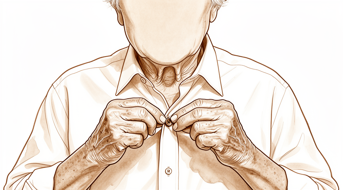
疼痛往往在您使用手指后加重,早晨醒来时也可能感到酸痛。需要用到指尖的精细动作会变得困难:扣衬衫纽扣、捡起小硬币、穿针,或捏住钥匙开锁。手写也可能引起酸痛。随着时间推移,该关节可能变得僵硬并失去部分弯曲能力,手指也可能慢慢偏离正常对线。
有些人会注意到指甲附近有一个充满液体的小肿块,称为黏液囊肿。它是从下方患关节炎的关节长出来的,因此即使抽液引流后也常常复发。
如果关节炎是在手指受伤之后出现的,例如锤状指(负责伸直的肌腱撕裂,或撕脱下一小块骨头),该关节可能会比原本磨损得更快。这种磨损会带来同样的疼痛和僵硬,即使损伤本身已经愈合,手指也可能失去部分活动度。
请留意以下几个警示征象。如果您的手指变得发热、发红、肿胀并疼痛,尤其是伴有发烧时,请当天前往急诊科。这可能是需要当天处理的感染,您无需先获得全科医生的转诊。如果疼痛数周仍未缓解、正在加重,或在夜间把您痛醒,请咨询您的全科医生或要求专科医生评估。
实际发生了什么
每根手指都是由一系列小骨组成的链条。最末端的关节就在指甲后方,是最后一节骨头与其上方那节骨头相接的地方。两块骨头的末端都覆盖着一层光滑、润滑的组织,让它们能够彼此滑动。可以把它想象成一个内置的减震器,同时还能保持关节面光滑。
患关节炎时,这层润滑组织会变薄或破损。于是骨头之间相互摩擦,关节的反应是在其边缘长出额外的骨质。这些骨刺就是您在指甲附近能摸到的坚硬肿块,也就是上一节所描述的那些肿块。摩擦会引起酸痛,而磨损的关节面和骨刺共同解释了为什么关节会肿胀、僵硬并慢慢偏离正常对线。
该关节还由两侧的小束组织带,以及一条从关节上方经过、附着在关节稍远处的扁平肌腱纤维带固定在一起。当关节炎改变了关节的结构时,这些组织带和肌腱可能会被牵拉而失去平衡。这就是为什么手指可能在中间关节处过度弯曲,同时指尖向下弯曲的原因之一,这种形状改变往往随着关节炎的进展而出现。
有时,这种磨损是继发于损伤,而不仅仅是普通的磨损和老化。一次重击、挤压伤,或锤状指之类的肌腱损伤,都可能损伤关节面。随后该关节会比原本更早地磨损,这就是为什么在损伤看似已经愈合多年之后,仍可能出现疼痛和僵硬。
该关节的关节炎还可能在指甲旁形成一个充满液体的小肿块,即前面提到的黏液囊肿。它是从受刺激的关节本身长出来的,这就是它在抽液引流后往往会复发的原因。
好消息是,这个关节很少承担用力抓握的工作。它的作用是指尖的精细控制,因此治疗的重点在于缓解疼痛,并让这种精细控制继续发挥作用。
我们如何处理该问题
Mater Private Hospital Rockhampton 的上肢外科医生 Kieran Hirpara 医生会从适合您病情的最微创方案入手。患者通常由全科医生转诊至我们的诊所;如果理疗师建议您就诊,您仍需获得全科医生的转诊才能符合 Medicare 报销资格。首次就诊时,我们会采集病史,检查您的手指,并在需要时安排影像学检查。由于这是一种长期存在的磨损问题,我们通常首先尝试非手术治疗,只有在其改善不足时才讨论手术。
第一步通常是使用一个小支具,将指尖关节保持在伸直位。佩戴支具可以缓解该关节的疼痛,并有助于手指伸直。它简单又安全,您可以在日常活动中一直佩戴。我们通常会同时使用抗炎药,即能减轻肿胀和疼痛的药片。在刺激症状缓解期间,手部治疗也可以帮助您保持指尖完成精细动作的能力。在决定下一步之前,请先充分尝试这些措施。
如果在此之后疼痛和僵硬仍然限制您的活动,就可以考虑手术。标准手术是融合术,也称为关节融合术:切除磨损的关节面,并将两块骨头连接起来,使它们愈合成一体。术后该关节不再能弯曲,但它无痛且稳定,手指会被固定在一个便于完成日常事务的位置。融合术还能矫正已经偏离对线的手指,并且是能阻止黏液囊肿复发的治疗方法。如果对您来说,保留指尖的部分活动度比完全消除疼痛更重要,还有另一种手术可以修整骨性肿块并清理关节,同时让关节保留活动。我们会与您讨论哪种方案适合您的手指,以及您希望双手完成哪些活动,并共同作出决定。
预期情况
指尖关节的关节炎通常在数年间缓慢发展。疼痛往往时好时坏:在您使用手指后加重,休息后又再次缓解。如果不治疗,该关节常常会变得更加僵硬,手指也可能随着时间推移更加偏离正常对线。有些人发现随着关节变僵硬,疼痛会自行减轻;但对许多人来说,疼痛会持续困扰,尤其是在扣衬衫纽扣或转动钥匙等精细动作时。
大多数人通过前面所述的简单措施就能得到良好的缓解:支具、抗炎药和手部治疗。这些措施能在您继续用手指完成日常事务的同时缓解疼痛。如果在充分尝试后这些措施仍无效,手术通常能很好地缓解疼痛。融合后的指尖关节无痛且稳定,人们通常能恢复正常用手,只是指尖不再能弯曲。如果保留部分活动度对您更重要,关节清理手术可以让手指保留弯曲能力,其疼痛缓解效果良好,但并不总是彻底。
手术后的恢复需要数周至数月,而不是几天。起初手指需要保持不动,让骨头愈合连接,手部治疗师会指导您逐步恢复握力和精细控制。大多数人发现,最终结果是一根能够正常发挥作用且不再酸痛的手指,尽管指尖比以前更直。
有几点需要坦诚提醒。融合术并不总是一次就能愈合,有些人需要再次手术以取出金属内固定物或重新进行融合。吸烟会减慢骨愈合,因此在手术前戒烟能让您的手指获得最佳的愈合机会。如果疼痛数周仍未缓解、正在加重,或在夜间把您痛醒,请咨询您的全科医生或要求专科医生评估。
何时就医
大多数指尖关节炎是缓慢发展的,因此一开始去全科医生处常规就诊就足够了。如果疼痛数周仍未缓解、正在加重,或在夜间把您痛醒,请咨询您的全科医生。如果手指影响您工作,或妨碍您完成扣衬衫纽扣之类的精细动作,也同样如此。
有些征象需要更快就医。如果您的手指变得发热、发红、肿胀并疼痛,尤其是伴有发烧时,请当天前往急诊科。这种表现提示感染,您无需先获得全科医生的转诊。
如果受伤后关节上方的皮肤破损、骨头外露,或手指明显变形,请前往急诊科。如果手指变得苍白、发凉、发白或发青,或者在碰撞后手指突然失去感觉或无法活动,也请前往急诊科。
黏液囊肿反复出现,或手指正在偏离正常对线,值得请专科医生评估。
深入探讨
Advanced reading: the deeper science (optional)
本节内容超出了您自身治疗决策所需的范围。指尖关节的关节炎值得额外阅读,因为标准手术——即关节融合术,通常被描述为简单直接——其并发症发生率高于其声誉所暗示的水平,并且存在一种很少被提及的保留关节活动度的替代方案。
关节融合术并非听起来那般平稳
指间关节融合术被描述为一种可靠的手术,就疼痛缓解而言确实如此。 但其并发症谱并不那么温和。一项针对 173 例患者危险因素的分析直接指出:远端指间关节融合术常导致并发症,并识别出 骨关节炎、翻修融合术和吸烟 为危险因素 [1]。
吸烟出现在该列表中值得采取行动,因为这是患者唯一可控的因素。 骨愈合依赖于血液供应,而这是一个位于手指血供末端的微小关节,其软组织包膜很薄。
愈合效果更优的植入物也会引发廉价替代品所不具备的问题
关于固定方式的争论有一个异常清晰的答案,且这并非胜负之分,而是一种权衡。在 1,125 名患者中,无头加压螺钉似乎具有更高的愈合率,但与并发症相关,而这些并发症在其他成熟且更廉价的技术中并未见到, 除愈合率外,证据不足以证明螺钉具有优越性 [2]。
螺钉特有的并发症源于解剖结构。螺钉沿指尖轴线运行,因此紧贴甲床,可能导致甲畸形; 在非常小的远节指骨中,可能缺乏足够的骨质来固定螺钉。钢丝更廉价,可避免这些特定 问题,但愈合可靠性略低。
当愈合率是唯一优先事项时,在翻修融合术或吸烟患者中,螺钉的优势最为相关。 当骨质较小且指甲外观重要时,其并非明确的最佳选择。
保留活动度的替代方案
关节融合并非唯一选择,且替代方案鲜少被广泛讨论。对于主要诉求是骨性隆起及其引发的疼痛,而非整个关节的关节炎的患者,可以在保留关节完整性的同时切除突出的骨赘。
指尖关节的开放关节缘切除术被描述为在希望保留关节活动度的症状性骨关节炎患者中,关节融合的安全且有效的替代方案,涉及78**例患者 [3]。
这一点至关重要,因为指尖关节对握力的贡献甚微,但对精细操作和手部外观却有较大影响。对于症状主要由赫伯登结节本身驱动的患者而言,用永久性僵硬的指尖来换取这些结节,其代价可能比听起来更大,且存在一种折中的选择。
常伴随出现的囊肿
该关节的关节炎常导致黏液囊肿,这是一种源自关节炎性关节的小液性肿胀,通常位于指甲旁。由于其由潜在的关节病变驱动,因此在这一点上表现得类似腕部腱鞘囊肿:仅抽吸囊肿可消除肿胀,却无法解决根本病因。该囊肿将在其他章节单独讨论,但了解其与关节炎的关联至关重要,因为反复发作的囊肿提示其下方存在关节炎,而非孤立性问题。
参考文献
[1] Runkel A, Bonaventura B, Sundermann B, Zajonc H, Eisenhardt S, Leibig N. 手部远端指间关节融合术的危险因素:一项回顾性研究。J Hand Surg Eur Vol. 2022;47(9):907-14. https://doi.org/10.1177/17531934221111641
[2] Dickson D, Mehta S, Nuttall D, Ng C. 远端指间关节融合术的系统性综述。J Hand Microsurg. 2014;6(2):74-84. https://doi.org/10.1007/s12593-014-0163-1
[3] Lin EA, Papatheodorou LK, Sotereanos DG. 关节成形术治疗有症状的远端指间关节骨关节炎:78例病例的回顾。J Hand Surg Am. 2017;42(11):889-93. https://doi.org/10.1016/j.jhsa.2017.07.006
Evidence & references
This is the clinical evidence summary written for health professionals. It is technical, and it lists the research this page was built from. You do not need to read it to understand your treatment or to make a decision about it.
Overview
- Osteoarthritis is the most common indication for distal interphalangeal joint arthrodesis [19].
- In a cohort of 149 cases, postoperative complications for distal interphalangeal and thumb interphalangeal joint arthrodesis occurred at a rate similar to that reported in existing literature [19].
- Finger distal interphalangeal and thumb interphalangeal joint arthrodesis generally results in favorable outcomes in terms of bony union regardless of underlying medical condition or technical details of the surgical operation [6].
- The X-fuse implant provides excellent stability with minimal hardware problems and a high rate of union in patients with finger distal interphalangeal and thumb interphalangeal joint arthritis [1].
- In select patients, percutaneous distal interphalangeal joint arthrodesis is advantageous in comparison with open fusion techniques [2].
- Reamed percutaneous distal interphalangeal joint arthrodesis is associated with higher union rates achieved in a shorter time and reduced postoperative pain compared with the non-reamed percutaneous technique [8].
- Successful fusions of the distal interphalangeal joint at an angle up to 35 degrees can be achieved using small diameter headless compression screws [14].
- The Mini-Acutrak screw is suitable for distal interphalangeal joint fusion in all fingers with the exception of the small finger [22].
- The nonaxial multiple small screws technique is a feasible option for distal interphalangeal and thumb interphalangeal joint arthrodesis, especially when a small finger is indicated and a significant flexion angle is required [24].
- Given the lack of difference in biomechanical performance between K-wires and compression screws, consideration should be given to other factors such as cost and complication profiles when choosing an implant for distal interphalangeal joint fusion [34].
- The smile incision and reverse shotgun approach may be a good surgical option for distal interphalangeal joint arthrodesis when more volar part joint preparation and more volar implant insertion sites are necessary [17].
- Results from a lateral approach and plate fixation for distal interphalangeal joint arthrodesis are equivalent to traditional methods but with fewer major complications [11].
- For persistently symptomatic, unstable distal interphalangeal joints, arthrodesis should be considered [4].
- In the distal interphalangeal joint, there is probably good reason to proceed to arthrodesis immediately for synovial chondromatosis [3].
- The swan neck deformity can progress significantly with time because of increasing distal interphalangeal joint flexion contracture [5].
- Silicone interpositional arthroplasty of the distal interphalangeal joint is an acceptable alternative to arthrodesis, achieving excellent pain relief and a range of movement of 30–40 degrees with a low overall complication rate of 5% [13].
- Swanson hinge implant arthroplasty can be the surgical treatment of choice for the patient who desires continued mobility, albeit limited, of the involved osteoarthritic distal interphalangeal joint [9].
- Open distal interphalangeal joint cheilectomy is a safe and effective alternative to distal interphalangeal joint arthrodesis in patients with symptomatic osteoarthritis who wish to preserve joint motion [28].
- Denervation with cheilectomy of the distal interphalangeal joint presents a compelling motion-preserving alternative to arthrodesis for symptomatic distal interphalangeal joint osteoarthritis [10].
- A surgical technique for treating symptomatic distal interphalangeal joint arthritis reduces pain while preserving distal interphalangeal joint motion [18].
- The authors recommend simultaneous surgical intervention in case of severe painful osteoarthritis of the proximal interphalangeal and distal interphalangeal joints of the same digit [7].
- The combination of distal interphalangeal arthrodesis and proximal interphalangeal Swanson arthroplasty resulted in a favorable outcome in terms of simultaneous bony union and flexibility [12].
- Combined distal interphalangeal arthrodesis and proximal interphalangeal procedures present unique challenges regarding hardware conflict, where K-wires offer the easiest compatibility and headless screws must ideally not reach proximal to the midpoint of the middle phalanx [90].
- A customized structural bone graft addresses issues of bone stock loss and medullary absence in failed distal interphalangeal joint silicone arthroplasty, achieving reliable union rates and high patient satisfaction [21].
Anatomy & Pathophysiology
Bony Anatomy & Morphology
- The skeleton of the hand consists of 27 bones, of which 19 are long bones [47].
- The skeleton is divided into five rays, each ray making up a polyarticulated chain comprising the metacarpals and phalanges [47].
- The thumb ray is made up of only three bones—a metacarpal and two phalanges [47].
- The other four digital rays are formed by four skeletal segments—a metacarpal and three phalanges [47].
- The thumb metacarpal is the shortest, and the index metacarpal is by far the longest [47].
- The proximal and middle phalanges of the long and ring fingers are longer than those of the index finger [47].
- The epiphyseal plates are located at the proximal ends of the phalanges and the first metacarpal [47].
- An examination of 100 specimens of the terminal joints of human fingers was described in a 1982 study [75].
- A substantial number of distal phalanges are too small to accommodate commonly available headless compression screws, particularly in females and the small finger [39].
- Understanding of DIP joint morphology may lend insight into the biomechanics and disease progression within the DIP joints [23].
Soft Tissue Anatomy & Extensor Mechanism
- Distal interphalangeal joint extension is achieved through the conjoined lateral bands that are composed of tendinous slips from the extrinsic and intrinsic tendons [46].
- The principal bony insertion of the extrinsic digital extensors is on the dorsal proximal aspect of the middle phalanx [46].
- The deep head of each dorsal interosseous muscle forms a lateral tendon, or lateral band, at the level of the MP joint [59].
- The lateral bands are joined by the lateral slips of the extensor tendon to form the conjoined lateral band [59].
- The two conjoined lateral bands to each finger unite at the distal third of the middle phalanx to form the terminal tendon [59].
- The terminal tendon inserts at the base of the distal phalanx to extend it [59].
- The flexor digitorum profundus inserts on the proximal volar aspect of the distal phalanx, flexing the distal interphalangeal joint as well as the proximal interphalangeal and metacarpophalangeal joints [46].
- The flexor digitorum profundus provides digital flexion at both the proximal and distal interphalangeal joints [66].
- The A5 annular pulley is located over the distal interphalangeal joint [66].
- The vinculum breve of the flexor digitorum profundus and check-rein ligaments may account for the greater amount of passive hyperextension that can be achieved at the distal than the proximal interphalangeal joint [80].
Biomechanics & Kinematics
- The wrist influences the position of the metacarpophalangeal joint; the metacarpophalangeal joint also affects the position of the proximal interphalangeal joint, which in turn affects the distal interphalangeal joint [67].
- Almost all movements in the hand are around oblique and variable axes, resulting in combined movements permitting optimal orientation of the phalanges at the time of prehension [67].
- The axes of flexion are so arranged that flexion of all the metacarpophalangeal and proximal interphalangeal joints causes the fingers to converge toward the scaphoid [70].
- In a combinatorial relationship, intrinsic muscles produce steep inclination of extensor forces at the DIP joint [73].
- There were no significant differences in comparisons among loads (200, 400, 600, and 800 g) regarding the inclination slope of intrinsic extensor forces at the DIP joint [73].
- Immobilization of the distal interphalangeal joint of any finger reduces the overall grip strength of the hand, with the effect becoming progressively more pronounced from the index to the little fingers [37].
- Positioning the middle finger DIP joint in either extension or 20° of flexion did not significantly affect grip strength or dexterity [49].
- Biomechanically, dynamic tenodesis for the DIP joint using the remaining FDP tendon results in a flexion angle greater than 30 degrees [76].
Pathophysiology & Etiology
- Osteoarthritis occurs with the highest prevalence in the distal interphalangeal joint of the hand and has been divided into an erosive and a nonerosive form [26].
- The pathogenesis of the early stages of osteoarthritis is poorly understood, but considerable emphasis has been placed on the role of cartilage and subchondral bone as well as soft tissue structures such as collateral ligaments and tendons [26].
- Osteoarthritis is a complex disease resulting in the failure of articular cartilage due to a combination of genetic, metabolic, biochemical, and biomechanical factors [26].
- Cartilage loss and reactive new bone formation at joint margins is accompanied by the proliferation of osteoarticular tissue in the capsule [26].
- Heberden's nodes are more common in women who engage in cooking in school and the incidence increases with the number of tasks involved [25].
- Cooking as an occupation is an aetiological factor in the pathogenesis of Heberden's nodes [25].
- Work-load, sports, and repeated minimal trauma contribute to the development of Heberden's nodes [25].
- Repetitive injuries to a joint lead to an increased risk of developing posttraumatic arthritis [48].
- Athletes have a higher incidence of degenerative joint changes compared to the general population [48].
- Mallet finger is caused by forced flexion of an extended fingertip [51].
- Less common hyperextension or hyperextension/axial loading tends to cause the larger fractures and subluxation in mallet injuries [51].
- One-third of all mallet fingers are associated with a fracture [51].
- Mallet fractures associated with large fragments may result in volar subluxation of the distal phalanx as the collateral ligaments remain attached to the fracture fragment [51].
- Fracture fragments that are less than 43% of the distal phalanx articular surface usually do not allow volar subluxation [51].
- Fracture fragments involving larger than 52% of the joint surface consistently allow subluxation [51].
- Palmar subluxation of a DIP joint without preexisting arthritic deformity is expected when more than one half of the dorsal articular surface is injured [20].
- The mechanism of displaced intra-articular osteochondral fracture causing irreducible dislocation of the DIP joint probably involved hyperextension of the DIP joint [15].
- The palmar plate was torn at the attachment to the distal phalanx in cases of displaced intra-articular osteochondral fracture [15].
- Attempts to perform a closed reduction of displaced intra-articular osteochondral fractures can lead to further displacement of the fracture, with folding of the fragment on the articular cartilage hinge [15].
- The cause of irreducible palmar dislocation of the DIP joint can be an entrapment of the extensor tendon in front of the head of the middle phalanx [41].
- Irreducibility was more commonly seen in dorsal than in volar dislocations of the DIP joint [85].
- Volar dislocations of the DIP joint carried a higher risk of instability immediately after reduction compared to dorsal dislocations [85].
- Some loss of motion in small joints of the fingers after hyperflexion injuries would be expected [79].
Classification
- Osteoarthritis of the distal interphalangeal joint has been divided into an erosive and a nonerosive form [26].
- Radiographic evaluation represents the most standardized method to quantify disease progression in distal interphalangeal joint osteoarthritis, with different systems developed for defining and grading radiographic features [26].
- The interrater reliability of the Kellgren & Lawrence and OARSI classification systems for post-traumatic osteoarthritis in the distal interphalangeal joint after mallet finger fractures is considerably lower than initially assumed [86].
Clinical Presentation
- Osteoarthritis occurs with the highest prevalence in the distal interphalangeal joint of the hand [26].
- Heberden's nodes are more common in women who engage in cooking in school, with incidence increasing with the number of tasks involved [25].
- There is no confirmed difference in the incidence of disease between right and left hands in elementary school cooks [25].
- Radiographic evaluation represents the most standardized method to quantify disease progression in distal interphalangeal joint osteoarthritis [26].
- The pathogenesis of the early stages of distal interphalangeal joint osteoarthritis is poorly understood [26].
- Considerable emphasis has been placed on the role of cartilage and subchondral bone in the pathogenesis of distal interphalangeal joint osteoarthritis [26].
- Soft tissue structures such as collateral ligaments and tendons play a role in the pathogenesis of distal interphalangeal joint osteoarthritis [26].
- Cartilage loss and reactive new bone formation at joint margins are accompanied by the proliferation of osteoarticular tissue in the capsule [26].
- Radiological osteoarthritis after a mallet finger fracture is similar to the natural degenerative process in the distal interphalangeal joint [27].
- Radiological osteoarthritis after a mallet finger fracture is accompanied by a decrease in range of motion of the distal interphalangeal joint [27].
- The decrease in range of motion following mallet finger fracture does not clinically affect patient-reported outcomes [27].
- Palmar subluxation of a distal interphalangeal joint without preexisting arthritic deformity is expected when more than one half of the dorsal articular surface is injured [20].
- Primary synovial chondromatosis of the distal interphalangeal joint is an extremely rare entity that requires accurate diagnosis to distinguish from other arthropathies [50].
- Adnexal tumours may masquerade as a distal interphalangeal joint ganglion and should enter the differential diagnosis when examining tumours of the digits [89].
- Floating distal interphalangeal joint injuries can be misdiagnosed initially due to minimal deformity [33].
- Osteoarthritis may develop following floating distal interphalangeal joint injuries [33].
- The mechanism of displaced intra-articular osteochondral fracture causing irreducible dislocation of the distal interphalangeal joint probably involved hyperextension of the joint [15].
- The palmar plate is torn at the attachment to the distal phalanx in displaced intra-articular osteochondral fractures of the distal interphalangeal joint [15].
- Attempts to perform a closed reduction of a displaced intra-articular osteochondral fracture can lead to further displacement of the fracture with folding of the fragment on the articular cartilage hinge [15].
- The cause of irreducibility in palmar dislocation of the distal interphalangeal joint can be entrapment of the extensor tendon in front of the head of the middle phalanx [41].
- Interrater reliability for measurements of the distal interphalangeal joint motions of the index and long digits is high [16].
- Interrater reliability for distal interphalangeal joint motion measurements is slightly higher for the dorsal method of placement than for the lateral method [16].
Investigations
- Radiographic evaluation is the most standardized method to quantify disease progression in distal interphalangeal joint osteoarthritis [26].
- Different systems have been developed for defining and grading radiographic features of distal interphalangeal joint osteoarthritis [26].
- Interrater reliability was high for measurements of the proximal and distal interphalangeal joint motions of the index and long digits [16].
- Interrater reliability was slightly higher for the dorsal method of goniometer placement than for the lateral method [16].
- Primary synovial chondromatosis of the distal interphalangeal joint requires accurate diagnosis to distinguish from other arthropathies [50].
- A distinct collagen septum exists between the extensor tendon and skin at the distal interphalangeal joint [53].
- The existence of a distinct collagen septum between the extensor tendon and skin at the distal interphalangeal joint was confirmed using MRI and histology [53].
Treatment
Non-Operative
- Splinting of the distal interphalangeal joint reduces pain and improves extension at the joint [29].
- Splinting for DIP joint arthritis does not give rise to non-compliance, increased stiffness, or restriction of range of motion [29].
- Splinting and anti-inflammatory medications are the mainstay of nonoperative treatment for DIP joint arthritis [48].
- Injection with Collagenase Clostridium histolyticum is an option for the treatment of DIP joint contractures in Dupuytren disease, though the potential risk for recurrence should be carefully weighed prior to its use [93].
Operative: Arthrodesis
- Arthrodesis is generally the most accepted surgical option for treatment of degenerative and traumatic conditions involving the distal interphalangeal joint of the fingers or the interphalangeal joint of the thumb [38].
- The ideal position of DIP arthrodesis is thought to be slightly flexed to improve power, fine pinch, and grip [38].
- In select patients, percutaneous DIP joint arthrodesis is advantageous in comparison with open fusion techniques [2].
- Reamed percutaneous DIPJ arthrodesis is associated with higher union rates achieved in a shorter time and reduced postoperative pain compared with the non-reamed percutaneous technique [8].
- The X-fuse implant provides excellent stability with minimal hardware problems and a high rate of union in patients with finger DIPJ and thumb IPJ arthritis [1].
- Finger DIP and thumb IP joint arthrodesis generally resulted in favourable outcome in terms of bony union regardless of the underlying medical condition or technical details of the surgical operation [6].
- Osteoarthritis was the most common indication for DIP and thumb IP arthrodesis, and postoperative complications occurred at a rate similar to that reported in existing literature [19].
- Successful fusions of the DIP joints at an angle up to 35 degrees were achieved using small diameter headless compression screws, providing benefits including early mobilization and favourable functional outcome scores [14].
- The Mini-Acutrak screw is suitable for DIPJ fusion in all fingers with the exception of the small finger [22].
- The nonaxial multiple small screws technique could be used as a feasible option in DIPJ and thumb IPJ arthrodesis, especially when a small finger is indicated and a significant flexion angle is required [24].
- Given the lack of difference in biomechanical performance between K-wires and compression screws, consideration should be given to other factors such as cost and complication profiles when choosing an implant for DIPJ fusion [34].
- DIP joint preparation is not necessary before performing arthrodesis in fingers with stage IV chondropathy [44].
- The use of a buried break-away compression screw avoids complications related to bulky hardware in DIP arthrodesis [44].
- Dorsal plate fixation allows for the performance of a distal finger joint arthrodesis in slight flexion [38].
- The smile incision and reverse shotgun approach may be a good surgical option for DIPJ arthrodesis when more volar part joint preparation and more volar implant insertion sites are necessary [17].
- Lateral approach and plate fixation for DIP joint arthrodesis yields results equivalent to traditional methods but with fewer major complications [11].
- For persistently symptomatic, unstable DIP joints, arthrodesis should be considered [4].
- In the DIP joint, there is probably good reason to proceed to arthrodesis immediately in cases of synovial chondromatosis [3].
- A customized structural bone graft addresses issues of bone stock loss and medullary absence in failed DIPJ silicone arthroplasty, achieving reliable union rates and high patient satisfaction [21].
- All joints had fused radiologically in a series of proximodistal interphalangeal arthrodesis of the little finger [32].
- Arthrodesis of the DIP joint provides stability and pain relief and can correct deformity [48].
- A painless, stable, distal interphalangeal joint that is in a functional position is more important than the ability to move the joint [48].
- The authors recommend simultaneous surgical intervention in case of severe painful OA of the PIP and DIP joints of the same digit [7].
- The combination of DIP arthrodesis and PIP Swanson arthroplasty resulted in a favourable outcome in terms of simultaneous bony union and flexibility [12].
- For patients prioritizing hand aesthetics or with unstable joints, distal interphalangeal joint arthrodesis is preferable [31].
Operative: Arthroplasty and Motion-Preserving
- Silicone interpositional arthroplasty of the DIP joint is an acceptable alternative to arthrodesis, achieving excellent pain relief and a range of movement of 30–40 degrees with a low overall complication rate of 5% [13].
- Swanson hinge implant arthroplasty can be the surgical treatment of choice for the patient who desires continued mobility, albeit limited, of the involved osteoarthritic DIP joint [9].
- Open DIP joint cheilectomy is a safe and effective alternative to DIP joint arthrodesis in patients with symptomatic osteoarthritis who wish to preserve joint motion [28].
- Denervation with cheilectomy of the distal interphalangeal joint presents a compelling motion-preserving alternative to arthrodesis for symptomatic DIP joint osteoarthritis [10].
- A surgical technique for DIP joint arthritis reduces pain while preserving DIP joint motion [18].
- Volar plate arthroplasty of the distal interphalangeal joint allows for a painless functional arc of motion without residual joint subluxation [40].
Complications
- The X-fuse implant provides excellent stability with minimal hardware problems [1].
- The X-fuse implant has a high rate of union in patients with finger DIPJ and thumb IPJ arthritis [1].
- Reamed percutaneous DIPJ arthrodesis is associated with reduced postoperative pain compared with the non-reamed percutaneous technique [8].
- The swan neck deformity progressed significantly with time because of increasing DIPJ flexion contracture [5].
- Postoperative complications occurred at a rate similar to that reported in the existing literature in a retrospective cohort of 149 cases of DIP and thumb IP joint arthrodesis [19].
- Arthrodesis of the distal interphalangeal joint often leads to complications [30].
- Silicone interpositional arthroplasty of the DIP joint has a low overall complication rate of 5% [13].
- Bony union of the joint was delayed in a series using the Herbert screw for DIPJ arthrodesis [56].
- Follow-up to 5 years of DIP joint flexible implant arthroplasty showed only one complication [92].
Recovery
Arthrodesis Outcomes and Complications
- Postoperative complications in DIP and thumb IP joint arthrodesis occurred at a rate similar to that reported in existing literature [19].
- Finger DIP and thumb IP joint arthrodesis generally resulted in favourable outcomes in terms of bony union regardless of underlying medical condition or technical details of the surgical operation [6].
- Reamed percutaneous DIPJ arthrodesis is associated with higher union rates achieved in a shorter time compared with the non-reamed percutaneous technique [8].
- Successful fusions of the DIP joints at an angle up to 35 degrees were achieved using small diameter headless compression screws [14].
- Small diameter headless compression screws for DIP arthrodesis provide benefits including early mobilization and favourable functional outcome scores [14].
- Although bony union of the joint was delayed in a minimally invasive technique using the Herbert Screw, firm bone union was ultimately obtained in all joints [56].
Arthroplasty Outcomes
- Silicone interpositional arthroplasty of the DIP joint achieves a range of movement of 30–40 degrees [13].
Non-Operative Management
- Splinting of the DIP joint does not give rise to non-compliance, increased stiffness, or restriction of range of motion [29].
Key Evidence
- [L4] The X-fuse implant seems to provide excellent stability with minimal hardware problems with a high rate of union in a wide array of patients with finger DIPJ and thumb IPJ arthritis. [1] (10.1097/bth.0b013e31829ba688)
- [L4] In select patients, this percutaneous DIP joint arthrodesis is advantageous in comparison with open fusion techniques. [2] (10.1007/s11552-010-9265-9)
- [L5] In the DIP joint there is probably good reason to proceed to arthrodesis immediately. [3] (10.1016/s0363-5023(09)91115-7)
- [L4] For persistently symptomatic, unstable DIP joints, arthrodesis should be considered. [4] (10.1016/j.jhsb.2005.09.003)
- [L5] The swan neck deformity in this individual progressed significantly with time because of increasing DIPJ flexion contracture. [5] (10.1016/j.jht.2009.11.005)
- [L3] In our cohort finger DIP and thumb IP joint arthrodesis generally resulted in favourable outcome in terms of bony union regardless of the underlying medical condition or technical details of the surgical operation. [6] (10.1142/s2424835520500216)
- [L3] The authors recommend simultaneous surgical intervention in case of severe painful OA of the PIP and DIP joints of the same digit. [7] (10.1177/17531934231191255)
- [L3] Reamed percutaneous DIPJ arthrodesis is associated with higher union rates achieved in a shorter time and reduced postoperative pain compared with the non-reamed percutaneous technique. [8] (10.1177/15589447261487482)
- [L5] Swanson hinge implant arthroplasty can be the surgical treatment of choice for the patient who desires continued mobility, albeit limited, of the involved osteoarthritic DIP joint. [9] (10.1016/s0894-1130(98)80061-6)
- [L4] It presents a compelling motion-preserving alternative to arthrodesis for symptomatic DIP joint osteoarthritis. [10] (10.1016/j.jhsa.2026.01.027)
- [L4] The results obtained in this small series are equivalent to the traditional methods of DIP joint arthrodesis but with fewer major complications. [11] (10.1016/j.jhsa.2007.09.004)
- [L4] The combination of DIP arthrodesis and PIP Swanson arthroplasty resulted in a favourable outcome in terms of simultaneous bony union and flexibility. [12] (10.1177/17531934231215790)
- [L4] The study confirms that silicone interpositional arthroplasty of the DIP joint is an acceptable alternative to arthrodesis, achieving excellent pain relief and a range of movement of 30–40 degrees with a low overall complication rate of 5%. [13] (10.1177/1753193411422679)
- [L4] In this series, successful fusions of the DIP joints, at an angle up to 35 degrees were achieved using small diameter headless compression screws, which provided benefits including early mobilization and favourable functional outcome scores. [14] (10.1142/s2424835518500406)
- [L5] [15] (10.1016/s0363-5023(82)80018-x)
- [L5] Interrater reliability was high for measurements of the PIP and DIP joint motions of the index and long digits and slightly higher for the dorsal method of placement than for the lateral method. [16] (10.1016/s0894-1130(01)80021-1)
- [L4] This technique may be a good surgical option for DIPJ arthrodesis when more volar part joint preparation and more volar implant insertion sites are necessary. [17] (10.1186/s12891-024-08016-6)
- [L4] This surgical technique reduces pain while preserving DIP joint motion. [18] (10.1177/1558944716660555i)
- [L3] Osteoarthritis was the most common indication for arthrodesis and postoperative complications occurred at a rate similar to that reported in the existing literature. [19] (10.1186/s12891-024-07361-w)
- [L5] Palmar subluxation of a DIP joint without preexisting arthritic deformity is expected when more than one half of the dorsal articular surface is injured. [20] (10.1016/j.jhsa.2007.09.006)
- [L4] A customized structural bone graft using the described technique addresses issues of bone stock loss and medullary absence in failed DIPJ silicone arthroplasty, achieving reliable union rates and high patient satisfaction. [21] (10.1177/17531934231151217)
- [L4] The authors conclude that the Mini-Acutrak screw is suitable for DIPJ fusion in all fingers with the exception of the small finger. [22] (10.1016/j.jhsa.2005.09.009)
- [L5] Our understanding of morphology may lend insight into the biomechanics and disease progression within the DIP joints. [23] (10.1007/s11552-014-9605-2)
- [L4] Thus, the NMSS technique could be used as a feasible option in DIPJ and thumb IPJ arthrodesis, especially when a small finger is indicated and a significant flexion angle is required. [24] (10.1186/s12891-022-05473-9)
- [L4] [25] (10.1016/0266-7681(93)90167-e)
- [L5] [26] (10.1016/j.jhsa.2010.09.003)
- [L4] Radiological OA after an MFF is similar to the natural degenerative process in the DIP joint and is accompanied by a decrease in range of motion of the DIP joint, which does not clinically affect PROMs. [27] (10.1016/j.jhsa.2023.03.027)
- [L4] Open DIP joint cheilectomy is a safe and effective alternative to DIP joint arthrodesis in patients with symptomatic osteoarthritis who wish to preserve joint motion. [28] (10.1016/j.jhsa.2017.07.006)
- [L2] It does not give rise to non-compliance, increased stiffness or restriction of range of motion. [29] (10.1016/j.jht.2013.08.004)
- [L3] Arthrodesis of the distal interphalangeal joint often leads to complications. [30] (10.1177/17531934221111641)
- [L3] For patients prioritizing hand aesthetics or with unstable joints, distal interphalangeal joint arthrodesis is preferable. [31] (10.1177/1753193420917818)
- [L4] All joints had fused radiologically. [32] (10.1016/j.hansur.2016.06.003)
- [Case_report] Floating DIP joint injuries can be misdiagnosed initially due to minimal deformity; open reduction and internal fixation is a viable treatment option for chronic cases, though osteoarthritis may develop. [33] (10.1016/j.jhsa.2010.05.025)
- [L5] Given the lack of difference in biomechanical performance between K-wires and compression screws, consideration should be given to other factors such as cost and complication profiles when choosing an implant for DIPJ fusion. [34] (10.1177/1558944715627211)
- [L4] Immobilization of the distal interphalangeal joint of any finger reduces the overall grip strength of the hand, with the effect becoming progressively more pronounced from the index to the little fingers. [37] (10.1177/1753193418765068)
- [L4] [38] (10.1016/j.jhsa.2018.03.049)
- [L4] A substantial number of distal phalanges are too small to accommodate commonly available headless compression screws, particularly in females and the small finger. [39] (10.1007/s11552-014-9679-x)
- [L4] The technique allows for a painless functional arc of motion without residual joint subluxation. [40] (10.1053/jhsu.2001.26325)
- [L5] The cause of irreducibility was an entrapment of the extensor tendon in front of the head of the middle phalanx. [41] (10.1016/s0363-5023(87)80116-8)
- [L4] Our results show that DIP joint preparation is not necessary before performing arthrodesis in fingers with stage IV chondropathy and that the use of a buried break-away compression screw avoids complications related to bulky hardware. [44] (10.1016/j.main.2015.03.002)
- [L4] [48] (10.1142/s0218810417500149)
- [L2] Positioning the middle finger DIP joint in either extension or 20° of flexion did not significantly affect grip strength or dexterity, allowing other considerations such as appearance to be prioritized. [49] (10.1016/j.jhsa.2014.06.021)
- [Case_report] Primary synovial chondromatosis of the distal interphalangeal joint is an extremely rare entity that requires accurate diagnosis to distinguish from other arthropathies. [50] (10.1177/15589447211049520)
- [L5] [51] (10.1177/1753193414554772)
- [L5] We confirmed the existence of a distinct collagen septum between the extensor tendon and skin at the DIP joint using MRI and histology. [53] (10.1016/j.jhsa.2008.11.030)
- [L4] Although bony union of the joint was delayed, firm bone union was ultimately obtained in all joints. [56] (10.1097/00130911-200212000-00008)
- [L5] [73] (10.1002/jor.22021)
- [L5] An examination of 100 specimens of the terminal joints of human fingers is described. [75] (10.1016/s0363-5023(82)80084-1)
- [L5] Biomechanically, dynamic tenodesis for the DIP joint using the remaining FDP tendon is a valuable procedure because it results in a flexion angle greater than 30 degrees. [76] (10.1016/j.jhsg.2020.08.007)
- [L5] Therefore, some loss of motion in small joints of the fingers after hyperflexion injuries would be expected. [79] (10.1142/s2424835516720206)
- [L5] This may account for the greater amount of passive hyperextension that can be achieved at the distal than the proximal interphalangeal joint. [80] (10.1016/0266-7681(91)90058-v)
- [L4] Irreducibility was more commonly seen in dorsal than in volar dislocations, while volar dislocations carried a higher risk of instability immediately after reduction. [85] (10.1177/1753193415616957)
- [L4] The interrater reliability of the Kellgren & Lawrence and OARSI classification systems for post-traumatic osteoarthritis in the distal interphalangeal joint after mallet finger fractures is considerably lower than initially assumed. [86] (10.1016/j.jhsa.2024.03.012)
- [L5] Adnexal tumours may masquerade as a DIPJ ganglion, and should enter the differential diagnosis of the hand surgeon when examining tumours of the digits. [89] (10.1177/1753193415620179)
- [L5] Combined DIP arthrodesis and PIP procedures present unique challenges regarding hardware conflict; K-wires offer the easiest compatibility, while headless screws must ideally not reach proximal to the midpoint of the middle phalanx. [90] (10.1016/j.jhsa.2024.08.006)
- [L4] Follow-up to 5 years showed results that were good to excellent, with only one complication, and a high degree of patient satisfaction. [92] (10.1016/0363-5023(89)90184-6)
- [L4] Injection with CCH is an option for the treatment of DIP joint contractures in Dupuytren disease, though the potential risk for recurrence should be carefully weighed prior to its use. [93] (10.1016/j.jhsa.2018.07.004)
References
[1] Distal Interphalangeal Joint Arthrodesis Using Nitinol Intramedullary Fixation Implants. Techniques in Hand & Upper Extremity Surgery. 2013. DOI: 10.1097/bth.0b013e31829ba688
[2] Treatment of Symptomatic Distal Interphalangeal Joint Arthritis with Percutaneous Arthrodesis: A Novel Technique in Select Patients. HAND. 2010. DOI: 10.1007/s11552-010-9265-9
[3] Synovial chondromatosis in the distal interphalangeal joint. The Journal of Hand Surgery. 1990. DOI: 10.1016/s0363-5023(09)91115-7
[4] Management of Tophaceous Gout of the Distal Interphalangeal Joint. Journal of Hand Surgery. 2006. DOI: 10.1016/j.jhsb.2005.09.003
[5] Swan Neck Deformity after Distal Interphalangeal Joint Flexion Contractures: A Biomechanical Analysis. Journal of Hand Therapy. 2010. DOI: 10.1016/j.jht.2009.11.005
[6] Factors Influencing Bone Union in Finger Distal Interphalangeal and Thumb Interphalangeal Joint Arthrodesis. The Journal of Hand Surgery (Asian-Pacific Volume). 2020. DOI: 10.1142/s2424835520500216
[7] Does distal interphalangeal joint arthrodesis affect proximal interphalangeal joint arthroplasty outcomes in the same finger?. Journal of Hand Surgery (European Volume). 2023. DOI: 10.1177/17531934231191255
[8] Reamed Percutaneous Arthrodesis for Distal Interphalangeal Joint Arthritis Improves Bone Fusion and Pain Compared With a Non-Reamed Technique. HAND. 2026. DOI: 10.1177/15589447261487482
[9] Distal interphalangeal joint implant arthroplasty in a musician. Journal of Hand Therapy. 1998. DOI: 10.1016/s0894-1130(98)80061-6
[10] Denervation with Cheilectomy of the Distal Interphalangeal Joint: Technique and Medium-Term Results. The Journal of Hand Surgery. 2026. DOI: 10.1016/j.jhsa.2026.01.027
[11] Alternative to the Distal Interphalangeal Joint Arthrodesis: Lateral Approach and Plate Fixation. The Journal of Hand Surgery. 2008. DOI: 10.1016/j.jhsa.2007.09.004
[12] Simultaneous anterograde screw arthrodesis of distal interphalangeal joint and silastic proximal interphalangeal joint replacement for osteoarthritis. Journal of Hand Surgery (European Volume). 2023. DOI: 10.1177/17531934231215790
[13] Joint replacement in 131 painful osteoarthritic and post-traumatic distal interphalangeal joints. Journal of Hand Surgery (European Volume). 2011. DOI: 10.1177/1753193411422679
[14] A Functional Angle of Up to 35° at the Distal Interphalangeal Joint Can Be Achieved with Headless Compression Screw Fusion. The Journal of Hand Surgery (Asian-Pacific Volume). 2018. DOI: 10.1142/s2424835518500406
[15] Displaced intra-articular osteochondral fracture—Cause for irreducible dislocation of the distal interphalangeal joint. The Journal of Hand Surgery. 1982. DOI: 10.1016/s0363-5023(82)80018-x
[16] Goniometry of the proximal and distal interphalangeal joints, part II: Placement preferences, interrater reliability, and concurrent validity. Journal of Hand Therapy. 2001. DOI: 10.1016/s0894-1130(01)80021-1
[17] Smile incision and reverse shotgun approach in distal interphalangeal joint arthrodesis. BMC Musculoskeletal Disorders. 2024. DOI: 10.1186/s12891-024-08016-6
[18] Treatment of Symptomatic Distal Interphalangeal Joint Arthritis. HAND. 2016. DOI: 10.1177/1558944716660555i
[19] Arthrodesis of distal interphalangeal and thumb interphalangeal joint: a retrospective cohort study of 149 cases. BMC Musculoskeletal Disorders. 2024. DOI: 10.1186/s12891-024-07361-w
[20] A Biomechanical Study of Distal Interphalangeal Joint Subluxation After Mallet Fracture Injury. The Journal of Hand Surgery. 2008. DOI: 10.1016/j.jhsa.2007.09.006
[21] Salvage of failed Swanson’s arthroplasty of the distal interphalangeal joint. Journal of Hand Surgery (European Volume). 2023. DOI: 10.1177/17531934231151217
[22] Use of a Headless Compressive Screw for Distal Interphalangeal Joint Arthrodesis in Digits: Clinical Outcome and Review of Complications. The Journal of Hand Surgery. 2006. DOI: 10.1016/j.jhsa.2005.09.009
[23] Curvatures of the DIP Joints of the Hand. HAND. 2014. DOI: 10.1007/s11552-014-9605-2
[24] Distal interphalangeal joint arthrodesis with nonaxial multiple small screws: a biomechanical analysis with axial headless compression screw and clinical result of 15 consecutive cases. BMC Musculoskeletal Disorders. 2022. DOI: 10.1186/s12891-022-05473-9
[25] The Aetiological Significance of Work-Load in the Development of Osteoarthritis of the Distal Interphalangeal Joint. Journal of Hand Surgery. 1993. DOI: 10.1016/0266-7681(93)90167-e
[26] Osteoarthritis of the Distal Interphalangeal Joint. The Journal of Hand Surgery. 2010. DOI: 10.1016/j.jhsa.2010.09.003
[27] Posttraumatic Osteoarthritis of the Distal Interphalangeal Joint: A Follow-Up Study of 12 Years After Nonsurgical Treatment of Mallet Finger Fractures. The Journal of Hand Surgery. 2023. DOI: 10.1016/j.jhsa.2023.03.027
[28] Cheilectomy for Treatment of Symptomatic Distal Interphalangeal Joint Osteoarthritis: A Review of 78 Patients. The Journal of Hand Surgery. 2017. DOI: 10.1016/j.jhsa.2017.07.006
[29] Splinting of the Distal Interphalangeal Joint Reduces Pain and Improves Extension at the Joint; Results Front the Splint-OA Study. Journal of Hand Therapy. 2014. DOI: 10.1016/j.jht.2013.08.004
[30] Risk factors in distal interphalangeal joint arthrodesis in the hand: a retrospective study of 173 cases. Journal of Hand Surgery (European Volume). 2022. DOI: 10.1177/17531934221111641
[31] Silicone arthroplasty versus screw arthrodesis in distal interphalangeal joint osteoarthritis. Journal of Hand Surgery (European Volume). 2020. DOI: 10.1177/1753193420917818
[32] Proximodistal interphalangeal arthrodesis of the little finger: A series of 7 cases. Hand Surgery and Rehabilitation. 2016. DOI: 10.1016/j.hansur.2016.06.003
[33] Floating Distal Interphalangeal Joint Injury: Case Report. The Journal of Hand Surgery. 2010. DOI: 10.1016/j.jhsa.2010.05.025
[34] Biomechanical Analysis of Internal Fixation Methods for Distal Interphalangeal Joint Arthrodesis. HAND. 2016. DOI: 10.1177/1558944715627211
[37] Effect of immobilization of the distal interphalangeal joint of fingers on grip strength. Journal of Hand Surgery (European Volume). 2018. DOI: 10.1177/1753193418765068
[38] Dorsal Plate Fixation for Distal Interphalangeal Joint Arthrodesis of the Fingers and Thumb. The Journal of Hand Surgery. 2018. DOI: 10.1016/j.jhsa.2018.03.049
[39] Dimensional Analysis of the Distal Phalanx with Consideration of Distal Interphalangeal Joint Arthrodesis Using a Headless Compression Screw. HAND. 2014. DOI: 10.1007/s11552-014-9679-x
[40] Volar plate arthroplasty of the distal interphalangeal joint. The Journal of Hand Surgery. 2001. DOI: 10.1053/jhsu.2001.26325
[41] Irreducible palmar dislocation of the distal interphalangeal joint of the finger. The Journal of Hand Surgery. 1987. DOI: 10.1016/s0363-5023(87)80116-8
[44] Comparison of distal interphalangeal fusion with and without joint preparation in cases of stage IV chondropathy. Chirurgie de la Main. 2015. DOI: 10.1016/j.main.2015.03.002
[46] A Lange Medical Book Current Diagnosis Treatment In Orthopedics Fifth Edition. 9Hand Surgery > Image DISORDERS OF THE MUSCULATURE OF THE HAND.
[47] Exam Of The Hand Wrist 2Ed. 1.1 SKELETON OF THE HAND > The osseous skeleton.
[48] Arthrodesis of Little Finger Distal Interphalangeal Joint in Flexion to Regain Sporting Ability. The Journal of Hand Surgery (Asian-Pacific Volume). 2017. DOI: 10.1142/s0218810417500149
[49] Simulated Distal Interphalangeal Joint Fusion of the Index and Middle Fingers in 0° and 20° of Flexion: A Comparison of Grip Strength and Dexterity. The Journal of Hand Surgery. 2014. DOI: 10.1016/j.jhsa.2014.06.021
[50] Primary Distal Interphalangeal Joint Tenosynovial Chondromatosis of the Small Finger: A Case Report With Literature Review. HAND. 2022. DOI: 10.1177/15589447211049520
[51] Mallet fingers with bone avulsion and DIP joint subluxation. Journal of Hand Surgery (European Volume). 2014. DOI: 10.1177/1753193414554772
[53] Dorsal Digital Septum of the Distal Interphalangeal Joint. The Journal of Hand Surgery. 2009. DOI: 10.1016/j.jhsa.2008.11.030
[56] Distal Interphalangeal Joint Arthrodesis Using a Minimally Invasive Technique with the Herbert Screw. Techniques in Hand and Upper Extremity Surgery. 2002. DOI: 10.1097/00130911-200212000-00008
[59] Green S Operative Hand Surgery. Interosseous and Hypothenar Muscles.
[66] A Lange Medical Book Current Diagnosis Treatment In Orthopedics Fifth Edition. 9Hand Surgery > FLEXOR TENDON INJURY.
[67] Exam Of The Hand Wrist 2Ed. 1.3 MOVEMENTS OF THE HAND AND WRIST > Gliding mechanisms.
[70] Exam Of The Hand Wrist 2Ed. The digital rays.
[73] Combinatorial roles of extrinsic and intrinsic muscles in extension strength of the distal interphalangeal joint. Journal of Orthopaedic Research. 2011. DOI: 10.1002/jor.22021
[75] The distal interphalangeal joints of human fingers. The Journal of Hand Surgery. 1982. DOI: 10.1016/s0363-5023(82)80084-1
[76] The Effect of Flexor Digitorum Profundus Dynamic Tenodesis on the Distal Interphalangeal Joint: A Cadaver Study. Journal of Hand Surgery Global Online. 2020. DOI: 10.1016/j.jhsg.2020.08.007
[79] Simultaneous Volar Dislocation of Distal Interphalangeal Joint and Volar Fracture-Subluxation of Proximal Interphalangeal Joint of Little Finger: A New Mechanism of Injury. The Journal of Hand Surgery (Asian-Pacific Volume). 2016. DOI: 10.1142/s2424835516720206
[80] Accessory Roles of the Vinculum Breve of the Flexor Digitorum Profundus and Check-Rein Ligaments at the Distal Interphalangeal Joint. Journal of Hand Surgery. 1991. DOI: 10.1016/0266-7681(91)90058-v
[85] Differences between dorsal and volar dislocations of the distal interphalangeal joint of fingers: a report of 30 cases. Journal of Hand Surgery (European Volume). 2016. DOI: 10.1177/1753193415616957
[86] Rater Agreement of Post-Traumatic Osteoarthritis of the Distal Interphalangeal Joint 12 Years After a Mallet Finger Fracture. The Journal of Hand Surgery. 2024. DOI: 10.1016/j.jhsa.2024.03.012
[89] An adnexal tumour masquerading as a distal interphalangeal joint ganglion. Journal of Hand Surgery (European Volume). 2016. DOI: 10.1177/1753193415620179
[90] Combined Distal Interphalangeal Joint Arthrodesis With Proximal Interphalangeal Joint Arthroplasty or Arthrodesis: Technical Considerations. The Journal of Hand Surgery. 2025. DOI: 10.1016/j.jhsa.2024.08.006
[92] Distal interphalangeal joint flexible implant arthroplasty. The Journal of Hand Surgery. 1989. DOI: 10.1016/0363-5023(89)90184-6
[93] Collagenase Clostridium histolyticum for the Treatment of Distal Interphalangeal Joint Contractures in Dupuytren Disease. The Journal of Hand Surgery. 2019. DOI: 10.1016/j.jhsa.2018.07.004