Patients › Shoulder
肱骨近端骨折
Proximal humerus fractures — Neer classification, sling management, and surgical options.
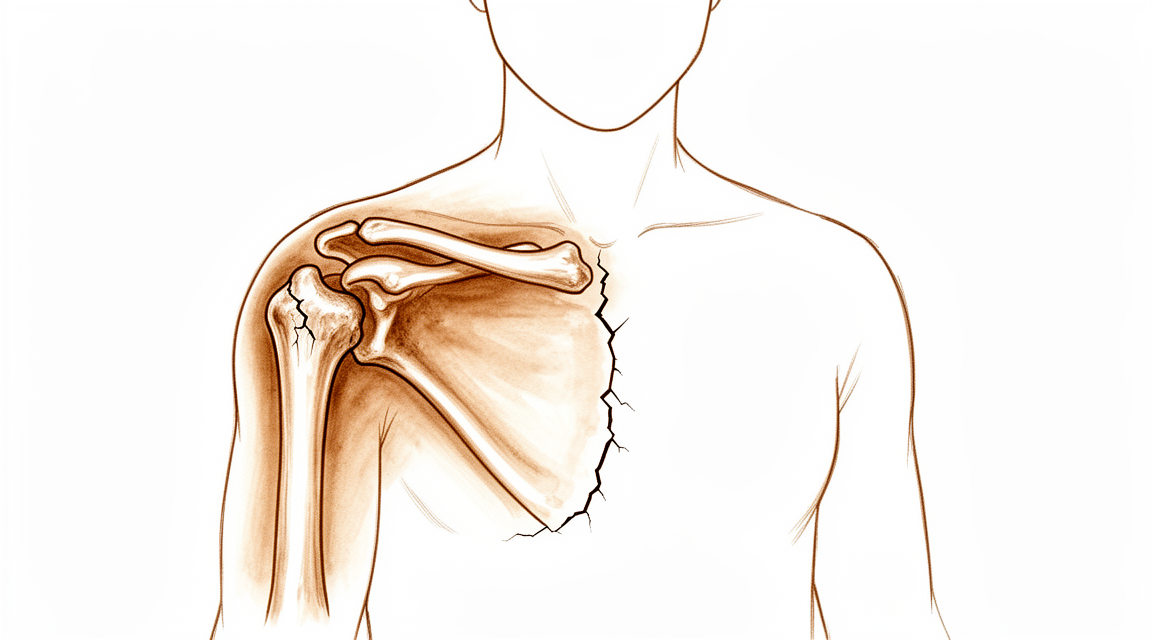
您的感受
肱骨近端骨折是指上臂骨顶部、紧邻肩部下方的骨折。它通常发生在跌倒时,常常是肩部着地或伸出的手臂撑地。骨折是突然发生的,而不是随时间慢慢形成的。
您首先感到的是上臂顶部和肩部的疼痛。疼痛常常在您跌倒的那一刻就开始了。在接下来的一两天里,肩部可能会肿胀并出现瘀青,瘀青可能会向下蔓延到手臂。如果骨折块发生了移位,您的手臂可能看起来变形。您可能不想活动手臂,简单的事情也会变得困难:穿衣服、伸手到橱柜里拿东西、提购物袋,或在床上翻身侧卧到患侧。
在最初几天,疼痛往往在夜间更严重,可能很难找到舒适的姿势。起初活动手臂会痛。在最初几周里,随着骨骼开始愈合,疼痛通常会逐渐缓解。到疼痛减轻时,肩部常常感到僵硬,因为它一直没有活动。
大多数此类骨折发生在 70 岁以上的女性中,并且常见于随着年龄增长而变薄、变脆的骨骼(这被称为骨质疏松症)。如果您以前骨折过,或者发生了这次骨折,可能提示您将来再次骨折的几率更高。这一点值得向您的全科医生提出,他们可以检查您的骨骼健康状况。
有些骨折碎成数块,有些则发生移位。您的外科医生会通过体格检查和X光片来判断骨折类型,有时还会进行CT扫描,这是一种能更清楚显示骨骼的精细扫描。大多数此类骨折无需手术即可愈合,以这种方式治疗的病例中,超过90%的骨骼能够连接。
请留意几种需要当天就医的征兆。如果您的手或手指变得苍白、冰冷、发白或发青,或在受伤后手臂突然失去感觉或活动能力,请前往急诊科。如果骨折处的皮肤破损,或有骨头外露,也同样如此。
实际发生了什么
您上臂骨的顶端形状像一个球,位于肩胛骨上的一个浅窝中。这个窝很小,大约只有球的三分之一大,因此关节可以自由活动,但要依靠软组织来保持稳定。四条肌腱包绕着这个球,在较大的肌肉抬起手臂时将球向下压住。骨骼断裂时,这种协同配合就被打乱了。
骨折通常是因为跌倒的力量把上臂骨撞向关节窝,而关节窝比它周围的骨骼更坚硬。上臂骨就像锤子砸在铁砧上一样,在骨骼最薄弱的地方断裂。随后,骨折块会被附着在其上的肌腱牵拉。每条肌腱把它所附着的骨块拉向各自的方向,这就是为什么有些骨折最终会成为数块移位的骨块。
即使骨折块仍靠近原来的位置,肩部也会在一段时间内无法正常工作。关节周围的肌腱需要光滑的滑动面才能发挥作用,而肿胀和断骨会扰乱这一点。让肌腱在骨面上滑动的衬垫囊可能会增厚并形成瘢痕,这是肩部变僵硬的原因之一。一旦外科医生认为安全,尽早活动手臂有助于减少这种瘢痕形成。
骨骼通过重新连接而愈合,就像身体其他任何部位的骨折一样。新骨在骨折处形成,并在数周到数月内逐渐变得坚固。如果某条肌腱连同一块骨头一起被拉脱,那么随着这块骨头重新连接到原位,肌腱也会随之愈合。即使愈合后略有歪斜,肩部通常也能很好地耐受,因为它不是负重关节,而且周围的肌肉承担了大部分工作。
有几种骨折类型会改变情况。正好位于球底部的骨折可能会切断球的血液供应,从而影响愈合。位置较低的骨折通常不会影响血液供应。如果骨折块严重移位或不稳定,或者球本身碎裂,则更有可能考虑手术。
我们如何处理
Mater Private Hospital Rockhampton 的上肢外科医生 Kieran Hirpara 医生会根据您的具体伤情制定治疗方案。有些骨折无需手术即可愈合,另一些则需要在受伤后尽快手术,因此及时评估很重要。我们会观察骨头碎成了几块、骨折块移位了多远、骨折是否稳定,以及您的手臂需要完成哪些活动。您自身的健康状况,以及您愿意忍受多大程度的疼痛,也是这一决定的考虑因素。
许多此类骨折采用非手术治疗,包括一些骨折块已经移位的骨折。非手术治疗是指在骨骼愈合期间用吊带固定手臂,而不是使用夹板或石膏。这并不是对您置之不理:我们会定期为您复诊,并重复拍摄X光片检查骨折,以确保它在可接受的位置上愈合。一旦外科医生认为骨骼已经准备好,物理治疗师会分阶段指导您恢复活动,先进行温和的辅助活动,然后随着骨骼变得坚固再进行主动活动。以这种方式治疗的病例中,十例中有超过九例骨骼能够连接,达到牢固愈合的平均时间为 14 周。骨骼愈合缓慢或不愈合的几率为 7%,以这种方式治疗的人中约有 5.6% 最终仍需要手术。
对于某些损伤,从一开始就推荐手术:严重移位且不稳定的骨折、关节的球碎裂的骨折、关节同时脱出的骨折脱位,以及手臂需要承担更多活动的较年轻或较活跃者的损伤。手术的目的是在骨折块愈合期间将其固定在更好的位置;对于年长患者的某些严重骨折类型,则是置换关节。在某些情况下,这确实需要共同做出选择:非手术治疗也许可行,但治疗过程中的疼痛或肩部最终的位置可能是您无法接受的。在做出任何决定之前,我们会与您详细讨论这两种方式。
无论您选择哪种方式,最初几周的情况都相似。早期控制疼痛很重要,我们会帮助您找到合适的姿势和简单的方法,让您能够睡觉并应付日常事务。在骨骼愈合期间,您要保护手臂,避免提举东西和突然的动作。物理治疗会在适合您伤情的阶段开始,由您的物理治疗师指导,这是治疗中您最能掌控的部分。
患者通常由全科医生(GP)转诊至我们的诊所;如果物理治疗师建议您就诊,您仍需获得全科医生的转诊,才有资格享受 Medicare 报销。
预期情况
骨骼愈合得很慢。对于大多数未接受手术治疗的人,达到牢固愈合的平均时间为 14 周,十例中有超过九例骨骼能够连接。在最初几周,疼痛会逐渐缓解,不过到那时肩部常常感到僵硬。在随后的几个月里,物理治疗师会分阶段指导您恢复活动,随着骨骼变得坚固,穿衣和提轻物等日常活动也会逐渐恢复。
如果您接受手术,最初几周的情况相似:在愈合期间保护手臂,然后在物理治疗师的指导下分阶段恢复活动。肩部可能需要数月才能再次感到稳定,在此过程中常会出现僵硬。手术能在骨折块愈合期间将其固定在更好的位置,但手术本身也有风险,包括日后需要再次手术的几率更高。对于年长患者的某些严重骨折类型,关节置换旨在获得更好的长期效果,不过置换术后的恢复本身也是一个过程。
有几件事值得坦诚告知。骨骼有可能愈合缓慢或不愈合,一些未接受手术治疗的人最终仍需要手术。愈合后略有歪斜的肩部通常仍能良好工作,因为该关节不负重,而且周围的肌肉承担了大部分工作。无论选择哪种方式,僵硬都是最常见的问题,这就是为什么活动锻炼如此重要。
这种损伤的恢复通常以月而不是以周来计算。大多数人能够恢复舒适地日常使用手臂,但有些人会留有持续的僵硬或酸痛。如果您的症状没有缓解、在数周内逐渐加重,或使您无法使用这只手臂,请去看您的全科医生或要求专科评估。
何时就医
如果受伤后您的手臂明显变形、骨折处的皮肤破损、有骨头外露,或者您的手或手指变得苍白、冰冷、发白或发青,请立即寻求紧急救治。受伤后手臂突然失去感觉或活动能力,也需要在当天前往急诊科。如果您联系不上诊所,请前往离您最近的急诊科。
如果疼痛没有缓解,或者随着骨骼愈合,肿胀、活动或手臂的使用没有逐周改善,请去看您的全科医生或要求专科评估。症状在数周内逐渐加重、在夜间把您弄醒,或使您无法工作或无法使用这只手臂,也需要复查。
深入探讨
Advanced reading: the deeper science (optional)
本节内容超出了您自身治疗决策所需的范围。晚年发生的肩关节骨折值得额外阅读,因为它是骨科领域中最清晰的案例之一,其中循证证据与日常临床实践并不一致,且听起来更严重的治疗方案并非能带来更好手臂功能的治疗方案。
对于大多数老年患者,手术并不能改善预后
这一比较已反复进行。一项系统综述汇总了 1,743 例患者,建议对平均年龄超过 65 岁且伴有移位性肱骨近端骨折的普通患者采取非手术治疗,指出观察性研究的汇总效应与随机对照试验的结果一致 [1]。此前一项针对 486 例患者的综述已发现,手术管理与保守管理在骨折结局方面无可证实的差异 [2]。
这有悖直觉,因为移位性肱骨近端骨折的 X 光片看起来令人担忧。骨骼呈多块碎裂且明显错位。患者和临床医生往往本能地认为,如此明显的错误必须予以纠正。
试验结果表明,肩关节对不完美骨性对位具有异常的宽容性。它不是负重关节,周围的肩袖和三角肌承担了大部分功能,且骨折块由软组织包裹,从而维持了其血供。即使肩关节愈合时略有歪斜,其活动和感觉往往与经过固定者非常相似,且无需经历切口、植入物或下述风险。
该试验改变了证据,而非临床实践
这是值得深思的部分。PROFHER 是一项大型随机对照试验,针对移位性肱骨近端骨折,比较了手术治疗与非手术治疗,结果显示手术并无获益 [6]。
随后,一项针对 116,868 名患者的研究探讨了该试验发表后临床实践是否实际发生了改变。结果并未改变:PROFHER 并未显著影响手术治疗率,每年手术治疗率无显著变化 [3]。
这是一个关于医学而非关于您肩部的发现,患者了解这一情况是合理的。如果您因该骨折被建议手术,正确的问题并非手术是否永远适用(有时手术显然适用),而是关于 您 的骨折和 您 的手臂,具体有哪些因素使您不属于那些已证明手术无益的人群。
当选择手术时,手术方式已发生转变
上述任何情况并不意味着手术永远不适用。劈裂型骨折、骨折脱位、开放性损伤以及需求较高的年轻患者,均属于不同的临床情境。
在老年患者中实施手术时,该领域已明确转向反向全肩关节置换术。一项纳入 228,523 例患者的荟萃分析显示,对于患有此类骨折的老年患者,反向全肩关节置换术在功能预后和并发症发生率方面优于半肩关节置换术,且在翻修手术方面比钢板固定术更具优势 [4]。
其逻辑在于,反向置换术不依赖于大结节和小结节在良好位置上的愈合,而结节愈合恰恰是骨质疏松性肩关节中不可靠的因素。它消除了使传统手术结果不可预测的变量。
真正预测您康复情况的因素
主要并非骨折模式。一项系统综述考察了4,323名患者,研究影响康复的生物-心理-社会预测因子,发现术前功能状态——即受伤前手臂及患者整体的功能表现——能够预测功能康复结果 [5]。
这一点值得正确理解。这并不意味着康复取决于态度。其含义是,决定最终结局的最强单一预测因子是起始状态,这既支持设定切合实际的预期,也强调在肩部感觉僵硬、进展看似不可见的数月里,必须认真对待康复治疗。
参考文献
[1] Beks RB, Ochen Y, Frima H, Smeeing DP, van der Meijden O, Timmers TK, et al. 肱骨近端骨折的手术治疗与非手术治疗:系统综述、荟萃分析及观察性研究与随机对照试验的比较. J Shoulder Elbow Surg. 2018;27(8):1526-34. https://doi.org/10.1016/j.jse.2018.03.009
[2] Nanidis TG, Majed A, Liddle AD, Constantinides VA, Sivagnanam P, Tekkis PP, et al. 复杂肱骨近端骨折的保守治疗与手术治疗:荟萃分析. Shoulder Elbow. 2010;2(3):166-74. https://doi.org/10.1111/j.1758-5740.2010.00075.x
[3] Cheesman JS, Englert CH, Yang Q, Yoo JU, Nazir OF, Mirarchi AJ. PROFHER对美国肱骨近端骨折治疗趋势的影响. Shoulder Elbow. 2025;18(3):476-84. https://doi.org/10.1177/17585732251359178
[4] Mekhail J, Mullan R, Cross JL, Jahagirdar O, Luo X, Salameh M. 反式全肩关节置换术与其他手术固定方法治疗肱骨近端骨折的结局:系统综述和荟萃分析. JSES Rev Rep Tech. 2026;6(2):100644. https://doi.org/10.1016/j.xrrt.2025.100644
[5] Varahra A, MacDermid JC, Szekeres M. 肱骨近端骨折后恢复的生物心理社会预后因素的系统综述. J Hand Ther. 2023;36(4):825-44. https://doi.org/10.1016/j.jht.2023.06.005
[6] Rangan A, Handoll H, Brealey S, Jefferson L, Keding A, Martin BC, et al. 成人移位性肱骨近端骨折的手术治疗与非手术治疗:PROFHER随机临床试验. JAMA. 2015;313(10):1037-47. https://doi.org/10.1001/jama.2015.1629
Evidence & references
This is the clinical evidence summary written for health professionals. It is technical, and it lists the research this page was built from. You do not need to read it to understand your treatment or to make a decision about it.
Overview
Non-Operative Management
- Non-operative management is associated with good outcomes in the majority of proximal humerus fractures in adults [1].
- Patients with proximal humerus fractures that would have normally necessitated surgical treatment showed favorable outcomes following nonsurgical treatment [2].
- Most proximal humeral fractures in elderly patients can be treated nonoperatively with good functional outcomes [10].
- Over the past decade, most older adults who sustain proximal humerus fractures continue to receive nonoperative treatment [6].
- Most one-part proximal humerus fractures are amenable to non-operative treatment with positive outcomes reported in the vast majority of cases [16].
- Most pediatric patients with proximal humerus fractures have favorable results, and complications are infrequent [17].
Operative Management
- Percutaneous treatment of selected proximal humeral fractures results in predictable union and good clinical results with a low rate of complications [32].
- Nail fixation for proximal humerus fractures is a very good technique if the indication is well defined [47].
- The selection of reverse total shoulder arthroplasty over other surgical options is a current, reasonable, and safe option to treat proximal humerus fractures, particularly in those with higher Neer grades and/or in older patients [55].
- Hemiarthroplasty and reverse prosthesis are indicated for complex proximal humerus fractures in patients no younger than 70 years of age [8].
- Patients with a proximal humerus fracture undergoing reverse total shoulder arthroplasty have significantly worse perioperative outcomes, including higher rates of complications, longer hospital stays, and higher costs, compared to patients with other indications [156].
Treatment Considerations and Evidence Gaps
- Treatment algorithms and outcomes following proximal humerus fractures in patients less than or equal to 60 years of age are distinctly different from that of a more elderly population [4].
- Both age and gender have an association with the definitive treatment patients received for proximal humerus fractures over the last decade [5].
- The available literature does not demonstrate a clear clinical benefit of operative treatment over nonoperative management of proximal humeral fractures in adult patients younger than 65 years [21].
- Evidence-based recommendations to guide treatment of proximal humerus fractures are lacking, and no good evidence exists whether surgery is clearly superior to nonoperative treatment [34].
- No single fixation method is a panacea for proximal humeral fractures; choice of implant and method should be selected according to individual patient and fracture pattern characteristics based on clearly defined indications and contraindications [51].
- Prospective clinical trials with longer-term follow-up are required for definitive assessment of the ideal fixation construct for surgical management of two-part proximal humerus fractures [24].
- Besides age, most randomized controlled trials on surgical management of proximal humerus fractures do not include patient-specific variables within their inclusion and exclusion criteria [23].
- There are conflicting opinions on what outcome measure is best to assess function following the treatment of proximal humerus fractures [11].
- The development of an evidence-based clinical protocol for the treatment of proximal humerus fractures is long overdue, requiring a thoughtful, all-inclusive, randomized multicenter trial to determine the best treatment options [60].
- Guidelines and treatment algorithms for native humerus fractures may not be generalizable for those of pathologic origin [70].
Outcomes and Complications
- Mortality at 1 year for fragility proximal humerus fractures is universally high regardless of risk factors [18].
- After surgical treatment, patients with pathologic humerus fractures had significantly higher complication rates compared with native humerus fractures [70].
Anatomy & Pathophysiology
Bony Anatomy
- The proximal humerus comprises four main parts: the humeral head, greater tuberosity (GT), lesser tuberosity (LT), and humeral shaft [76].
- The articular head of the proximal humerus is spherical with a diameter of 37 to 57 mm [76].
- The most superior portion of the articular surface of the humeral head averages 8 mm above the greater tuberosity [76].
- Humeral version averages 29.8 degrees, with a range of 10 to 55 degrees [76].
- The humeral head is inclined approximately 130 degrees with respect to the humeral shaft [76].
- The anatomic neck is located at the junction of the articular surface and the tuberosities [76].
- The surgical neck represents an indistinct region, or metadiaphyseal junction, below the tuberosities but above the humeral shaft [76].
- The greater tuberosity is located in a posterior-superior location with respect to the humeral shaft [76].
- The lesser tuberosity is located on the anterior aspect of the proximal humerus [76].
- The bicipital groove lies between the greater and lesser tuberosities and serves as a pathway for the long head of the biceps [76].
- The distal aspect of the bicipital groove is internally rotated with respect to the proximal portion [76].
- The glenoid is a convex structure of shallow depth shaped like an inverted pear [76].
- The acromion, coracoacromial ligament, and coracoid process form the coracoacromial arch, a rigid bony-ligamentous structure that imparts stability to the shoulder girdle [76].
- The humeral head averages 19° of retroversion and 41° of inclination (neck-shaft angle) [79].
- The proximal humerus has three centers of ossification: the humeral head (4 to 6 months), the greater tuberosity (1 to 3 years), and the lesser tuberosity (3 to 5 years) [79].
- The proximal humeral ossification centers fuse to the shaft at age 17 to 20 years [79].
- The humeral head is retroverted 30 degrees relative to the transepicondylar axis of the humerus [90].
- Head height is approximately 5.6 cm above the superior border of the pectoralis major tendon [90].
- The anatomic neck is directly below the humeral head and serves as an attachment for the shoulder capsule [90].
- The surgical neck is more distal than the anatomic neck and is more often involved in fractures [90].
- The transverse humeral ligament is an important stabilizer of the biceps tendon [90].
- The articular surface of the humeral head is essentially spherical, with an arc of approximately 160 degrees covered by articular cartilage [88].
- The radius of curvature of the humeral head is approximately 25 mm and is slightly larger in men than in women [88].
- The average neck-shaft angle is 45 degrees (±5 degrees), with a range of 30 to 50 degrees [88].
- The superior margin of the humeral head articular surface is normally superior to the top of the greater tuberosity by 8 to 10 mm [88].
- The distance from the lateral base of the coracoid process to the lateral margin of the greater tuberosity is called the lateral humeral offset [88].
- Proximal humeral retroversion is highly variable, ranging from 0 to 55 degrees depending on the method used for measurement [88].
- The glenoid cavity is a shallow socket, approximately one third the size of the humeral head [77].
- The neck-shaft angle measures an average of 135 degrees [77].
- The humeral head is retroverted an average of 30 degrees [77].
- The proximal humeral physis closes by 14 to 17 years of age in girls and by 16 to 18 years in boys [83].
- Humeral retroversion averages 65 degrees in infants and young children and gradually decreases, approaching adult values by 11 years of age [83].
- Eighty percent of subsequent humeral growth comes from the proximal humeral physis, accounting for approximately 40% of the growth of the entire upper extremity [83].
- The periosteum is thicker and stronger in the posteromedial portion of the proximal humerus compared to the anterolateral portion, which is often quite thin [83].
- The proximal humeral physis is irregularly shaped, with its apex located on the posteromedial portion of the proximal humerus [83].
Soft Tissue & Vascular Anatomy
- The greater tuberosity serves as the attachment site for the supraspinatus, infraspinatus, and teres minor tendons of the rotator cuff [76].
- The lesser tuberosity serves as the attachment site for the subscapularis tendon [76].
- The rotator cuff consists of four muscles: the subscapularis, supraspinatus, infraspinatus, and teres minor [77].
- The teres major is not a rotator cuff muscle [77].
- The infraspinatus and teres minor are external rotators, while the subscapularis is an internal rotator of the humerus [77].
- The deltoid and pectoralis major muscles, along with the rotator cuff, cause predictable displacement of fractures around the proximal humerus [77].
- The subscapularis originates from the anterior scapula and inserts anteriorly onto the lesser tuberosity [83].
- The greater tuberosity provides attachment superiorly and posteriorly for the supraspinatus, infraspinatus, and teres minor [83].
- The deltoid courses from the clavicle and acromion superiorly into a common tendinous insertion onto the lateral upper third of the humeral shaft [83].
- The pectoralis major inserts anteriorly onto the lateral wall of the bicipital groove and forms the roof of the distal continuation of the bicipital tunnel [83].
- The proximal humerus receives its blood supply from the anterior and posterior humeral circumflex branches from the third division of the axillary artery [76].
- The posterior humeral circumflex artery travels with the axillary nerve, enters the quadrilateral space posteriorly, and anastomoses with a branch of the anterior circumflex to supply the posterior cuff [76].
- The anterior humeral circumflex artery arises from the axillary artery at the inferior border of the subscapularis [76].
- The anterior humeral circumflex artery provides vascular inflow to the humeral head by way of its terminal anterolateral branch known as the artery of Laing (also known as the arcuate artery) [76].
- The ascending branch of the anterior humeral circumflex artery courses parallel to the lateral aspect of the long head biceps tendon and enters the humeral head at the interface of the bicipital groove and greater tuberosity [76].
- The major blood supply to the humeral head is through the ascending branch of the anterior humeral circumflex artery, which penetrates the head at the bicipital groove and becomes the arcuate artery [77].
- The anterolateral ascending branch of the anterior humeral circumflex artery provides the primary blood supply to the humeral head [79].
- The terminal intraosseous portion of the anterior humeral circumflex artery enters at the proximal aspect of the intertubercular groove as the arcuate artery [79].
- Quantitative assessment has shown that 64% of the humeral head blood supply arises from the posterior humeral circumflex artery [83].
- The brachial plexus and axillary artery lie anterior to the coracoid process of the scapula and humeral head [77].
- The axillary nerve circles the humeral neck just inferior to the glenohumeral joint as it courses posteriorly [83].
- The axillary nerve is a terminal branch coming off the posterior cord of the brachial plexus just proximal to the coracoid process [82].
- The axillary nerve passes beneath the conjoined tendon anterior to the subscapularis 3 to 5 mm medial to the musculotendinous junction and then adjacent to the inferior capsule before entering the quadrilateral space posteriorly [82].
- The axillary nerve splits into the anterior and posterior branches within the quadrangular space [82].
- The anterior and middle deltoid muscle receives sole innervation from the anterior branch of the axillary nerve [82].
- Posterior deltoid muscle innervation varies, with supply only from the anterior branch in 2.3% of cases, from the posterior branch in 8.5%, and from both branches in 89.1% [82].
- The posterior branch of the axillary nerve branches to supply the teres minor muscle and then terminates as the superior lateral brachial cutaneous nerve [82].
- The subacromial bursa separates the rotator cuff tendons from the coracoacromial arch, allowing them to glide [84].
- The subscapular bursa lies between the subscapularis tendon and the neck of the scapula and communicates with the joint cavity between the superior and middle glenohumeral ligaments [80].
- The subscapular bursa often houses loose bodies in the shoulder and is a region in which synovitis of the shoulder may be most intense [80].
- The rotator interval is defined medially by the base of the coracoid, superiorly by the supraspinatus tendon, and inferiorly by the subscapularis tendon [79].
- The rotator interval contains the coracohumeral ligament, the superior glenohumeral ligament, and the intra-articular portion of the long head of the biceps tendon [79].
- The tendons of the infraspinatus and supraspinatus muscles join approximately 15 mm proximal to their insertion and cannot be readily separated by blunt dissection [89].
- The infraspinatus and teres minor fuse near their musculotendinous junctions [89].
- The supraspinatus and subscapularis tendons join as a sheath that surrounds the biceps tendon at the entrance of the bicipital groove [89].
- The roof of the biceps sheath consists of a portion of the supraspinatus tendon, and a sheet of the subscapularis tendon forms the floor [89].
- The coracohumeral ligament is a thick band of fibrous tissue extending from the coracoid process along the surface of the capsule to the tuberosities between the supraspinatus and subscapularis tendons [89].
Pathophysiology & Biomechanics
- Stability and function of the glenohumeral joint are provided by the interaction of structures that promote a near global range of motion and purposeful function [76].
- External loads transferred to the shoulder girdle are initially offset by joint surface anatomy, joint volume, atmospheric pressure, and joint fluid cohesion and adhesion [76].
- Moderate and large loads are counterbalanced by the deltoid and rotator cuff and by the capsulolabral and bone structures, respectively [76].
- Proximal humerus fractures alter complex interactions of the shoulder girdle, resulting in pain, decreased range of motion and stiffness, and disability [76].
- Following a fracture of the proximal humerus, displacement of each "part" occurs in a predictable manner based on the deforming forces created by the tendinous insertions of the pectoralis major, subscapularis, supraspinatus, and infraspinatus [76].
- The subscapularis inserts on the lesser tuberosity and causes medial displacement [76].
- The supraspinatus and infraspinatus insert on the greater tuberosity and cause superior and posterior displacement [76].
- The pectoralis major inserts on the humeral shaft and displaces it medially [76].
- A fracture involving the anatomic neck is prognostically worse than fractures involving other regions of the proximal humerus with respect to the potential disruption of the vascular supply to the humeral head and the subsequent development of avascular necrosis [76].
- Injury to the arcuate artery may result in osteonecrosis of the humeral head [76].
- Additional extraosseous collateral branches can permit humeral head perfusion despite complete ligation of the arcuate artery [76].
- Fractures of the anatomic neck have a poor prognosis because of complete disruption of the blood supply to the head [77].
- Surgical neck fractures are common, and with these, the blood supply to the head is preserved [77].
- Displaced proximal humerus fractures can impede normal movement of structures passing underneath the coracoacromial arch, causing impingement and disruption of normal glenohumeral motion [76].
- In displaced and nondisplaced proximal humerus fractures, the subdeltoid and subacromial bursae can become thickened and fibrotic, forming adhesions that can limit normal glenohumeral motion [76].
- Early range of motion exercises after a fracture have been hypothesized to decrease the formation of such adhesions [76].
- The glenoid bone is generally harder and denser than the proximal humerus and acts as an 'anvil' on which the proximal humerus is impacted during injury [64].
- The combination of the direction of the blow to the humerus, quality of bone in the proximal humerus, and the pull of the soft tissues produces various types of fracture patterns [64].
- Medical comorbidities increase the risk for fracture and type of fracture sustained [64].
- Proximal humerus fractures are seen in a greater frequency in patients with a depleted neuromuscular response [64].
- The close anatomical relationship between the proximal humerus, axillary artery, and brachial plexus predisposes these structures to combined injury patterns that can threaten limb viability [43].
- The incidence of neurovascular injury is increased in fracture-dislocations [27].
- The axillary nerve is most commonly injured in proximal humerus fractures [27].
- An axillary nerve injury from proximal humeral fracture or fracture-dislocation would result in paralysis of the deltoid muscle and anesthesia over the “badge” region at the lateral proximal arm [77].
- The brachial plexus is prone to injury when the proximal humerus is injured in fractures or dislocations, or during traction [83].
- Adhesions in the humeroscapular motion interface can limit shoulder mobility, with examples including scarring after proximal humerus fracture trauma [82].
- The humeroscapular motion interface lies between the inner structures of the proximal humerus, rotator cuff, coracohumeral ligament, and biceps tendon sheath and the superficial layer of the acromion, deltoid, coracoacromial ligament, coracoid process, and conjoined tendon [82].
- Smooth, unrestricted motion at the humeroscapular motion interface is vital to shoulder mobility [82].
- The axillary nerve has an intimate relationship within the humeroscapular motion interface [82].
- Positioning the arm in abduction and internal rotation may help mitigate deforming muscular forces in proximal humerus fractures [132].
- Bone quality significantly impacts implant anchorage in osteosynthesis of proximal humerus fractures [144].
- Biomechanical changes associated with inferior tuberosity positioning after hemiarthroplasty may explain diminished function in patients with proximal humerus fractures [117].
- Anatomic reconstruction of tuberosity fragments produced results indistinguishable from normal shoulder controls in biomechanical studies [143].
- The double plate strategy can increase the stability of the medial column of the proximal humerus and enhance the overall biomechanical property of the repaired proximal humerus [153].
- Varus-displacing forces to the humeral head were superiorly reduced in settings utilizing a specific proximal anchoring point for intramedullary nailing [149].
- The Humerusblock NG allows for angular stable dynamic fixation of two-part proximal humeral fractures [161].
- Proper technique for internal fixation of the proximal humerus requires an understanding of osseous and neurovascular anatomy, with biomechanical studies showing that locked plating provides stable fixation [166].
- The biphasic plate concept is aimed at improving the biomechanics of locked plating [174].
- A biomechanically efficient nail without increased neurological risks can improve the pullout strength of the screws to provide more secure fixation of proximal humeral fractures [165].
Classification
Classification Systems and Reliability
- The Neer classification categorizes displaced proximal humerus fractures from two to four parts according to anatomic segments [169].
- In the Neer classification, displacement is defined as separation of a fragment greater than 1 cm or angulation of a fragment greater than 45° [169].
- Fracture lines in nondisplaced segments are not included in the Neer classification [169].
- The AO classification is based on the vascular supply of the articular segments [169].
- The AO classification is divided into three categories (A, B, C) of increasing severity, with each category further split into numerical subgroupings [169].
- Evaluation of classification systems for fractures of the proximal humerus using plain radiographs has yielded low interobserver reliability [41].
- The Mayo-FJD classification system for proximal humerus fractures allows high intraobserver and interobserver agreement using both radiographs and computed tomography [129].
- Reported mean kappa values for interobserver agreement on the AO classification have varied between 0.26 and 0.53 [173].
- Mean kappa values for interobserver agreement on the AO classification decreased from 0.53 for AO Types to 0.2 for AO Groups [173].
- Morphologic classification of proximal humerus fractures as the sole basis for treatment algorithms and surgical success should be scrutinized [59].
- Current diagnosis coding practices do not adequately capture the fracture complexity needed to conduct subgroup analysis for proximal humerus fractures [163].
Epidemiology and Demographics
- Proximal humerus fractures are osteoporotic injuries with increasing incidence due to aging populations [12].
- In a cohort of 688 patients, 49.2% of fractures were grouped into AO-OTA type A, 43.1% into type B, and 7.7% into type C [177].
- In a cohort of 688 patients, 24.3% of proximal humeral fractures were non-displaced or minimally displaced according to the Neer classification [177].
- In a cohort of 688 patients, 32.3% of proximal humeral fractures were two-part displaced according to the Neer classification [177].
- In a cohort of 688 patients, 30.9% of proximal humeral fractures were three-part according to the Neer classification [177].
- In a cohort of 688 patients, 6.9% of proximal humeral fractures were four-part according to the Neer classification [177].
- In a cohort of 688 patients, 5.6% of proximal humeral fractures were associated with a glenohumeral dislocation according to the Neer classification [177].
- There was no statistically significant association between gender and the AO-OTA classification or the Neer classification [177].
- There was no statistically significant association between the type of trauma and the AO-OTA classification or the Neer classification [177].
- There was no statistically significant association between the diagnosis of osteoporosis and the AO-OTA classification or the Neer classification [177].
- There was a statistically significant association between the AO-OTA and Neer classifications and age grouped by decades [177].
- There was a statistically significant association between the AO-OTA and Neer classifications and the type of treatment performed [177].
Clinical Evaluation and Imaging
- Accurate clinical evaluation, imaging, and classification are paramount for informed treatment decisions regarding proximal humerus fractures [12].
- The use of artificial intelligence can accurately detect and classify proximal humerus fractures on plain shoulder AP radiographs [69].
- The type of proximal humerus fracture depends on the amount and direction of applied force, the quality of bone, and the position of the shoulder at the time of injury [181].
- Neer defined significant displacement as greater than 1 cm of translation, or angulation greater than 45° for any of the major fracture fragments [181].
- The AO/ASIF group labeled valgus impaction injuries as Type C (C2.1, C2.2) fractures of the proximal humerus in their classification system [181].
Clinical Presentation
History and Mechanism
- The fracture results most commonly from an indirect mechanism, such as a fall on the outstretched upper extremity [27].
- Other indirect mechanisms, such as seizures or electrical shocks, are uncommon [27].
- Direct mechanisms from a blow to the shoulder occur much less frequently than the indirect mechanism [27].
- When obtaining a history from a patient with a suspected proximal humerus fracture, it is important to record the hand dominance, occupation, and mechanism of injury [27].
Physical Examination
- Gross deformity is not often appreciated because of the soft tissues surrounding the proximal humerus [27].
- Swelling and tenderness to palpation are typically present in patients with proximal humerus fractures [27].
- Ecchymosis extending distally along the extremity and along the chest wall is often present a few days following injury [27].
- Assessment of fracture stability is an important part of the examination [27].
- With one hand palpating the humeral head, the humeral shaft should be gently internally and externally rotated to assess stability [27].
- If the proximal and distal fragments move as a unit, the fracture is considered stable [27].
- A thorough neurovascular examination is crucial due to the close proximity of the brachial plexus and the axillary artery [27].
- Deltoid motor function and sensation over the lateral aspect of the shoulder should be assessed during the neurovascular examination [27].
Epidemiology and Risk Factors
- Proximal humerus fractures are now typically osteoporotic fractures in women over 70, with prevalence increasing due to an aging population in poor general condition [45].
- The incidence of proximal humerus fracture increased from 104.7/100,000 in 2008 to 124.7/100,000 in 2012 in women [115].
- The incidence of proximal humerus fracture increased from 45.3/100,000 in 2008 to 52.0/100,000 in 2012 in men [115].
- Proximal humerus fracture increased by 40.5% over the 5 year of study from 10,135 in 2008 to 14,238 in 2012 [115].
- The number of individuals 50 years or older in the general population increased 19.5% from 13,103,814 in 2008 to 15,657,674 in 2012 [115].
- The incidence of proximal humerus fracture in women (19.1%) was more increased than that in men (14.8%) from 2008 to 2012 [115].
- Combined fractures with femoral or vertebral fractures are associated with significantly higher mortality and morbidity compared with isolated proximal humerus fractures [130].
Complications and Prognosis
- Complications associated with proximal humerus fractures are varied and can be categorized as occurring at the time of initial injury, during operative management, or as delayed sequelae [13].
- Neurovascular injuries associated with proximal humerus fractures represent a rare yet clinically significant complication with potential for devastating functional outcomes [43].
- The multifactorial etiology of neurovascular injuries encompasses direct trauma from displaced fracture fragments and indirect injury due to the close anatomical relationship between the proximal humerus, axillary artery, and brachial plexus [43].
- Most nerve injuries, particularly involving the axillary nerve, demonstrate favorable outcomes with conservative management [43].
- Vascular injuries associated with proximal humerus fractures demand urgent multidisciplinary intervention to restore perfusion and prevent irreversible ischemia [43].
- Axillary artery injury associated with a proximal humerus fracture is a rare occurrence but can have profound consequences [131].
- There is a substantial mortality in patients with a proximal humerus fracture [14].
- Surviving patients with proximal humerus fractures frequently have persistent symptoms that can be predicted as early as after 1 year [14].
Investigations
Imaging Modalities and Protocols
- At least two X-ray views should be obtained for proximal humerus fractures: an anteroposterior view in the plane of the glenoid and an axillary projection with the arm in abduction [93].
- The recommended radiographic series for proximal humerus fractures is the Neer trauma series, consisting of an AP view, a lateral view in the scapular plane, and a Velpeau modified axillary view [95].
- The combination of the three views in the Neer trauma series allows evaluation of the shoulder joint in three separate perpendicular planes [95].
- The axillary view is important for evaluating the glenoid articular surface and the relationship of the humeral head anteriorly and posteriorly [95].
- Computed tomography (CT) is helpful for planning fracture surgery and shoulder joint replacement [93].
- On occasion, CT scanning may be necessary for detailing bony anatomy in proximal humerus fractures [95].
- CT imaging is frequently used to evaluate fractures of the shoulder [100].
- Magnetic resonance imaging (MRI) is useful to identify osteonecrosis of the humeral head or a bone tumour [93].
- MRI can identify labral tears and rotator cuff tears, although accuracy for these is enhanced by combining the scan with arthrography [93].
- MRI is the modality of choice for evaluating the rotator cuff, biceps, and subacromial/subdeltoid bursa [100].
- T2-weighted MRI provides better visualization of full thickness rotator cuff tears [100].
- Ultrasound is a simple and accurate test for identifying rotator cuff tears and calcific tendinitis [93].
- Ultrasound can be useful in guiding injections or barbotage [93].
- Ultrasonography is a low-cost alternative to MRI and arthrography for evaluating both skeletal and soft-tissue structures of the shoulder [100].
- Ultrasonography can provide immediate, real-time visualization of the rotator cuff, biceps tendon, and calcific deposits [100].
- Ultrasonography is highly operator dependent and is not as useful for evaluating labral tears or rotator cuff tears that are very small or larger than 3 cm [100].
- Zero-echo time (ZTE) MRI presents a viable alternative to CT in the evaluation of proximal humerus fractures [170].
Classification and Diagnostic Accuracy
- Evaluation of the classification systems for fractures of the proximal humerus with plain radiographs has yielded low interobserver reliability [41].
- Computed tomography scan was more specific than radiographs in the assessment of proximal humerus fracture sequelae [44].
- Convolutional neural networks (CNNs) proficiently rule out proximal humerus fractures on plain radiographs [190].
- ChatGPT-5 is highly inaccurate at identifying proximal humerus fractures on shoulder x-rays, characterizing fracture patterns, and providing accurate interpretations [207].
- The routine use of 3D-printed models may not be beneficial for classifying proximal humeral fracture patterns beyond the information gained from currently available imaging modalities [203].
- The use of 3D-printed models as the sole determinant for recommending surgical intervention should be avoided at this time [203].
Clinical Evaluation and Associated Injuries
- Accurate clinical evaluation, imaging, and classification are paramount for informed treatment decisions in proximal humerus fractures [12].
- Rotator cuff injuries can be expected with fractures of the tuberosities but can also result from strictly soft-tissue injuries such as shoulder dislocations [95].
- Evaluation of the integrity of the rotator cuff may be difficult in the acute setting [95].
- Ultrasound, MRI, arthrogram, or arthroscopy may be valuable in making a diagnosis of rotator cuff injury in the acute setting [95].
- Axillary artery injuries generally result from fractures or fracture-dislocations in which a medial bone spike injures or penetrates the axillary artery [95].
- The index of suspicion for axillary artery injury is high if the arm shows significant color differences compared with the uninjured arm [95].
- Pulses should be palpated and evaluated by Doppler studies when axillary artery injury is suspected [95].
- In addition to palpation and anteroposterior and lateral humeral x-ray, bilateral anteroposterior shoulders x-ray is suggested routinely to confirm the shoulder location in children with shoulder dislocation combined with proximal humerus fracture [205].
Treatment
Non-Operative Management
- In the vast majority of cases, proximal humerus fractures may be treated nonoperatively [3].
- A majority of patients with proximal humeral fractures underwent non-operative treatment [22].
- Nonsurgical treatment should have a more prominent role in the treatment of proximal humeral fractures [57].
- Approximately 80% of proximal humerus fractures are minimally displaced low energy injuries and are at low risk for future displacement, nonunion, or avascular necrosis, and have a high union rate with conservative management [65].
- Nonsurgical management of proximal humerus fractures decreased during the study period [110].
- There was no statistical difference in functional outcomes between non-displaced, 2-part and 3-part fractures of the proximal humerus treated non-operatively [114].
- Patients with non-operatively managed proximal humerus fractures continue to improve up to, and beyond 6 months post-injury [114].
- Non-operative treatment (NOT) involves a period of immobilization, such as an arm sling, followed by physiotherapy and exercise [116].
- Non-operative treatment is generally the accepted treatment option for minimally displaced fractures and often used also for people with displaced fractures [116].
- In a randomized controlled trial, there was no significant difference in clinical outcomes at 2 years between surgery and non-operative treatment in patients 60 years of age or older with displaced 2-part fractures of the proximal humerus [29].
- The ProFHER trial showed that conservative management is as effective as surgical intervention for displaced proximal humerus fractures [104].
- Prolonged sling use in conservatively managed displaced proximal humerus fractures can be associated with increased pain, stiffness, risk of falls, delayed return to function, and burden on healthcare systems [104].
- Supervised rehabilitation is comparable to a single advice session after nonoperative treatment of displaced proximal humerus fracture [126].
- Nonoperative treatment is recommended for the average elderly patient (aged > 65 years) with a displaced proximal humeral fracture [178].
- Proximal humerus fractures in children have tremendous potential for remodeling, making non-operative management the treatment of choice for most fractures [138].
Operative Management: Indications and General Principles
- Patients who have sustained an open fracture, vascular injuries, or those that have repairable neurologic injuries, are usually indicated for acute operative intervention [65].
- Operative fixation can provide stability if there is a need for any vascular or nerve repair procedures [65].
- Patients who have preexisting neurologic impairment on the side of injury resulting from a stroke or a traumatic spine injury, or who lead very inactive lifestyles, may not benefit from any acute intervention and can be managed nonoperatively [65].
- Patients who are medically unstable can be treated conservatively or treated in a delayed fashion once they are more physiologically stable [65].
- Consensus when managing proximal humerus fractures is limited to specific scenarios, whereas lack of consensus still exists in others [15].
- Surgical treatment of proximal humerus fractures remains far from straightforward, with unpredictable outcomes where factors associated with poor results include being a woman, four-part fracture dislocation, and absence of metaphyseal head extension [28].
- Delaying surgery for proximal humerus fracture is likely to increase inpatient morbidity, postoperative length of stay and non-routine discharge [167].
- The results of a prospective, randomized controlled trial will provide Level 1 evidence to guide decision-making in the treatment of proximal humerus fractures in the elderly population [103].
- Besides age, most RCTs on surgical management of proximal humerus fractures do not include patient-specific variables within their inclusion and exclusion criteria [23].
Operative Management: Internal Fixation (ORIF)
- The early, published results to date are encouraging in the treatment of three-part proximal humerus fractures [35].
- MIPO is a safe and effective option for the treatment of proximal humerus fractures, with good functional recovery and fewer complications, which are typically technique dependent [133].
- Although the less-invasive surgical procedure is a feasible treatment option in proximal humeral fractures with acceptable complications and considerable improvement during the first six months, a lengthy recovery time is required [140].
- Osteosynthesis of the proximal humerus in osteoporotic bone typically produces inferior results to that in younger subjects with better bone stock [109].
- In a study of the PlantTan Fixator Plate, there were no cases of infection, impingement, avascular necrosis or malunion in the population under 70 years of age [109].
- In a study of the PlantTan Fixator Plate, there was a significant proportion of patients with avascular necrosis and implant migration in the group over 70 years [109].
- Open reduction internal fixation (ORIF) is one of the common operative treatments for displaced and unstable fractures and those with more complex fracture patterns [116].
- In a network meta-analysis, the rank of treatments in terms of high Constant score was: RSA, ORIF, IN, NOT and HA [116].
- In a network meta-analysis, the rank for reduction in total incidence of complications was: RSA, NOT, HA, IN and ORIF [116].
- In a network meta-analysis, the rank for lowering the risk of additional surgery was: RSA, NOT, HA, IN and ORIF [116].
Operative Management: Arthroplasty
- Reverse total shoulder replacement is a promising treatment for geriatrics with three- and four-part proximal humerus fractures aiming for a better long-term functional outcome [26].
- The selection of RTSA over other surgical options is a current, reasonable, and safe option to treat proximal humerus fractures, particularly in those with higher Neer grades and/or in older patients [55].
- While the main potential advantage of the pyrolytic carbon head (PCH)—reduced glenoid erosion—will require further investigation with longer follow-up, this is the first study to demonstrate the safety and short-term outcomes of the PCH in treating proximal humerus fractures [61].
- These findings should be considered when treatment is selected for acute three- and four-part proximal humerus fractures [72].
- In a network meta-analysis, RSA had significantly the highest Constant score and lower total incidence of complications than ORIF, HA and IN [116].
- In a network meta-analysis, RSA resulted in a lower incidence of additional surgery than ORIF and IN [116].
- RSA had the highest probability for improving functional outcome and reduction in the total incidence of complications and requiring additional surgery among the five interventions for treating adults with displaced proximal humeral fracture [116].
- RTSA is an effective treatment option for selected patients with acute proximal humerus fractures [135].
- RTSA has shown to provide reproducible functional outcomes and is a good treatment option for elderly patients with 3-part and 4-part proximal humerus fractures [136].
- A meta-analysis demonstrates no significant differences in clinical outcomes or complication rates between standard components and fracture-specific components in RSA, suggesting comparable performance in the treatment of proximal humerus fractures [134].
Rehabilitation and Post-Treatment Care
- There is substantial variation in the literature regarding the optimal management for PHFs, with studies supporting nonoperative management, ORIF, and arthroplasty [127].
- The inconsistency between studies may in part be explained by heterogeneity in rehabilitation protocols, which are seldom evaluated [127].
- There is some evidence that early intensive mobilization yields similar outcomes compared to later or conventional mobilization after operative treatment (both plate fixation and hemiarthroplasty) and conservative treatment [127].
- It has remained unclear how other aspects of rehabilitation impact outcomes such as sling usage and timing of physical therapy [127].
- Participants allocated to nonsurgical treatment in the ProFHER trial were given a sling for the injured arm for as long as deemed necessary (3 weeks was suggested), followed by active rehabilitation [123].
- Rehabilitation care in the ProFHER trial was provided by physiotherapists in inpatient, outpatient, and community settings [123].
Complications
General Classification and Epidemiology
- Complications associated with proximal humerus fractures are categorized as occurring at the time of initial injury, during operative management, or as delayed sequelae [13].
- Proximal humerus fractures are typically osteoporotic fractures in women over 70, with prevalence increasing due to an aging population in poor general condition [45].
Mortality and Morbidity
- There is substantial mortality in patients with a proximal humerus fracture, and surviving patients frequently have persistent symptoms that can be predicted as early as after 1 year [14].
- The adjusted one-year mortality rate following a proximal humerus fracture was 13.05%, which is significantly higher than other upper extremity fractures but lower than hip fractures [68].
Non-Operative Complications
- The prevalence of nonunion after proximal humeral fracture is higher than previously reported, with most patients having a very low risk but a smaller subgroup at much higher risk [74].
Operative Complications: Osteosynthesis
- Fixation of proximal humerus fractures with proximal humerus locking plates is associated with a high rate of complications and reoperation [151].
- Selective Glenohumeral External Rotation Deficit (SGERD) is a new shoulder evaluation symptom identified as a sequela of post-ORIF deltoid adhesions after treatment of the proximal humerus fracture [71].
- Predictive models constructed using machine learning techniques demonstrated favorable discrimination and satisfactory-to-excellent performance in forecasting prolonged length of stay and serious adverse complications occurring within 30 days of surgical intervention for proximal humerus fracture [66].
- Percutaneous proximal humerus fixation offers less complications compared to other methods [158].
- After one-year, long-term follow-up of fixed proximal humerus fractures may be unnecessary for those without symptoms [48].
Operative Complications: Arthroplasty
- Hemiarthroplasty for the treatment of complex proximal humerus fractures yields variable long-term clinical outcomes and high rates of failure, with the majority due to greater tuberosity malunion or nonunion [63].
- Low arthroplasty survival is observed after treatment for proximal humerus fracture sequelae, as reported by the Nordic Arthroplasty Register Association [7].
- Hemiarthroplasty outcomes for acute proximal humerus fractures and fracture sequelae did not differ significantly, supporting the use of hemiarthroplasty in both settings with modest clinical outcomes [150].
- The main potential advantage of pyrolytic carbon head hemiarthroplasty—reduced glenoid erosion—will require further investigation with longer follow-up [61].
Pathologic Fractures
- After surgical treatment, patients with pathologic humerus fractures had significantly higher complication rates compared with native humerus fractures, suggesting that guidelines and treatment algorithms for native humerus fractures may not be generalizable for those of pathologic origin [70].
Recovery
Non-operative Management
- A multicenter randomized controlled trial found no significant difference in clinical outcomes at 2 years between surgery and non-operative treatment in patients 60 years of age or older with displaced 2-part fractures of the proximal humerus [29].
Operative Management
- Minimally invasive treatment of displaced proximal humeral fractures in patients younger than 70 years using the Humerusblock yields good midterm clinical and radiological results [37].
- The locking plate provides satisfactory functional outcomes after a mid-term follow-up in patients with displaced proximal humerus fractures [50].
- Double-plating of proximal humeral fractures yields good clinical mid- to long-term results in complex and highly unstable fractures [141].
- Timing of surgery did not impact outcomes of patients who underwent ORIF for proximal humerus fractures [206].
- Early operative intervention does not appear to decrease the rate of development of avascular necrosis after proximal humeral fracture [211].
Arthroplasty
- Long-term treatment with reverse shoulder arthroplasty (RSA) for displaced 3- or 4-part proximal humerus fractures provides better functional outcomes compared to nonoperative treatment, a difference attributed to the deterioration of functional outcomes of the nonoperative treatment over time [56].
- Functional outcomes of proximal humerus fractures treated with reverse shoulder arthroplasty improve with surgical experience, and outcomes become less variable after approximately 20 procedures [67].
- Patients in the proximal humerus fracture cohort were less likely to report persistent shoulder pain at all evaluated time points compared to the osteoarthritis cohort [201].
Prognosis and Complications
- Surviving patients with a proximal humerus fracture frequently have persistent symptoms that can be predicted as early as after 1 year [14].
- Post-traumatic osteonecrosis of the proximal humerus is a challenging problem commonly seen following multi-fragmentary fractures, affecting long-term functional recovery [147].
- If patients do not follow the usual course of improvement after a proximal humerus fracture from a superior traction mechanism, consideration should be given to associated superior labral tears that may require surgical intervention [210].
Follow-up and Assessment
Key Evidence
- [L4] Non-operative management is associated with good outcomes in the majority of proximal humerus fractures in adults. [1] (10.5312/wjo.v5.i5.685)
- [L4] Patients with proximal humerus fractures that would have normally necessitated surgical treatment showed favorable outcomes following nonsurgical treatment. [2] (10.1097/bte.0000000000000174)
- [L4] In the vast majority of cases, proximal humerus fractures may be treated nonoperatively. [3] (10.1155/2012/861598)
- [L4] Treatment algorithms and outcomes following proximal humerus fractures in patients less than or equal to 60 years of age are distinctly different from that of a more elderly population. [4] (10.1016/j.xrrt.2023.01.002)
- [L3] Both age and gender have an association with the definitive treatment patients received for proximal humerus fractures over the last decade. [5] (10.1016/j.jseint.2021.11.007)
- [L4] Over the past decade, most older adults who sustain proximal humerus fractures continue to receive nonoperative treatment. [6] (10.1016/j.jseint.2021.08.006)
- [L3] These results are pertinent when deciding on the treatment of proximal humerus fracture sequelae. [7] (10.1080/17453674.2020.1793548)
- [L4] They are indicated for complex proximal humerus fractures in patients no younger than 70 years of age. [8] (10.1016/j.otsr.2008.09.002)
- [L5] Most proximal humeral fractures in elderly patients can be treated nonoperatively with good functional outcomes. [10] (10.2106/jbjs.l.01293)
- [L4] Additionally, there are conflicting opinions on what outcome measure is best to assess function following the treatment of proximal humerus fractures. [11] (10.1007/s00264-017-3569-0)
- [L3] Our results suggest that there is a substantial mortality in patients with a proximal humerus fracture, as we have previously reported, and that surviving patients frequently have persistent symptoms that can be predicted as early as after 1 year. [14] (10.1080/17453670510041295)
- [L5] Consensus when managing proximal humerus fractures is limited to specific scenarios, whereas lack of consensus still exists in others. [15] (10.1016/j.jse.2024.12.005)
- [L5] Most pediatric patients with proximal humerus fractures have favorable results, and complications are infrequent. [17] (10.5435/jaaos-d-14-00033)
- [L3] Mortality at 1 year for fragility proximal humerus fractures is universally high regardless of risk factors. [18] (10.1016/j.jse.2022.03.006)
- [L1] The available literature does not demonstrate a clear clinical benefit of operative treatment over nonoperative management of proximal humeral fractures in adult patients younger than 65 years. [21] (10.1016/j.xrrt.2021.04.014)
- [L3] A majority of patients with proximal humeral fractures underwent non-operative treatment. [22] (10.1186/s12891-019-2812-9)
- [L2] Besides age, most RCTs on surgical management of proximal humerus fractures do not include patient-specific variables within their inclusion and exclusion criteria. [23] (10.1016/j.xrrt.2025.07.023)
- [L3] However, prospective clinical trials with longer-term follow-up are required for definitive assessment of the ideal fixation construct for surgical management of two-part proximal humerus fractures. [24] (10.1016/j.injury.2013.08.024)
- [L3] It is a promising treatment for geriatrics with three- and four-part proximal humerus fractures aiming for a better long-term functional outcome. [26] (10.1186/s12891-023-06669-3)
- [L5] [27] (10.1097/00132589-200212000-00003)
- [L5] Surgical treatment of proximal humerus fractures remains far from straightforward, with unpredictable outcomes where factors associated with poor results include being a woman, four-part fracture dislocation, and absence of metaphyseal head extension. [28] (10.1097/corr.0000000000002242)
- [L1] This trial found no significant difference in clinical outcomes at 2 years between surgery and non-operative treatment in patients 60 years of age or older with displaced 2-part fractures of the proximal humerus. [29] (10.1371/journal.pmed.1002855)
- [L4] Percutaneous treatment of selected proximal humeral fractures results in predictable union and good clinical results with a low rate of complications. [32] (10.1016/j.jse.2006.09.006)
- [L4] Evidence-based recommendations to guide treatment of proximal humerus fractures are lacking, and no good evidence exists whether surgery is clearly superior to nonoperative treatment. [34] (10.1016/j.ocl.2008.06.003)
- [L5] The early, published results to date are encouraging in the treatment of three-part proximal humerus fractures. [35] (10.1097/00132589-200206000-00007)
- [L4] Minimally invasive treatment of displaced proximal humeral fractures in patients younger than 70 years using the Humerusblock yields good midterm clinical and radiological results. [37] (10.1016/j.injury.2015.05.017)
- [L5] Evaluation of the classification systems for fractures of the proximal humerus with plain radiographs has yielded low interobserver reliability. [41] (10.1016/j.ocl.2008.05.002)
- [L5] [43] (10.1016/j.xrrt.2026.100825)
- [L2] Computed tomography scan was more specific than radiographs in the assessment of proximal humerus fracture sequelae. [44] (10.1177/17585732221150785)
- [L2] Proximal humerus fractures are now typically osteoporotic fractures in women over 70, with prevalence increasing due to an aging population in poor general condition. [45] (10.1016/j.otsr.2012.05.013)
- [Paper] Nail fixation for proximal humerus fractures is a very good technique if the indication is well defined. [47] (10.1097/bte.0b013e31817303af)
- [L3] After one-year, long-term follow-up of fixed proximal humerus fractures may be unnecessary for those without symptoms. [48] (10.1007/s00590-021-03099-6)
- [L4] The locking plate provides satisfactory functional outcomes after a mid-term follow-up in patients with displaced proximal humerus fractures. [50] (10.1007/s00590-010-0655-z)
- [L4] No single fixation method is a panacea for proximal humeral fractures; choice of implant and method should be selected according to individual patient and fracture pattern characteristics based on clearly defined indications and contraindications. [51] (10.1016/j.injury.2010.10.016)
- [L5] The selection of RTSA over other surgical options is a current, reasonable, and safe option to treat proximal humerus fractures, particularly in those with higher Neer grades and/or in older patients. [55] (10.1097/corr.0000000000002430)
- [L1] Long-term treatment with RSA for displaced 3- or 4-part proximal humerus fractures provides better functional outcomes compared to nonoperative treatment, a difference attributed to the deterioration of functional outcomes of the nonoperative treatment over time. [56] (10.1016/j.jse.2024.09.032)
- [L3] Nonsurgical treatment should have a more prominent role in the treatment of proximal humeral fractures. [57] (10.1016/j.jse.2011.01.025)
- [L2] Morphologic classification of proximal humerus fractures as the sole basis for treatment algorithms and surgical success should be scrutinized. [59] (10.1016/j.jseint.2022.02.006)
- [L5] The development of an evidence-based clinical protocol for the treatment of proximal humerus fractures is long overdue, requiring a thoughtful, all-inclusive, randomized multicenter trial to determine the best treatment options. [60] (10.1016/j.injury.2014.05.017)
- [L2] While the main potential advantage of the PCH—reduced glenoid erosion—will require further investigation with longer follow-up, this is the first study to demonstrate the safety and short-term outcomes of the PCH in treating proximal humerus fractures. [61] (10.1016/j.jse.2025.07.032)
- [L5] Hemiarthroplasty for the treatment of complex proximal humerus fractures yields variable long-term clinical outcomes and high rates of failure, with the majority due to greater tuberosity malunion or nonunion. [63] (10.1016/j.xrrt.2025.100616)
- [L5] [64] (10.1007/978-3-319-08951-5_2)
- [L4] [65] (10.1007/s12178-012-9130-2)
- [L3] Predictive models constructed using ML techniques demonstrated favorable discrimination and satisfactory-to-excellent performance in forecasting prolonged LOS and serious adverse complications occurring within 30 days of surgical intervention for proximal humerus fracture. [66] (10.1016/j.jseint.2024.02.005)
- [L4] Functional outcomes of proximal humerus fractures treated with reverse shoulder arthroplasty improve with surgical experience, and outcomes become less variable after approximately 20 procedures. [67] (10.1016/j.jseint.2021.07.008)
- [L3] The adjusted one-year mortality rate following a proximal humerus fracture was 13.05%, which is significantly higher than other upper extremity fractures but lower than hip fractures. [68] (10.1016/j.jse.2015.11.031)
- [L4] The use of artificial intelligence can accurately detect and classify proximal humerus fractures on plain shoulder AP radiographs. [69] (10.1080/17453674.2018.1453714)
- [L3] After surgical treatment, patients with pathologic humerus fractures had significantly higher complication rates compared with native humerus fractures, suggesting that guidelines and treatment algorithms for native humerus fractures may not be generalizable for those of pathologic origin. [70] (10.1016/j.jse.2020.10.024)
- [L4] These observations allow the identification of a new shoulder evaluation symptom: Selective Glenohumeral External Rotation Deficit (SGERD). [71] (10.1186/s12891-020-03634-2)
- [L4] These findings should be considered when treatment is selected for acute three- and four-part proximal humerus fractures. [72] (10.1016/s1058-2746(05)80020-5)
- [L3] The prevalence of nonunion after proximal humeral fracture is higher than previously reported, with most patients having a very low risk but a smaller subgroup at much higher risk. [74] (10.2106/jbjs.20.01139)
- [L1] The results of this trial will provide Level 1 evidence to guide decision-making in the treatment of proximal humerus fractures in the elderly population. [103] (10.1186/s12891-018-2223-3)
- [L4] [104] (10.1177/17585732241239011)
- [L4] [109] (10.1016/j.injury.2005.05.030)
- [L4] Nonsurgical management of proximal humerus fractures decreased during the study period. [110] (10.1016/j.jhsa.2020.03.022)
- [L5] [114] (10.1016/j.injury.2004.11.026)
- [L4] [115] (10.11005/jbm.2015.22.1.17)
- [L1] [116] (10.1371/journal.pone.0166801)
- [Abstract] These biomechanical changes may explain diminished function in patients with inferior tuberosity positioning after hemiarthroplasty for proximal humerus fractures. [117] (10.1016/j.jse.2007.02.027)
- [L1] [123] (10.1001/jama.2015.1629)
- [L1] [126] (10.1016/j.jse.2025.11.013)
- [L4] [127] (10.1177/17585732231182374)
- [L4] The Mayo-FJD classification system for proximal humerus fractures seems to allow high intraobserver and interobserver agreement using both radiographs and computed tomography. [129] (10.1016/j.jse.2023.02.035)
- [L3] Combined fractures with femoral or vertebral fractures are associated with significantly higher mortality and morbidity compared with isolated proximal humerus fractures. [130] (10.1016/j.jse.2025.04.013)
- [L5] Axillary artery injury associated with a proximal humerus fracture is a rare occurrence but can have profound consequences. [131] (10.1016/s1058-2746(98)90058-1)
- [L5] These findings suggest that positioning the arm in abduction and internal rotation may help mitigate deforming muscular forces in proximal humerus fractures. [132] (10.5397/cise.2022.00885)
- [L4] MIPO is a safe and effective option for the treatment of proximal humerus fractures, with good functional recovery and fewer complications, which are typically technique dependent. [133] (10.1016/j.aott.2016.10.003)
- [L1] This meta-analysis demonstrates no significant differences in clinical outcomes or complication rates between standard components and fracture-specific components in RSA, suggesting comparable performance in the treatment of proximal humerus fractures. [134] (10.1302/0301-620x.107b9.bjj-2024-1508.r2)
- [Abstract] RTSA is an effective treatment option for selected patients with acute proximal humerus fractures. [135] (10.1016/j.jse.2014.06.021)
- [L4] RTSA has shown to provide reproducible functional outcomes and is a good treatment option for elderly patients with 3-part and 4-part proximal humerus fractures. [136] (10.1097/bot.0000000000000607)
- [L3] Although the less-invasive surgical procedure is a feasible treatment option in proximal humeral fractures with acceptable complications and considerable improvement during the first six months, a lengthy recovery time is required. [140] (10.1186/s12891-015-0618-y)
- [Abstract] Double-plating of proximal humeral fractures yields good clinical mid- to long-term results in complex and highly unstable fractures. [141] (10.1016/j.jse.2022.01.036)
- [L5] In contrast, anatomic reconstruction produced results indistinguishable from normal shoulder controls. [143] (10.1067/mse.2001.113962)
- [L4] The paper reviews the biology and biomechanics of osteosynthesis for proximal humerus fractures, emphasizing that bone quality significantly impacts implant anchorage. [144] (10.1007/s00068-007-7089-2)
- [L4] Post-traumatic osteonecrosis of the proximal humerus is a challenging problem commonly seen following multi-fragmentary fractures, affecting long-term functional recovery. [147] (10.1016/j.injury.2015.06.026)
- [L5] Varus-displacing forces to the humeral head were superiorly reduced in this setting. [149] (10.1007/s00264-017-3498-y)
- [L3] Hemiarthroplasty outcomes for acute proximal humerus fractures and fracture sequelae did not differ significantly, supporting the use of hemiarthroplasty in both settings with modest clinical outcomes. [150] (10.1016/j.jseint.2022.10.009)
- [L4] Fixation of proximal humerus fractures with proximal humerus locking plates is associated with a high rate of complications and reoperation. [151] (10.1016/j.injury.2010.11.058)
- [L5] The double plate strategy can increase the stability of the medial column of the proximal humerus, and enhance the overall biomechanical property of the repaired proximal humerus. [153] (10.1186/s12891-024-08216-0)
- [Abstract] Patients with a proximal humerus fracture undergoing reverse total shoulder arthroplasty have significantly worse perioperative outcomes, including higher rates of complications, longer hospital stays, and higher costs, compared to patients with other indications. [156] (10.1016/j.jse.2015.05.005)
- [L4] This study explains positive experience with percutaneous proximal humerus fixation, suggesting it offers less complications compared to other methods, and encourages continuing the technique with longer term follow-up. [158] (10.1016/j.jse.2021.03.017)
- [L5] The Humerusblock NG allows for angular stable dynamic fixation of two-part proximal humeral fractures. [161] (10.1007/s00402-012-1503-x)
- [L3] Current diagnosis coding practices do not adequately capture the fracture complexity needed to conduct subgroup analysis for proximal humerus fractures. [163] (10.1016/j.jse.2023.08.022)
- [L5] The study introduces a biomechanically efficient nail without increased neurological risks to improve the pullout strength of the screws to provide more secure fixation of proximal humeral fractures. [165] (10.1016/j.clinbiomech.2015.12.005)
- [L5] Proper technique for internal fixation of the proximal humerus requires an understanding of osseous and neurovascular anatomy, with biomechanical studies showing that locked plating provides stable fixation. [166] (10.5435/jaaos-d-20-00558)
- [Abstract] Delaying surgery for proximal humerus fracture is likely to increase inpatient morbidity, postoperative length of stay and non-routine discharge. [167] (10.1016/j.jse.2014.11.011)
- [L5] [169] (10.21037/aoj-20-42)
- [L4] ZTE MRI presents a viable alternative to CT in the evaluation of proximal humerus fractures (PHF). [170] (10.1016/j.jseint.2024.08.111)
- [L2] [173] (10.1016/j.injury.2011.08.025)
- [L5] The biphasic plate concept is aimed at improving the biomechanics of locked plating. [174] (10.1016/j.injury.2020.04.032)
- [L4] [177] (10.1186/s13018-021-02551-x)
- [L1] We recommend nonoperative treatment for the average elderly patient (aged > 65 years) with a displaced proximal humeral fracture. [178] (10.1016/j.jse.2018.03.009)
- [L5] [181] (10.1097/01.blo.0000194675.64387.33)
- [L3] CNNs proficiently rule out proximal humerus fractures on plain radiographs. [190] (10.1302/0301-620x.106b11.bjj-2024-0264.r1)
- [L3] Patients in the proximal humerus fracture (PHF) cohort were less likely to report persistent shoulder pain at all evaluated time points compared to the osteoarthritis (OA) cohort, suggesting that symptom relief following treatment of traumatic pathology may differ fundamentally from that of chronic degenerative disease. [201] (10.1016/j.jsea.2026.100012)
- [L5] The routine use of 3D-printed models may not be beneficial for classifying proximal humeral fracture patterns beyond the information gained from currently available imaging modalities, and their use as the sole determinant for recommending surgical intervention should be avoided at this time. [203] (10.1097/corr.0000000000002017)
- [L5] In addition to palpation and anteroposterior and lateral humeral x-ray, we suggest adding bilateral anteroposterior shoulders xray routinely to confirm the shoulder location. [205] (10.1097/md.0000000000008977)
- [L3] Timing of surgery did not impact outcomes of patients who underwent ORIF for proximal humerus fractures. [206] (10.1016/j.jse.2025.02.019)
- [L4] This study demonstrates that ChatGPT-5 is highly inaccurate at identifying proximal humerus fractures on shoulder x-rays, characterizing fracture patterns, and providing accurate interpretations. [207] (10.1016/j.jseint.2025.101426)
- [L4] If patients do not follow the usual course of improvement after a proximal humerus fracture from a superior traction mechanism, consideration should be given to associated superior labral tears that may require surgical intervention. [210] (10.1016/j.arthro.2006.08.010)
- [L4] Early operative intervention does not appear to decrease the rate of development of avascular necrosis after proximal humeral fracture. [211] (10.1007/s12306-016-0425-0)
References
[1] Management of proximal humerus fractures in adults. World Journal of Orthopedics. 2014. DOI: 10.5312/wjo.v5.i5.685
[2] Clinical Results of Conservative Treatment of Severe Proximal Humerus Fractures Requiring Surgical Treatment in Old Patients. Techniques in Shoulder & Elbow Surgery. 2019. DOI: 10.1097/bte.0000000000000174
[3] Evaluation and Management of Proximal Humerus Fractures. Advances in Orthopedics. 2012. DOI: 10.1155/2012/861598
[4] Proximal humerus fracture management and outcomes are distinctly different for individuals 60 years of age or younger: a systematic review. JSES Reviews, Reports, and Techniques. 2023. DOI: 10.1016/j.xrrt.2023.01.002
[5] How age and gender influence proximal humerus fracture management in patients older than fifty years. JSES International. 2022. DOI: 10.1016/j.jseint.2021.11.007
[6] Trending a decade of proximal humerus fracture management in older adults. JSES International. 2022. DOI: 10.1016/j.jseint.2021.08.006
[7] Low arthroplasty survival after treatment for proximal humerus fracture sequelae: 3,245 shoulder replacements from the Nordic Arthroplasty Register Association. Acta Orthopaedica. 2020. DOI: 10.1080/17453674.2020.1793548
[8] Three or four parts complex proximal humerus fractures: Hemiarthroplasty versus reverse prosthesis: A comparative study of 40 cases. Orthopaedics & Traumatology: Surgery & Research. 2009. DOI: 10.1016/j.otsr.2008.09.002
[10] Proximal Humeral Fracture Treatment in Adults. Journal of Bone and Joint Surgery. 2014. DOI: 10.2106/jbjs.l.01293
[11] Orthopaedic surgeons’ opinions surrounding the management of proximal humerus fractures: an international survey. International Orthopaedics. 2017. DOI: 10.1007/s00264-017-3569-0
[12] 1. Clinical Evaluation, Imaging, and Classification of Proximal Humerus Fractures. 2011.
[13] 6. Complications of Proximal Humerus Fractures: Evaluation and Management. 2011.
[14] Long-term outcome of a proximal humerus fracture predicted after 1 year. Acta Orthopaedica. 2005. DOI: 10.1080/17453670510041295
[15] Consensus statement on the treatment of proximal humerus fractures: a Delphi approach by the Neer Circle of the American Shoulder and Elbow Surgeons. Journal of Shoulder and Elbow Surgery. 2025. DOI: 10.1016/j.jse.2024.12.005
[16] 2. Non-operative Management of Proximal Humerus Fractures: Indications, Protocols, and Outcomes. 2011.
[17] Evaluation and Management of Pediatric Proximal Humerus Fractures. Journal of the American Academy of Orthopaedic Surgeons. 2015. DOI: 10.5435/jaaos-d-14-00033
[18] Morbidity and mortality of fragility proximal humerus fractures: a retrospective cohort study of patients presenting to a level one trauma center. Journal of Shoulder and Elbow Surgery. 2022. DOI: 10.1016/j.jse.2022.03.006
[21] Analyzing outcomes after proximal humerus fractures in patients <65 years: a systematic review and meta-analysis. JSES Reviews, Reports, and Techniques. 2021. DOI: 10.1016/j.xrrt.2021.04.014
[22] Readmissions, revisions, and mortality after treatment for proximal humeral fractures in three large states. BMC Musculoskeletal Disorders. 2019. DOI: 10.1186/s12891-019-2812-9
[23] Randomized controlled trials investigating proximal humerus fractures lack consensus in inclusion criteria. JSES Reviews, Reports, and Techniques. 2025. DOI: 10.1016/j.xrrt.2025.07.023
[24] A comprehensive update on current fixation options for two-part proximal humerus fractures. Injury. 2014. DOI: 10.1016/j.injury.2013.08.024
[26] Rehabilitation progress following reverse total shoulder replacement and internal fixation for geriatric three and four-part proximal humerus fractures – a propensity score matched comparison. BMC Musculoskeletal Disorders. 2023. DOI: 10.1186/s12891-023-06669-3
[27] Decision Making for the Treatment of Proximal Humerus Fractures. Techniques in Shoulder and Elbow Surgery. 2002. DOI: 10.1097/00132589-200212000-00003
[28] CORR Insights®: What Factors Are Associated With Poor Shoulder Function and Serious Complications After Internal Fixation of Three-part and Four-part Proximal Humerus Fracture-dislocations?. Clinical Orthopaedics & Related Research. 2022. DOI: 10.1097/corr.0000000000002242
[29] Operative versus non-operative treatment for 2-part proximal humerus fracture: A multicenter randomized controlled trial. PLOS Medicine. 2019. DOI: 10.1371/journal.pmed.1002855
[32] Outcomes after percutaneous reduction and fixation of proximal humeral fractures. Journal of Shoulder and Elbow Surgery. 2007. DOI: 10.1016/j.jse.2006.09.006
[34] Open Reduction and Internal Fixation of Proximal Humerus Fractures. Orthopedic Clinics of North America. 2008. DOI: 10.1016/j.ocl.2008.06.003
[35] Operative Fixation of Three-Part Proximal Humerus Fractures. Techniques in Shoulder and Elbow Surgery. 2002. DOI: 10.1097/00132589-200206000-00007
[37] Midterm outcome and complications after minimally invasive treatment of displaced proximal humeral fractures in patients younger than 70 years using the Humerusblock. Injury. 2015. DOI: 10.1016/j.injury.2015.05.017
[41] Classification and Imaging of Proximal Humerus Fractures. Orthopedic Clinics of North America. 2008. DOI: 10.1016/j.ocl.2008.05.002
[43] Neurovascular Injuries Associated with Proximal Humerus Fractures: A Review of the Current Literature. JSES Reviews, Reports, and Techniques. 2026. DOI: 10.1016/j.xrrt.2026.100825
[44] Computed tomography improves the diagnostic accuracy but not the interobserver reliability of the Boileau classification of proximal humerus fracture sequelae. Shoulder & Elbow. 2023. DOI: 10.1177/17585732221150785
[45] Epidemiology of proximal humerus fractures managed in a trauma center. Orthopaedics & Traumatology: Surgery & Research. 2012. DOI: 10.1016/j.otsr.2012.05.013
[47] Nail Fixation of Proximal Humerus Fractures. Techniques in Shoulder & Elbow Surgery. 2008. DOI: 10.1097/bte.0b013e31817303af
[48] No change in outcome ten years following locking plate repair of displaced proximal humerus fractures. European Journal of Orthopaedic Surgery & Traumatology. 2021. DOI: 10.1007/s00590-021-03099-6
[50] Results of 131 consecutive operated patients with a displaced proximal humerus fracture: an analysis with more than two years follow-up. European Journal of Orthopaedic Surgery & Traumatology. 2010. DOI: 10.1007/s00590-010-0655-z
[51] New trends in fixation of proximal humeral fractures: A review. Injury. 2011. DOI: 10.1016/j.injury.2010.10.016
[55] CORR Insights®: Short-term Complications for Proximal Humerus Fracture Surgery Have Decreased: An Analysis of the National Surgical Quality Improvement Program Database. Clinical Orthopaedics & Related Research. 2022. DOI: 10.1097/corr.0000000000002430
[56] Long-term outcomes of reverse shoulder arthroplasty versus nonoperative treatment for 3- or 4-part proximal humerus fractures in elderly patients: results from a prior randomized clinical trial. Journal of Shoulder and Elbow Surgery. 2025. DOI: 10.1016/j.jse.2024.09.032
[57] Locking plate versus nonsurgical treatment for proximal humeral fractures: better midterm outcome with nonsurgical treatment. Journal of Shoulder and Elbow Surgery. 2011. DOI: 10.1016/j.jse.2011.01.025
[59] The reliability of the Neer classification for proximal humerus fractures: a survey of orthopedic shoulder surgeons. JSES International. 2022. DOI: 10.1016/j.jseint.2022.02.006
[60] Proximal humerus fractures: Is there more than one way to skin a cat?. Injury. 2014. DOI: 10.1016/j.injury.2014.05.017
[61] Pyrolytic carbon head hemiarthroplasty vs. cobalt-chromium head for proximal humerus fractures: a short-term follow-up study. Journal of Shoulder and Elbow Surgery. 2026. DOI: 10.1016/j.jse.2025.07.032
[63] Long-term outcomes of hemiarthroplasty for complex proximal humerus fractures: a systematic review of clinical studies with minimum 10-year follow-up. JSES Reviews, Reports, and Techniques. 2026. DOI: 10.1016/j.xrrt.2025.100616
[64] Nonoperative Treatment of Proximal Humerus Fractures. Proximal Humerus Fractures. 2014. DOI: 10.1007/978-3-319-08951-5_2
[65] Proximal humerus fractures. Current Reviews in Musculoskeletal Medicine. 2012. DOI: 10.1007/s12178-012-9130-2
[66] Preoperative factors predict prolonged length of stay, serious adverse complications, and readmission following operative intervention of proximal humerus fractures: a machine learning analysis of a national database. JSES International. 2024. DOI: 10.1016/j.jseint.2024.02.005
[67] Surgical learning curve in reverse shoulder arthroplasty for proximal humerus fractures. JSES International. 2021. DOI: 10.1016/j.jseint.2021.07.008
[68] Mortality after proximal humerus fractures. Journal of Shoulder and Elbow Surgery. 2016. DOI: 10.1016/j.jse.2015.11.031
[69] Automated detection and classification of the proximal humerus fracture by using deep learning algorithm. Acta Orthopaedica. 2018. DOI: 10.1080/17453674.2018.1453714
[70] Morbidity and mortality of surgically treated pathologic humerus fractures compared to native humerus fractures. Journal of Shoulder and Elbow Surgery. 2021. DOI: 10.1016/j.jse.2020.10.024
[71] Selective Glenohumeral external rotation deficit – sequelae of post-ORIF deltoid adhesions after treatment of the proximal humerus fracture. BMC Musculoskeletal Disorders. 2020. DOI: 10.1186/s12891-020-03634-2
[72] Late prosthetic shoulder arthroplasty for displaced proximal humerus fractures. Journal of Shoulder and Elbow Surgery. 1995. DOI: 10.1016/s1058-2746(05)80020-5
[74] Prediction of Nonunion After Nonoperative Treatment of a Proximal Humeral Fracture. Journal of Bone and Joint Surgery. 2021. DOI: 10.2106/jbjs.20.01139
[76] Rockwood And Matsen S The Shoulder. Shoulder and Elbow Specialty Clinic Workers’ Survey > ANATOMY.
[77] A Lange Medical Book Current Diagnosis Treatment In Orthopedics Fifth Edition. 2Musculoskeletal Trauma Surgery > SHOULDER AND ARM INJURIES.
[79] Aaos Comprehensive Orthopaedic Review 3. Anatomy of the Shoulder, Arm, and Elbow > I. Shoulder.
[80] Rockwood And Matsen S The Shoulder. Developmental Anatomy of the Shoulder and Anatomy of the Glenohumeral Joint > Bursae.
[82] Rockwood And Matsen S The Shoulder. Developmental Anatomy of the Shoulder and Anatomy of the Glenohumeral Joint > EDITOR COMMENTARY.
[83] Rockwood And Matsen S The Shoulder. Fractures, Dislocations, and Acquired Problems of the Shoulder in Children > FRACTURES OF THE PROXIMAL HUMERUS.
[84] Apley And Solomon S Concise System Of Orthopaedics And Trauma. DISORDERS OF THE ROTATOR CUFF.
[88] Campbell S Operative Orthopaedics 4 Volume Set. RECONSTRUCTIVE PROCEDURES OF THE SHOULDER AND ELBOW IN ADULTS > ANATOMY AND BIOMECHANICS.
[89] Campbell S Operative Orthopaedics 4 Volume Set. ANTERIOR CRUCIATE LIGAMENT RECONSTRUCTION WITH BONE-PATELLAR TENDON-BONE GRAFT > SHOULDER INJURIES > ANATOMY AND BIOMECHANICS.
[90] Miller S Review Of Orthopaedics. Genetics of musculoskeletal conditions and abnormalities are summarized in Table 1.27 > UPPER EXTREMITY > SHOULDER.
[93] Apley And Solomon S Concise System Of Orthopaedics And Trauma. INVESTIGATION.
[95] A Lange Medical Book Current Diagnosis Treatment In Orthopedics Fifth Edition. 2Musculoskeletal Trauma Surgery > FRACTURES AND DISLOCATIONS AROUND THE SHOULDER.
[100] Orthopaedic Knowledge Update 13 Ebook Without Multimedia. Shoulder Anatomy and Biomechanics, Clinical Evaluation, Imaging > Clinical Evaluation > Imaging.
[103] Open reduction internal fixation vs non-operative management in proximal humerus fractures: a prospective, randomized controlled trial protocol. BMC Musculoskeletal Disorders. 2018. DOI: 10.1186/s12891-018-2223-3
[104] How long is the arm immobilised after a conservatively managed displaced proximal humerus fracture and does early mobilisation effect complication rates: A systematic review. Shoulder & Elbow. 2024. DOI: 10.1177/17585732241239011
[109] Early experience with the PlantTan Fixator Plate for 2 and 3 part fractures of the proximal humerus. Injury. 2005. DOI: 10.1016/j.injury.2005.05.030
[110] Cost-Minimization Analysis and Treatment Trends of Surgical and Nonsurgical Treatment of Proximal Humerus Fractures. The Journal of Hand Surgery. 2020. DOI: 10.1016/j.jhsa.2020.03.022
[114] Proximal humerus fracture in the Osteoprotic bone: should one use a nail or plate device? - a biomechanical study. Injury. 2005. DOI: 10.1016/j.injury.2004.11.026
[115] Incidence and Mortality after Proximal Humerus Fractures Over 50 Years of Age in South Korea: National Claim Data from 2008 to 2012. Journal of Bone Metabolism. 2015. DOI: 10.11005/jbm.2015.22.1.17
[116] Effectiveness and Safety of Interventions for Treating Adults with Displaced Proximal Humeral Fracture: A Network Meta-Analysis and Systematic Review. PLOS ONE. 2016. DOI: 10.1371/journal.pone.0166801
[117] Biomechanical Assessment Of Inferior Tuberosity Placement During Hemiarthroplasty For 4-Part Proximal Humerus Fractures. Journal of Shoulder and Elbow Surgery. 2007. DOI: 10.1016/j.jse.2007.02.027
[123] Surgical vs Nonsurgical Treatment of Adults With Displaced Fractures of the Proximal Humerus. JAMA. 2015. DOI: 10.1001/jama.2015.1629
[126] Supervised rehabilitation comparable to single advice session after nonoperative treatment of displaced proximal humerus fracture: a randomized controlled trial. Journal of Shoulder and Elbow Surgery. 2026. DOI: 10.1016/j.jse.2025.11.013
[127] Rehabilitation protocols in proximal humerus fracture management: A systematic review. Shoulder & Elbow. 2023. DOI: 10.1177/17585732231182374
[129] The Mayo-FJD Classification System For Proximal Humerus Fractures: Intra And Interobserver Agreement. Journal of Shoulder and Elbow Surgery. 2023. DOI: 10.1016/j.jse.2023.02.035
[130] Combined proximal humerus fractures are associated with greater mortality and morbidity compared with isolated fractures: a retrospective cohort study. Journal of Shoulder and Elbow Surgery. 2026. DOI: 10.1016/j.jse.2025.04.013
[131] Axillary artery injury as a complication of proximal humerus fractures. Journal of Shoulder and Elbow Surgery. 1998. DOI: 10.1016/s1058-2746(98)90058-1
[132] Biomechanical investigation of arm position on deforming muscular forces in proximal humerus fractures. Clinics in Shoulder and Elbow. 2022. DOI: 10.5397/cise.2022.00885
[133] Minimally invasive plate osteosynthesis with PHILOS plate for proximal humerus fractures. Acta Orthopaedica et Traumatologica Turcica. 2020. DOI: 10.1016/j.aott.2016.10.003
[134] Standard compared with fracture-specific components in reverse shoulder arthroplasty for proximal humerus fractures. The Bone & Joint Journal. 2025. DOI: 10.1302/0301-620x.107b9.bjj-2024-1508.r2
[135] Reverse Total Shoulder Arthroplasty for Acute Proximal Humerus Fracture: Is There a Benefit in Using a Fracture-Specific Stem?. Journal of Shoulder and Elbow Surgery. 2014. DOI: 10.1016/j.jse.2014.06.021
[136] Reverse Total Shoulder Arthroplasty for a 4-Part Proximal Humerus Fracture. Journal of Orthopaedic Trauma. 2016. DOI: 10.1097/bot.0000000000000607
[138] 24. Proximal Humerus Fractures in the Adolescent Patient: Diagnosis, Management, and Complications. 2009.
[140] Mid-term results of a less-invasive locking plate fixation method for proximal humeral fractures: a prospective observational study. BMC Musculoskeletal Disorders. 2015. DOI: 10.1186/s12891-015-0618-y
[141] Three-Dimensional Measurement Of Bone Fragment Displacement In Proximal Humerus Fractures: A Computerized Analysis. Journal of Shoulder and Elbow Surgery. 2022. DOI: 10.1016/j.jse.2022.01.036
[143] Biomechanical effects of malposition of tuberosity fragments on the humeral prosthetic reconstruction for four-part proximal humerus fractures. Journal of Shoulder and Elbow Surgery. 2001. DOI: 10.1067/mse.2001.113962
[144] Biology and Biomechanics in Osteosynthesis of Proximal Humerus Fractures. European Journal of Trauma and Emergency Surgery. 2007. DOI: 10.1007/s00068-007-7089-2
[147] Post-traumatic osteonecrosis of the proximal humerus. Injury. 2015. DOI: 10.1016/j.injury.2015.06.026
[149] Biomechanical evaluation of straight antegrade nailing in proximal humeral fractures: the rationale of the “proximal anchoring point”. International Orthopaedics. 2017. DOI: 10.1007/s00264-017-3498-y
[150] Hemiarthroplasty for proximal humerus fractures and for fracture sequelae: did not differ in their outcomes. JSES International. 2023. DOI: 10.1016/j.jseint.2022.10.009
[151] A systematic review of locking plate fixation of proximal humerus fractures. Injury. 2011. DOI: 10.1016/j.injury.2010.11.058
[153] Biomechanical study of two different fixation methods for the treatment of Neer III proximal humerus fractures. BMC Musculoskeletal Disorders. 2024. DOI: 10.1186/s12891-024-08216-0
[156] Reverse Total Shoulder Arthroplasty Patients with a Proximal Humerus Fracture Have Significantly Worse Perioperative Outcomes than Other Indications: An Analysis of 5644 Cases. Journal of Shoulder and Elbow Surgery. 2015. DOI: 10.1016/j.jse.2015.05.005
[158] Revisiting Percutaneous Fixation for Proximal Humerus Fractures. Journal of Shoulder and Elbow Surgery. 2021. DOI: 10.1016/j.jse.2021.03.017
[161] The Humerusblock NG: a new concept for stabilization of proximal humeral fractures and its biomechanical evaluation. Archives of Orthopaedic and Trauma Surgery. 2012. DOI: 10.1007/s00402-012-1503-x
[163] ICD-10 diagnosis codes in electronic health records do not adequately capture fracture complexity for proximal humerus fractures. Journal of Shoulder and Elbow Surgery. 2024. DOI: 10.1016/j.jse.2023.08.022
[165] Anatomical and biomechanical evaluation of an intramedullary nail for fractures of proximal humerus fractures based on tuberosity fixation. Clinical Biomechanics. 2016. DOI: 10.1016/j.clinbiomech.2015.12.005
[166] Principles of Locking Plate Fixation of Proximal Humerus Fractures. Journal of the American Academy of Orthopaedic Surgeons. 2021. DOI: 10.5435/jaaos-d-20-00558
[167] Does the Timing of Surgery for Proximal Humerus Fracture Affect Inpatient Outcomes?. Journal of Shoulder and Elbow Surgery. 2015. DOI: 10.1016/j.jse.2014.11.011
[169] Locking plate fixation for proximal humerus fractures—when do I use a fibular strut?. Annals of Joint. 2020. DOI: 10.21037/aoj-20-42
[170] "Preliminary Study Of Integrating ZTE MRI In Proximal Humerus Fractures: Bridging The Gap Between CT And MRI". JSES International. 2024. DOI: 10.1016/j.jseint.2024.08.111
[173] Benefits and harms of locking plate osteosynthesis in intraarticular (OTA Type C) fractures of the proximal humerus: A systematic review. Injury. 2012. DOI: 10.1016/j.injury.2011.08.025
[174] Biphasic Plating – In vivo study of a novel fixation concept to enhance mechanobiological fracture healing. Injury. 2020. DOI: 10.1016/j.injury.2020.04.032
[177] Epidemiology of proximal humerus fractures. Journal of Orthopaedic Surgery and Research. 2021. DOI: 10.1186/s13018-021-02551-x
[178] Operative versus nonoperative treatment of proximal humeral fractures: a systematic review, meta-analysis, and comparison of observational studies and randomized controlled trials. Journal of Shoulder and Elbow Surgery. 2018. DOI: 10.1016/j.jse.2018.03.009
[181] Evaluation and Management of Valgus Impacted Four-part Proximal Humerus Fractures. Clinical Orthopaedics & Related Research. 2006. DOI: 10.1097/01.blo.0000194675.64387.33
[190] Detection, classification, and characterization of proximal humerus fractures on plain radiographs. The Bone & Joint Journal. 2024. DOI: 10.1302/0301-620x.106b11.bjj-2024-0264.r1
[201] Complication rates following total shoulder arthroplasty for osteoarthritis versus proximal humerus fracture: a propensity-matched cohort comparison of 9,190 patients. Journal of Shoulder and Elbow Arthroplasty. 2026. DOI: 10.1016/j.jsea.2026.100012
[203] CORR Insights®: 3D-printed Handheld Models Do Not Improve Recognition of Specific Characteristics and Patterns of Three-part and Four-part Proximal Humerus Fractures. Clinical Orthopaedics & Related Research. 2021. DOI: 10.1097/corr.0000000000002017
[205] Shoulder dislocation combined with proximal humerus fracture in children. Medicine. 2017. DOI: 10.1097/md.0000000000008977
[206] Delays beyond 5 days to surgery does not affect outcome following plate and screw fixation of proximal humerus fractures. Journal of Shoulder and Elbow Surgery. 2025. DOI: 10.1016/j.jse.2025.02.019
[207] Bot vs. doc—who is better at reading proximal humerus fracture x-rays?. JSES International. 2026. DOI: 10.1016/j.jseint.2025.101426
[210] SLAP Tear Associated With a Minimally Displaced Proximal Humerus Fracture. Arthroscopy. 2007. DOI: 10.1016/j.arthro.2006.08.010
[211] Rate of avascular necrosis and time to surgery in proximal humerus fractures. MUSCULOSKELETAL SURGERY. 2016. DOI: 10.1007/s12306-016-0425-0