Patients › Shoulder
钙化性肌腱炎
Calcific tendinitis causes shoulder pain from calcium deposits; treatment ranges from observation to washing out the calcium.
您的感受
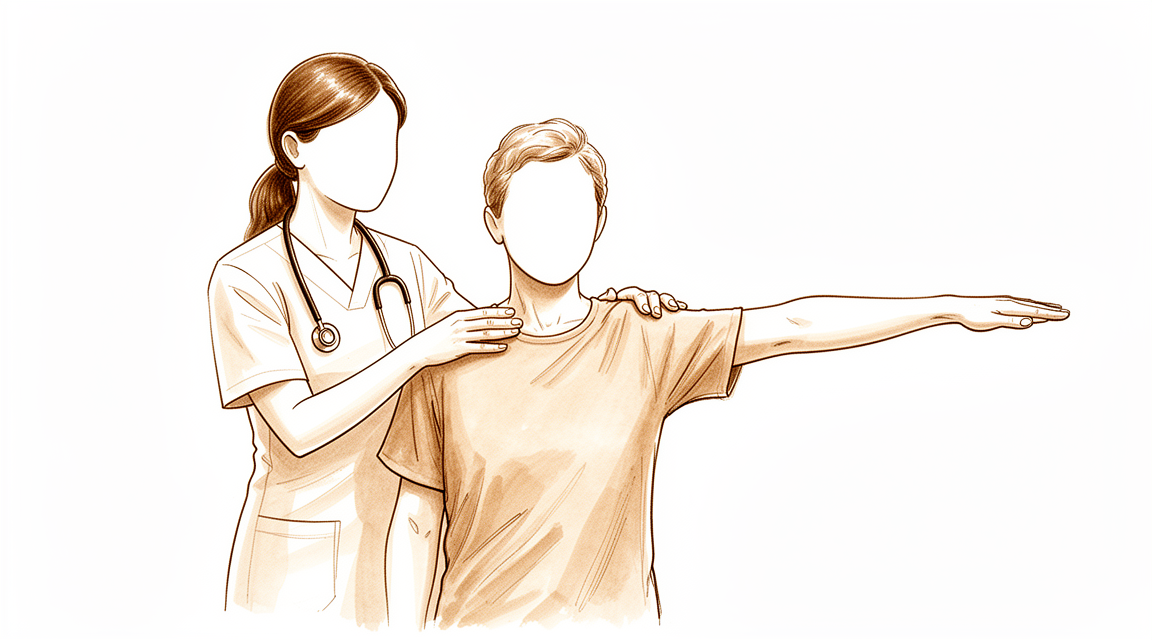
钙化性肌腱炎是指钙质在肩袖(一组固定并带动肩部活动的肌腱)的某一条肌腱内部少量积聚。疼痛通常位于肩部的侧面或顶部,即冈上肌腱所在的位置,这是最常受累的肌腱。向侧方或过头抬起手臂往往会使疼痛加重,够高处的架子或晾衣服可能会变得很吃力。
疼痛常在夜间加重,尤其是当您侧卧在患侧肩部时,并可能把您痛醒。疼痛也可能在活动后或清晨起床时出现。有些人发现疼痛是阵发性的,中间有较为平静的间歇期,而且可能拖延数月。这种长时间的不适可能意味着需要请假,并可能逐渐降低您的生活质量。
这种疾病最常见于30至60岁之间的女性,但其他人也会患上。沉积物本身的长度通常在0.5至1.5厘米之间,大多数致密而非呈绒毛状。约9%的人有不止一条肌腱受累,这可能使疼痛扩散到肩部周围。
值得了解的一点是:影像学检查发现钙化沉积物,并不能排除同一条肌腱存在撕裂。如果您的疼痛没有缓解、在数周内不断加重、在夜间把您痛醒,或使您无法工作或使用手臂,请咨询您的全科医生或要求专科医生评估。
实际发生了什么
您的肩袖是一组四条肌腱,负责固定并带动肩部活动。这些肌腱在肩部顶部一个小骨拱下方的狭窄空间中滑动,两者之间有一个充满液体的薄垫。您可以把肌腱想象成一根穿过狭窄滑轮的绳子。
在钙化性肌腱炎中,这根绳子的一部分会发生改变。肌腱内部的细胞慢慢转变为类似软骨的细胞,钙质开始沉积在这块发生改变的组织中。这一过程分阶段进行:首先肌腱发生改变,然后钙质形成并停留在那里,最后身体将沉积物分解并吸收。这是一个活跃的过程,而不是简单的磨损,它也不同于您可能在身体其他部位老化、磨损的肌腱中看到的钙质积聚。
阶段之所以重要,是因为每个阶段的感受都不同。当钙质静静地停留在肌腱中时,您可能几乎没有疼痛或完全没有疼痛。当身体开始吸收钙质时,疼痛往往会变得突然而剧烈,并且可能在夜间最严重。沉积物本身也可能因其大小而带来麻烦:当它在狭窄的空间内肿胀时,可能会顶住上方的骨拱,阻碍手臂顺畅活动。这就是为什么向侧方抬起手臂时会感觉僵硬或被卡住。
沉积物最常位于冈上肌腱,距离其在上臂骨顶部附着处约1.5至2厘米。这个位置解释了疼痛为何出现在那里,以及为何过头伸手是最困难的动作。
还有一点值得了解:许多人带有这些沉积物却完全没有疼痛,只有约三分之一的沉积物会引起症状。因此,影像学检查上的钙质是一个真实的发现,但它是否是您疼痛的原因,取决于它所处的阶段以及您的身体对它的反应。
我们能做什么
Mater Private Hospital Rockhampton 的上肢外科医生 Kieran Hirpara 博士会从适合您病情的微创方案入手。患者通常由全科医生转诊至我们的诊所;如果物理治疗师建议您就诊,您仍需获得全科医生的转诊才能符合 Medicare 报销资格。在您的首次就诊时,我们会采集病史,检查您的肩部,并在必要时安排影像学检查。对于这类长期存在的问题,我们通常先尝试非手术治疗,当其未能带来足够改善时才考虑手术。
第一步很简单:调整会引发疼痛的活动,让肩部暂停过头工作、得到休息。物理治疗旨在缓解疼痛,并重建手臂顺畅、受控的活动。在评判其效果之前,请给它足够的时间,因为这种疾病可能拖延数月。如果您的症状没有缓解、在数周内不断加重、在夜间把您痛醒,或使您无法工作或使用手臂,请咨询您的全科医生或要求专科医生评估。
抗炎药物是缓解疼痛的常用一线治疗。如果药物不够,我们可以提供直接针对沉积物本身的操作。一种选择是冲击波疗法,即从体外将声波对准钙质,以分解沉积物。它是非侵入性的,即不会刺穿皮肤,并以设定的强度和脉冲次数进行,使钙质松解。另一种选择是超声引导下的穿刺,即借助超声(一种可显示肌腱实时图像的扫描)将针头置入沉积物中,以破碎并抽出钙质。这两种方法都能缓解疼痛、恢复功能并清除钙质。如果症状持续,穿刺可以重复进行,两次操作之间至少间隔六周。
当这些治疗无效时,才会考虑手术。手术包括从肌腱中去除钙化沉积物,如果肌腱受累严重,还会同时修复肌腱。如果在认真尝试上述方案后您的疼痛仍未缓解,我们会与您讨论手术是否适合您,并共同做出决定。
预期情况
在大多数情况下,钙化性肌腱炎的前景是良好的。许多人带有这些沉积物却从不知道,而其中只有约三分之一会引起疼痛。当沉积物确实引起症状时,从长期来看很有可能完全消退。您的身体可以自行吸收钙质,而且这一过程从沉积物形成的那一刻起就已经开始了。
尽管如此,这一过程并不总是很快。症状可能拖延数月,这段不适期可能意味着需要请假,并对您的生活质量造成实际影响。疼痛常常是阵发性的,中间有较为平静的间歇期,当身体开始分解沉积物时,疼痛可能突然而剧烈。有些沉积物在数月内消退,有些则需要更长时间,目前没有可靠的方法能准确预测您的沉积物会如何变化。
大多数人无需手术即可好转。保守治疗(即不涉及手术的治疗)通常是首选,并能为大多数人带来明显改善:以这种方式治疗的人中,72%报告效果良好或极佳,无论沉积物的大小或位置如何。冲击波疗法和超声引导下的穿刺都能为许多人缓解疼痛并清除钙质,而且两者都有良好的安全记录。当症状出现的时间较短、沉积物较小时,穿刺的效果往往最好。如果其中某种操作不适合您的沉积物,我们会与您讨论其他选择。
手术仅用于在认真尝试这些治疗后疼痛仍未缓解的人。手术从肌腱中去除钙化沉积物,对于经其他治疗仍疼痛不止的人,88.5%能获得良好效果。术后恢复是渐进的:肩部功能在最初六个月内缓慢改善,舒适度和活动度的提升大多在这段时间内逐步累积,而不是一下子出现。
如果您的症状没有缓解、在数周内不断加重、在夜间把您痛醒,或使您无法工作或使用手臂,请咨询您的全科医生或要求专科医生评估。
何时就医
大多数钙化沉积物会自行消退,许多根本不会引起任何问题。但这种疾病可能拖延数月,可能让您耽误工作,并逐渐降低您的生活质量。如果您的肩部疼痛没有缓解、在数周内不断加重、在夜间把您痛醒,或使您无法工作或使用手臂,请咨询您的全科医生或要求专科医生评估。如果向侧方抬起手臂时感觉疼痛被阻挡或卡住,也请告诉医生,因为肿胀的沉积物可能在物理上阻止肌腱在其上方的骨拱下方活动。还有一点值得提出:影像学检查发现钙化沉积物,并不能排除同一条肌腱存在撕裂,因此仅凭影像学发现并不能确定诊断。如果您在非工作时间或周末无法联系到诊所,请前往离您最近的急诊科。
深入探讨
Advanced reading: the deeper science (optional)
本节内容超出了您做出自身治疗决策所需的深度。钙化性肌腱炎值得额外阅读,因为其表现与大多数肩部疾病不同:钙化沉积物在完全无痛的人群中也很常见,身体通常无需干预即可自行溶解钙化灶,且去除钙化的治疗方法并非最快缓解疼痛的方法。
钙化沉积物是常见发现,并不自动等同于诊断
影像学检查报告中提到的钙化沉积物很容易被解读为问题的病因,因为它清晰可见且疼痛确实存在。流行病学数据使这一解读变得复杂。在1,219名成年人中,7.8%的无症状人群和42.5%的肩峰下疼痛综合征患者存在沉积物 [1]。在另一组302例肩关节病例中,沉积物在普通人群中很常见,且仅约三分之一伴有疼痛 [2]。
因此,沉积物确实会显著增加其作为疼痛来源的可能性,但大约每十三例无痛肩关节中就有一例存在沉积物。使沉积物从无症状转变为有症状的因素部分取决于其大小和位置:真正有症状的疾病在30至60岁、伴有肩峰下疼痛且沉积物长度超过1.5厘米的女性中可能性最高 [1],且疼痛与冈上肌部位及累及多条肌腱相关 [2]。
它通常会自行溶解,而这决定了所有治疗的意义
其自然病程趋向于吸收,这正是许多治疗看似有效的原因。最清晰的例证来自一项随机对照试验,该试验中所有患者均接受了针刺和冲洗,随后被随机分配至激素注射组或生理盐水注射组:在12个月时,生理盐水组的钙化灶在83%的患者中已吸收,而激素组为74% [3]。
请仔细研读这一结果,因为它包含了一个反直觉的结论。激素使疼痛在六周内改善,功能在三个月内改善,但对钙质是否消失无显著影响 [3]。疼痛缓解与钙质清除是两个独立的过程。一种治疗可能仅实现其中之一而缺乏另一者,且影像学上可见的病灶并非每周驱动症状的因素。
这正是非手术治疗方案疗效相似的原因
如果钙化灶无论采取何种治疗大多都会自行消退,那么这些治疗手段的目的并非治愈,而是让症状间歇期变得可以耐受。比较研究的结果正说明了这一点。在 257 名患者中,物理治疗、皮质类固醇注射和超声引导下的冲洗术(barbotage)在避免手术方面的 成功率相似 [4]。在 239 名患者中,上述三种方法大多取得成功,其中仅进行物理治疗者的失败率最高 [5]。
在手术/操作类选项中,高能冲击波疗法是研究最为透彻的微创治疗,已被证明在短期至中期安全且有效;而超声引导下的针刺疗法 并未 显示出优于超声引导下的肩峰下注射 [6]。汇总 1,258 名患者的数据显示,冲击波疗法、针刺疗法和关节镜手术均取得了良好的临床疗效 [7];在 908 名患者中,冲洗术(barbotage)安全且成功率高,但从未与其他主要选项进行过头对头比较 [8]。
手术带来的益处,精确表述
手术并非没有优势,且其优势幅度值得引用具体数据而非仅作定性描述。 汇总来自随机对照试验的 2,352 名患者,手术治疗在 功能评分改善幅度 上优于非手术治疗,且 疼痛缓解程度 与非手术治疗相当,尤其是超声引导下的针刺治疗,两种治疗途径均达到了具有临床意义的改善 [9]。 在手术技术本身之间 无显著差异,仅清除钙化沉积物与清除沉积物联合肩峰下减压术的效果相似 [10]。
功能与疼痛的分离是关键的细节。如果疼痛是主要主诉,证据并未明确支持手术。如果僵硬和功能丧失占主导且持续存在,则更倾向于支持手术。
最常被忽视的关联
钙化性肌腱炎并非均匀分布。在一项针对 102 名患者的研究中,伴有内分泌疾病(主要是甲状腺疾病和糖尿病)的患者,其症状出现年龄更轻,病程显著更长,且更常需要手术治疗 [11]。
如果您的病程异常漫长或发病较早,这一点值得向您的全科医生提出。这并非因为治疗内分泌疾病能解决肩部问题,而是因为它改变了现实时间表的预期。被告知某种疾病具有自限性,与持续两年的疼痛难以调和;内分泌疾病的关联是解释为何常规安慰不适用于所有病例的一个原因。
参考文献
[1] Louwerens JK, Sierevelt IN, van Hove RP, van den Bekerom MP, van Noort A. 伴有和不伴有肩峰下疼痛综合征的成人肩袖肌腱内钙化沉积物的患病率:1219例患者的临床和影像学分析. J Shoulder Elbow Surg. 2015;24(10):1588-93. https://doi.org/10.1016/j.jse.2015.02.024
[2] Sansone V, Consonni O, Maiorano E, Meroni R, Goddi A. 肩袖钙化性肌腱病:有症状和无症状女性肩部疼痛与影像学特征之间的相关性. Skeletal Radiol. 2015;45(1):49-55. https://doi.org/10.1007/s00256-015-2240-3
[3] Darrieutort-Laffite C, Varin S, Coiffier G, Albert J, Planche L, Maugars Y, et al. 钙化性肌腱炎穿刺冲洗后是否需要皮质类固醇注射?随机、双盲、非劣效性试验. Ann Rheum Dis. 2019;78(6):837-43. https://doi.org/10.1136/annrheumdis-2018-214971
[4] Gilbert R, Dadoo S, Lin R, Bhardwaj N, McMahon S, Steuer F, et al. 物理治疗、皮质类固醇注射和超声引导下高压冲洗在钙化性肌腱炎非手术治疗中的比较. Orthop J Sports Med. 2026;14(4). https://doi.org/10.1177/23259671261434919
[5] Drummond M, Ayinon C, Lin A, Dunn R. 钙化性肌腱炎三种非手术治疗的相对疗效:物理治疗与类固醇注射与高压冲洗. Orthop J Sports Med. 2021;9(7_suppl4). https://doi.org/10.1177/2325967121S00210
[6] Louwerens JK, Sierevelt IN, van Noort A, van den Bekerom MP. 微创疗法在慢性肩袖钙化性肌腱病管理中的证据:系统综述和荟萃分析. J Shoulder Elbow Surg. 2014;23(8):1240-9. https://doi.org/10.1016/j.jse.2014.02.002
[7] Louwerens JK, Veltman ES, van Noort A, van den Bekerom MP. 高能体外冲击波疗法与超声引导下穿刺与关节镜手术在慢性钙化性肩袖肌腱病管理中的有效性:系统综述. Arthroscopy. 2015;32(1):165-75. https://doi.org/10.1016/j.arthro.2015.06.049
[8] Gatt DL, Charalambous CP. 超声引导下高压冲洗治疗肩部钙化性肌腱炎:包含908例患者的系统综述. Arthroscopy. 2014;30(9):1166-72. https://doi.org/10.1016/j.arthro.2014.03.013
[9] Angileri HS, Gohal C, Comeau-Gauthier M, Owen MM, Shanmugaraj A, Terry MA, et al. 慢性肩袖钙化性肌腱炎:比较手术和非手术干预的随机对照试验的系统综述和荟萃分析. J Shoulder Elbow Surg. 2023;32(8):1746-60. https://doi.org/10.1016/j.jse.2023.03.017
[10] Anam E, Zahran S, Roy A, Daneshvar P, Bicknell RT, Janssen I. 肩部钙化性肌腱炎的手术入路:系统综述和荟萃分析. JSES Rev Rep Tech. 2024;4(3):353-8. https://doi.org/10.1016/j.xrrt.2024.03.013
[11] Harvie P, Pollard TC, Carr AJ. 钙化性肌腱炎:自然病程及其与内分泌疾病的相关性. J Shoulder Elbow Surg. 2007;16(2):169-73. https://doi.org/10.1016/j.jse.2006.06.007
Evidence & references
This is the clinical evidence summary written for health professionals. It is technical, and it lists the research this page was built from. You do not need to read it to understand your treatment or to make a decision about it.
Overview
Epidemiology and Natural History
- Calcific tendon deposits of the shoulder are a frequent occurrence in the general population [2].
- Only one third of calcific tendon deposits in the shoulder are painful [2].
- Symptomatic calcific tendinitis of the shoulder has a good likelihood to completely resolve in the long-term [3].
Non-Operative Treatment
- The primary choice of treatment for calcific tendinitis is conservative, especially in patients with acute calcific tendinitis [26].
- Ultrasound treatment helps resolve calcifications and is associated with short-term clinical improvement in patients with symptomatic calcific tendinitis of the shoulder [1].
- Conservative treatment for calcific tendinitis of the shoulder showed clinically significant improvement, with 72% of excellent or good results regardless of the location, radiologic type and size, and initial symptoms of calcific deposits [5].
- Extracorporeal shock wave therapy (ESWT) has produced a high rate of success in pain relief and functional restoration with negligible associated complications [11].
- Shock wave therapy is a safe and effective noninvasive treatment for patients with calcific tendinitis of the shoulder, producing a high rate of success in pain relief and functional restoration with negligible complications [19].
- ESWT can be considered as an alternative treatment for chronic calcific tendinitis of the shoulder due to its good tolerance, safety, and clinical radiological response [25].
- Both ultrasound-guided needling and extracorporeal shock wave therapy improved clinical outcomes and eliminated calcium deposits in the treatment of calcific tendinitis [6].
- Utilization of barbotage as a treatment for calcific tendonitis of the shoulder appears to produce notable pain reduction in the short term (specifically at the 2-month follow-up), but begins to lose some efficacy over long-term evaluation [27].
- Patients with calcific tendinitis of the shoulder who have the factors identified for a poor outcome after ESWT should undergo a different procedure [42].
Operative Treatment
- Surgical treatment of calcific tendinitis gives good results in 88.5% of patients resistant to medical treatment [12].
- The aim of arthroscopic treatment in calcific tendinitis is to remove the calcific deposit [15].
- Arthroscopic removal of calcification leads to improved clinical outcomes in patients with chronic calcific tendinitis, but at least 6 months of follow-up is needed for these improvements to become statistically significant [13].
- Patients undergoing arthroscopic treatment of a calcific deposit in the shoulder had satisfactory clinical and radiological outcomes at the final follow-up, with functional scores improving slowly and reaching more than 75 percent at six months after surgery [7].
- Arthroscopic treatment of calcifying tendinitis provides good to excellent clinical results [20].
- Arthroscopic treatment of chronically painful calcific tendinitis of the rotator cuff promises to be successful in more than 90% of the patients if conservative treatment fails [37].
- Arthroscopic removal of calcific deposits of the shoulder shows good clinical results for pain reduction, shoulder function and patient satisfaction [44].
- Endoscopic and open surgery are equally effective in the treatment of chronic calcifying tendinopathy, showing similar clinical and sonographic results [34].
- Preoperative ultrasound-guided marking of calcific deposits is a procedure that statistically significantly improves the clinical results of arthroscopic surgery as seen at 6 weeks and 2 years [33].
- The short-term functional outcome of patients with calcific tendonitis after arthroscopic bursectomy and debridement of the calcific deposit is not influenced if performed in combination with or without a subacromial decompression [40].
- Routine diagnostic glenohumeral exploration does not appear beneficial in arthroscopic treatment of calcific tendinitis due to the low prevalence of intraarticular pathologies which most frequently do not require surgical treatment [23].
Prognostic Factors
- Patients with calcific lesions >1 cm had a 2.8 increased likelihood to undergo operative treatment in the setting of calcific tendinitis of the shoulder [4].
Anatomy & Pathophysiology
Epidemiology & Demographics
- Calcific tendon deposits of the shoulder are a frequent occurrence in the general population, although only one third are painful [2].
- In approximately 80% of patients, calcifying tendinitis occurs in the supraspinatus tendon [115].
- Most patients with calcifying tendinitis are 30-50 years old [115].
- Women are approximately 1.5 times more often affected by calcifying tendinitis than men [115].
- The incidence of calcifying tendinitis in the general population is 2.5-20% [115].
- In about 10% of individuals, calcific deposits are found bilaterally [17].
- In 20% of cases, subjects with rotator cuff calcific tendinopathy are asymptomatic [17].
- Subjects with rotator cuff calcific tendinopathy were often female aged between 30 and 60 [17].
- Demographic, radiographic, and clinical features of calcific tendinitis of the shoulder in the Korean population were not different from those of Western populations [14].
Anatomical Location & Structures
- Calcific deposits are most commonly located at the level of the supraspinatus tendon [17].
- In calcifying tendinitis, deposits are typically located 1.5-2 cm from the tendinous insertion at the greater tuberosity [115].
- The greater tuberosity serves as the attachment site for the supraspinatus, infraspinatus, and teres minor tendons of the rotator cuff [70].
- The rotator cuff consists of four muscles: the subscapularis, supraspinatus, infraspinatus, and teres minor [71].
- The acromion, the coracoacromial ligament, and the coracoid process form the coracoacromial arch [70].
- The rotator cuff, subacromial bursa, and subdeltoid bursa pass underneath the coracoacromial arch [70].
- The subacromial bursa separates the rotator cuff tendons from the coracoacromial arch and allows them to glide [78].
- The infraspinatus and teres minor fuse near their musculotendinous junctions [83].
- The tendons of the infraspinatus and supraspinatus muscles join approximately 15 mm proximal to their insertion [83].
Pathogenesis & Staging
- The pathogenesis of calcific tendinitis remains unclear, and different theories have been proposed [30].
- Calcific tendinitis of the rotator cuff tendons is thought to have a different pathophysiologic mechanism than calcific tendinitis at other sites, such as the Achilles or patellar tendons [30].
- Uhthoff and Loehr described an active, cell-mediated process that is now widely accepted for the pathogenesis of calcific tendinitis [30].
- Uhthoff and Loehr described cartilage metaplasia as a reactionary process in an active biologic environment [30].
- The reactive calcification theory proposed by Uthoff in 1997 consists of a three-stages process: pre-calcific, calcific, and post-calcific stage [17].
- The precalcific stage consists of predominantly fibrocartilaginous metaplasia presumably within less vascular areas of the tendon [30].
- During the pre-calcific stage, tenocytes undergo a metaplastic transformation into chondrocytes within the site where the calcium deposits will subsequently form [17].
- The calcific stage is subdivided into three separate phases: formative, resting, and resorptive [30].
- In the formative phase of the calcific stage, matrix vesicles unite to become calcific deposits that are separated by fibrocartilage [30].
- The resting phase is characterized by the presence of calcium deposits in the rotator cuff [17].
- The resorptive phase is associated with acute, sudden onset of extremely severe pain [49].
- Calcific deposits consist of two different forms of hydroxyapatite: A type and B type [30].
- The composition of an individual calcific deposit can have a mixture of the two types of hydroxyapatite [30].
- Each individual phase of calcific tendinitis has a different composition of hydroxyapatite [30].
- Calcium is deposited in the fibrocartilaginous matrix of the tendon as calcium carbonate apatite [49].
- Endocrine and metabolic disorders were often described as comorbidity in subjects with rotator cuff calcific tendinopathy [17].
- Endocrine disorders probably play an important role in the development of rotator cuff calcific tendinopathy, but the effect of this process is still unknown [17].
- Shorter length of the infraspinatus tendon and specific subacromial bursa anatomy may explain the intramuscular migration of calcium deposits leading to the lesion [10].
Clinical Presentation & Pain Mechanisms
- Subjects with rotator cuff calcific tendinopathy often reported nightly, acute, unilateral and severe pain with spontaneous onset [17].
- Subjects with rotator cuff calcific tendinopathy often reported deficit in active and passive range of motion, mainly in abduction and flexion [17].
- Calcific tendinitis of the supraspinatus does not typically cause loss of external rotation but is frequently associated with mild isolated restriction of abduction [22].
- Patients with calcific tendinitis had less passive abduction, forward flexion, internal rotation, and external rotation than the control group [65].
- The main mechanism of abduction contracture lies in the mechanical blocking between the bulged calcified deposits and the coracoacromial arch [45].
- Abduction contracture is presumably caused by mechanical blocking by the calcified deposits and the coracoacromial arch and by reflex muscle spasm caused by pain [125].
- Patients with calcific tendinitis had significantly greater nerve counts compared with the rotator cuff repair group and the control group [65].
- A greater nerve count positively correlated with a greater degree of neovascularization and also positively correlated with greater frequency of extreme pain [65].
- Neovascularization positively correlated with more frequent pain during sleep and more frequent extreme pain [65].
- The pathophysiology of calcific tendinitis remains largely unknown, with proposed theories including degenerative calcification, repetitive trauma, necrosis of tenocytes and intracellular calcium accumulation, reactive calcification, endochondral ossification, and chondral metaplasia [65].
- Calcific tendinitis of the rotator cuff with tuberosity osteolysis is a distinctive form of calcific tendinitis that should be considered in clinical and surgical practice [28].
- A rotator cuff tear was the only factor affecting the complete recovery of shoulder function in patients treated for calcific tendinitis [113].
Classification
- The supraspinatus is the most frequently affected tendon in rotator cuff calcific tendinopathy, followed by the infraspinatus and subscapularis [38].
- In 9% of patients with rotator cuff calcific deposits, two or more tendons are affected [38].
- The median length of calcific deposits in the rotator cuff is 1.16 cm [38].
- 55.8% of calcific deposits in the rotator cuff have a length between 0.5 and 1.5 cm [38].
- 28.6% of calcific deposits in the rotator cuff have a length greater than 1.5 cm [38].
- Gartner type II morphology is the most common radiographic classification for calcific deposits, present in 47.0% of cases [38].
- Gartner type I morphology is present in 38.4% of calcific deposits [38].
- Gartner type III morphology is present in 14.6% of calcific deposits [38].
- Approximately 76% of calcifications in the shoulder are characterized as dense (type A or B) using the Mole et al. classification scheme [39].
- Approximately 24% of calcifications in the shoulder are characterized as fluffy (type C) using the Mole et al. classification scheme [39].
- Type A calcific deposits are dense, rounded, and sharply delineated [67].
- Type B calcific deposits are multilobular in appearance, radiodense, and sharply outlined [67].
- Type C calcific deposits are more radiolucent and heterogeneous with irregular outlines [67].
- Type D calcific deposits are dystrophic calcific lesions of the tendon insertion [67].
- The pathogenesis of calcific tendinitis involves a three-stage process: precalcific, calcific, and postcalcific [30].
- The calcific stage is subdivided into three phases: formative, resting, and resorptive [30].
- The precalcific stage consists of predominantly fibrocartilaginous metaplasia within less vascular areas of the tendon [30].
- In the formative phase of the calcific stage, matrix vesicles unite to become calcific deposits separated by fibrocartilage [30].
- The composition of an individual calcific deposit can have a mixture of A type and B type hydroxyapatite [30].
- Disease evolution in calcific tendinitis can be classified into four stages: precalcific, calcific, resorptive, and postcalcific [107].
- The resorptive phase of calcific tendinitis is the most painful stage owing to vascular invasion, increased phagocytic cells, and edema from intratendinous pressure [107].
Clinical Presentation
- Subjects complaining of rotator cuff calcific tendinopathy were often female aged between 30 and 60 [17].
- Calcific tendinitis is a poorly understood condition in which symptoms can be protracted, resulting in time off work and impaired quality of life [8].
- The calcium deposits are most commonly located at the level of the supraspinatus tendon [17].
- The supraspinatus was the most frequently affected tendon in 82.7% of patients with calcific deposits, followed by the subscapularis tendon in 8.9% and the infraspinatus in 8.4% [38].
- Two or more tendons were affected in 9% of patients with calcific deposits [38].
- The median length of the calcific deposit was 1.16 cm, with 55.8% ranging from 0.5 to 1.5 cm and 28.6% greater than 1.5 cm [38].
- Approximately 76% of calcifications were characterized as dense (type A or B) and 24% as fluffy (type C) [39].
- Recognition of atypical presentations of calcific tendinitis with bone erosion may prevent unnecessary biopsy and overtreatment [16].
- The presence of a calcium deposit in the rotator cuff of a patient with painful shoulder does not rule out the possibility of a coexisting rotator cuff tear [60].
- Calcific tendinitis in the elbow of a child has not been reported previously [24].
- Atypical calcific tendinitis involving teres minor can affect overhead movement [9].
- Calcific tendinitis of the trapezius insertion is a recognized presentation [18].
- Of twelve histologically verified cases of calcific periarthritis adjacent to joints other than the shoulder, in only one patient was the preoperative clinical diagnosis accurate [59].
Investigations
Plain Radiography
- Standardized plain films are almost always sufficient to garner the information needed for shoulder imaging [47].
- The purpose of imaging the shoulder is to help establish the diagnosis, determine the severity of the pathoanatomy, assist in surgical planning, and enable the surgeon to illustrate the condition of the shoulder to the patient [47].
- At least two X-ray views should be obtained for shoulder imaging: an anteroposterior in the plane of the glenoid and an axillary projection with the arm in abduction [87].
- The axillary view is taken with the arm in the functional position of elevation in the plane of the scapula and oriented so that both the spinoglenoid notch and the scapular neck are visible [47].
- The axillary view demonstrates glenohumeral relationships in the functional position of elevation, which is referred to as the "truth view" [47].
- CT scans may offer a few degrees of increased precision in the measurement of glenoid version, but this precision does not improve the quality of the surgery or the clinical outcome [47].
- The temptation to "overimage" should be resisted, obtaining only the scans or reconstructions that are necessary for the care of the patient [47].
- Calcific tendinitis can be observed on plain radiograph [94].
- Calcific deposits in the fingers may not be seen on standard views and oblique views may be necessary [111].
Ultrasonography
- Ultrasound is a simple and accurate test for identifying rotator cuff tears and calcific tendinitis [87].
- Ultrasound can be useful in guiding injections or barbotage (aspirating calcific deposits in the rotator cuff) [87].
- Ultrasonography is a low-cost alternative to MRI and arthrography for evaluating both skeletal and soft-tissue structures of the shoulder [94].
- Ultrasonography can provide immediate, real-time visualization of the rotator cuff, biceps tendon, and calcific deposits [94].
- Ultrasonography can be used to measure the subacromial space and detect atrophy of rotator cuff muscles [94].
- Ultrasonography can evaluate impingement in various positions and motions due to real-time imaging [94].
- Ultrasonography is highly operator dependent and is not as useful for evaluating labral tears or rotator cuff tears that are very small or larger than 3 cm [94].
- The accuracy of rotator cuff ultrasonography depends on the skill of the scanner operator and an awareness of pitfalls that are encountered [85].
- US elastography is recommended for all patients with calcific tendinosis, except for those with the arc morphological type, before making management decisions [31].
- The surgeon performing ultrasound of the shoulder can diagnose intraosseous calcifying tendinitis [56].
- Calcific tendinitis of the supraspinatus is frequently associated with mild isolated restriction of abduction but does not typically cause loss of external rotation [22].
Magnetic Resonance Imaging
- MRI is the modality of choice for evaluating the rotator cuff, biceps, and subacromial/subdeltoid bursa [94].
- T2-weighted MRI provides better visualization of full thickness rotator cuff tears [94].
- MRI can identify labral tears and rotator cuff tears, although the accuracy for these is enhanced by combining the scan with arthrography [87].
- Shoulder surgeons should be aware of the accuracy limitations of sonographic or MRI evaluation regarding rotator cuff tears as a comorbidity in calcific tendinitis [32].
- The incidence of rotator cuff tears in cases of calcific tendonitis is higher than previously reported in cohorts undergoing MRI [41].
Computed Tomography
- CT imaging is frequently used to evaluate fractures of the shoulder, to assess for bony lesions in recurrent instability cases, or for preoperative templating for shoulder arthritis [94].
- CT is helpful for planning fracture surgery and shoulder joint replacement [87].
Diagnostic Findings and Pathology
- The paper illustrates the dynamic pathological process of calcific tendinopathy, including migration patterns of calcium deposits [29].
Treatment
Non-Operative Management
- Nonoperative treatment consists of physical therapy, therapeutic modalities, and injections [49].
- Anti-inflammatory medications and steroid use are a common first line treatment option [36].
- Extracorporeal shock wave therapy (ESWT) is a safe and effective noninvasive treatment for patients with calcific tendinitis of the shoulder, producing a high rate of success in pain relief and functional restoration with negligible complications [19].
- Treatment of calcific tendinitis of the shoulder with shock waves has produced a high rate of success in pain relief and functional restoration with negligible associated complications [11].
- The effectiveness of ESWT in calcific tendinitis of the shoulder is dose-dependent [43].
- Disintegration of calcific deposits via ESWT requires an energy of at least 0.42 mJ/mm² and 2000 impulses [58].
- Shock wave therapy is a heterogeneous but effective treatment for calcifying tendinitis of the shoulder [53].
- ESWT in calcific tendinitis of the shoulder is very effective [55].
- Both ultrasound-guided needling and extracorporeal shock wave therapy improved clinical outcomes and eliminated calcium deposits in patients with calcific tendinitis [6].
- Ultrasound-guided needling showed a higher decrease in calcium deposits compared to radial shockwave therapy at 6 weeks [104].
- Ultrasound-guided needling showed a significantly higher improvement in the Oxford Score pre- and post-treatment compared to radial shockwave therapy [104].
- After one year, there was no significant difference in NRS and Oxford Score between ultrasound-guided needling and radial shockwave therapy groups [104].
- Patients with calcific tendinitis of the shoulder who have factors identified for a poor outcome after ESWT should undergo a different procedure [42].
- Needle aspiration of calcific deposits (NACD) is safe and effective for calcific tendinitis [35].
- In case of persisting symptoms, patients may be scheduled for another NACD procedure with a minimal term of six weeks between procedures [35].
- Percutaneous needle barbotage of calcium deposits has been demonstrated to provide pain relief and improved symptoms in patients suffering from calcific tendonitis [36].
- Symptoms of calcific tendinitis of the trapezius insertion responded to needle aspiration and injection, with the patient reporting normal shoulder function approximately 1 year after the procedure [18].
Operative Management
- Arthroscopic or open removal of the deposit is occasionally necessary for calcific tendinitis [49].
- In athletes with calcifying tendinitis of the supraspinatus tendon with failed nonoperative treatment, complete arthroscopic removal of calcific deposits and tendon repair without acromioplasty results in significant pain relief and improvement in functional outcomes [102].
- Arthroscopic debridement of calcific tendinitis with intraosseous involvement is a safe and effective treatment method similar to that of pure tendinous involvement [100].
- Complete removal of calcium was achieved in 44 patients (88%) after surgery, with faint specks apparent in 6 (12%) [39].
- Routine exploration of the gleno-humeral joint during arthroscopic removal of rotator cuff calcifications is not advisable as it significantly increases the risk of adhesive capsulitis without identifying a sufficient number of lesions requiring a therapeutic procedure [110].
- The rotator cuff should be repaired if it is significantly involved during treatment for calcific tendinitis [49].
Complications
- Shoulder surgeons should be cautious about rotator cuff tears as a comorbidity in calcific tendinitis and aware of the accuracy limitations of sonographic or MRI evaluation [32].
- The incidence of rotator cuff tears in cases of calcific tendonitis is higher than previously reported in a cohort of patients who underwent MRI [41].
- The incidence and type of intraarticular lesions in calcifying tendinitis are comparable to age-matched shoulders with partial- rather than full-thickness rotator cuff tears [61].
- It is speculated that the main mechanism of abduction contracture lies in the mechanical blocking between the bulged calcified deposits and the coracoacromial arch [45].
- Periarticular calcific deposits are self-limiting and respond to conservative treatment, whereas recurrence and persistent symptoms are not rare in patients with peritendinous calcific deposits in the hand and wrist [21].
Recovery
Natural History and Prognosis
- Symptoms of calcific tendinitis can be protracted, resulting in time off work and impaired quality of life [8].
- The onset of calcium resorption in acute calcific tendinitis occurs most frequently in the summer in Japan [130].
Conservative and Minimally Invasive Management
- Extracorporeal shock wave therapy (ESWT) can be considered as an alternative treatment for chronic calcific tendinitis of the shoulder due to its good tolerance, safety, and clinical radiological response [25].
- Calcific deposits disappeared in the same percentage of patients in both groups of a randomized clinical trial comparing two different energy levels of extracorporeal shock-wave therapy [69].
- Percutaneous needle aspiration and lavage is effective in the short term and in the long term in calcific tendinitis of the shoulder, with results similar to or better than those published for other techniques, and it is only slightly invasive and painful [62].
- There was no significant difference in short- and long-term clinical outcomes between single- and double-needle US-guided percutaneous irrigation of calcific tendinopathy [63].
- A symptom duration of ≤10 months or calcification size of ≤10.82 mm represented the clinical scenarios most likely to show resorption after ESWT [126].
- In SWT studies, Type III calcifications and shorter symptom duration were associated with better outcomes [129].
Operative Management
- Improved Constant scores at 24 months after arthroscopic treatment were inversely related to the number and size of residual calcifications in all patients [67].
Prognostic Factors and Complications
Key Evidence
- [L1] In patients with symptomatic calcific tendinitis of the shoulder, ultrasound treatment helps resolve calcifications and is associated with short-term clinical improvement. [1] (10.1056/nejm199905203402002)
- [L3] Calcific tendon deposits of the shoulder are a frequent occurrence in the general population, although only one third are painful. [2] (10.1007/s00256-015-2240-3)
- [L1] Symptomatic calcific tendinitis of the shoulder has a good likelihood to completely resolve in the long-term. [3] (10.1097/phm.0000000000000939)
- [L3] Patients with calcific lesions >1 cm had a 2.8 increased likelihood to undergo operative treatment in the setting of calcific tendinitis of the shoulder. [4] (10.1016/j.jseint.2021.01.013)
- [L2] Conservative treatment for calcific tendinitis of the shoulder showed clinically significant improvement, with 72% of excellent or good results regardless of the location, radiologic type and size, and initial symptoms of calcific deposits. [5] (10.1016/j.jse.2009.07.008)
- [L2] Both treatment modalities for calcific tendinitis improved clinical outcomes and eliminated calcium deposits. [6] (10.1016/j.jse.2014.06.036)
- [Paper] Patients undergoing arthroscopic treatment of a calcific deposit in the shoulder had satisfactory clinical and radiological outcomes at the final follow-up, with functional scores improving slowly and reaching more than 75 percent at six months after surgery. [7] (10.1016/j.otsr.2020.03.005)
- [L3] Calcific tendinitis is a poorly understood condition in which symptoms can be protracted, resulting in time off work and impaired quality of life. [8] (10.1016/j.jse.2006.06.007)
- [Case_report] [9] (10.1016/j.jisako.2025.101055)
- [L4] Shorter length of the infraspinatus tendon and specific subacromial bursa anatomy may explain the intramuscular migration of calcium deposits leading to the lesion. [10] (10.1016/j.jse.2022.01.092)
- [L2] Treatment of calcific tendinitis of the shoulder with shock waves has produced a high rate of success in pain relief and functional restoration with negligible associated complications. [11] (10.1016/j.jse.2007.03.023)
- [L4] Surgical treatment of calcific tendinitis gives good results in 88.5% of patients resistant to medical treatment. [12] (10.1007/s100670050108)
- [L4] Arthroscopic removal of calcification leads to improved clinical outcomes in patients with chronic calcific tendinitis, but at least 6 months of follow-up is needed for these improvements to become statistically significant. [13] (10.5397/cise.2018.21.2.75)
- [L4] This study reported demographic, radiographic, and clinical features of calcific tendinitis of the shoulder in the Korean population, which were not different from those of Western populations. [14] (10.5397/cise.2020.00010)
- [L4] The aim of arthroscopic treatment in calcific tendinitis is to remove the calcific deposit. [15] (10.1016/s1058-2746(98)90180-x)
- [L5] Recognition of atypical presentations of calcific tendinitis with bone erosion may prevent unnecessary biopsy and overtreatment. [16] (10.1016/j.jse.2009.02.009)
- [L3] [17] (10.1177/17585732241244515)
- [L5] Symptoms responded to needle aspiration and injection, with the patient reporting normal shoulder function approximately 1 year after the procedure. [18] (10.1016/s1058-2746(99)90011-3)
- [L3] Shock wave therapy is a safe and effective noninvasive treatment for patients with calcific tendinitis of the shoulder, producing a high rate of success in pain relief and functional restoration with negligible complications. [19] (10.1177/03635465030310031701)
- [L3] Arthroscopic treatment of calcifying tendinitis provides good to excellent clinical results. [20] (10.1177/03635465211037690)
- [L3] Periarticular calcific deposits are self-limiting and respond to conservative treatment, whereas recurrence and persistent symptoms are not rare in patients with peritendinous calcific deposits in the hand and wrist. [21] (10.1177/1753193413478393)
- [L3] Calcific tendinitis of the supraspinatus does not typically cause loss of external rotation but is frequently associated with mild isolated restriction of abduction. [22] (10.1177/2325967117752907)
- [L3] Routine diagnostic glenohumeral exploration does not appear beneficial in arthroscopic treatment of calcific tendinitis due to the low prevalence of intraarticular pathologies which most frequently do not require surgical treatment. [23] (10.1186/s12891-017-1839-z)
- [L5] Calcific tendinitis in the elbow of a child has not been reported previously. [24] (10.1067/mse.2002.119854)
- [L2] Because of its good tolerance, safety, and clinical radiological response, ESWT can be considered as an alternative treatment for chronic calcific tendinitis of the shoulder. [25] (10.1136/ard.62.3.248)
- [L5] The primary choice of treatment for calcific tendinitis is conservative, especially in patients with acute calcific tendinitis. [26] (10.5397/cise.2020.00318)
- [L4] Utilization of barbotage as a treatment for calcific tendonitis of the shoulder appears to produce notable pain reduction in the short term (specifically at the 2-month follow-up), but begins to lose some efficacy over long-term evaluation. [27] (10.1016/j.jseint.2024.06.005)
- [L2] Imaging and functional data indicate that calcific tendinitis of the rotator cuff with tuberosity osteolysis is a distinctive form of calcific tendinitis that should be considered in clinical and surgical practice. [28] (10.1016/j.jse.2008.09.016)
- [L5] The paper illustrates the dynamic pathological process of calcific tendinopathy, including migration patterns of calcium deposits, and discusses clinical and sonographic assessment to optimize diagnosis, rehabilitation, and interventional management for functional recovery. [29] (10.3390/diagnostics12123097)
- [L5] [30] (10.5435/jaaos-22-11-707)
- [L4] We recommend that US elastography be performed for all patients with calcific tendinosis, except for those with the arc morphological type, before making management decisions. [31] (10.1016/j.jcma.2015.05.006)
- [L3] Shoulder surgeons should be cautious about rotator cuff tears as a comorbidity in calcific tendinitis and aware of the accuracy limitations of sonographic or MRI evaluation. [32] (10.5397/cise.2021.00094)
- [L3] Preoperative ultrasound-guided marking of calcific deposits is a procedure that statistically significantly improves the clinical results of arthroscopic surgery as seen at 6 weeks and 2 years. [33] (10.1016/j.arthro.2006.08.005)
- [L1] Endoscopic and open surgery are equally effective in the treatment of chronic calcifying tendinopathy, showing similar clinical and sonographic results. [34] (10.1097/01.blo.0000063786.32430.22)
- [L4] [35] (10.1016/j.ejrad.2016.01.018)
- [L4] [36] (10.1177/2325967121s00655)
- [L4] This study confirmed that if conservative treatment fails, arthroscopic treatment of chronically painful calcific tendinitis of the rotator cuff promises to be successful in more than 90% of the patients. [37] (10.1016/j.arthro.2006.01.012)
- [L3] [38] (10.1016/j.jse.2015.02.024)
- [L3] [39] (10.1016/j.jse.2010.10.038)
- [L1] This study has demonstrated that the short-term functional outcome of patients with calcific tendonitis after arthroscopic bursectomy and debridement of the calcific deposit is not influenced if performed in combination with or without a subacromial decompression. [40] (10.1016/j.arthro.2015.05.015)
- [L4] The incidence of rotator cuff tears in cases of calcific tendonitis in this cohort of patients who underwent MRI is higher than previously reported. [41] (10.1016/j.arthro.2019.11.127)
- [L3] Patients with calcific tendinitis of the shoulder who have the factors identified for a poor outcome after ESWT should undergo a different procedure. [42] (10.1302/0301-620x.99b12.bjj-2016-1178.r1)
- [L2] The study confirms the effectiveness of ESWT in CT of the shoulder and demonstrates that the results are dose-dependent. [43] (10.1067/mse.2002.126614)
- [L3] Arthroscopic removal of calcific deposits of the shoulder shows good clinical results for pain reduction, shoulder function and patient satisfaction. [44] (10.1007/s00167-008-0507-0)
- [L4] It is speculated that the main mechanism of abduction contracture lies in the mechanical blocking between the bulged calcified deposits and the coracoacromial arch. [45] (10.1016/s1058-2746(96)80285-0)
- [L1] Shock wave therapy is a heterogeneous but effective treatment for calcifying tendinitis of the shoulder. [53] (10.1177/1941738108331197)
- [L1] ESWT in calcific tendinitis of the shoulder is very effective. [55] (10.1007/s00256-004-0849-8)
- [L4] This complex case report illustrates the value of the surgeon performing ultrasound of the shoulder to diagnose an intraosseous calcifying tendinitis. [56] (10.1016/j.xrrt.2023.09.012)
- [L2] [58] (10.1007/s00776-003-0720-0)
- [L4] Of twelve histologically verified cases of calcific periarthritis adjacent to joints other than the shoulder, in only one patient (toe) was the preoperative clinical diagnosis accurate, which signals the need for greater awareness of this entity as a differential diagnostic option. [59] (10.2106/jbjs.k.00874)
- [L4] The presence of a calcium deposit in the rotator cuff of a patient with painful shoulder does not rule out the possibility of a coexisting rotator cuff tear (RCT). [60] (10.1016/s1058-2746(09)80095-5)
- [L3] The incidence and type of intraarticular lesions in calcifying tendinitis are comparable to age-matched shoulders with partial- rather than full-thickness rotator cuff tears. [61] (10.1007/s00402-011-1263-z)
- [L4] Percutaneous needle aspiration and lavage is effective in the short term and in the long term in calcific tendinitis of the shoulder, with results similar to or better than those published for other techniques, and it is only slightly invasive and painful. [62] (10.2214/ajr.07.2254)
- [L1] There was no significant difference in short- and long-term clinical outcomes between single- and double-needle US-guided percutaneous irrigation of calcific tendinopathy. [63] (10.1148/radiol.2017162888)
- [L3] [65] (10.2106/jbjs.o.00417)
- [L4] [67] (10.1016/j.jse.2004.04.001)
- [L2] Calcific deposits disappeared in the same percentage of patients in both groups. [69] (10.2522/ptj.20110252)
- [L3] Arthroscopic debridement of calcific tendinitis with intraosseous involvement is a safe and effective treatment method similar to that of pure tendinous involvement. [100] (10.1007/s00167-022-06870-2)
- [L4] In athletes with calcifying tendinitis of the supraspinatus tendon with failed nonoperative treatment, complete arthroscopic removal of calcific deposits and tendon repair without acromioplasty results in significant pain relief and improvement in functional outcomes. [102] (10.1177/2325967116669310)
- [L2] [104] (10.1016/j.jor.2017.07.011)
- [L1] [107] (10.1148/radiol.11111157)
- [L3] Routine exploration of the gleno-humeral joint during arthroscopic removal of rotator cuff calcifications is not advisable as it significantly increases the risk of adhesive capsulitis without identifying a sufficient number of lesions requiring a therapeutic procedure. [110] (10.1016/j.otsr.2021.102915)
- [L5] The calcification may not be seen on standard views and oblique views may be necessary. [111] (10.1016/0266-7681(85)90036-1)
- [L3] A rotator cuff tear was the only factor affecting the complete recovery of shoulder function. [113] (10.1177/03635465231217733)
- [L2] [115] (10.1177/0269215510396740)
- [L4] The abduction contracture is presumably caused by mechanical blocking by the calcified deposits and the coracoacromial arch and by reflex muscle spasm caused by pain. [125] (10.1016/s1058-2746(97)90074-4)
- [L3] A symptom duration of ≤10 months or calcification size of ≤10.82 mm represented the clinical scenarios most likely to show resorption after ESWT. [126] (10.1177/23259671241231609)
- [L3] In SWT studies, Type III calcifications and shorter symptom duration were associated with better outcomes. [129] (10.1177/17585732251414964)
- [L3] The onset of calcium resorption in acute calcific tendinitis occurs most frequently in the summer in Japan; however, the reasons for seasonal variation remain unclear, and further studies will be needed. [130] (10.1186/s12891-020-03773-6)
References
[1] Ultrasound Therapy for Calcific Tendinitis of the Shoulder. New England Journal of Medicine. 1999. DOI: 10.1056/nejm199905203402002
[2] Calcific tendinopathy of the rotator cuff: the correlation between pain and imaging features in symptomatic and asymptomatic female shoulders. Skeletal Radiology. 2015. DOI: 10.1007/s00256-015-2240-3
[3] Long-Term Course of Shoulders After Ultrasound Therapy for Calcific Tendinitis. American Journal of Physical Medicine & Rehabilitation. 2018. DOI: 10.1097/phm.0000000000000939
[4] Predictive factors for failure of conservative management in the treatment of calcific tendinitis of the shoulder. JSES International. 2021. DOI: 10.1016/j.jseint.2021.01.013
[5] Radiologic course of the calcific deposits in calcific tendinitis of the shoulder: Does the initial radiologic aspect affect the final results?. Journal of Shoulder and Elbow Surgery. 2010. DOI: 10.1016/j.jse.2009.07.008
[6] Which method is more effective in treatment of calcific tendinitis in the shoulder? Prospective randomized comparison between ultrasound-guided needling and extracorporeal shock wave therapy. Journal of Shoulder and Elbow Surgery. 2014. DOI: 10.1016/j.jse.2014.06.036
[7] Recovery pattern after arthroscopic treatment for calcific tendinitis of the shoulder. Orthopaedics & Traumatology: Surgery & Research. 2020. DOI: 10.1016/j.otsr.2020.03.005
[8] Calcific tendinitis: Natural history and association with endocrine disorders. Journal of Shoulder and Elbow Surgery. 2007. DOI: 10.1016/j.jse.2006.06.007
[9] Atypical calcific tendinitis involving teres minor which affects overhead movement: A case report. Journal of ISAKOS. 2026. DOI: 10.1016/j.jisako.2025.101055
[10] Infraspinatus Myotendinous Junction Tears: Analysis Of A Consecutive Series And Their Relationship With Calcifying Tendonitis. Journal of Shoulder and Elbow Surgery. 2022. DOI: 10.1016/j.jse.2022.01.092
[11] Extracorporeal shock wave therapy for calcifying tendinitis of the shoulder. Journal of Shoulder and Elbow Surgery. 2008. DOI: 10.1016/j.jse.2007.03.023
[12] Surgical Management of Calcific Tendinitis of the Shoulder: An Analysis of 26 Cases. Clinical Rheumatology. 1999. DOI: 10.1007/s100670050108
[13] Functional Recovery of the Shoulder after Arthroscopic Treatment for Chronic Calcific Tendinitis. Clinics in Shoulder and Elbow. 2018. DOI: 10.5397/cise.2018.21.2.75
[14] Calcific tendinitis of the shoulder in the Korean population: demographics and its relation with coexisting rotator cuff tear. Clinics in Shoulder and Elbow. 2021. DOI: 10.5397/cise.2020.00010
[15] Arthroscopic treatment of calcific tendinitis of the shoulder. Journal of Shoulder and Elbow Surgery. 1998. DOI: 10.1016/s1058-2746(98)90180-x
[16] Calcific tendinitis of the rotator cuff associated with intraosseous loculation: Two case reports. Journal of Shoulder and Elbow Surgery. 2009. DOI: 10.1016/j.jse.2009.02.009
[17] Clinical features in rotator cuff calcific tendinopathy: A scoping review. Shoulder & Elbow. 2024. DOI: 10.1177/17585732241244515
[18] Calcific tendinitis of the trapezius insertion. Journal of Shoulder and Elbow Surgery. 1999. DOI: 10.1016/s1058-2746(99)90011-3
[19] Shock Wave Therapy for Calcific Tendinitis of the Shoulder. The American Journal of Sports Medicine. 2003. DOI: 10.1177/03635465030310031701
[20] Clinical and Structural Results of Rotator Cuff Repair Compared With Rotator Cuff Debridement in Arthroscopic Treatment of Calcifying Tendinitis of the Shoulder. The American Journal of Sports Medicine. 2021. DOI: 10.1177/03635465211037690
[21] Acute calcium deposits in the hand and wrist; comparison of acute calcium peritendinitis and acute calcium periarthritis. Journal of Hand Surgery (European Volume). 2013. DOI: 10.1177/1753193413478393
[22] Restriction of Passive Glenohumeral Abduction Combined With Normal Passive External Rotation Is a Diagnostic Feature of Calcific Tendinitis. Orthopaedic Journal of Sports Medicine. 2018. DOI: 10.1177/2325967117752907
[23] Examination of concomitant glenohumeral pathologies in patients treated arthroscopically for calcific tendinitis of the shoulder and implications for routine diagnostic joint exploration. BMC Musculoskeletal Disorders. 2017. DOI: 10.1186/s12891-017-1839-z
[24] Calcific tendinitis at the biceps brachii insertion of a child: A case report. Journal of Shoulder and Elbow Surgery. 2002. DOI: 10.1067/mse.2002.119854
[25] Extracorporeal shock wave therapy for chronic calcific tendinitis of the shoulder: single blind study. Annals of the Rheumatic Diseases. 2003. DOI: 10.1136/ard.62.3.248
[26] Diagnosis and treatment of calcific tendinitis of the shoulder. Clinics in Shoulder and Elbow. 2020. DOI: 10.5397/cise.2020.00318
[27] Determining the efficacy of barbotage for pain relief in calcific tendinitis. JSES International. 2024. DOI: 10.1016/j.jseint.2024.06.005
[28] Osteolytic lesion of greater tuberosity in calcific tendinitis of the shoulder. Journal of Shoulder and Elbow Surgery. 2009. DOI: 10.1016/j.jse.2008.09.016
[29] Clinical/Sonographic Assessment and Management of Calcific Tendinopathy of the Shoulder: A Narrative Review. Diagnostics. 2022. DOI: 10.3390/diagnostics12123097
[30] Calcific Tendinitis of the Rotator Cuff. Journal of the American Academy of Orthopaedic Surgeons. 2014. DOI: 10.5435/jaaos-22-11-707
[31] Management of rotator cuff calcific tendinosis guided by ultrasound elastography. Journal of the Chinese Medical Association. 2015. DOI: 10.1016/j.jcma.2015.05.006
[32] Is common the rotator cuff tear in the calcific tendinitis?. Clinics in Shoulder and Elbow. 2021. DOI: 10.5397/cise.2021.00094
[33] Value of Preoperative Ultrasound Marking of Calcium Deposits in Patients Who Require Surgical Treatment of Calcific Tendinitis of the Shoulder. Arthroscopy. 2007. DOI: 10.1016/j.arthro.2006.08.005
[34] Prospective Randomized Surgical Treatments for Calcifying Tendinopathy. Clinical Orthopaedics & Related Research. 2003. DOI: 10.1097/01.blo.0000063786.32430.22
[35] Needle aspiration of calcific deposits (NACD) for calcific tendinitis is safe and effective: Six months follow-up of clinical results and complications in a series of 431 patients. European Journal of Radiology. 2016. DOI: 10.1016/j.ejrad.2016.01.018
[36] Paper 92: Outcomes of Percutaneous Barbotage for Calcific Tendonitis of the Shoulder. Orthopaedic Journal of Sports Medicine. 2022. DOI: 10.1177/2325967121s00655
[37] Arthroscopic Treatment of Chronically Painful Calcifying Tendinitis of the Supraspinatus Tendon. Arthroscopy. 2006. DOI: 10.1016/j.arthro.2006.01.012
[38] Prevalence of calcific deposits within the rotator cuff tendons in adults with and without subacromial pain syndrome: clinical and radiologic analysis of 1219 patients. Journal of Shoulder and Elbow Surgery. 2015. DOI: 10.1016/j.jse.2015.02.024
[39] Calcific tendonitis of the shoulder: is subacromial decompression in combination with removal of the calcific deposit beneficial?. Journal of Shoulder and Elbow Surgery. 2011. DOI: 10.1016/j.jse.2010.10.038
[40] Short‐Term Outcome After Arthroscopic Bursectomy Debridement of Rotator Cuff Calcific Tendonopathy With and Without Subacromial Decompression: A Prospective Randomized Controlled Trial. Arthroscopy. 2015. DOI: 10.1016/j.arthro.2015.05.015
[41] Calcific Tendonitis of the Shoulder: Protector or Predictor of Cuff Pathology? A Magnetic Resonance Imaging–Based Study. Arthroscopy. 2019. DOI: 10.1016/j.arthro.2019.11.127
[42] Prognostic factors for the outcome of extracorporeal shockwave therapy for calcific tendinitis of the shoulder. The Bone & Joint Journal. 2017. DOI: 10.1302/0301-620x.99b12.bjj-2016-1178.r1
[43] Long-term effects of extracorporeal shockwave therapy in chronic calcific tendinitis of the shoulder. Journal of Shoulder and Elbow Surgery. 2002. DOI: 10.1067/mse.2002.126614
[44] Influence of deposit stage and failed ESWT on the surgical results of arthroscopic treatment of calcifying tendonitis of the shoulder. Knee Surgery, Sports Traumatology, Arthroscopy. 2008. DOI: 10.1007/s00167-008-0507-0
[45] Calcific tendinitis of the rotator cuff showing abduction contracture. Journal of Shoulder and Elbow Surgery. 1996. DOI: 10.1016/s1058-2746(96)80285-0
[47] Rockwood And Matsen S The Shoulder. Arthroscopic Management of Prearthritic and Arthritic Conditions of the Shoulder and the Postarthroplasty Shoulder > Radiographic Evaluation.
[49] Miller S Review Of Orthopaedics. ACROMIOCLAVICULAR AND STERNOCLAVICULAR INJURIES > 5. Calcific tendinitis.
[53] Focused Extracorporeal Shock Wave Therapy in Calcifying Tendinitis of the Shoulder: A Meta-Analysis. Sports Health: A Multidisciplinary Approach. 2009. DOI: 10.1177/1941738108331197
[55] Extracorporeal shock wave therapy in calcific tendinitis of the shoulder. Skeletal Radiology. 2004. DOI: 10.1007/s00256-004-0849-8
[56] Intraosseous migration of supraspinatus calcification: benefits of intraoperative ultrasound technique. JSES Reviews, Reports, and Techniques. 2024. DOI: 10.1016/j.xrrt.2023.09.012
[58] Efficacy of extracorporal shock-wave treatment for calcific tendinitis of the shoulder: experimental and clinical results. Journal of Orthopaedic Science. 2003. DOI: 10.1007/s00776-003-0720-0
[59] Calcific Periarthritis: More Than a Shoulder Problem. Journal of Bone and Joint Surgery. 2012. DOI: 10.2106/jbjs.k.00874
[60] Calcific tendinitis and rotator cuff tearing: A clinical and radiographic study. Journal of Shoulder and Elbow Surgery. 1994. DOI: 10.1016/s1058-2746(09)80095-5
[61] Intraarticular lesions in calcifying tendinitis: incidence and association with the acromion index. Archives of Orthopaedic and Trauma Surgery. 2011. DOI: 10.1007/s00402-011-1263-z
[62] Sonographically Guided Percutaneous Needle Lavage in Calcific Tendinitis of the Shoulder: Short- and Long-Term Results. American Journal of Roentgenology. 2007. DOI: 10.2214/ajr.07.2254
[63] Rotator Cuff Calcific Tendinopathy: Randomized Comparison of US-guided Percutaneous Treatments by Using One or Two Needles. Radiology. 2017. DOI: 10.1148/radiol.2017162888
[65] Are the Symptoms of Calcific Tendinitis Due to Neoinnervation and/or Neovascularization?. Journal of Bone and Joint Surgery. 2016. DOI: 10.2106/jbjs.o.00417
[67] Arthroscopic treatment of calcifying tendinitis of the shoulder: Clinical and ultrasonographic follow-up findings at two to five years. Journal of Shoulder and Elbow Surgery. 2004. DOI: 10.1016/j.jse.2004.04.001
[69] Extracorporeal Shock-Wave Therapy for Supraspinatus Calcifying Tendinitis: A Randomized Clinical Trial Comparing Two Different Energy Levels. Physical Therapy. 2012. DOI: 10.2522/ptj.20110252
[70] Rockwood And Matsen S The Shoulder. Shoulder and Elbow Specialty Clinic Workers’ Survey > ANATOMY.
[71] A Lange Medical Book Current Diagnosis Treatment In Orthopedics Fifth Edition. 2Musculoskeletal Trauma Surgery > SHOULDER AND ARM INJURIES.
[78] Apley And Solomon S Concise System Of Orthopaedics And Trauma. DISORDERS OF THE ROTATOR CUFF.
[83] Campbell S Operative Orthopaedics 4 Volume Set. ANTERIOR CRUCIATE LIGAMENT RECONSTRUCTION WITH BONE-PATELLAR TENDON-BONE GRAFT > SHOULDER INJURIES > ANATOMY AND BIOMECHANICS.
[85] Orthopaedic Knowledge Update Sports Medicine 6. Diagnostic Ultrasonography and Ultrasonography-Guided Procedures > Annotated References.
[87] Apley And Solomon S Concise System Of Orthopaedics And Trauma. INVESTIGATION.
[94] Orthopaedic Knowledge Update 13 Ebook Without Multimedia. Shoulder Anatomy and Biomechanics, Clinical Evaluation, Imaging > Clinical Evaluation > Imaging.
[100] Intraosseous calcific tendinitis of the rotator cuff yields similar outcomes to those of intratendinous lesions despite worse preoperative scores. Knee Surgery, Sports Traumatology, Arthroscopy. 2022. DOI: 10.1007/s00167-022-06870-2
[102] Return to Sports After Arthroscopic Treatment of Rotator Cuff Calcifications in Athletes. Orthopaedic Journal of Sports Medicine. 2016. DOI: 10.1177/2325967116669310
[104] Ultrasound guided Needling vs Radial Shockwave Therapy in calcific tendinitis of the shoulder: A prospective randomized trial. Journal of Orthopaedics. 2017. DOI: 10.1016/j.jor.2017.07.011
[107] Rotator Cuff Calcific Tendinitis: Does Warm Saline Solution Improve the Short-term Outcome of Double-Needle US-guided Treatment?. Radiology. 2012. DOI: 10.1148/radiol.11111157
[110] Is routine gleno-humeral exploration a risk factor for adhesive capsulitis after arthroscopic removal of rotator cuff calcifications? A comparative retrospective study in 340 cases. Orthopaedics & Traumatology: Surgery & Research. 2021. DOI: 10.1016/j.otsr.2021.102915
[111] Acute calcification in the fingers. The Journal of Hand Surgery: Journal of the British Society for Surgery of the Hand. 1985. DOI: 10.1016/0266-7681(85)90036-1
[113] Arthroscopic Surgery Versus Nonoperative Treatment for Calcific Tendinitis of the Shoulder: A Retrospective Cohort Study. The American Journal of Sports Medicine. 2024. DOI: 10.1177/03635465231217733
[115] Arm position during extracorporeal shock wave therapy for calcifying tendinitis of the shoulder: a randomized study. Clinical Rehabilitation. 2011. DOI: 10.1177/0269215510396740
[125] Calcific tendinitis of the rotator cuff showing a contracted state of abduction: A report of four cases. Journal of Shoulder and Elbow Surgery. 1997. DOI: 10.1016/s1058-2746(97)90074-4
[126] Treatment Algorithm for the Resorption of Calcific Tendinitis Using Extracorporeal Shockwave Therapy: A Data Mining Study. Orthopaedic Journal of Sports Medicine. 2024. DOI: 10.1177/23259671241231609
[129] Prognostic factors for outcomes following needling and shockwave therapy in rotator cuff calcific tendinitis: A systematic review. Shoulder & Elbow. 2026. DOI: 10.1177/17585732251414964
[130] Seasonal variation in the onset of acute calcific tendinitis of rotator cuff. BMC Musculoskeletal Disorders. 2020. DOI: 10.1186/s12891-020-03773-6