Patients › Shoulder
肱二头肌腱固定术
Biceps tenodesis and tenotomy — when, where (suprapectoral vs subpectoral) and what to expect afterwards.
为何建议进行此手术
Mater Private Hospital Rockhampton 的上肢外科医生 Kieran Hirpara 医生会从适合您病情的最微创方案开始。患者通常由其全科医生转诊至我们的诊所;如果物理治疗师建议您就诊,您仍需获得全科医生的转诊才能符合 Medicare 报销资格。就诊时,我们会采集病史、检查您的肩部,并在需要查明疼痛原因时安排影像学检查。
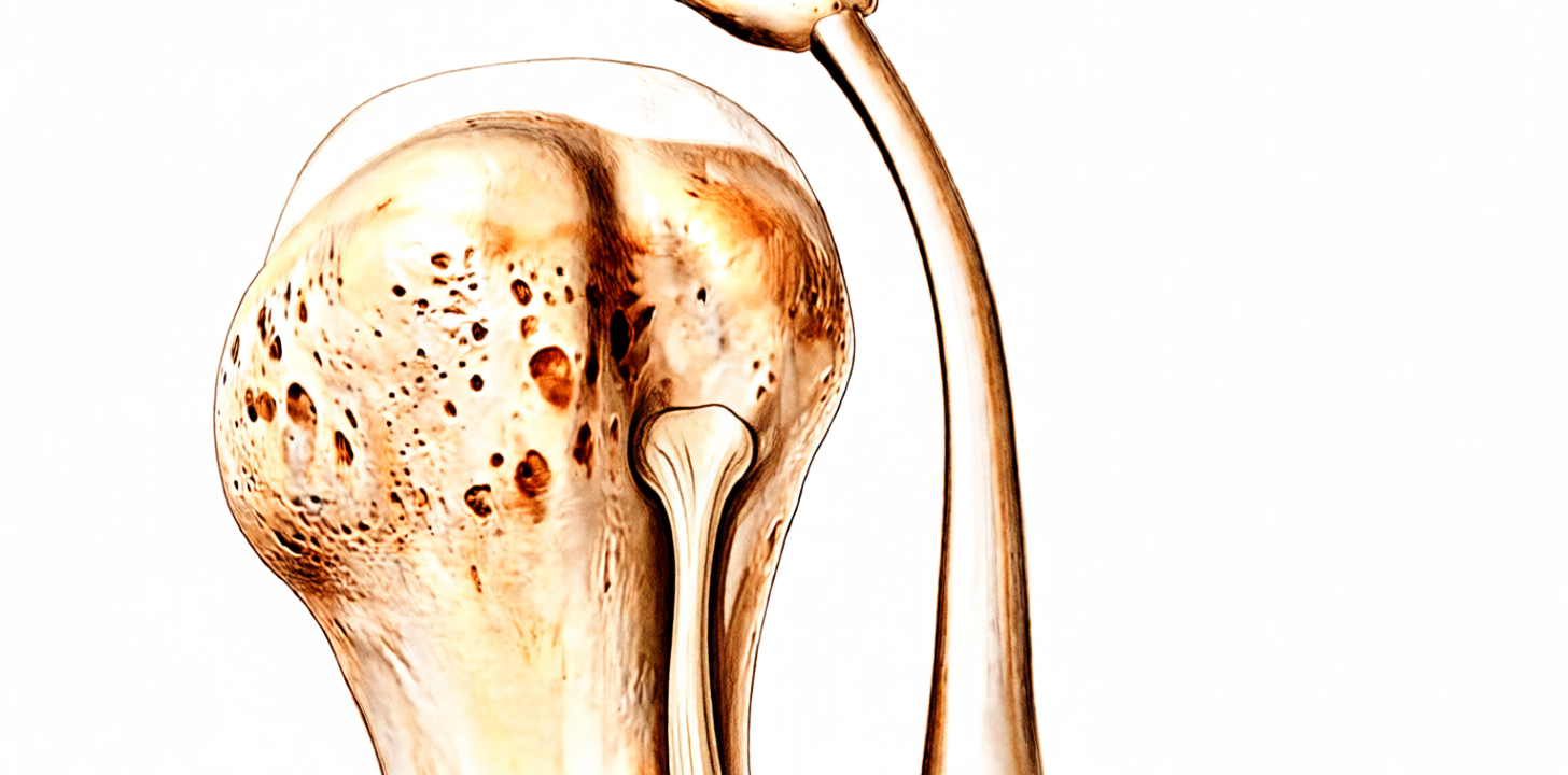
肱二头肌腱固定术是将肱二头肌长头腱(位于肩部前方的肌腱)重新固定到肱骨上一个新位置的手术。当调整活动方式和物理治疗等较简单的治疗未能带来足够改善,或者您本来就要接受另一项肩部手术而肱二头肌腱是问题的一部分时,我们通常会建议进行此手术。大多数人在术后 5 至 8 个月之间获得明显改善,到 13 个月时效果趋于稳定。手术旨在缓解您的疼痛,帮助您的肩部恢复正常功能。
术前
手术前,我们会依据影像学检查来规划手术,例如 X 线、MRI 或超声检查,这些检查可能已在您首次就诊时安排好。在手术前一周,您会收到我们团队的明确指示。您需要在手术前七小时停止进食和饮水。我们要求七小时而不是更短的时间,是为了在手术室排程提前时,您的手术也可以提前进行。请告诉我们您所服用的全部药物,包括任何抗凝药(血液稀释剂),并在手术当天携带一份书面药物清单。有些药物需要在手术前暂停,我们会告诉您是哪些药物以及何时暂停。请安排他人在术后驾车送您回家。请穿着宽松舒适的衣物。如果您有其他健康状况,可能需要进行血液检查,或接受麻醉医生(负责为您实施麻醉的医生)的评估,但大多数人两者都不需要。
手术当日
手术当天,您将前往医院的手术入院单元。我们的团队会为您办理入院手续,并为您进入手术室做好准备。麻醉医生将在手术前与您见面,并向您详细讲解这两部分麻醉方案。本手术在全身麻醉联合区域神经阻滞下进行。随后,您将被带入手术室进行手术。
手术结束后,您将在复苏区苏醒。护士会在此监测您的情况,直至麻醉作用消退。待您的情况平稳后,根据您所接受的手术及恢复情况,您将被转入病房或直接回家。
手术内容
这是一种关节镜(微创)手术。您的外科医生会在肩部周围做几个小切口,包括后方的一个切口,并借助一个伸入关节内的小型摄像头进行操作。外科医生会通过这些切口找到肱二头肌长头腱,即位于肩部前方、一直引起您疼痛的那条肌腱。
外科医生会将受损的肌腱从其磨损的部位松解,然后重新固定到肱骨上更低处的一个新位置。肌腱通过植入骨中的小型锚钉或螺钉固定在那里。这样可以让肌腱在一个新的、健康的位置上愈合,有点像把一根磨损的绳子移到柱子上更结实的地方。肌腱仍然附着在骨上,因此肱二头肌能保持其外形和力量。
如果您同时接受另一项肩部手术,例如肩袖修复术,肱二头肌的处理将在您已身处手术室时,通过相同的关节镜切口完成。您的外科医生会在术前与您详细讨论这一点。
手术结束时,器械会被取出,小切口会用缝线关闭。缝线位于敷料之下,我们的团队会与您一起护理敷料。您回家时会收到关于敷料和最初几天注意事项的书面说明。
肌腱的具体固定方式可能有所不同,您的外科医生会选择适合您的肩部和个人情况的方法。不变的是目标:将肌腱的疼痛部分移出关节,并锚定在坚固的骨骼上,使其能够平复下来,不再疼痛。
术后
大多数患者在此手术后需住院一晚,但部分患者可当日出院。您将在复苏室醒来,护士会密切观察您的情况,并给您用药让您保持舒适。您的手臂将佩戴一个简单的吊带以提供支撑,清洗和锻炼时可取下。请安排有人在前 24 小时内陪伴您。敷料需保留约 10 天;除非我们另行通知,否则请勿提前拆除。我们将在复诊时为您更换或拆除敷料。从第一天起您就可以在家中走动,但至少六周内不得驾驶。一旦外科医生确认您可以驾驶(通常在术后六周复诊时),您就可以重新开始驾驶。详情请参阅上肢手术后驾驶。
恢复
术后最初几天,您的肩部会感到疼痛,并可能伴有肿胀感。这些症状会在随后的几周内逐渐缓解。止痛药、休息和冰敷有助于让您保持舒适。您的手臂佩戴一个简单的吊带,清洗和锻炼时可取下。
从第一天起您就可以在家中走动。您的物理治疗师起初会指导您进行轻柔的活动,然后随着肌腱愈合逐步增强您的力量。对肱二头肌施加负荷的强化锻炼,例如抗阻屈肘或伸直手臂向前方举物,需推迟到肌腱牢固愈合之后进行。一旦外科医生确认您可以驾驶(通常在术后六周复诊时),您就可以重新开始驾驶。详情请参阅上肢手术后驾驶。
轻度日常活动会最先恢复。随着时间一周周过去,您会注意到疼痛逐渐减轻,穿衣、伸手够物等日常动作也会变得更容易。较重的工作、体育运动和健身房锻炼则会在之后恢复,前提是您的力量和活动度已经建立起来,并且您的物理治疗师和外科医生对您的进展感到满意。起初睡觉可能会不舒服;在肩部平复之前,靠坐在椅子上或仰卧睡觉通常会有帮助。
每个人的恢复情况各不相同。您的恢复时间线可能有所不同,您的外科医生和物理治疗师会在每一步为您提供指导。
可能出现的问题
大多数患者恢复良好,但偶尔也会出现并发症。您的外科医生和医疗团队会密切监测您的状况,以便尽早发现任何问题。
任何手术都有感染的风险。您可能会注意到从伤口向外扩散的发红、伤口渗液或流脓,或者出现发烧。如果发生这种情况,请在当天致电诊所。大多数伤口感染可通过伤口护理或抗生素得到控制,但少数需要进一步治疗。
神经在手术区域附近走行,因此神经可能在手术过程中受到刺激。这可能表现为手臂或手部麻木、刺痛或无力。神经阻滞后最初 24 小时内出现一些麻木和无力是预料之中的正常现象。在神经阻滞作用消退后(约 24 小时),如果您的手臂、手部或手指仍然麻木,或者无法活动,请致电诊所。
此手术后肩部可能会变得僵硬。您可能会发现难以向上够物、将手伸到背后或向侧方伸展,而且活动可能感觉紧绷或受阻。请在下次复诊时提出这一问题,因为在物理治疗师指导下尽早活动有助于使肩部放松。
固定肌腱的螺钉或锚钉偶尔会引起问题。肌腱可能在固定处受损,或者固定可能失效,这可能表现为突然“啪”的一下、屈肘时出现新的无力,或者肱二头肌在肩部前方附近隆起成团。在极少数情况下,上臂的肱骨也可能在植入物附近发生骨折,这会导致突然剧烈疼痛、肿胀以及手臂外形改变。如果您的手臂外形突然改变或完全无法使用,请前往急诊科。如果出现“啪”的一下或新的无力,请致电诊所。
有些人即使在愈合后,肩部前方仍持续疼痛,或者肱二头肌出现痉挛。请在复诊时提及这一点。如果疼痛持续,有时需要再做一次手术。
如果您想了解具体数据,本页的并发症表格列出了典型的并发症发生率。
何时联系我们
大多数问题会在最初几周内出现。了解需要留意的情况有助于您及早采取行动。
如果您出现小腿肿胀或疼痛,或者呼吸急促或胸痛,请前往急诊科。这些可能是血栓的迹象。如果您的手指或手部变得苍白、冰冷、发白、发青或发黑,或者您的手臂外形突然改变且完全无法使用,也请前往急诊科。
如果您出现发烧、伤口周围发红扩散,或者伤口渗液或流脓,请在当天致电诊所。如果尽管服用了止痛药,疼痛仍持续加重,也请在当天致电诊所。
神经阻滞后最初 24 小时内出现一些麻木和无力是预料之中的正常现象。在神经阻滞作用消退后(约 24 小时),如果您的手臂、手部或手指仍然麻木,或者无法活动,请致电诊所。
如果在下班时间或周末无法联系到诊所,请前往离您最近的急诊科。
关于该疾病的更多阅读
本页主要介绍手术本身。关于该手术所治疗的疾病,包括现有证据显示手术在何时有效、何时无效,将在肱二头肌肌腱病及长头撕裂页面中作更详细的介绍。
Evidence & references
This is the clinical evidence summary written for health professionals. It is technical, and it lists the research this page was built from. You do not need to read it to understand your treatment or to make a decision about it.
Overview
- Arthroscopic suprapectoral biceps tenodesis using standard portals aims to maximize outcomes and minimize common complications associated with biceps tenodesis [1].
- Good clinical results have been shown with the use of several soft tissue tenodesis techniques for tenodesis of the long head of the biceps [2].
- A single portal technique for proximal biceps tenodesis in the bicipital groove using an all-suture anchor has been demonstrated [3].
- Arthroscopic keyhole biceps tenodesis is hypothesized to be a safe, reproducible, and cost-effective technique [4].
- In situ subpectoral biceps tenodesis with a cortical button offers potential advantages including in situ biceps tendon fixation, creation of a smaller cortical defect, and subcortical attachment of a nonbioreactive device [5].
- A technique for arthroscopicly aided biceps tenodesis has been developed and presented [6].
- The described arthroscopic suprapectoral biceps tenodesis onlay technique is safe, simple, reproducible, and reduces the risk of complications related to open subpectoral tenodesis and arthroscopic intraarticular biceps tendon fixation [7].
- Arthroscopic extra-articular suprapectoral biceps tenodesis is considered an excellent option to address biceps pathologies, especially in active patients [8].
- Arthroscopic biceps tenodesis is a safe and reliable treatment for managing intra-articular biceps tendon pathology [9].
- Tenodesis of the long head of the biceps tendon can give excellent and good results in the long term with an average follow-up of 7 years [10].
- Mini-open biceps tenodesis using an onlay technique with enthesis growth augment offers a reproducible and biologically enhanced alternative that may optimize enthesis healing and reduce the risk of failure [11].
- Revision biceps tenodesis with tibialis anterior allograft tendon augmentation is an effective surgical technique for symptomatic failed biceps tenodesis in a young active patient [12].
- Suprapectoral biceps tenodesis during total shoulder arthroplasty using an onlay technique has good outcomes and low rates of overall and biceps-related complications [13].
- The described proximal biceps tenodesis fixation in double-row rotator cuff repair is simple and cost-effective, with no need for additional anchors [14].
- An all-arthroscopic suprapectoral biceps tenodesis technique utilizing a knotless locking button has been described [15].
- Patients undergoing simultaneous rotator cuff repair and biceps tenodesis demonstrate similar patient-reported and objective outcomes for both lateral-row tenodesis and in-the-groove tenodesis techniques [17].
- An in situ variation of arthroscopic suprapectoral biceps tenodesis using a double loop-and-tack knotless suture anchor provides an option for inclusion in the surgical armamentarium [18].
- A biceps tenodesis technique that can be performed percutaneously using needle arthroscopy under regional anesthesia has been described [19].
- The gripping biceps knot technique provides a safe, efficient, and effective approach to enhancing biomechanical integrity and minimizing repair failures in proximal biceps tenodesis [20].
- Both tenotomy and tenodesis produced good clinical outcomes in the treatment of concurrent long head of biceps tendon lesions and rotator cuff tears [29].
- Available randomized controlled trials comparing suprapectoral and subpectoral approaches to tenodesis lack specific matching of the uniformity of the particular techniques [35].
Anatomy & Pathophysiology
Bony Anatomy and Tendon Origin
- The long head of the biceps originates from the bicipital tubercle at the superior rim of the glenoid and along the posterior superior rim of the glenoid and labrum [43].
- 40% to 60% of the biceps tendon attaches to the supraglenoid tubercle 5 mm medial to the superior glenoid rim, with the remainder attaching directly to the superior glenoid labrum [41].
- The biceps tendon typically attaches entirely (type I) or predominantly posterior (type II) on the superior labrum [41].
- The labral attachment of the biceps tendon may have equal anterior and posterior contributions (type III) or, less commonly, predominantly anterior (type IV) [41].
- Type I attachment involves all of the labral part attaching to the posterior labrum with none to the anterior labrum [56].
- Type II attachment involves most of the labral contribution attaching to the posterior labrum with a small contribution to the anterior labrum [56].
- Type III attachment involves equal contributions to both the anterior and posterior parts of the labrum [56].
- Type IV attachment involves most of the labral contribution attaching to the anterior labrum with a small contribution to the posterior labrum [56].
- The biceps tendon is an intra-articular but extrasynovial structure within the glenohumeral joint [41].
- The superior labrum inserts on the superior glenoid rim, medial to the articular cartilage margin, through a transitional zone of fibrocartilage [41].
- A normal synovial recess exists between the meniscoid or triangular superior labrum and the articular cartilage extension over the superior glenoid rim [41].
- The glenoid labrum consists of parallel collagen fibers that course around the circumference of the glenoid [41].
- The superior labrum is usually triangular but can have a meniscoid shape, and commonly attaches medial to the articular margin of the glenoid rim [44].
- Anatomic variants in the superior labrum include a sublabral foramen or absence of the superior labrum, often seen together with a cordlike middle glenohumeral ligament (MGHL) [44].
- In a cohort of 73 shoulders, 3.3% had a sublabral foramen, 8.6% had a sublabral foramen with cordlike MGHL (Buford complex), and 1.5% had an absent anterosuperior labrum [44].
Vascularity and Innervation
- Vascularity to the glenoid labrum originates from the scapular, circumflex scapular, and posterior circumflex humeral arteries via capsular or periosteal vessels [41].
- The suprascapular artery, the circumflex scapular branch of the subscapular artery, and the posterior humeral circumflex artery provide the labrum’s vascular supply [44].
- The inner portion of the labrum is avascular, and the superior labrum is less vascular compared with the inferior and posterior labrum [44].
- Vascularity of the biceps tendon is provided primarily by the ascending branch of the anterior humeral circumflex artery, which travels within the bicipital groove [41].
- An avascular zone exists at the proximal portion of the biceps tendon, close to the superior glenoid [41].
- Blood is supplied to the long head of the biceps (LHB) tendon from the thoracoacromial and brachial arteries via the osteotendinous and musculotendinous junctions, respectively [44].
- A hypovascular zone found near the tendon origin at the superior glenoid attachment corresponds to where it commonly tears at the LHB pulley near the proximal groove [44].
- The LHB tendon is innervated by thinly myelinated sensory neurons, with most innervation occurring at the LHB origin [44].
- Pathology in the region of the LHB origin can generate pain due to this innervation [44].
- Innervation of the biceps is supplied by branches of the musculocutaneous nerve (C5 and C6) [43].
Bicipital Groove and Pulley Anatomy
- The biceps tendon passes through the bicipital groove, or intertubercular groove, between the greater and lesser tuberosities [41].
- Stability of the biceps within the bicipital groove is afforded by the biceps sling, or pulley, consisting of fibers from the subscapularis tendon, supraspinatus tendon, coracohumeral ligament, and superior glenohumeral ligament [41].
- The LHB pulley is a capsuloligamentous complex comprising the superior glenohumeral ligament, the coracohumeral ligament, and fibers from the subscapularis and supraspinatus tendons [44].
- The biceps pulley stabilizes the proximal LHB as the tendon enters the bicipital groove [44].
- The bicipital tendon does not move up and down in the groove; rather, the humerus moves down and up with adduction and abduction relative to the tendon [43].
- The biceps tendon is retained within the groove by a pulley made up of fibers from the coracohumeral and superior glenohumeral ligaments, with some reinforcement from adjacent tendons [43].
- The rotator cuff interval is a triangular region bounded medially by the coracoid process, superiorly by the anterior margin of the supraspinatus, and inferiorly by the superior margin of the subscapularis [45].
- The biceps tendon traverses the rotator cuff interval, where it is held in place by the biceps pulley, before exiting the joint via the bicipital groove [45].
- Zone 1 and zone 2 of the extra-articular bicipital tunnel contain synovial tissue, which may generate pain [44].
- Zone 2 of the extra-articular bicipital tunnel cannot be visualized by arthroscopy from above or with an open approach from below the zone [44].
Biomechanics and Function
- The biceps has its main action at the elbow rather than the shoulder and is considered primarily an elbow muscle [43].
- Loss of the long head attachment is manifested mainly as loss of supination strength (20%) with a smaller loss (8%) of elbow flexion strength [43].
- The role of the biceps tendon on the static and dynamic stability of the shoulder is controversial [58].
- Andrews noted a dynamic tensioning of the biceps-labral complex with electric stimulation of the biceps tendon [58].
- From full adduction to full elevation of the arm, the groove moves a distance of up to 2 to 5 cm along the tendon [58].
- Maximal excursion of the humeral head along the tendon results when the shoulder is in a position of maximal external rotation [58].
- Minimal excursion is seen when the shoulder is in a position of maximal internal rotation [58].
- The synovial pouch extends from the shoulder joint, lining the greater part of the intertubercular groove to facilitate excursion along the tendon [58].
- With the arm in full abduction, 1.3 cm of the long head of the biceps tendon (LHBT) lies within the shoulder joint [58].
- When the arm is adducted and externally rotated, the length of tendon within the joint increases to 5 cm [58].
- In external rotation, the long head of the biceps acts as a head depressor at the shoulder to enhance abduction strength [58].
- The biceps can potentially act as a static humeral head depressor, preventing migration of the humeral head into the acromion with contraction of the deltoid [58].
- The function of the biceps tendon as a humeral head depressor increased in the context of a chronic rotator cuff tear [58].
- Both the long and short heads of the biceps function as anterior stabilizers of the glenohumeral joint with the arm in abduction and external rotation [58].
- With increasing instability from sectioning of the inferior glenohumeral ligament, both heads of the biceps have an increased stabilizing function to resist anterior displacement of the humeral head [58].
- Severing the LHBT while both heads were tensed caused significant upward migration of the humeral head [58].
- The long head of the biceps is important in stabilizing the humeral head in the glenoid during powerful elbow flexion and forearm supination [58].
- In shoulders with cuff tears, muscle activity of the long head of the biceps increased in internal rotation at the upper arm elevation to compensate for cuff insufficiency [75].
Pathophysiology and Instability
- SLAP tears can be caused by forceful traction to the arm, direct compression loads, and repetitive overhead throwing [46].
- Increased external rotation of the shoulder in the late cocking phase increases torsional force at the LHB root, resulting in a peel-back injury to the posterosuperior labrum [46].
- Injuries can result from repetitive contact of the posterosuperior labrum with the undersurface of the rotator cuff in the late cocking phase, known as internal impingement [46].
- SLAP tears are seen more frequently in the late cocking position, occurring because of an adaptive posterior capsular contracture [46].
- Throwing athletes demonstrate increased shoulder external rotation and decreased internal rotation in abduction, which causes posterosuperior migration of the humeral head in the late cocking phase [46].
- Increased external rotation results in greater torsional loads across the superior labrum from the more posteriorly oriented LHB tendon, causing the labrum and LHB tendon to displace medially over the glenoid rim [46].
- The proximal LHB tendon has been recognized as a source of substantial anterior shoulder pain [46].
- Pathology of the LHB tendon includes tendinitis, tendinopathy, tears, subluxation, entrapment, delamination, and dislocation out of the bicipital groove [46].
- Because of the relatively anterior position of the bicipital groove along the humeral head combined with humeral retroversion, the tendon is exposed to medial instability, which can increase the risk of tendon degeneration [46].
- Variations of bicipital groove morphology can also increase the risk of LHB tendon pathology [46].
- Isolated LHB tendon pathology can occur but frequently is associated with other shoulder pathologies, especially rotator cuff pathology [46].
- Primary LHB tendinitis usually occurs in younger patients who participate in overhead activities such as volleyball and baseball [46].
- With LHB tendon instability, the patient describes a clicking or snapping with overhead motions [46].
- A subscapularis tear is associated with LHB medial instability and a supraspinatus tear is associated with posterolateral instability [46].
- Bicipital instability is usually associated with rotator interval injury or subscapularis tendon injury, or both [47].
- Biceps tendinitis is rarely the primary cause of shoulder pain and is usually secondarily involved as a part of an impingement syndrome or degenerative lesions of the rotator cuff [47].
- A shallow bicipital groove and supratubercular ridge above the lesser tubercle were thought to predispose to biceps tendon pathology [43].
- A shallower bicipital groove may be more likely to expose the long head of the biceps to impingement [43].
- The medial wall of the bicipital groove was higher, with an opening angle of 30 to 40 degrees in the largest fraction of patients [43].
- Subluxation type I involves a partial or complete tear of the rotator interval sling (superior glenohumeral and coracohumeral ligaments) resulting in loss of restraint above the entrance to the groove [63].
- Subluxation type II involves a lesion below the entrance to the bony groove where the tendon slips over the medial rim of the bony groove and rides on the border of the lesser tuberosity [63].
- The causal lesion for subluxation type II is a detachment of the outermost fibres of the subscapularis tendon [63].
- Subluxation type III involves malunion and nonunion of the lesser tuberosity that compromises the medial bony restraint of the long biceps tendon [63].
- Dislocation type I involves extraarticular dislocation combined with a partial tear of the subscapularis tendon [57].
- In dislocation type I, the biceps tendon is displaced over the anterior wall of the groove and slips or glides medially over the torn fibres of the subscapularis tendon [57].
- Dislocation type II involves intraarticular dislocation of the long biceps tendon combined with a complete tear of the subscapularis tendon [57].
- In dislocation type II, the biceps tendon is interposed into the joint space and displaced inferomedially, with entrapment occurring with each internal rotational movement of the humerus [57].
- Dislocation of the long biceps tendon over a completely intact subscapularis tendon is very rare, occurring in only 2 patients (3%) in a series of 70 patients with subluxation and dislocations [57].
Classification
- Arthroscopic suprapectoral biceps tenodesis can be performed using standard arthroscopic portals [1].
- Soft tissue tenodesis techniques have demonstrated good clinical results for tenodesis of the long head of the biceps [2].
- A single portal technique exists for proximal biceps tenodesis in the bicipital groove using an all-suture anchor [3].
- An arthroscopicly aided biceps tenodesis technique has been developed [6].
- Arthroscopic suprapectoral biceps tenodesis using an onlay technique is described as safe, simple, reproducible, and reduces risk of complications related to open subpectoral tenodesis and arthroscopic intraarticular biceps tendon fixation [7].
- Proximal biceps tenodesis fixation in double-row rotator cuff repair is simple and cost-effective with no need for additional anchors [14].
- An all-arthroscopic suprapectoral biceps tenodesis technique utilizes a knotless locking button [15].
- All-arthroscopic falciform portal biceps tenodesis allows for improved management of more distal biceps lesions while avoiding potential complications associated with an open approach [16].
- An in situ variation of arthroscopic suprapectoral biceps tenodesis is provided by a technique using a double loop-and-tack knotless suture anchor [18].
- A biceps tenodesis technique can be performed percutaneously using needle arthroscopy under regional anesthesia [19].
- An all-arthroscopic in situ biceps tenodesis technique is performed at the inferior portion of the bicipital groove with the biceps tendon still attached proximally, maintaining the tendon’s in situ length-tension relationship [23].
- Functional results of the open interference screw technique for biceps tenodesis are difficult to elucidate as patient results are typically more related to the rotator cuff repair [25].
- Arthroscopic bicortical biceps anchorage for subpectoral biceps tenodesis underscores the need for further biomechanical and clinical evaluation [26].
- A modified docking configuration into the intraosseous canal decreases stresses at the bone-tendon interface in a cost-effective, implant-free, all-suture modified subpectoral biceps tenodesis technique [28].
- Contemporary literature suggests no clear superiority of one specific biceps tenodesis technique over others [31].
- A network meta-analysis found no significant differences in multiple outcome measures when comparing open versus arthroscopic biceps tenodesis [31].
- A systematic review found no significant difference in Constant, American Shoulder and Elbow Surgeons, or Single Assessment Numeric Evaluation scores between arthroscopic and open biceps tenodesis [31].
- Biomechanical evaluation found no significant differences in construct strength when comparing suprapectoral versus subpectoral fixation locations or between different fixation types including interference screws, suture anchors, and cortical buttons [31].
- Contemporary all-suture anchors appear not only equivalent but superior to more classical metal anchors with respect to fixation strength [31].
- Data suggests that greater tendon migration correlates with lower patient-reported outcomes [31].
- A reproducible systematic technique exists for open repair of teres major and latissimus dorsi tendon ruptures with accompanying biceps tenodesis using cortical suspensory fixation buttons [33].
- An arthroscopic double-cinch double-cerclage technique for proximal biceps tenodesis incorporates a unique construct of circumferential and trans-tendon suture passing at the bicipital groove [34].
- An arthroscopic inlay biceps tenodesis technique uses a tendon-docking anchor [40].
- An arthroscopically assisted mini-open transdeltoid biceps tenodesis technique has been described [51].
Clinical Presentation
- Painful long head of the biceps tendon (LHBT) tendinitis may ensue from tears about the rotator interval or with any chronic inflammatory pathology of the glenohumeral joint [71].
- Clinical tests including the O’Brien, Yergason, Speed, and direct palpation tests have limited specificity for diagnosing LHBT pathology [71].
- A history of radiating anterior shoulder pain may inform the examiner of pain generation from the LHBT when combined with clinical tests [71].
- MRI, ultrasonography, and arthroscopic examination are tools used to evaluate biceps pathology [71].
- Arthroscopic examination is limited to the intra-articular LHBT and the proximal groove, missing less common distal biceps groove lesions [71].
- Isolated traumatic tears of the LHBT are generally treated nonsurgically [71].
- Tenodesis for isolated traumatic tears is a rare exception indicated for the dominant arm of a laborer or an individual who cannot tolerate deformity [71].
- Arthroscopic tenotomy is acceptable for less physically demanding individuals who may tolerate deformity [71].
- Tenotomy results in cosmetic deformity (Popeye) about 30% of the time [71].
- Vigorous activity following tenotomy may result in cramping pain of the biceps muscle belly [71].
- Arthroscopic suprapectoral tenodesis may be performed for SLAP tears or in conjunction with rotator cuff repair for a patient who needs full supination strength and endurance [71].
- Open or arthroscopic-assisted subpectoral tenodesis are options if biceps groove pathology is a concern [71].
Investigations
Clinical Examination
- The "3-pack" examination for biceps-labrum complex disease consists of the active compression test, throwing test, and bicipital tunnel palpation [42].
- The 3-pack tests demonstrated high sensitivity ranging from 73% to 98% for biceps-labrum complex disease [42].
- No single physical examination finding is completely accurate for the diagnosis of a SLAP tear [48].
- A combined physical examination approach aids in the diagnosis of SLAP or long head of biceps pathology [48].
- The proximal long head of biceps tendon is a source of substantial anterior shoulder pain [46].
- Clinical diagnosis of SLAP tears or symptomatic long head of biceps tendinopathy is challenging because findings are similar to other pathologies within the glenohumeral joint [48].
Imaging
- Plain radiographs, including scapular Y, AP, and axillary lateral views, are used to assess the glenohumeral joint for abnormalities [65].
- MRI is used to assess the long head of biceps tendon, associated fluid, possible synovitis, bicipital groove morphology, and the presence of bony osteophytes [65].
- Studies have demonstrated poor correlation between MRI and arthroscopic findings regarding long head of biceps pathology [65].
- MRI has poor to moderate sensitivity for inflammation, partial-thickness tendon tears, and tendon ruptures of the long head of biceps [65].
- Magnetic resonance arthrography is more specific and sensitive for long head of biceps pathology and SLAP tears than MRI alone [65].
- On MRA in patients with no pathology, the biceps tendon is surrounded by contrast fluid and resembles a kidney bean [65].
- Both MRI and MRA should be performed in the sagittal oblique and axial planes because long head of biceps subluxation and dislocation are often associated with partial-thickness and full-thickness subscapularis tendon tears [65].
- Ultrasonography is accurate and cost-effective in the diagnosis of long head of biceps dislocation, subluxation, and rupture [65].
- Ultrasonography is not as accurate as other modalities in diagnosing partial-thickness tendon tears of the long head of biceps [65].
- Proton density–weighted sequences with fat suppression have the greatest sensitivity for detecting tendon degeneration [45].
- Tendon caliber change is more specific than signal intensity for detecting tendon degeneration [45].
- Half of biceps tendon partial tears at the groove entrance show an associated caliber change [45].
- Evaluation in all imaging planes aids in the identification of a biceps groove entrance lesion [45].
- MRA has a sensitivity of 82% to 89% and a specificity of 87% to 98% in the evaluation of the biceps pulley [45].
- Diagnostic criteria for biceps pulley evaluation on MRA include nonvisualization or discontinuity of the superior glenohumeral ligament, medial subluxation of the biceps tendon on axial images, biceps tendinopathy, and inferior displacement on oblique sagittal images [45].
- The Fisk view is a radiographic technique used to evaluate the anatomy of the bicipital groove [47].
Treatment
Operative Techniques and Fixation
- A single portal technique for proximal biceps tenodesis in the bicipital groove using an all-suture anchor has been described [3].
- An arthroscopicly aided biceps tenodesis technique has been developed and presented [6].
- Arthroscopic suprapectoral biceps tenodesis using an onlay technique is safe, simple, reproducible, and reduces the risk of complications related to open subpectoral tenodesis and arthroscopic intraarticular biceps tendon fixation [7].
- Revision biceps tenodesis with tibialis anterior allograft tendon augmentation is an effective surgical technique for the rare case of symptomatic failed biceps tenodesis in a young active patient [12].
- A simplified proximal biceps tenodesis fixation in double-row rotator cuff repair is simple and cost-effective, with no need for additional anchors [14].
- All-arthroscopic falciform portal biceps tenodesis allows for improved management of more distal biceps lesions while avoiding the potential complications associated with an open approach [16].
- An in situ variation of arthroscopic suprapectoral biceps tenodesis using a double loop-and-tack knotless suture anchor provides an option for the surgical armamentarium [18].
- The gripping biceps knot provides a safe, efficient, and effective approach to enhancing biomechanical integrity and minimizing repair failures in proximal biceps tenodesis [20].
- In clinical scenarios where an upper border subscapularis tear is also to be repaired, either a soft tissue or bony technique can be employed that effectively addresses both the subscapularis tear and a symptomatic biceps tendon [21].
- An all-arthroscopic biceps tenodesis technique performed in the in situ position at the inferior portion of the bicipital groove maintains the tendon’s in situ length-tension relationship [23].
- Subpectoral biceps tenodesis using dynamic endobutton fixation in a humeral bone tunnel with interference screw augmentation provides a safe, minimally invasive fixation that may help accelerate return to demanding professional and athletic activities [24].
- Arthroscopic suprapectoral biceps tenodesis below the groove places the bony anchor with minimal soft tissue disruption, minimal risk for major postoperative complications, and comparable biomechanical outcomes to other techniques [27].
- A cost-effective, implant-free, all-suture modified subpectoral biceps tenodesis technique uses a docking configuration into the intraosseous canal that decreases stresses at the bone-tendon interface [28].
- A high-in-the-groove biceps tenodesis using a loop-and-tack technique offers a simple, effective, and reproducible approach to treat long head of biceps tendon and/or superior labral pathology [31].
- Teres major and latissimus dorsi repair with biceps tenodesis utilizing cortical suspensory fixation buttons is a reproducible systematic technique [33].
- An arthroscopic double-cinch double-cerclage technique for proximal biceps tenodesis at the bicipital groove incorporates a unique construct of circumferential and trans-tendon suture passing [34].
- A suprapectoral double-row technique with all-suture anchors for long head of biceps tenodesis aims to maintain inherent length and uniform tension at the bicipital groove [35].
- An all-arthroscopic simple double 360° lasso loop technique for suprapectoral biceps tenodesis requires further clinical and biomechanical studies to evaluate reliability [36].
- Subpectoral biceps tenodesis using an all-suture knotless anchor involves a lateral decubitus position with standard posterior and direct anterior portals [52].
- The BITER device can be a useful tool for both arthroscopic and open shoulder surgery during tendon extraction for biceps tenodesis [53].
- Tenodesis can be performed with a PEEK tenodesis screw, two suture anchors, or a FiberSnare, with comparable resistance to cyclic loading and stronger ultimate pull-out strength for the biotenodesis screw compared to suture anchors [66].
- Long-term results are comparable whether biceps tenodesis is done arthroscopically or through a mini-open approach with a small anterior or subpectoral incision [66].
- For arthroscopic or mini-open biceps tenodesis with screw fixation, the bone tunnel is reamed 10 to 15 mm below the insertion of the supraspinatus lateral to the subscapularis insertion at the level of the transverse humeral ligament [66].
- The depth of insertion for the tenodesis reamer is 20 mm, with an 8-mm reamer used for most men and a 7-mm reamer for most women [66].
- The tenodesis screw is inserted flush with the cortex, and stability is checked by rotating the humerus [66].
- In the rare instance when no pathologic condition exists in the rotator cuff, an anterior approach through the deltopectoral interval is used to identify the long head of the biceps tendon and perform tenodesis [73].
- If a pathologic process of the rotator cuff is present with a subluxing biceps tendon, an anterosuperior approach is used to expose the deltoid, perform acromioplasty, and tenodese the biceps tendon to the humerus with interference or tenodesis screws or suture anchors [73].
- Alternatively, the proximal biceps tendon attachment can be released arthroscopically followed by a tenodesis procedure or a subpectoral technique [73].
Biomechanics and Comparative Outcomes
- Available randomized controlled trials comparing suprapectoral and subpectoral tenodesis approaches lack specific matching of the uniformity of the particular techniques [35].
- Functional results of the open interference screw technique are difficult to elucidate as patients’ results are typically more related to the rotator cuff repair [25].
Non-Operative Management
- Ultrasound-guided biceps tenotomy combined with corticosteroid injection can be an optimal option for patients who need nontraditional management for rotator cuff tears [30].
Postoperative Care
- If only a biceps tenodesis was performed, postoperative management is the same as for arthroscopic acromioplasty [66].
- Strengthening activities related to elbow flexion or forward elevation of the arm with the elbow extended should be restricted until 6 weeks after the biceps tenodesis [66].
- A shoulder immobilizer is worn for 2 weeks, followed by a sling for an additional 2 weeks, after which active use and exercises are begun [73].
- If the rotator cuff was repaired during biceps tenodesis, rehabilitation depends on the size of the tear [73].
Complications
- Simultaneous musculocutaneous nerve entrapment and radial nerve traction injury can occur after open subpectoral biceps tenodesis [32].
- A rope-effect mechanism has been demonstrated in cases of simultaneous musculocutaneous nerve entrapment and radial nerve traction injury following open subpectoral biceps tenodesis [32].
- Suprapectoral biceps tenodesis during total shoulder arthroplasty using an onlay technique has low rates of overall and biceps-related complications [13].
- Revision biceps tenodesis with tibialis anterior allograft tendon augmentation is an effective surgical technique for the rare case of symptomatic failed biceps tenodesis [12].
- A single failure was observed in a 65-year-old patient with a long head of biceps rupture present for about 8 years following infrapectoral biceps tenodesis [72].
- The arthroscopic keyhole biceps tenodesis technique is hypothesized to be safe [4].
- The all-arthroscopic suprapectoral biceps tenodesis technique utilizing a knotless locking button is described as a method for the procedure [15].
- The mini-open biceps tenodesis using an onlay technique with enthesis growth augment may reduce the risk of failure [11].
- The arthroscopic suprapectoral biceps tenodesis using standard portals aims to minimize common complications associated with biceps tenodesis [1].
- The arthroscopic suprapectoral biceps tenodesis using an onlay technique reduces the risk of complications related to open subpectoral tenodesis and arthroscopic intraarticular biceps tendon fixation [7].
- The all-arthroscopic falciform portal biceps tenodesis technique avoids the potential complications associated with an open approach [16].
- The arthroscopic suprapectoral biceps tenodesis below the groove technique carries a minimal risk for major postoperative complications [27].
- The gripping biceps knot technique aims to minimize repair failures in proximal biceps tenodesis [20].
Recovery
- Biomechanical evaluation found no significant differences in construct strength when comparing suprapectoral versus subpectoral fixation locations [31].
- Biomechanical evaluation found no significant differences in construct strength between different fixation types, including interference screws, suture anchors, and cortical buttons [31].
- Contemporary all-suture anchors appear superior to more classical metal anchors with respect to fixation strength [31].
- Greater tendon migration correlates with lower patient-reported outcomes [31].
Key Evidence
- [L5] This technique simplifies the procedure to be performed from standard arthroscopic portals and aims to maximize outcomes and minimize common complications associated with biceps tenodesis. [1] (10.1016/j.eats.2023.04.002)
- [Paper] Good clinical results have been shown with the use of several soft tissue tenodesis techniques for tenodesis of the long head of the biceps. [2] (10.1097/bte.0b013e31816408ee)
- [L5] The presented technique demonstrates a single portal technique for a proximal biceps tenodesis in the bicipital groove using an all-suture anchor. [3] (10.1016/j.eats.2021.11.023)
- [L4] The hypothesis was that the arthroscopic keyhole biceps tenodesis can be a safe, reproducible, and cost-effective technique. [4] (10.1097/bte.0b013e3182116be8)
- [L5] This method has potential advantages over existing subpectoral biceps tenodesis techniques such as in situ biceps tendon fixation, creation of a smaller cortical defect, and subcortical attachment of a nonbioreactive device. [5] (10.1097/bte.0b013e3181a94599)
- [Paper] A technique for an arthroscopicly aided biceps tenodesis has been developed and is presented. [6] (10.1016/s1058-2746(95)80204-5)
- [L5] The technique described is safe, simple, reproducible, and reduces risk of complications related to open subpectoral tenodesis and arthroscopic intraarticular biceps tendon fixation. [7] (10.1016/j.eats.2024.103123)
- [L5] We believe arthroscopic extra-articular suprapectoral biceps tenodesis is an excellent option to address biceps pathologies, especially in active patients. [8] (10.1016/j.eats.2024.102922)
- [L5] Arthroscopic biceps tenodesis is a safe and reliable treatment for managing intra-articular biceps tendon pathology. [9] (10.1016/j.eats.2024.103207)
- [L4] Tenodesis of the long head of the biceps tendon can give excellent and good results in the long term (average follow-up, 7 years). [10] (10.1016/s1058-2746(05)80034-5)
- [L5] This method offers a reproducible and biologically enhanced alternative for proximal biceps tenodesis that may optimize enthesis healing and reduce the risk of failure. [11] (10.1002/atn2.70167)
- [L4] Revision biceps tenodesis with tibialis anterior allograft tendon augmentation is an effective surgical technique for the rare case of symptomatic failed biceps tenodesis in a young active patient. [12] (10.1016/j.eats.2021.12.029)
- [L3] Suprapectoral biceps tenodesis during TSA using an onlay technique has good outcomes and low rates of overall and biceps-related complications. [13] (10.5435/jaaosglobal-d-25-00369)
- [L5] The described proximal biceps tenodesis fixation is simple and cost-effective, with no need for additional anchors. [14] (10.1016/j.eats.2025.103634)
- [L5] We describe an all-arthroscopic suprapectoral biceps tenodesis technique utilizing a knotless locking button. [15] (10.1016/j.eats.2025.103498)
- [Paper] This technique allows for improved management of more distal biceps lesions while avoiding the potential complications associated with an open approach. [16] (10.1016/j.eats.2023.09.017)
- [L3] Patients undergoing simultaneous RCR and BT demonstrate similar patient-reported and objective outcomes for both LR tenodesis and in-the-groove tenodesis techniques. [17] (10.1016/j.jses.2019.09.008)
- [L5] This technique provides an in situ variation of arthroscopic suprapectoral biceps tenodesis for inclusion in the surgical armamentarium. [18] (10.1016/j.eats.2023.04.014)
- [L5] The purpose of this article is to describe a biceps tenodesis technique that can be performed percutaneously using needle arthroscopy under regional anesthesia. [19] (10.1016/j.eats.2024.103414)
- [L5] This technique provides a safe, efficient, and effective approach to enhancing biomechanical integrity and minimizing repair failures in proximal biceps tenodesis. [20] (10.1016/j.eats.2025.103831)
- [L5] In clinical scenarios in which an upper border subscapularis tear is also to be repaired, either a soft tissue or bony technique can be employed that effectively addresses both the subscapularis tear and a symptomatic biceps tendon. [21] (10.1016/j.eats.2025.103724)
- [L5] The authors describe a unique all-arthroscopic biceps tenodesis technique performed in the in situ position at the inferior portion of the bicipital groove with the biceps tendon still attached proximally, thereby maintaining the tendon’s in situ length-tension relationship. [23] (10.1097/bte.0b013e318182c3bf)
- [L5] This novel technique provides a safe, minimally invasive fixation of the long head of the biceps and may help to accelerate return to demanding professional and athletic activities. [24] (10.1097/bte.0b013e3182270fab)
- [L4] Functional results of this technique are difficult to elucidate, as the patients’ results are typically more related to the rotator cuff repair. [25] (10.1097/00132589-200312000-00006)
- [L5] These considerations underscore the need for further biomechanical and clinical evaluation. [26] (10.1002/atn2.70089)
- [L5] The technique places the bony anchor for the long head of the biceps tendon below the bicipital groove with minimal soft tissue disruption, minimal risk for major postoperative complications, and comparable biomechanical outcomes to other techniques. [27] (10.1016/j.eats.2025.103707)
- [L5] The modified docking configuration into the intraosseous canal decreases stresses at the bone-tendon interface. [28] (10.1016/j.eats.2023.11.001)
- [L1] Both tenotomy and tenodesis produced good clinical outcomes in the treatment of concurrent LHBT lesions and rotator cuff tears. [29] (10.1097/bte.0000000000000142)
- [L4] Ultrasound-guided biceps tenotomy combined with corticosteroid injection can be an optimal option for patients who need nontraditional management for rotator cuff tears. [30] (10.1016/j.eats.2023.09.022)
- [L5] [31] (10.1002/atn2.70105)
- [L5] [32] (10.1016/j.xrrt.2026.100806)
- [L5] The authors present a reproducible systematic technique for open repair of teres major and latissimus dorsi tendon ruptures with accompanying biceps tenodesis using cortical suspensory fixation buttons. [33] (10.1016/j.eats.2022.10.017)
- [L5] This technical note introduces an arthroscopic technique for proximal biceps tenodesis at the bicipital groove that incorporates a unique construct of circumferential and trans-tendon suture passing. [34] (10.1016/j.eats.2025.103464)
- [L5] The available randomized controlled trials comparing the approaches to tenodesis, namely suprapectoral and subpectoral, lack specific matching of the uniformity of the particular techniques. [35] (10.1016/j.eats.2024.103130)
- [L5] Further clinical and biomechanical studies are needed to evaluate the reliability of this tenodesis technique. [36] (10.1016/j.eats.2023.02.008)
- [Paper] [40] (10.1016/j.eats.2024.103284)
- [L4] [51] (10.1097/bte.0b013e318297c451)
- [L5] [52] (10.1016/j.eats.2023.02.030)
- [L5] The BITER can be a useful device for both arthroscopic and open shoulder surgery. [53] (10.1016/j.eats.2023.09.020)
- [L4] [72] (10.1097/bte.0b013e3182443d1d)
- [L4] We suggest that in the shoulders with cuff tears, muscle activity of the LHB increased in internal rotation at the upper arm elevation to compensate the cuff insufficiency. [75] (10.1016/s1058-2746(96)80562-3)
References
[1] Arthroscopic Suprapectoral Biceps Tenodesis Using Standard Portals. Arthroscopy Techniques. 2023. DOI: 10.1016/j.eats.2023.04.002
[2] Biceps Tenodesis. Techniques in Shoulder & Elbow Surgery. 2008. DOI: 10.1097/bte.0b013e31816408ee
[3] Single‐Portal Proximal Biceps Tenodesis Using an All‐Suture Anchor. Arthroscopy Techniques. 2022. DOI: 10.1016/j.eats.2021.11.023
[4] Preliminary Result of Arthroscopic Keyhole Biceps Tenodesis. Techniques in Shoulder & Elbow Surgery. 2011. DOI: 10.1097/bte.0b013e3182116be8
[5] In Situ Subpectoral Biceps Tenodesis With a Cortical Button. Techniques in Shoulder & Elbow Surgery. 2009. DOI: 10.1097/bte.0b013e3181a94599
[6] Arthroscopicly-aided biceps tenodesis. Journal of Shoulder and Elbow Surgery. 1995. DOI: 10.1016/s1058-2746(95)80204-5
[7] Arthroscopic Suprapectoral Biceps Tenodesis Using an Onlay Technique. Arthroscopy Techniques. 2024. DOI: 10.1016/j.eats.2024.103123
[8] Arthroscopic Suprapectoral Retensioning Biceps Tenodesis. Arthroscopy Techniques. 2024. DOI: 10.1016/j.eats.2024.102922
[9] Arthroscopic In Situ Biceps Tenodesis Using a Double Loop‐and‐Tack Knotless Suture Anchor. Arthroscopy Techniques. 2024. DOI: 10.1016/j.eats.2024.103207
[10] Tenodesis of the long head of biceps brachii in the painful shoulder: Improving results in the long term. Journal of Shoulder and Elbow Surgery. 1995. DOI: 10.1016/s1058-2746(05)80034-5
[11] Mini‐Open Biceps Tenodesis Using an Onlay Technique With Enthesis Growth Augment. Arthroscopy Techniques. 2026. DOI: 10.1002/atn2.70167
[12] Revision Open Subpectoral Biceps Tenodesis With Allograft Tendon Reconstruction for Symptomatic Failed Biceps Tenodesis. Arthroscopy Techniques. 2022. DOI: 10.1016/j.eats.2021.12.029
[13] Suprapectoral Onlay Biceps Tenodesis With Metal Button or Soft-Body Anchor Is Safe and Effective During Total Shoulder Arthroplasty. JAAOS: Global Research and Reviews. 2026. DOI: 10.5435/jaaosglobal-d-25-00369
[14] Simplified Proximal Biceps Tenodesis in Double‐Row Fixation of Rotator Cuff Repair. Arthroscopy Techniques. 2025. DOI: 10.1016/j.eats.2025.103634
[15] All‐Arthroscopic Suprapectoral Biceps Tenodesis With Knotless Unicortical Locking Button Technique. Arthroscopy Techniques. 2025. DOI: 10.1016/j.eats.2025.103498
[16] All‐Arthroscopic Falciform Portal Biceps Tenodesis. Arthroscopy Techniques. 2024. DOI: 10.1016/j.eats.2023.09.017
[17] Case-control comparison of “in-the-groove” and lateral-row arthroscopic biceps tenodesis with concomitant rotator cuff repair. JSES Open Access. 2019. DOI: 10.1016/j.jses.2019.09.008
[18] Arthroscopic In Situ Biceps Tenodesis Using a Double Loop‐and‐Tack Knotless Suture Anchor. Arthroscopy Techniques. 2023. DOI: 10.1016/j.eats.2023.04.014
[19] Percutaneous Biceps Tenodesis Using Needle Arthroscopy and Regional Anesthesia: The Infinity Technique. Arthroscopy Techniques. 2025. DOI: 10.1016/j.eats.2024.103414
[20] The Gripping Biceps Knot: All‐Arthroscopic Self‐Cinching Knot for Proximal Biceps Tenodesis. Arthroscopy Techniques. 2025. DOI: 10.1016/j.eats.2025.103831
[21] Arthroscopic Suprapectoral Biceps Tenodesis Techniques: Soft‐Tissue and Bony Technique Options. Arthroscopy Techniques. 2025. DOI: 10.1016/j.eats.2025.103724
[23] All-Arthroscopic In Situ Biceps Tenodesis. Techniques in Shoulder & Elbow Surgery. 2008. DOI: 10.1097/bte.0b013e318182c3bf
[24] Subpectoral Biceps Tenodesis Using Dynamic Endobutton Fixation in a Humeral Bone Tunnel With Interference Screw Augmentation. Techniques in Shoulder & Elbow Surgery. 2011. DOI: 10.1097/bte.0b013e3182270fab
[25] Open Biceps Tenodesis: The Interference Screw Technique. Techniques in Shoulder and Elbow Surgery. 2003. DOI: 10.1097/00132589-200312000-00006
[26] Arthroscopic Bicortical Biceps Anchorage: An Arthroscopic‐Assisted Technique for Subpectoral Biceps Tenodesis. Arthroscopy Techniques. 2026. DOI: 10.1002/atn2.70089
[27] Arthroscopic Suprapectoral Biceps Tenodesis Below the Groove: A Surgical Technique. Arthroscopy Techniques. 2025. DOI: 10.1016/j.eats.2025.103707
[28] Cost‐Effective, Implant‐Free, All‐Suture Modified Subpectoral Biceps Tenodesis Technique. Arthroscopy Techniques. 2024. DOI: 10.1016/j.eats.2023.11.001
[29] Treatment of Long Head of Biceps Tendon Lesions Together With Rotator Cuff Tears: Which Method is Preferred? Tenotomy or Tenodesis. Techniques in Shoulder & Elbow Surgery. 2018. DOI: 10.1097/bte.0000000000000142
[30] Nonsurgical Management of Shoulder Pain in Rotator Cuff Tears: Ultrasound‐Guided Biceps Tenotomy Combined With Corticosteroid Injection. Arthroscopy Techniques. 2024. DOI: 10.1016/j.eats.2023.09.022
[31] Arthroscopic High‐in‐the‐Groove Biceps Tenodesis: Loop‐and‐Tack Technique. Arthroscopy Techniques. 2026. DOI: 10.1002/atn2.70105
[32] Simultaneous musculocutaneous nerve entrapment and radial nerve traction injury after open subpectoral biceps tenodesis: a unique case demonstrating a rope-effect mechanism. JSES Reviews, Reports, and Techniques. 2026. DOI: 10.1016/j.xrrt.2026.100806
[33] Teres Major and Latissimus Dorsi Repair With Biceps Tenodesis Utilizing Cortical Suspensory Fixation Buttons. Arthroscopy Techniques. 2023. DOI: 10.1016/j.eats.2022.10.017
[34] Arthroscopic Double‐Cinch Double‐Cerclage Technique for Proximal Biceps Tenodesis. Arthroscopy Techniques. 2025. DOI: 10.1016/j.eats.2025.103464
[35] Long Head of Biceps Tenodesis for Maintaining Inherent Length and Uniform Tension at the Bicipital Groove: Suprapectoral Double‐Row Technique With All‐Suture Anchors. Arthroscopy Techniques. 2024. DOI: 10.1016/j.eats.2024.103130
[36] An All‐Arthroscopic Simple Double 360° Lasso Loop Technique for Supraopectoral Biceps Tenodesis. Arthroscopy Techniques. 2023. DOI: 10.1016/j.eats.2023.02.008
[40] Arthroscopic Inlay Biceps Tenodesis Using a Tendon‐Docking Anchor. Arthroscopy Techniques. 2024. DOI: 10.1016/j.eats.2024.103284
[41] Aaos Comprehensive Orthopaedic Review 3. Superior Labrum Anterior to Posterior Tears and Lesions of the Proximal Biceps Tendon > II. Anatomy.
[42] Orthopaedic Knowledge Update Sports Medicine 6. Superior Labrum and Biceps Pathology > Annotated References.
[43] Rockwood And Matsen S The Shoulder. Developmental Anatomy of the Shoulder and Anatomy of the Glenohumeral Joint > Biceps Brachii.
[44] Orthopaedic Knowledge Update Sports Medicine 6. Superior Labrum and Biceps Pathology > Anatomy.
[45] Orthopaedic Knowledge Update Sports Medicine 6. Magnetic Resonance Imaging of the Glenohumeral Joint > The Biceps Tendon and Rotator Cuff Interval.
[46] Orthopaedic Knowledge Update Sports Medicine 6. Superior Labrum and Biceps Pathology > Pathophysiology.
[47] Rockwood And Matsen S The Shoulder. Developmental Anatomy of the Shoulder and Anatomy of the Glenohumeral Joint > BICEPS TENDON.
[48] Orthopaedic Knowledge Update Sports Medicine 6. Superior Labrum and Biceps Pathology > Summary.
[51] Arthroscopically Assisted Mini-Open Transdeltoid Biceps Tenodesis. Techniques in Shoulder & Elbow Surgery. 2013. DOI: 10.1097/bte.0b013e318297c451
[52] Subpectoral Biceps Tenodesis Using an All‐Suture Knotless Anchor. Arthroscopy Techniques. 2023. DOI: 10.1016/j.eats.2023.02.030
[53] One‐Step Release Technique for Tendon Extraction During Biceps Tenodesis. Arthroscopy Techniques. 2024. DOI: 10.1016/j.eats.2023.09.020
[56] Classifications And Scores Of The Shoulder. 6.1 Variants of the origin of the long head of the biceps from the scapula and glenoid labrum according to Vangsness et al. [131]*.
[57] Classifications And Scores Of The Shoulder. 6.9 Classification of dislocation of the long head of the biceps tendon according to Walch [54]*.
[58] Rockwood And Matsen S The Shoulder. Arthroscopic Management of Prearthritic and Arthritic Conditions of the Shoulder and the Postarthroplasty Shoulder > FUNCTION OF THE BICEPS TENDON (AND THE BICEPS-LABRAL COMPLEX) > Direct Observation.
[63] Classifications And Scores Of The Shoulder. 6.8 Classification of subluxation of the long head of the biceps tendon according to Walch [54]*.
[65] Orthopaedic Knowledge Update Sports Medicine 6. Superior Labrum and Biceps Pathology > Imaging.
[66] Campbell S Operative Orthopaedics 4 Volume Set. ARTHROSCOPIC BICEPS TENODESIS: PERCUTANEOUS INTRAARTICULAR TRANSTENDON TECHNIQUE > BICEPS TENODESIS: ARTHROSCOPIC OR MINI-OPEN TECHNIQUE WITH SCREW FIXATION.
[71] Orthopaedic Knowledge Update 13 Ebook Without Multimedia. Shoulder Instability, Rotator Cuff Disorders, Muscular Ruptures, Adhesive Capsulitis, Calcific Tendinitis > Rotator Cuff Tears > Role of the Biceps Tendon.
[72] Infrapectoral Biceps Tenodesis for Chronic Long Head of Biceps Rupture. Techniques in Shoulder & Elbow Surgery. 2012. DOI: 10.1097/bte.0b013e3182443d1d
[73] Campbell S Operative Orthopaedics 4 Volume Set. ACHILLES TENDON AUGMENTATION OF SUPERIOR PERONEAL RETINACULUM REPAIR > TREATMENT OF BICEPS BRACHII TENDON DISPLACEMENT.
[75] Role of long head of biceps brachii in rotator cuff tendon failure: An EMG study. Journal of Shoulder and Elbow Surgery. 1996. DOI: 10.1016/s1058-2746(96)80562-3