Patients › Shoulder
肩峰骨
Patient-facing topic on os acromiale — failed fusion of an acromial ossification centre that may cause shoulder pain and contribute to subacromial impingement.
您的感受
患有肩峰骨(os acromiale)时,疼痛通常位于肩部的前方和外侧,即关节骨性顶盖的上方。举手过头的活动会使疼痛加重。够取高处的架子、晾晒衣物或将物品举过肩部高度都可能诱发疼痛。许多人还会注意到夜间疼痛,侧躺压在该肩上时可能会使您痛醒。
疼痛来自肩部顶端的一小块骨头,它在生长发育期间始终未与其余部分连接。当您抬起手臂时,一块肌肉会牵拉这块松动的骨片,使其轻微移动,并压迫其下方的肌腱。两块骨头相接处也可能有触痛,直接按压该处可能诱发疼痛。
在日常生活中,您可能会发现将手臂向侧方抬起或向外旋转时感到无力。疼痛弧很常见:抬起手臂到一半时肩部疼痛,继续抬高时疼痛又会缓解。由于这些症状与肩部顶盖下方肌腱的磨损性病变非常相似,这种病症很容易被误认为肩袖问题,而且即使在针对肩袖问题进行治疗之后,它仍可能持续引起疼痛。
大多数有这种骨骼结构的人完全没有任何感觉。它常常是在因其他原因进行的扫描检查中被偶然发现的。当它确实引起疼痛时,往往出现在较年轻的成年人身上,通常没有任何受伤经历;如果您的肩痛经过物理治疗、暂停运动或注射后仍未缓解,就值得怀疑是这种情况。
如果您的症状没有缓解、在数周内逐渐加重、在夜间使您痛醒,或使您无法工作或使用手臂,请去看全科医生或要求专科医生评估。
实际发生了什么
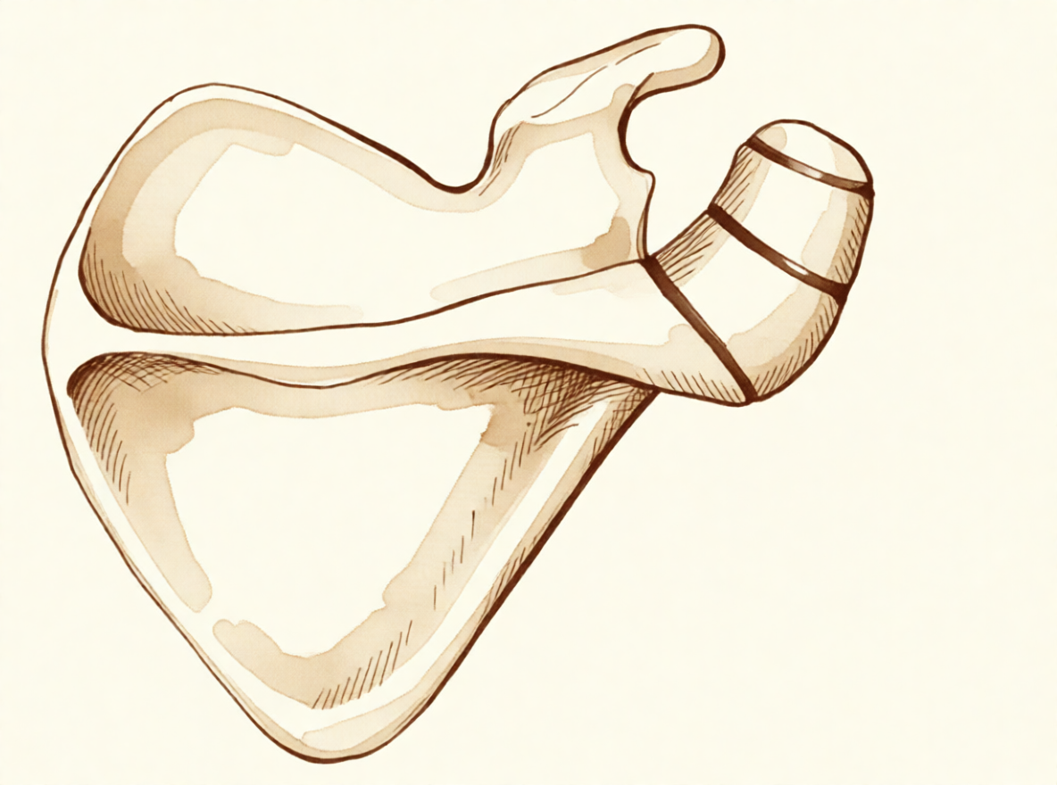
您肩部的骨性顶盖并不是由一整块骨头形成的。它起初是三个独立的骨化中心,从大约 15 岁开始逐渐融合,通常在 25 岁时完成。约 8% 的人中,其中一处连接始终没有闭合。间隙被坚韧的、类似软骨的组织而非骨骼填充,使顶盖的一小块通过一个可活动的连接而非坚实的骨骼相连。
可以把它想象成一块用一颗松动的螺丝固定在墙上的木板。它仍然固定着,但您靠在上面时它会轻微移动。将手臂向侧方抬起的肌肉附着在这个顶盖上,因此每次您抬起手臂时,这块肌肉都会牵拉松动的骨片,使其轻微弯曲。这种活动会向下压迫在顶盖下方走行的肌腱,而摩擦会刺激肌腱。这就是举手过头的活动会引起疼痛、且疼痛位于肩部前方和外侧的原因。
这个可活动的连接本身也可能是问题所在。它本身可能出现触痛,很像一个磨损的关节,按压它会诱发疼痛。大多数有这种骨骼结构的人完全没有感觉,而且它本身似乎并不会引起肩痛,也不会增加肌腱撕裂的可能性。但当松动的骨片开始比平常活动得更多时,无论是由于其形成方式,还是在该部位受到撞击之后,它都可能在您每次抬起手臂时压迫肌腱。这就是上文所述的疼痛弧和夜间疼痛的成因。
由于松动的骨片恰好位于肌腱所在的位置,这两种问题常常同时出现。如果您的肩部已经接受过肌腱问题的治疗而疼痛仍未缓解,就值得检查是否存在这块未融合的骨片,因为仅对肌腱进行手术并不能让其上方一块会活动的骨头安分下来。
我们能做什么
Mater Private Hospital Rockhampton 的上肢外科医生 Kieran Hirpara 医生会从适合您病情的最微创方案入手。患者通常由全科医生转诊至我们的诊所;如果物理治疗师建议您就诊,您仍需获得全科医生的转诊,才有资格享受 Medicare 报销。在您的首次就诊时,我们会采集病史,检查您的肩部,并在必要时安排影像学检查。
第一步通常是改变您使用肩部的方式,并认真尝试物理治疗。物理治疗旨在缓解骨性顶盖下方的刺激,并增强肩部周围的肌肉,使其能更好地应对举手过头的工作。在考虑其他选择之前,请先充分尝试这些方法。
止痛药可以帮助您在此期间保持活动。按照全科医生的建议服用简单的抗炎药,可以缓解酸痛,使您能够舒适地进行锻炼。如果疼痛没有缓解、在数周内逐渐加重、在夜间使您痛醒,或使您无法工作或使用手臂,请去看全科医生或要求专科医生评估。
当非手术治疗未能带来足够的改善时,我们会考虑手术。在此之前,我们会通过普通 X 线片确认诊断,X 线片几乎总能显示出这块未融合的骨片。从腋下拍摄的视图能清楚显示关节的形态和顶盖的前部,这一点很重要,因为损伤可能破坏两块骨头之间的连接处。超声检查是检查顶盖下方肌腱的一种简单而准确的方法,而磁共振成像(MRI)扫描能更全面地显示该肌腱及周围软组织的情况。
我们最常提供的手术是:先通过锁孔(关节镜)对关节进行评估并行滑囊切除术,即清除顶盖下方空间内发炎的组织;然后通过一个小的开放切口进行植骨和固定,即将松动的骨片固定住,使其能够与骨头的其余部分愈合。将其固定住可使两块骨头长在一起。如果松动的骨片较小,我们可能会将其切除而不是固定。固定较大的骨片需要谨慎,因为抬起手臂的肌肉会持续牵拉它,而且骨片很薄。有时骨片在固定后仍无法愈合,这称为骨不连。我们会与您讨论哪种方案适合您的肩部,并共同做出决定。
预期情况
大多数有这种骨骼结构的人从未出现任何问题,也无需治疗。当它确实引起疼痛时,预后取决于松动的骨片是否稳定,以及它是否确实是您疼痛的来源。
对于引起疼痛的情况,治疗通常从非手术方法开始。改变使用肩部的方式、物理治疗和简单的止痛措施可以使许多人的症状缓解。如果在充分尝试后仍无效,手术可以明显减轻疼痛并改善肩部功能。当松动的骨片被固定住并与骨头的其余部分愈合时,患者在肩部功能评估上的表现要好于两块骨头仍保持分离的情况。不过,手术并不总是解决办法。在部分同时患有这种病症和肌腱撕裂的人中,术后结果好坏参半,总体上有 53% 的人获得满意的结果。
如果不加处理而它持续困扰您,疼痛通常不会自行消失。它也很容易被漏诊:当肩痛在治疗后(包括肌腱问题手术后)仍未缓解时,就值得怀疑是这种病症,因为仅对肌腱进行手术并不能让其上方一块会活动的骨头安分下来。
术后恢复是循序渐进的。您将与物理治疗师合作,先恢复活动度,再增强力量,举手过头提物的能力会在数周至数月内慢慢恢复。目标是让您的肩部能够应对日常生活,不再出现您就诊时那种抬臂时的剧痛。
如果您注意到手指、手或手臂变得发烫、发红、肿胀且疼痛,尤其是伴有发烧时,请当天前往急诊科。无需全科医生转诊。
何时就医
大多数有这种骨骼结构的人从不需要治疗,但如果您的肩痛没有缓解、在数周内逐渐加重、在夜间使您痛醒,或使您无法工作或使用手臂,请去看全科医生或要求专科医生评估。如果疼痛位于肩部前方和外侧、在举手过头活动时加重,或在您侧躺压在该肩上时反复使您痛醒,请尽早要求评估。如果肩部顶端的痛点按压时有触痛、将手臂向侧方抬起或向外旋转时感觉无力,或抬起手臂到一半时肩部疼痛,也值得接受评估。有这些症状且没有受伤经历的较年轻成年人应接受检查,排查是否患有这种病症,尤其是在物理治疗、暂停运动或注射未能起效的情况下。如果手指、手或手臂变得发烫、发红、肿胀且疼痛,尤其是伴有发烧时,请当天前往急诊科;无需全科医生转诊。
深入探讨
Advanced reading: the deeper science (optional)
本节内容超出了您做出自身治疗决策所需的深度。肩峰骨(Os acromiale)值得额外阅读,因为它是相当一部分人群中的正常解剖变异,通常不引起任何症状,且常被错误地归咎于其并非责任方的肩部疼痛。
定义与患病率
肩峰是肩关节上方的骨性顶盖,由多个独立的骨化中心形成,这些中心通常在青少年晚期或二十岁出头时融合。在部分人群中,其中一个中心未能融合,从而留下一个永久的纤维性连接。这就是肩峰骨(os acromiale),它自青春期起即存在,而非后天获得。
患病率因人群而异。一项针对 6,842 名患者的多中心研究发现,肩峰骨与肩袖损伤相关,所研究的日本人群中该病的患病率低于非亚洲人群,且骨碎片尺寸往往较小 [1]。
这种与肩袖损伤的关联值得仔细解读。它确立的是两者共同发生的频率高于随机概率,而非肩峰骨导致撕裂。合理的机制是双向的:未融合的碎片在三角肌牵拉下可能轻微移动,从而刺激其下方的肌腱——或者,导致肩袖疾病的相同肩部生物力学机制使得不稳定的碎片出现症状。
是否为疼痛来源才是核心问题
由于肩峰骨在影像学上可见、呈永久性且外观异常,它往往被直接报告为肩痛的病因。大多数肩峰骨无症状,且大多数携带者终生对此毫不知情。
有症状的肩峰骨的特征是:骨片正上方存在局限性压痛,按压该处可诱发疼痛;若使用诊断性注射,则特异性地表现为向该骨性连接处(而非其下方的肩峰下间隙)注射后疼痛缓解。若缺乏上述表现,该影像学发现最好被视为偶然发现。
亚型通常根据所涉及的融合部位进行分类 [2],这在外科上具有重要意义,因为活动性骨片的大小决定了是将其切除还是进行固定。
为何使其他肩部手术复杂化
肩峰骨(os acromiale)的实际重要性往往不在于其自身的症状,而在于它对为其他目的而计划的手术所产生的影响。
三角肌附着于肩峰,因此未融合的骨片会在可活动的连接处承受该牵拉力。在肩峰成形术中,从下表面去除骨质会使本已不稳定的骨片变薄,可能将无症状的变异转化为有症状的病变,或导致骨不连。这是术前影像学检查中识别肩峰骨的最强理由:并非为了治疗它,而是为了避免使其不稳定。
当确实需要治疗时,选择在于切除小骨片还是固定大骨片。已知跨越此连接处的固定具有挑战性,因为骨片较薄,三角肌持续对其施加牵拉力,且骨不连率相当可观。这种困难本身就是一个理由,要求在对其实施手术前,确信肩峰骨确实是疼痛的真正来源。
参考文献
[1] Kozono N, Nishii A, Ishitani E, Mizuki Y, Kimura T, Yamamoto S, et al. 肩胛冈骨(os acromiale)的患病率及相关因素:一项多中心研究. JSES Int. 2025;9(5):1541-5. https://doi.org/10.1016/j.jseint.2025.05.015
[2] Boehm TD, Matzer M, Brazda D, Gohlke FE. 肩胛冈骨(os acromiale)伴肩袖撕裂的手术治疗。33例患者回顾. J Bone Joint Surg Br. 2003;85(4):545-9. https://doi.org/10.1302/0301-620X.85B4.13634
Evidence & references
This is the clinical evidence summary written for health professionals. It is technical, and it lists the research this page was built from. You do not need to read it to understand your treatment or to make a decision about it.
Overview
- A symptomatic, unstable os acromiale is relatively rare despite the radiographic appearance of an os acromiale being not uncommon in patients presenting with shoulder pain [1].
- An os acromiale should always be suspected in cases of failed rotator cuff surgery or ongoing shoulder pain after failed treatment [2].
- The prevalence of os acromiale was 2.13% in Thai patients with shoulder problems who required MRI evaluation [3].
- Os acromiale is associated with rotator cuff injuries [7].
- The greater the distance of the acromioclavicular joint from the anterior edge of the acromion, the higher the likelihood of an os acromiale [15].
- The authors recommend that the acromion be carefully examined roentgenographically prior to operation in any patient with a rotator cuff tear [10].
- Surgical treatment is usually not indicated for os acromiale in the professional tennis player [9].
- Operative management of a symptomatic os acromiale that has failed initial nonoperative treatment leads to decreased symptoms and improvement in clinical outcomes [4].
- Surgical options for symptomatic os acromiale include arthroscopic sub-total excision, arthroscopic subacromial decompression of stable fragments, and open reduction and internal fixation of unstable fragments [13].
- Patients with a united os acromiale had a significantly better functional outcome as measured by the Constant score (P = .0169) [12].
- The advantages of arthroscopic internal fixation include improved visualization of the undersurface of the acromion, allowing excellent reduction, the possibility of treating concomitant shoulder pathologies, and to avoid increased morbidity of the open approach [14].
- Postoperative local tenderness at the os acromiale can be expected in 1 out of 4 patients but resolves spontaneously over time in the majority of patients [5].
- The presence of os acromiale does not appear to have a negative impact on the clinical outcomes after surgery and rTSA remains a safe and effective treatment option [6].
- The outcome of RTSA does not seem to be negatively affected by the presence of an os acromiale [11].
- Ipsilateral os acromiale may be a relative contraindication to the clavicle hook plate [16].
Anatomy & Pathophysiology
Bony Anatomy
- The acromion has three ossification centers: the metacromion (base), the mesoacromion (middle), and the preacromion (tip) [38].
- Failure of fusion of the acromial ossification centers results in os acromiale [38].
- Os acromiale is defined as incomplete fusion of secondary ossification centers, most commonly between the mesoacromion and meta-acromion [49].
- The unfused segment in os acromiale is most commonly the meso-acromion, a configuration often termed a meso-type os acromiale [22].
- The acromion, coracoacromial ligament, and coracoid process form the coracoacromial arch, a rigid bony-ligamentous structure that imparts stability to the shoulder girdle [35].
- The rotator cuff, subacromial bursa, and subdeltoid bursa pass underneath the coracoacromial arch [35].
- The scapula is attached to the axial skeleton by the acromioclavicular and sternoclavicular joints [37].
- The acromion is a flattened bony process that curves forwards from the scapular spine [37].
Prevalence and Associations
- In Thai patients with shoulder problems who required MRI evaluation, the prevalence of os acromiale was 2.13% [3].
- The radiographic appearance of an os acromiale is not uncommon in patients presenting with shoulder pain [1].
- A symptomatic, unstable os acromiale is relatively rare [1].
Pathophysiology
- Most diagnoses of os acromiale are made incidentally on axillary view radiographs of the shoulder [22].
- Primary shoulder pain is usually unrelated to the unfused os acromiale [22].
- When an os acromiale drives symptoms, the two principal causes are motion at the non-union site or an impingement-type syndrome resulting from flexion of the os fragment during deltoid contraction and arm elevation [22].
- Pain from an os acromiale due to these mechanisms has been reported in a variety of sports, particularly among throwing athletes and swimmers [22].
- Previously stable non-unions can become unstable following an episode of blunt trauma to the region [22].
- The synchondrosis of an os acromiale can be injured following trauma [24].
- Extrinsic impingement occurs when the space available for the rotator cuff is diminished, with examples including acromial fracture or pathologic os acromiale [62].
Epidemiology and Prevalence
- Liberson reviewed 1800 shoulder girdles and identified an incidence of os acromiale of 1.4% [21].
- The lesion was bilateral in 62% of patients in Liberson's review of 1800 shoulder girdles [21].
Associated Pathology and Risk Factors
- The greater the distance of the AC joint from the anterior edge of the acromion, the higher the likelihood of an os acromiale [15].
Diagnostic Considerations
- Awareness of the os acromiale in the young athlete, appropriate clinical examination, and image studies are crucial to confirm diagnosis [19].
- Appropriate radiographic investigation including axillary views is emphasized for the diagnosis of os acromiale injuries [24].
Clinical Presentation
- An os acromiale should always be suspected in cases of failed rotator cuff surgery [2].
- An os acromiale should always be suspected in cases of ongoing shoulder pain after failed treatment [2].
- Postoperative local tenderness at the os acromiale can be expected in 1 out of 4 patients [5].
- Postoperative local tenderness at the os acromiale resolves spontaneously over time in the majority of patients [5].
- The lesion was bilateral in 62% of patients in Liberson's review [21].
- The unfused segment is most commonly the meso-acromion [22].
- Any primary shoulder pain is usually unrelated to the unfused os [22].
- If the os acromiale is driving symptoms, one principle cause is motion at the non-union site [22].
- If the os acromiale is driving symptoms, another principle cause is an impingement-type syndrome resulting from flexion of the os fragment during deltoid contraction and arm elevation [22].
- Pain from an os acromiale by the mechanism of impingement has been reported in a variety of sports [22].
- Pain from an os acromiale by the mechanism of impingement has been reported in particular among throwing athletes and swimmers [22].
- Meta–os acromiale is the rarest subtype of os acromiale [23].
Investigations
Prevalence and Demographics
- Liberson reviewed 1800 shoulder girdles and identified an incidence of os acromiale of 1.4%, with the lesion being bilateral in 62% of patients [21].
- A multicenter study aimed to determine the prevalence of and factors associated with os acromiale in the Japanese population [8].
- The radiographic appearance of an os acromiale is not uncommon in patients presenting with shoulder pain, but a symptomatic, unstable os acromiale is relatively rare [1].
Radiographic Evaluation
- The acromion should be carefully examined roentgenographically prior to operation in any patient with a rotator cuff tear [10].
- Appropriate radiographic investigation for os acromiale includes axillary views [24].
- Awareness of the os acromiale in the young athlete requires appropriate clinical examination and image studies to confirm diagnosis [19].
- The standard shoulder series should include orthogonal views of the shoulder, including a true AP view in the scapular plane, an AP view, an axillary view, and a scapular Y view [60].
- The scapular Y view provides visualization of the coracoacromial arch and can reveal coracoacromial spurs, which have been closely associated with the presence of rotator cuff pathology [60].
- The scapular Y view can also show acromial shape [60].
- At least two X-ray views should be obtained for shoulder imaging: an anteroposterior in the plane of the glenoid and an axillary projection with the arm in abduction to show the relationship of the humeral head to the glenoid [52].
- The purpose of imaging of the shoulder is to help establish the diagnosis, determine the severity of the pathoanatomy, assist in surgical planning, and enable the surgeon to illustrate the condition of the shoulder to the patient [30].
- Standardized plain films are almost always sufficient to garner the information needed for shoulder evaluation [30].
- The first key view for shoulder imaging is the anteroposterior (AP) in the plane of the scapula taken so that the x-ray beam passes through the glenohumeral joint [30].
- The second key view for shoulder imaging is the axillary view taken with the arm in the functional position of elevation in the plane of the scapula and oriented so that both the spinoglenoid notch and the scapular neck are visible [30].
- The axillary view is referred to as the "truth view" because it demonstrates the glenohumeral relationships in the functional position of elevation [30].
- CT scans have the disadvantage of being taken with the arm in the adducted position, unlike the axillary truth view [30].
- The temptation to "overimage" should be resisted, obtaining only the scans or reconstructions that are necessary for the care of the patient [30].
Magnetic Resonance Imaging
- In Thai patients with shoulder problems, os acromiale was identified via Magnetic Resonance Imaging evaluation [3].
- MRI is useful to identify labral tears and rotator cuff tears, although the accuracy for these is enhanced by combining the scan with arthrography [52].
- MRI is the modality of choice for evaluating the rotator cuff, biceps, and subacromial/subdeltoid bursa [59].
- T2-weighted MRI provides better visualization of full thickness rotator cuff tears [59].
- MR arthrography is considered the benchmark for evaluation for labral tears and rarely is indicated for evaluation of rotator cuff pathology [59].
- When MRI or MR arthrography is contraindicated, CT arthrography is indicated [59].
Ultrasonography
- Ultrasonography is a simple and accurate test for identifying rotator cuff tears and calcific tendinitis [52].
- Ultrasonography is a low-cost alternative to MRI and arthrography for evaluating both skeletal and soft-tissue structures of the shoulder [59].
- Ultrasonography can provide immediate, real-time visualization of the rotator cuff, biceps tendon, and calcific deposits [59].
- Ultrasonography can be used to measure the subacromial space and detect atrophy of rotator cuff muscles [59].
- Ultrasonography is highly operator dependent and is not as useful for evaluating labral tears or rotator cuff tears that are very small or larger than 3 cm [59].
- The most commonly performed joint examination using ultrasonography is the shoulder examination, and accuracy depends on the skill of the scanner operator and an awareness of pitfalls that are encountered [50].
Clinical Associations and Diagnostic Implications
- The synchondrosis of an os acromiale can be injured following trauma, though rarely [24].
Treatment
Non-Operative
- A symptomatic os acromiale in a competitive female fastball pitcher was treated successfully with open reduction and internal fixation [27].
Operative
- Symptomatic unstable os acromiale can be effectively treated with autogenous bone-grafting and internal fixation using a rigid tension-band construct and cannulated screws, which results in a high rate of union [65].
- This new arthroscopic technique of fixation of os acromiale with absorbable screws provides promising clinical, cosmetic, and radiologic results with high patient satisfaction [28].
- After 3 to 6 years of follow-up, all 6 shoulders were rated as excellent according to criteria similar to that of the American Shoulder and Elbow Surgeons [18].
- Modified arthroscopic subacromial decompression can provide results similar to standard arthroscopic subacromial decompression for routine impingement [25].
- Meta–os acromiale is the rarest subtype of os acromiale, and special consideration must be given to the type of tension-band construct used to achieve adequate compression and fixation [23].
Complications
Post-operative and Surgical Complications
- Postoperative local tenderness at the os acromiale can be expected in 1 out of 4 patients following reverse total shoulder arthroplasty [5].
Diagnostic and Clinical Implications
Pathophysiology and Mechanisms of Symptom
- Pain from an os acromiale by the mechanism of impingement has been reported in a variety of sports, in particular among throwing athletes and swimmers [22].
Recovery
- After 3 to 6 years of follow-up, all 6 shoulders treated with osteosynthesis of an unstable mesoacromion were rated as excellent according to criteria similar to that of the American Shoulder and Elbow Surgeons [18].
- At a two to five year follow-up, all patients treated for fusion of the unstable mesoacromion had fused [69].
Key Evidence
- [L4] While the radiographic appearance of an os acromiale is not uncommon in patients presenting with shoulder pain, a symptomatic, unstable os acromiale is relatively rare. [1] (10.1097/00132589-200412000-00006)
- [L4] An os acromiale should always be suspected in cases of failed rotator cuff surgery or ongoing shoulder pain after failed treatment. [2] (10.1097/00132589-200609000-00006)
- [L3] In Thai patients with shoulder problems who required MRI evaluation, the prevalence of os acromiale was 2.13%. [3] (10.1177/23259671221078806)
- [L4] Operative management of a symptomatic os acromiale that has failed initial nonoperative treatment leads to decreased symptoms and improvement in clinical outcomes. [4] (10.1016/j.jse.2019.05.047)
- [L3] Postoperative local tenderness at the os acromiale can be expected in 1 out of 4 patients but resolves spontaneously over time in the majority of patients. [5] (10.1177/2325967120965131)
- [L4] The presence of os acromiale does not appear to have a negative impact on the clinical outcomes after surgery and rTSA remains a safe and effective treatment option. [6] (10.1016/j.xrrt.2025.01.002)
- [L3] The study supports previous findings that os acromiale is associated with rotator cuff injuries. [7] (10.1016/j.jseint.2025.05.015)
- [L3] This multicenter study aimed to determine the prevalence of and factors associated with os acromiale in the Japanese population. [8] (10.1016/j.jse.2025.01.008)
- [L4] Surgical treatment is usually not indicated for os acromiale in the professional tennis player. [9] (10.1177/2325967118773723)
- [L4] The authors recommend that the acromion be carefully examined roentgenographically prior to operation in any patient with a rotator cuff tear. [10] (10.2106/00004623-198466080-00029)
- [L4] The outcome of RTSA does not seem to be negatively affected by the presence of an os acromiale. [11] (10.1016/j.jse.2017.02.012)
- [L4] Patients with a united os acromiale had a significantly better functional outcome as measured by the Constant score (P = .0169). [12] (10.1016/s1058-2746(98)90008-8)
- [L5] Surgical options for symptomatic os acromiale include arthroscopic sub-total excision, arthroscopic subacromial decompression of stable fragments, and open reduction and internal fixation of unstable fragments. [13] (10.5435/jaaos-d-17-00011)
- [L5] The advantages of this technique are improved visualization of the undersurface of the acromion, allowing excellent reduction, the possibility of treating concomitant shoulder pathologies, and to avoid increased morbidity of the open approach. [14] (10.1097/bte.0000000000000191)
- [L4] Our data suggest that the greater the distance of the AC joint from the anterior edge of the acromion, the higher the likelihood of an os acromiale. [15] (10.1067/mse.2003.128136)
- [L4] Ipsilateral os acromiale may be a relative contraindication to the clavicle hook plate. [16] (10.1186/s12891-021-04841-1)
- [L4] After 3 to 6 years of follow-up, all 6 shoulders were rated as excellent according to criteria similar to that of the American Shoulder and Elbow Surgeons. [18] (10.1016/s1058-2746(99)90004-6)
- [L4] Awareness of the os acromiale in the young athlete, appropriate clinical examination, and image studies are crucial to confirm diagnosis. [19] (10.1016/j.jseint.2020.02.008)
- [L4] [22] (10.1302/2058-5241.4.180100)
- [L4] Meta–os acromiale is the rarest subtype of os acromiale, and special consideration must be given to the type of tension-band construct used to achieve adequate compression and fixation. [23] (10.1177/03635465211028238)
- [L4] This case highlights that the synchondrosis of an os acromiale can be injured following trauma, though rarely, and emphasizes the need for appropriate radiographic investigation including axillary views and a flexible surgical approach. [24] (10.1016/j.jse.2008.02.012)
- [L5] Increased awareness of the diagnosis and improved techniques have shown that modified arthroscopic subacromial decompression can provide results similar to standard arthroscopic subacromial decompression for routine impingement. [25] (10.1097/00132589-200206000-00003)
- [L4] A symptomatic os acromiale in a competitive female fastball pitcher was treated successfully with open reduction and internal fixation. [27] (10.1177/0363546506288305)
- [L4] This new arthroscopic technique of fixation of os acromiale with absorbable screws provides promising clinical, cosmetic, and radiologic results with high patient satisfaction. [28] (10.1016/j.jse.2011.12.011)
- [L4] Symptomatic unstable os acromiale can be effectively treated with autogenous bone-grafting and internal fixation using a rigid tension-band construct and cannulated screws, which results in a high rate of union. [65] (10.2106/00004623-199809000-00011)
- [L4] At a two to five year followup all had fused. [69] (10.1016/s1058-2746(95)80192-8)
References
[1] Treatment of the Symptomatic Os Acromiale. Techniques in Shoulder and Elbow Surgery. 2004. DOI: 10.1097/00132589-200412000-00006
[2] Management of Os Acromiale. Techniques in Shoulder and Elbow Surgery. 2006. DOI: 10.1097/00132589-200609000-00006
[3] Prevalence of Os Acromiale in Thai Patients With Shoulder Problems: A Magnetic Resonance Imaging Study. Orthopaedic Journal of Sports Medicine. 2022. DOI: 10.1177/23259671221078806
[4] Os acromiale: systematic review of surgical outcomes. Journal of Shoulder and Elbow Surgery. 2020. DOI: 10.1016/j.jse.2019.05.047
[5] Os Acromiale in Reverse Total Shoulder Arthroplasty: A Cohort Study. Orthopaedic Journal of Sports Medicine. 2020. DOI: 10.1177/2325967120965131
[6] Clinical implications of reverse total shoulder arthroplasty with an os acromiale: a systematic review. JSES Reviews, Reports, and Techniques. 2025. DOI: 10.1016/j.xrrt.2025.01.002
[7] Prevalence and factors associated with os acromiale: a multicenter study. JSES International. 2025. DOI: 10.1016/j.jseint.2025.05.015
[8] The prevalence and associated factors of os acromiale: a multicenter study. Journal of Shoulder and Elbow Surgery. 2025. DOI: 10.1016/j.jse.2025.01.008
[9] Os Acromiale in Professional Tennis Players. Orthopaedic Journal of Sports Medicine. 2018. DOI: 10.1177/2325967118773723
[10] Rotator cuff tears associated with os acromiale.. The Journal of Bone & Joint Surgery. 1984. DOI: 10.2106/00004623-198466080-00029
[11] Reverse shoulder arthroplasty in patients with os acromiale. Journal of Shoulder and Elbow Surgery. 2017. DOI: 10.1016/j.jse.2017.02.012
[12] Transacromial approach to obtain fusion of unstable os acromiale. Journal of Shoulder and Elbow Surgery. 1998. DOI: 10.1016/s1058-2746(98)90008-8
[13] Symptomatic, Unstable Os Acromiale. Journal of the American Academy of Orthopaedic Surgeons. 2018. DOI: 10.5435/jaaos-d-17-00011
[14] Arthroscopic Internal Fixation of Symptomatic Os Acromiale. Techniques in Shoulder & Elbow Surgery. 2020. DOI: 10.1097/bte.0000000000000191
[15] Relationship between os acromiale and acromioclavicular joint anatomic position. Journal of Shoulder and Elbow Surgery. 2003. DOI: 10.1067/mse.2003.128136
[16] Os acromiale may be a contraindication of the clavicle hook plate: case reports and literature review. BMC Musculoskeletal Disorders. 2021. DOI: 10.1186/s12891-021-04841-1
[18] Successful osteosynthesis of an unstable mesoacromion in 6 shoulders: A new technique. Journal of Shoulder and Elbow Surgery. 1999. DOI: 10.1016/s1058-2746(99)90004-6
[19] The unstable os acromiale: a cause of pain in the young athlete. JSES International. 2020. DOI: 10.1016/j.jseint.2020.02.008
[21] Types of os acromiale according to Liberson. 2006.
[22] Os acromiale: a review of its incidence, pathophysiology, and clinical management. EFORT Open Reviews. 2019. DOI: 10.1302/2058-5241.4.180100
[23] Rare Symptomatic Meta–Os Acromiale in an Athlete. The American Journal of Sports Medicine. 2021. DOI: 10.1177/03635465211028238
[24] Fracture of an os acromiale with associated rupture of the coracoclavicular ligaments. Journal of Shoulder and Elbow Surgery. 2008. DOI: 10.1016/j.jse.2008.02.012
[25] Arthroscopic Management of the Mesoacromion. Techniques in Shoulder and Elbow Surgery. 2002. DOI: 10.1097/00132589-200206000-00003
[27] Surgical Stabilization of Os Acromiale in a Fast-Pitch Softball Pitcher. The American Journal of Sports Medicine. 2006. DOI: 10.1177/0363546506288305
[28] Arthroscopically assisted internal fixation of the symptomatic unstable os acromiale with absorbable screws. Journal of Shoulder and Elbow Surgery. 2012. DOI: 10.1016/j.jse.2011.12.011
[30] Rockwood And Matsen S The Shoulder. Arthroscopic Management of Prearthritic and Arthritic Conditions of the Shoulder and the Postarthroplasty Shoulder > Radiographic Evaluation.
[35] Rockwood And Matsen S The Shoulder. Shoulder and Elbow Specialty Clinic Workers’ Survey > ANATOMY.
[37] Rockwood And Green S Fractures In Adults. 29: Principles of Nonunion and Bone Defect Treatment > Applied Anatomy Related to Scapular Fractures.
[38] Aaos Comprehensive Orthopaedic Review 3. Anatomy of the Shoulder, Arm, and Elbow > I. Shoulder.
[49] Miller S Review Of Orthopaedics. Genetics of musculoskeletal conditions and abnormalities are summarized in Table 1.27 > UPPER EXTREMITY > SHOULDER.
[50] Orthopaedic Knowledge Update Sports Medicine 6. Diagnostic Ultrasonography and Ultrasonography-Guided Procedures > Annotated References.
[52] Apley And Solomon S Concise System Of Orthopaedics And Trauma. INVESTIGATION.
[59] Orthopaedic Knowledge Update 13 Ebook Without Multimedia. Shoulder Anatomy and Biomechanics, Clinical Evaluation, Imaging > Clinical Evaluation > Imaging.
[60] Aaos Comprehensive Orthopaedic Review 3. Imaging of the Shoulder and Elbow > I. Shoulder.
[62] Campbell S Operative Orthopaedics 4 Volume Set. ANTERIOR CRUCIATE LIGAMENT RECONSTRUCTION WITH BONE-PATELLAR TENDON-BONE GRAFT > Developmental Stages of Impingement Syndrome.
[65] The Treatment of Symptomatic Os Acromiale. The Journal of Bone & Joint Surgery*. 1998. DOI: 10.2106/00004623-199809000-00011
[69] Successful fusion of the unstable mesoacromion: Technique and report of five cases. Journal of Shoulder and Elbow Surgery. 1995. DOI: 10.1016/s1058-2746(95)80192-8