Patients › Hand
Cisto mucoso
Mucous cysts – common bumps near finger joints, often linked to arthritis, and treatment options.
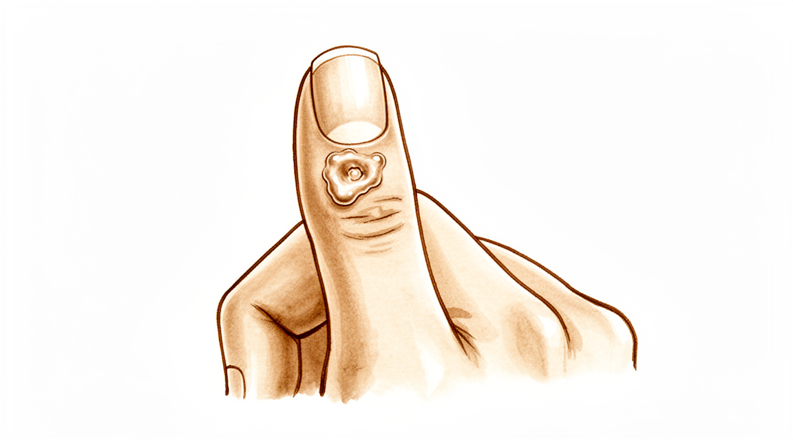
O que você está sentindo
Um cisto mucoso é um pequeno nódulo firme que aparece na parte de trás do dedo, geralmente logo atrás da unha. Ele é preenchido por um líquido espesso e transparente. O nódulo em si muitas vezes não dói, mas pode pressionar as estruturas vizinhas e causar desconforto.
Você pode notar alterações na própria unha. O cisto fica perto do leito ungueal, por isso pode pressionar a unha à medida que cresce. Isso muitas vezes causa um sulco ou uma crista ao longo de toda a unha. A pele sobre o cisto também pode ficar fina e brilhante. Em alguns casos, a pele se rompe e o cisto vaza líquido por um pequeno canal.
O nódulo costuma ficar bem onde o dedo dobra, na última articulação antes da unha. Isso torna algumas tarefas do dia a dia desajeitadas. Enfiar a mão num bolso apertado, segurar a tampa de um pote ou pegar moedas pequenas pode ser desconfortável. Digitar ou escrever pode pressionar o nódulo e deixá-lo dolorido.
O cisto está ligado à artrose (desgaste articular) nessa mesma articulação. Uma pequena saliência óssea, chamada osteófito, irrita a articulação e produz o líquido que enche o cisto. Por isso, a articulação pode doer depois que você usa muito a mão, e o nódulo pode ficar mais sensível nesses momentos.
Se a pele sobre o cisto se abrir e vazar líquido, mantenha a área limpa e procure atendimento rapidamente. Um canal entre a pele e a articulação do dedo pode deixar entrar uma infecção, e uma articulação infectada precisa de tratamento sem demora. Se o seu dedo ficar quente, vermelho, inchado e dolorido, especialmente com febre, vá a um pronto-socorro no mesmo dia. Não é necessário encaminhamento do médico de família.
Não fure nem esprema o cisto por conta própria. Isso pode deixar entrar uma infecção na articulação.
Se o nódulo estiver crescendo, se a deformidade da unha estiver incomodando você ou se os sintomas não estiverem melhorando ao longo das semanas, consulte o seu médico de família ou peça uma avaliação especializada.
O que está realmente acontecendo
A última articulação do seu dedo funciona como uma pequena dobradiça. Como qualquer articulação, ela é revestida por uma capa macia que produz um pouco de líquido para manter as superfícies deslizando suavemente. No cisto mucoso, essa articulação desenvolveu artrose por desgaste. O revestimento da articulação fica irritado e passa a produzir mais líquido do que a articulação precisa.
Ao mesmo tempo, a artrose muitas vezes forma uma pequena saliência óssea na borda da articulação. Essa saliência pressiona e desgasta a capa da articulação por dentro. O líquido então vaza pelo ponto desgastado e se acumula sob a pele, na parte de trás do dedo. O nódulo que você consegue ver e sentir é esse líquido retido, localizado ao lado da articulação, logo atrás da unha.
O cisto em si é uma bolsa de parede fina, e não um crescimento de tecido. A sua parede é formada por fibras de tecido de sustentação, e ele não tem um revestimento próprio. Como a parede é fina e a pele sobre ela muitas vezes é ainda mais fina, uma pequena pancada pode rompê-lo. Quando isso acontece, o líquido transparente com aspecto de gelatina que está dentro dele vaza.
Isso explica os sintomas sobre os quais você acabou de ler. O nódulo fica perto do leito ungueal, por isso a pressão do cisto pode formar um sulco na unha à medida que cresce. A articulação dói porque a artrose também está presente, e o cisto é um sinal dela. A saliência na borda da articulação é o que mantém o líquido fluindo; por isso, enquanto ela estiver lá, o cisto tende a voltar a se encher, mesmo depois de vazar ou ser drenado.
É também por isso que o tratamento se concentra na articulação, e não apenas no nódulo. A remoção da saliência óssea interrompe o vazamento na sua origem, e o cisto regride quando deixa de ser reabastecido.
O que podemos fazer a respeito
O Dr. Kieran Hirpara, cirurgião de membros superiores no Mater Private Hospital Rockhampton, começa com as opções menos invasivas adequadas ao seu caso. Em geral, os pacientes são encaminhados à nossa clínica pelo médico de família; caso um fisioterapeuta tenha sugerido que você nos procurasse, ainda assim será necessário um encaminhamento do seu médico de família para que você tenha direito ao reembolso do Medicare. Na consulta na clínica, colhemos o histórico, examinamos o seu dedo e solicitamos exames de imagem, se necessário. Como a maioria dos cistos mucosos está ligada à artrose (desgaste articular) da articulação, geralmente tentamos primeiro o tratamento não cirúrgico e consideramos a cirurgia quando ele não trouxe melhora suficiente.
O primeiro passo costuma ser simplesmente observar e aguardar. Alguns cistos continuam pequenos e causam poucos problemas. Proteger o dedo ajuda: evite pancadas repetidas no nódulo e tenha cuidado com tarefas que o pressionam. Se a articulação doer após um uso intenso, diminuir essa atividade pode acalmar as coisas. A terapia da mão pode ajudar com a rigidez e a manter o dedo se movimentando com conforto. Dê a essas medidas simples uma chance justa, ao longo de várias semanas, antes de decidir qualquer outra coisa.
O alívio da dor é simples. Analgésicos simples da farmácia, tomados conforme as instruções, podem acalmar uma articulação dolorida. Anti-inflamatórios podem aliviar a artrose por trás do cisto. Eles tratam o desconforto, e não o nódulo em si.
Uma opção que não oferecemos é drenar o cisto com uma agulha. É um procedimento prático, feito no consultório, mas o cisto volta em cerca de metade dos casos depois disso. Há também o risco de levar uma infecção para dentro da articulação, e é por isso que também recomendamos não furar nem espremer o cisto em casa.
A cirurgia é considerada quando o cisto continua voltando a se encher, quando a deformidade da unha incomoda você ou quando a pele sobre o cisto ficou fina ou está se rompendo. A operação trata a articulação, e não apenas o nódulo. A saliência óssea na borda da articulação é removida, o que interrompe o vazamento de líquido na sua origem, e o cisto regride quando deixa de ser reabastecido. Em alguns casos, o cisto e um pequeno retalho de pele vizinha também são removidos, o que é adequado para um cisto que afinou a pele sobre ele. A remoção da saliência óssea junto com o pedúnculo do cisto oferece a menor chance de o cisto voltar. Conversaremos sobre qual abordagem é adequada para o seu dedo e decidiremos juntos.
O que esperar
Se deixado sem tratamento, um cisto mucoso tende a continuar voltando, em vez de desaparecer. Enquanto a saliência óssea na borda da articulação estiver lá, ela continua reabastecendo o cisto com líquido. Alguns cistos continuam pequenos e causam poucos problemas por muito tempo. Outros crescem lentamente, aprofundam o sulco na unha ou afinam a pele sobre o nódulo até que ele vaze.
Sem tratamento, os principais riscos são os que você já leu: uma unha que continua com uma crista enquanto o cisto a pressiona, e uma pele que se rompe e abre um canal até a articulação. Esse canal deve ser levado a sério. Se o seu dedo ficar quente, vermelho, inchado e dolorido, especialmente com febre, vá a um pronto-socorro no mesmo dia. Não é necessário encaminhamento do médico de família.
Com um tratamento voltado para a articulação, as perspectivas são claras. A remoção da saliência óssea interrompe o vazamento de líquido na sua origem, e o cisto regride quando deixa de ser reabastecido. Quando a saliência óssea é removida, o cisto raramente volta. Quando o próprio cisto também é removido, a recorrência continua baixa: cerca de 2 em cada 100 cistos tratados com cirurgia voltam, e cerca de 1,4 em cada 100 quando se usa um retalho de pele vizinha para fechar a área. As pessoas tratadas dessa forma geralmente ficam satisfeitas com a aparência da cicatriz e passariam pelo procedimento novamente.
A recuperação não é algo complicado, mas também não é insignificante. O dedo muitas vezes fica rígido e sensível por algumas semanas, e tarefas do dia a dia, como segurar objetos ou fazer pinça, levam um tempo para voltar a parecer normais. A terapia da mão após a cirurgia é feita com Ruby Doolan, da Extend Rehabilitation. Ruby é terapeuta da mão: ela orienta os seus exercícios e confecciona qualquer tala de que você precise. A maioria das pessoas volta a usar a mão normalmente num período que vai de algumas semanas a poucos meses.
O sulco na unha geralmente também melhora quando a pressão sobre o leito ungueal desaparece. Tenha paciência com essa parte: o trecho deformado da unha precisa crescer até sair, o que leva vários meses.
Se os sintomas não estiverem melhorando ao longo das semanas, ou estiverem piorando, consulte o seu médico de família ou peça uma avaliação especializada.
Quando procurar ajuda médica
A maioria dos cistos mucosos não é urgente, e muitos melhoram com cuidados simples. Alguns sinais, porém, exigem ação.
Vá a um pronto-socorro no mesmo dia se o seu dedo ficar quente, vermelho, inchado e dolorido, especialmente com febre. Isso indica infecção na articulação, que precisa de tratamento sem demora. O mesmo vale se a pele sobre o cisto se abriu e está vazando líquido, porque essa abertura pode deixar entrar uma infecção.
Consulte o seu médico de família ou peça uma avaliação especializada se o nódulo continuar crescendo, se o sulco na unha estiver incomodando você ou se a pele sobre o cisto tiver ficado fina. Esses são os sinais de que vale a pena conversar sobre o tratamento. Se os sintomas não estiverem melhorando ao longo das semanas, ou estiverem piorando, o seu médico de família pode providenciar um encaminhamento.
Em maior profundidade
Advanced reading: the deeper science (optional)
Esta seção vai além do que você precisa saber para tomar decisões sobre o próprio tratamento. Os cistos mucosos merecem uma leitura mais aprofundada devido a uma constatação cirúrgica bem fundamentada: o cisto não é o problema, e a operação mais eficaz nem sempre consiste em removê-lo.
O esporão ósseo é a causa, não o cisto
O cisto mucoso surge a partir de uma articulação da ponta do dedo afetada por artrite. Um esporão ósseo, ou osteófito, irrita e perfura a cápsula articular; o líquido sinovial então escapa pelo defeito e se acumula sob a pele. O cisto é apenas o resultado visível desse processo, não sua origem.
Essa compreensão tem uma consequência cirúrgica direta: em uma série de casos, a excizão do osteófito sem excizão do cisto resultou em resolução completa na maioria dos pacientes, sendo considerada uma boa opção terapêutica por ser um método menos invasivo [1].
A remoção do esporão ósseo interrompe esse fluxo de líquido. Sem mais reposição de líquido, o cisto desaparece. Essa é a mesma lógica aplicada aos gânglios do punho: o “pedúnculo” do cisto, e não o saco em si, é o fator determinante para sua recorrência. Isso explica por que a simples drenagem ou punção de um cisto mucoso quase sempre falha.
Nos locais onde o cisto é excisado, os resultados também são bons
A abordagem alternativa consiste em remover o cisto juntamente com um retalho cutâneo local para fechar o defeito. Essa técnica também é confiável: em 69 pacientes, a excisão cirúrgica associada a um retalho de avançamento local apresentou taxa de recorrência de 1,4%, além de alta satisfação dos pacientes quanto à cicatriz e disposição para submeter-se novamente ao procedimento [2].
Ambas as abordagens são eficazes e atacam a articulação afetada. A diferença prática reside na quantidade de pele envolvida: um cisto de longa data torna a pele sobrejacente mais fina, às vezes a ponto de ocorrer extravasação de conteúdo; nesse caso, a pele afinada precisa ser excisada e substituída, independentemente do que se faça com o osso.
Por que a unha fica deformada e se ela volta ao normal
A presença de um sulco ou elevação ao longo de toda a unha é algo comum e preocupa mais as pessoas do que o próprio nódulo. A causa é mecânica: o cisto fica localizado exatamente sobre a matriz germinativa, ou seja, a parte do leito ungueal responsável pela formação da unha, exercendo pressão sobre ela; por isso a unha se desenvolve com um defeito.
O aspecto positivo é que se trata de uma pressão, e não de uma destruição real. Uma vez que o cisto é descomprimido, a unha geralmente volta a crescer normalmente; porém, leva vários meses para que a parte deformada seja eliminada na ponta da unha. Portanto, a deformidade ungueal é motivo para tratar o cisto, e não uma consequência permanente dele.
Por que é preciso ter cuidado ao perfurir um cisto
Um cisto mucoso se comunica diretamente com a articulação. Perfurá-lo, seja de forma intencional ou quando a pele se rompe sobre um cisto de grande tamanho, cria um canal entre o meio externo e a articulação do dedo; a artrite séptica em uma articulação pequena é um problema consideravelmente mais grave do que o próprio cisto.
Esse é o argumento prático contra a drenagem caseira de um cisto que parece ser apenas uma bolha de líquido, além da razão pela qual um cisto que drena espontaneamente deve ser tratado com urgência, em vez de apenas ser monitorado.
Referências
[1] Lee H, Kim P, Jeon I, Kyung H, Ra I, Kim T. Excisão de osteófitos sem excisão do cisto no tratamento de cistos mucosos digitais. J Hand Surg Eur Vol. 2013;39(3):258-61. https://doi.org/10.1177/1753193413478549
[2] Johnson SM, Treon K, Thomas S, Cox QGN. Um tratamento cirúrgico confiável para cistos mucosos digitais. J Hand Surg Eur Vol. 2013;39(8):856-60. https://doi.org/10.1177/1753193413508540
Evidence & references
This is the clinical evidence summary written for health professionals. It is technical, and it lists the research this page was built from. You do not need to read it to understand your treatment or to make a decision about it.
Overview
- The scientific data regarding mucous cysts consist almost entirely of retrospective studies, and much of what is done or recommended is based on expert opinion [3].
- Surgical treatment of mucous cysts is not a totally benign procedure, and complications do occur contrary to literature reporting glowing results [5].
- In-office excision is a safe and effective option for treating digital mucous cysts [6].
- Complications are rare and occur only in cysts associated with fistula, justifying their early surgical treatment [7].
- Surgery provided definitive treatment with no major long-term problems [10].
- Surgical excision was the primary treatment, with no reported recurrences [11].
- Incomplete excisions will invariably result in recurrence [12].
- Surgical excision with a local advancement skin flap is a reliable treatment for digital mucous cysts, demonstrating a low recurrence rate of 1.4% and high patient satisfaction regarding the scar and willingness to undergo the procedure again [13].
- The 2% recurrence rate (2 of 113) obtained in this series illustrates the advantage of this procedure [16].
- Osteophyte excision without cyst excision may be a good treatment choice for mucous cyst of the finger, providing a less invasive method with complete resolution in most cases [20].
- The final recurrence rate of less than 1.5% may be acceptable and comparable with other techniques [21].
- Excision with local flap cover offers benefit in terms of a reduced recurrence rate and is particularly valuable where the cyst has caused marked thinning of the skin [30].
- Excision with local flap cover would seem to offer benefit in terms of a reduced recurrence rate, and is particularly valuable where the cyst has caused marked thinning of the skin [34].
- Excision of the cyst and complete removal of the marginal osteophyte eradicates mucous cysts with extremely rare recurrence [1].
- A total dorsal capsulectomy alone was a simple treatment for mucous cysts and did not lead to any recurrence [8].
Anatomy & Pathophysiology
Clinical Presentation and Location
- Mucous cysts are small, firm, cystic masses that appear just distal to the distal interphalangeal joint [23].
- The lesion is always located to one side of the midline [23].
- Mucous cysts are rarely greater than fifteen millimeters in diameter [23].
- The skin over the mucous cyst is thinned out and occasionally may be ulcerated [23].
- Pressure on the mucous cyst does not usually result in a decrease in its size [23].
- Longitudinal grooving of the nail may be noted, occasionally occurring prior to the appearance of the cyst [23].
- Mucous cysts are usually small and located to one side as they emerge from the joint beside the extensor mechanism [28, 29].
- Occasionally, mucous cysts may spread across the whole of the dorsum of the digit between the distal interphalangeal joint and nail fold [28, 29].
- The overlying skin of a mucous cyst is frequently thin, and minimal trauma may result in rupture [28, 29].
Histology and Ultrastructure
- The histological appearance of a mucous cyst is essentially identical to that of ganglions, synovial cysts, and similar lesions appearing on other areas of the extremities [23].
- The mucous cyst is surrounded by an undemarcated fibrous capsule [23].
- The basic structure of the mucous cyst is myxomatous with interspersed fibroblasts [23].
- Areas of myxomatous degeneration tend to coalesce to form a multiloculated cyst [23].
- An epithelial lining has not been reported for mucous cysts [23].
- Digital mucous cysts and wrist ganglia show no anatomic difference at the light and scanning electron microscopic levels [19].
- The wall of a digital mucous cyst consists of multidirectional strata of collagen fibers with rare fibroblast-like cells on the inner surface [19].
- There are no signs of myxomatous degeneration in the wall of a digital mucous cyst [19].
- The surface of the digital mucous cyst wall is generally flat, with some areas showing localized elevations [19].
- Multifunctional mesenchymal cells are located beneath the lining of the ganglion and digital mucous cyst [19].
- These multifunctional mesenchymal cells contain abundant rough endoplasmic reticulum, smooth-walled vacuoles, and Golgi complexes [19].
- The ganglion fluid may be produced by these multifunctional cells and reaches the ganglion cavity by flowing through the porous collagen matrix of the ganglion wall [19].
- A digital mucous cyst has a large globular dome-shaped interconnecting cavity having a wall of collagen fibers covered by a fenestrated pseudomembrane [19].
- A porous network of collagen fibers is present in the wall of the digital mucous cyst on cross section [19].
Etiology and Pathogenesis
- Degenerative arthritis of the distal interphalangeal joint is a frequent associated finding with mucous cysts [23].
- An incidence of 78 percent for degenerative arthritis of the distal interphalangeal joint has been reported in association with mucous cysts [23].
- The etiology indicated by evidence is that the lesion arises from the joint capsule [23].
- Mucous cysts are often associated with degenerative changes at the distal interphalangeal joints [28, 29].
- Theories on the pathogenesis of mucous cysts include myxoid degeneration, repeated local trauma, vascular insufficiency, and synovial herniation [28, 29].
- Studies suggest that mucous cysts arise due to excessive hyaluronic acid production from a degenerate distal interphalangeal joint [28, 29].
Classification
- The lesion is a small, firm, cystic mass which appears just distal to the distal interphalangeal joint [23].
- The lesion is always located to one side of the mid-line [23].
- The lesion is rarely greater than fifteen millimeters in diameter [23].
- The skin over the lesion is thinned out and occasionally may be ulcerated [23].
- Pressure on the lesion does not usually result in a decrease in its size [23].
- Degenerative arthritis of the distal interphalangeal joint is a frequent associated finding [23].
- An incidence of 78 per cent has been reported for degenerative arthritis of the distal interphalangeal joint associated with mucous cysts [23].
- The histological appearance is essentially identical to that of ganglions, synovial cysts, and similar lesions appearing on other areas of the extremities [23].
- The lesion is surrounded by an undemarcated fibrous capsule [23].
- The basic structure is myxomatous with interspersed fibroblasts [23].
- An epithelial lining has not been reported [23].
- In spite of their different clinical settings, digital mucous cysts and wrist ganglia show no anatomic difference at the light and scanning electron microscopic levels [19].
- The wall of the digital mucous cyst consisted of multidirectional strata of collagen fibers with rare fibroblast-like cells on the inner surface [19].
- There were no signs of myxomatous degeneration in the wall of the digital mucous cyst [19].
- The surface of the digital mucous cyst was generally flat, with some areas showing localized elevations [19].
- Multifunctional mesenchymal cells beneath the lining of the ganglion are postulated to produce collagen fibers, elastic fibers, and the interfibrillary mucopolysaccharide matrix [19].
- The porous nature of the ganglion wall was confirmed in the ultrastructural study of digital mucous cysts [19].
- A previous ultrastructural study of digital mucous cysts demonstrated a large globular dome-shaped interconnecting cavity having a wall of collagen fibers covered by a fenestrated pseudomembrane [19].
- The current study confirmed the anatomic organization of a large globular dome-shaped interconnecting cavity having a wall of collagen fibers covered by a fenestrated pseudomembrane [19].
- A porous network of collagen fibers in the wall was revealed on cross section [19].
Clinical Presentation
- The mucous cyst presents as a small, firm, cystic mass located just distal to the distal interphalangeal joint [23].
- The lesion is easily distinguished from a Heberden's node by its placement distal to the distal interphalangeal joint [23].
- The histological appearance of the mucous cyst is essentially identical to that of ganglions, synovial cysts, and similar lesions appearing on other areas of the extremities [23].
- The basic structure of the lesion is myxomatous with interspersed fibroblasts [23].
- An epithelial lining has not been reported for the mucous cyst [23].
- The wall of the digital mucous cyst consists of a porous network of collagen fibers [19].
- The fluid within the digital mucous cyst is produced by underlying mesenchymal cells [19].
Investigations
- Ultrasound is a powerful modality for evaluation of pathologic conditions in the hand and wrist, providing a cost-effective and expedient alternative and/or adjunct to MRI [33].
- Ultrasound is best used when there is a specific clinical question regarding a well-localized abnormality [33].
- Ultra-sonography provides a new ability to study the complex anatomical structures of the hand, with the technique being quick and simple to perform [38].
- A careful physical examination is essential to direct care and future testing if indicated [35].
- Diagnostic tests such as imaging and serum laboratory studies are useful in this determination but can be expensive, time consuming, and often nonspecific [35].
Treatment
Operative Techniques
- Total dorsal capsulectomy alone is a simple treatment for mucous cysts that did not lead to any recurrence [8].
- Osteophyte-sparing treatment of mucous cysts has a final recurrence rate of less than 1.5% [21].
- Surgical excision was the primary treatment in a series with no reported recurrences [11].
- Surgery provided definitive treatment for mucous cysts with no major long-term problems [10].
- The 2% recurrence rate (2 of 113) obtained in a series of 191 excised cysts illustrates the advantage of the procedure [4, 16].
- The authors of a study on osteophyte excision without cyst excision state that their principle is not to do more damage to the friable skin because the skin is not the culprit of the cyst [15].
Outcomes and Complications
- Complications of surgical treatments include infection, recurrence, nail deformity, swelling, stiffness and pain [28].
- This report provides additional basis for prophylactic extirpation of mucous cyst of the finger [2].
- This case emphasizes the utility of a pathohistological analysis in cases where doubts arise about the initial diagnosis of a benign tumorous lesion [14].
Non-Operative and Adjunctive Considerations
- Aspiration with injection of steroid is a convenient technique that can be performed in an outpatient setting but is followed by a recurrence rate of 50% [28].
- Surgical treatment has a higher cure rate than aspiration with injection of steroid [28].
- Radical treatment with complete excision of the stalk and underlying osteophytes seems to reduce recurrence rates [28].
Complications
- Complications are rare and occur only in cysts associated with fistula [7].
- Aspiration with injection of steroid is followed by a recurrence rate of 50% [28].
- The 2% recurrence rate (2 of 113) was obtained in a series of surgically treated mucous cysts [16].
- Surgical excision with a local advancement skin flap demonstrated a low recurrence rate of 1.4% [13].
- A total dorsal capsulectomy alone did not lead to any recurrence [8].
- Eccrine porocarcinomas have a substantial risk of metastasis, high risk of local recurrence, and are potentially fatal [24].
Recovery
- Surgical excision with a local advancement skin flap demonstrated a low recurrence rate of 1.4% and high patient satisfaction regarding the scar and willingness to undergo the procedure again [13].
Key Evidence
- [L4] Excision of the cyst and complete removal of the marginal osteophyte eradicates mucous cysts with extremely rare recurrence. [1] (10.2106/00004623-197355030-00013)
- [L4] This report provides additional basis for prophylactic extirpation of mucous cyst of the finger. [2] (10.1016/s0363-5023(84)80152-5)
- [L4] The scientific data regarding mucous cysts consist almost entirely of retrospective studies, and much of what is done or recommended is based on expert opinion. [3] (10.1016/j.jhsa.2010.01.029)
- [L5] Of the 191 mucous cysts excised, 113 had at least 6 months of follow-up time. [4] (10.1016/j.jhsa.2010.07.028)
- [L4] Surgical treatment of mucous cysts is not a totally benign procedure, and complications do occur contrary to literature reporting glowing results. [5] (10.1016/s0266-7681(97)80067-8)
- [Paper] In-office excision is a safe and effective option for treating digital mucous cysts. [6] (10.1177/15589447251350168)
- [L4] Complications are rare and occur only in cysts associated with fistula, justifying their early surgical treatment. [7] (10.1016/j.main.2015.06.001)
- [L4] A total dorsal capsulectomy alone was a simple treatment for mucous cysts and did not lead to any recurrence. [8] (10.1016/j.jhsa.2014.03.004)
- [L4] Surgery provided definitive treatment with no major long-term problems. [10] (10.1053/jhsu.2003.50088)
- [L5] Incomplete excisions will invariably result in recurrence. [12] (10.1016/0363-5023(93)90302-j)
- [L4] Surgical excision with a local advancement skin flap is a reliable treatment for digital mucous cysts, demonstrating a low recurrence rate of 1.4% and high patient satisfaction regarding the scar and willingness to undergo the procedure again. [13] (10.1177/1753193413508540)
- [L5] This case emphasizes the utility of a pathohistological analysis in cases where doubts arise about the initial diagnosis of a benign tumorous lesion. [14] (10.1007/s00402-008-0794-4)
- [L5] The authors state that their principle is not to do more damage to the friable skin because the skin is not the culprit of the cyst. [15] (10.1177/1753193414546990)
- [L4] The 2% recurrence rate (2 of 113) obtained in this series illustrates the advantage of this procedure. [16] (10.1016/0363-5023(94)90071-x)
- [L5] [19] (10.1016/s0363-5023(88)80143-6)
- [L4] Osteophyte excision without cyst excision may be a good treatment choice for mucous cyst of the finger, providing a less invasive method with complete resolution in most cases. [20] (10.1177/1753193413478549)
- [L4] The final recurrence rate of less than 1.5% may be acceptable and comparable with other techniques. [21] (10.5435/jaaosglobal-d-21-00164)
- [L4] [23] (10.2106/00004623-197254070-00008)
- [L4] Prompt recognition and appropriate treatment are critical because eccrine porocarcinomas have a substantial risk of metastasis, high risk of local recurrence, and are potentially fatal. [24] (10.1016/j.jhsa.2016.07.112)
- [L4] [28] (10.1177/1753193408103498)
- [L4] [29] (10.1177/1753193409103498)
- [L4] Excision with local flap cover offers benefit in terms of a reduced recurrence rate and is particularly valuable where the cyst has caused marked thinning of the skin. [30] (10.1016/0266-7681(90)90064-b)
- [L5] Ultrasound is a powerful modality for evaluation of pathologic conditions in the hand and wrist, providing a cost-effective and expedient alternative and/or adjunct to MRI, best used when there is a specific clinical question regarding a well-localized abnormality. [33] (10.1016/j.jhsa.2009.02.010)
- [L4] Excision with local flap cover would seem to offer benefit in terms of a reduced recurrence rate, and is particularly valuable where the cyst has caused marked thinning of the skin. [34] (10.1016/0266-7681_90_90064-b)
- [L4] Ultra-sonography provides a new ability to study the complex anatomical structures of the hand, with the technique being quick and simple to perform. [38] (10.1016/0266-7681(90)90133-o)
References
[1] Marginal Osteophyte Excision in Treatment of Mucous Cysts. The Journal of Bone & Joint Surgery. 1973. DOI: 10.2106/00004623-197355030-00013
[2] Infected mucous cyst of the finger. The Journal of Hand Surgery. 1984. DOI: 10.1016/s0363-5023(84)80152-5
[3] Mucous Cysts. The Journal of Hand Surgery. 2010. DOI: 10.1016/j.jhsa.2010.01.029
[4] Mucous Cysts. The Journal of Hand Surgery. 2010. DOI: 10.1016/j.jhsa.2010.07.028
[5] Complications Following Mucous Cyst Excision. Journal of Hand Surgery. 1997. DOI: 10.1016/s0266-7681(97)80067-8
[6] Safety and Cost-Effectiveness of In-Office Digital Mucous Cyst Excisions. HAND. 2025. DOI: 10.1177/15589447251350168
[7] Surgical treatment of mucous cysts by subcutaneous excision and osteophyte resection: Results in 68 cases at a mean 6.63 years’ follow-up. Chirurgie de la Main. 2015. DOI: 10.1016/j.main.2015.06.001
[8] Total Dorsal Capsulectomy for the Treatment of Mucous Cysts. The Journal of Hand Surgery. 2014. DOI: 10.1016/j.jhsa.2014.03.004
[10] Treatment of mucous cysts of the fingers: Review of 134 cases with minimum 2-year follow-up evaluation. The Journal of Hand Surgery. 2003. DOI: 10.1053/jhsu.2003.50088
[11] 10.1177-15589447261433068. n.d..
[12] Juvenile hyaline fibromatosis of the hand in an adult. The Journal of Hand Surgery. 1993. DOI: 10.1016/0363-5023(93)90302-j
[13] A reliable surgical treatment for digital mucous cysts. Journal of Hand Surgery (European Volume). 2013. DOI: 10.1177/1753193413508540
[14] Malignant Natural-Killer cell neoplasm presenting as a mucous cyst on the distal interphalangeal joint of the finger. Archives of Orthopaedic and Trauma Surgery. 2008. DOI: 10.1007/s00402-008-0794-4
[15] Re: Lee, H.-J., Kim, P.-T., Jeon, I.-H., Kyung, H.-S., Ra, I.-H. and Kim, T.-K. Osteophyte excision without cyst excision for a mucous cyst of the finger. J Hand Surg Eur. 2014, 39: 258–61. Journal of Hand Surgery (European Volume). 2014. DOI: 10.1177/1753193414546990
[16] Outcome of surgically treated mucous cysts of the hand. The Journal of Hand Surgery. 1994. DOI: 10.1016/0363-5023(94)90071-x
[19] A surface ultrastructure study of ganglia and digital mucous cysts. The Journal of Hand Surgery. 1988. DOI: 10.1016/s0363-5023(88)80143-6
[20] Osteophyte excision without cyst excision for a mucous cyst of the finger. Journal of Hand Surgery (European Volume). 2013. DOI: 10.1177/1753193413478549
[21] Osteophyte-Sparing Treatment of Mucous Cysts: Case Analysis and Surgical Technique. JAAOS: Global Research and Reviews. 2021. DOI: 10.5435/jaaosglobal-d-21-00164
[23] Etiology and Treatment of the So-Called Mucous Cyst of the Finger. The Journal of Bone & Joint Surgery. 1972. DOI: 10.2106/00004623-197254070-00008
[24] Eccrine Porocarcinoma Presenting as a Hand Cyst. The Journal of Hand Surgery. 2016. DOI: 10.1016/j.jhsa.2016.07.112
[28] Use of Wolfe Graft for the Treatment of Mucous Cysts. Journal of Hand Surgery (European Volume). 2009. DOI: 10.1177/1753193408103498
[29] Use of Wolfe Graft for the Treatment of Mucous Cysts. Journal of Hand Surgery (European Volume). 2009. DOI: 10.1177/1753193409103498
[30] Mucous cyst of the distal interphalangeal joint: Treatment by simple excision or excision and rotation flap. The Journal of Hand Surgery: Journal of the British Society for Surgery of the Hand. 1990. DOI: 10.1016/0266-7681(90)90064-b
[33] Ultrasound of the Hand and Wrist. The Journal of Hand Surgery. 2009. DOI: 10.1016/j.jhsa.2009.02.010
[34] Mucous Cyst of the Distal Interphalangeal Joint: Treatment by Simple Excision or Excision and Rotation Flap. Journal of Hand Surgery. 1990. DOI: 10.1016/0266-7681_90_90064-b
[35] Orthopaedic Knowledge Update 13 Ebook Without Multimedia. Anatomy, Evaluation, Clinical Examination, and Imaging > Evaluation and Clinical Examination: Current Concepts.
[38] Diagnostic medical ultrasound in the management of hand injuries. The Journal of Hand Surgery: Journal of the British Society for Surgery of the Hand. 1990. DOI: 10.1016/0266-7681(90)90133-o