Patients › Shoulder
Os Acromiale
Patient-facing topic on os acromiale — failed fusion of an acromial ossification centre that may cause shoulder pain and contribute to subacromial impingement.
O que você está sentindo
No os acromiale, a dor geralmente fica na parte da frente e na lateral do ombro, sobre o teto ósseo da articulação. As atividades acima da cabeça a pioram. Alcançar uma prateleira alta, pendurar roupas para secar ou levantar algo acima da altura do ombro podem desencadeá-la. Muitas pessoas também notam dor à noite, e ela pode acordar você quando se deita sobre esse lado.
A dor vem de um pequeno pedaço de osso na parte superior do ombro que nunca se uniu ao restante durante o crescimento. Quando você levanta o braço, um músculo puxa esse pedaço solto e ele se move ligeiramente, pressionando o tendão que fica por baixo. A junção entre os dois pedaços de osso também pode ficar sensível ao toque, e pressioná-la diretamente pode provocar a dor.
No dia a dia, você pode sentir o braço fraco ao levantá-lo para o lado ou ao girá-lo para fora. Um arco doloroso é comum: o ombro dói em parte do movimento de elevação e depois melhora à medida que o braço sobe mais. Como os sintomas se parecem muito com o desgaste do tendão que fica sob o teto do ombro, essa condição é facilmente confundida com um problema do manguito rotador e pode continuar causando dor mesmo após o tratamento desse problema.
A maioria das pessoas com esse padrão ósseo nunca sente nada. Muitas vezes ele é descoberto por acaso em um exame de imagem feito por outro motivo. Quando causa dor, tende a aparecer em adultos mais jovens, muitas vezes sem nenhuma lesão, e vale a pena suspeitar dele se a dor no ombro não melhorou com fisioterapia, afastamento do esporte ou uma injeção.
Se os seus sintomas não estiverem melhorando, estiverem piorando ao longo de semanas, acordarem você à noite ou impedirem você de trabalhar ou de usar o braço, consulte o seu médico de família ou peça uma avaliação com especialista.
O que está realmente acontecendo
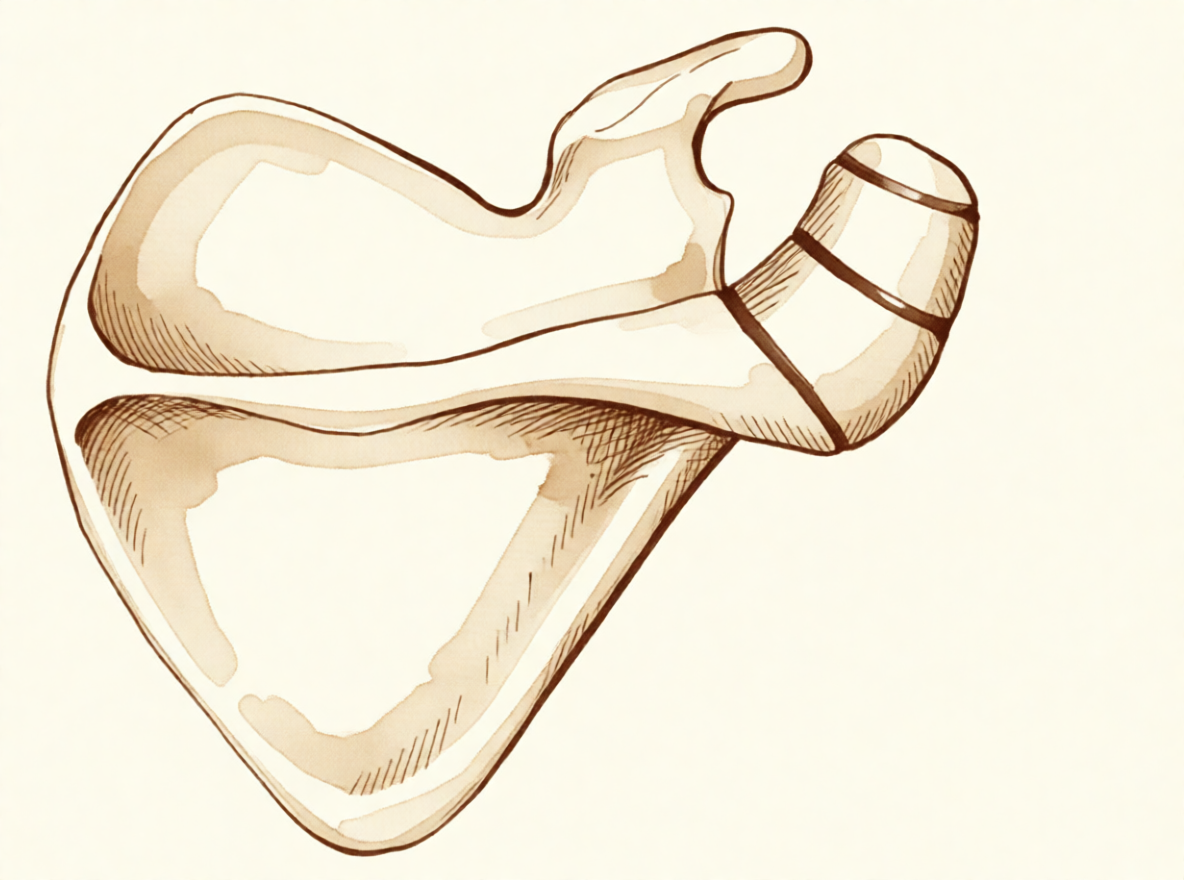
O teto ósseo do ombro não se forma como um único pedaço de osso. Ele começa como três centros separados que se unem lentamente, a partir de cerca dos 15 anos, e essa união geralmente termina por volta dos 25 anos. Em cerca de 8% das pessoas, uma dessas uniões nunca se fecha. O espaço é preenchido por um tecido resistente e fibroso, em vez de osso, deixando um pequeno pedaço do teto preso por uma ligação flexível, e não por osso sólido.
Pense nisso como uma tábua presa a uma parede com um parafuso frouxo. Ela se mantém no lugar, mas se move um pouco quando você se apoia nela. O músculo que levanta o braço para o lado se prende a esse teto; por isso, toda vez que você levanta o braço, esse músculo puxa o pedaço solto e ele se flexiona ligeiramente. Esse movimento pressiona para baixo o tendão que passa sob o teto, e o atrito o irrita. É por isso que as atividades acima da cabeça doem e que a dor fica na parte da frente e na lateral do ombro.
A própria ligação flexível também pode ser o problema. Ela pode ficar sensível por si só, como uma articulação desgastada, e pressioná-la provoca a dor. A maioria das pessoas com esse padrão ósseo nunca sente nada, e ele não parece causar dor no ombro por si só nem tornar mais provável uma ruptura do tendão. Mas, quando o pedaço solto começa a se mover mais do que o normal, seja pela forma como se formou, seja após uma pancada na região, ele pode pressionar o tendão toda vez que você levanta o braço. É isso que produz o arco doloroso e a dor noturna descritos acima.
Como o pedaço solto fica exatamente onde o tendão passa, os dois problemas muitas vezes aparecem juntos. Se o seu ombro já foi tratado por um problema no tendão e a dor não melhorou, vale a pena investigar esse pedaço não unido, pois operar apenas o tendão não vai acalmar um pedaço de osso que se move acima dele.
O que podemos fazer a respeito
O Dr. Kieran Hirpara, cirurgião de membro superior no Mater Private Hospital Rockhampton, começa com as opções menos invasivas adequadas ao seu caso. Em geral, os pacientes são encaminhados à nossa clínica pelo médico de família; caso um fisioterapeuta tenha sugerido que você nos procure, ainda assim será necessário um encaminhamento do seu médico de família para ter direito ao reembolso do Medicare. Na sua primeira consulta, colhemos o histórico clínico, examinamos o ombro e solicitamos exames de imagem, quando necessário.
O primeiro passo costuma ser mudar a forma como você usa o ombro e dar à fisioterapia uma chance de verdade. A fisioterapia tem como objetivo acalmar a irritação sob o teto ósseo e fortalecer os músculos ao redor do ombro, para que eles lidem melhor com atividades acima da cabeça. Faça uma tentativa razoável antes de pensar em qualquer outra opção.
Os analgésicos podem ajudar você a continuar se movimentando enquanto isso acontece. Anti-inflamatórios simples, tomados conforme a orientação do seu médico de família, aliviam a dor para que você possa fazer os exercícios com conforto. Se a dor não estiver melhorando, estiver piorando ao longo de semanas, acordar você à noite ou impedir você de trabalhar ou de usar o braço, consulte o seu médico de família ou peça uma avaliação com especialista.
Quando o tratamento não cirúrgico não trouxe melhora suficiente, consideramos a cirurgia. Antes disso, confirmamos o diagnóstico com radiografias simples, que quase sempre bastam para mostrar o pedaço não unido. Uma incidência feita por baixo da axila mostra claramente o formato da articulação e a parte da frente do teto, e isso é importante porque uma lesão pode danificar a junção entre os dois pedaços de osso. A ultrassonografia é uma forma simples e precisa de examinar o tendão que passa sob o teto, e a ressonância magnética oferece uma visão mais completa desse tendão e dos tecidos moles ao redor.
A operação que oferecemos com mais frequência é uma avaliação da articulação por pequenas incisões (artroscópica) com bursectomia, que consiste em remover o tecido inflamado do espaço sob o teto, seguida de uma pequena incisão aberta para enxerto ósseo e fixação, ou seja, manter o pedaço solto imóvel para que ele possa se unir ao restante do osso. Mantê-lo imóvel permite que os dois pedaços cresçam juntos. Se o pedaço solto for pequeno, podemos removê-lo em vez de fixá-lo. Fixar um pedaço maior exige cuidado, porque o músculo que levanta o braço o puxa constantemente e o pedaço é fino. Às vezes, o fragmento não se une após a fixação, o que se chama não união. Vamos conversar sobre qual opção é adequada ao seu ombro e decidir juntos.
O que esperar
A maioria das pessoas com esse padrão ósseo nunca tem problema algum, e ele não precisa de tratamento. Quando causa dor, o prognóstico depende de o pedaço solto estar estável e de ele ser realmente a fonte da sua dor.
No caso de um pedaço doloroso, o tratamento geralmente começa sem cirurgia. Mudar a forma como você usa o ombro, a fisioterapia e analgésicos simples aliviam o problema em muitas pessoas. Se isso não funcionou após uma tentativa razoável, a cirurgia pode levar a uma redução real da dor e a uma melhor função do ombro. Quando o pedaço solto é mantido imóvel e se une ao restante do osso, as pessoas obtêm melhores resultados nas medidas de função do ombro do que quando os dois pedaços continuam separados. Mas a cirurgia nem sempre é a solução. Em algumas pessoas com essa condição e também uma ruptura do tendão, os resultados após a cirurgia foram variados, com 53% obtendo resultados satisfatórios no geral.
Se não for tratado e continuar incomodando, a dor geralmente não desaparece sozinha. O problema também pode passar despercebido: vale a pena suspeitar dessa condição quando a dor no ombro não melhorou após o tratamento, inclusive após uma cirurgia para um problema no tendão, porque operar apenas o tendão não vai acalmar um pedaço de osso que se move acima dele.
A recuperação após a cirurgia é gradual. Você trabalhará com o seu fisioterapeuta, recuperando primeiro o movimento e depois a força, e a capacidade de levantar peso acima da cabeça voltará aos poucos, ao longo de semanas a meses. O objetivo é um ombro que dê conta da vida diária sem a dor aguda ao levantar o braço que trouxe você até nós.
Se você notar que um dedo, a mão ou o braço ficou quente, vermelho, inchado e dolorido, especialmente com febre, vá ao pronto-socorro no mesmo dia. Não é necessário encaminhamento do médico de família.
Quando procurar ajuda médica
A maioria das pessoas com esse padrão ósseo nunca precisa de tratamento, mas consulte o seu médico de família ou peça uma avaliação com especialista se a dor no ombro não estiver melhorando, estiver piorando ao longo de semanas, acordar você à noite ou impedir você de trabalhar ou de usar o braço. Peça uma avaliação mais cedo se a dor ficar na parte da frente e na lateral do ombro, piorar com atividades acima da cabeça ou continuar acordando você quando se deita sobre esse lado. Também vale a pena uma avaliação se o ponto dolorido na parte superior do ombro estiver sensível à pressão, se o braço parecer fraco ao levantá-lo para o lado ou ao girá-lo para fora, ou se o ombro doer em parte do movimento de elevação. Adultos mais jovens com esses sintomas e sem lesão devem ser avaliados quanto a essa condição, especialmente se a fisioterapia, o afastamento do esporte ou uma injeção não tiverem ajudado. Se um dedo, a mão ou o braço ficar quente, vermelho, inchado e dolorido, especialmente com febre, vá ao pronto-socorro no mesmo dia; não é necessário encaminhamento do médico de família.
Em maior profundidade
Advanced reading: the deeper science (optional)
Esta seção aborda conteúdos além do necessário para suas próprias decisões de tratamento. O acrômio acessório merece uma leitura mais aprofundada, pois é uma variante anatômica normal em uma parcela significativa da população; geralmente não causa nenhum problema e, muitas vezes, é erroneamente apontado como causa da dor no ombro, quando na verdade não é.
O que é e quão comum é
O acrômio, a estrutura óssea que forma o “teto” do ombro, se forma a partir de vários focos de ossificação que normalmente se fundem no final da adolescência ou início da idade adulta. Em algumas pessoas, um desses focos não se funde, resultando numa junção fibrosa permanente. Isso é o os acromiale, que está presente desde a adolescência e não é adquirido posteriormente.
A prevalência varia conforme a população. Um estudo multicêntrico com 6.842 pacientes constatou que o os acromiale está associado a lesões do manguito rotador, que a sua prevalência na população japonesa estudada era menor do que em populações não asiáticas, e que o tamanho do fragmento tendia a ser menor [1].
Essa associação com lesões do manguito merece atenção. Ela demonstra que ambos os quadros ocorrem juntos com maior frequência do que o esperado por acaso, mas não prova que o os acromiale seja a causa da lesão. O mecanismo plausível funciona nos dois sentidos: o fragmento não fundido se move ligeiramente sob a ação do músculo deltoide, podendo irritar o tendão abaixo; ou então, a mesma biomecânica do ombro que predispõe às lesões do manguito torna o fragmento instável e sintomático.
A questão central é se ele realmente é a causa da dor
Por ser visível, permanente e apresentar aspecto anormal nas imagens, o os acromiale costuma ser apontado como explicação para a dor no ombro. Na maioria dos casos, porém, ele não causa sintomas; a maioria das pessoas que o possuem nem sequer sabe disso.
O que diferencia um os acromiale sintomático são a sensibilidade localizada exatamente sobre o fragmento, a dor que surge ao pressioná-lo e, quando se utiliza injeção diagnóstica, o alívio obtido ao injetar justamente na região do fragmento, e não no espaço subacromial abaixo dele. Na ausência desses sinais, esse achado deve ser considerado incidental.
Os subtipos são convencionalmente classificados conforme o local da fusão envolvido [2]; isso é relevante do ponto de vista cirúrgico, pois o tamanho do fragmento móvel determina se ele pode ser removido ou se é necessário fixá-lo.
Por que isso complica outras cirurgias do ombro
A importância prática do os acromiale geralmente não está nos seus próprios sintomas, mas no impacto que exerce sobre cirurgias planejadas para outras condições.
O deltoide se insere no acrômio; portanto, um fragmento não fundido sofre essa tração numa região móvel. A remoção de osso da face inferior do acrômio, como na acromioplastia, torna ainda mais fino um fragmento que já é instável, podendo transformar uma variante assintomática em uma condição dolorosa ou provocar não união óssea. Este é o principal motivo para que o os acromiale seja identificado em exames de imagem pré-operatórios: não para tratá-lo, mas para evitar desestabilizá-lo.
Quando o tratamento se torna necessário, a escolha recai entre a excisão de um fragmento pequeno ou a fixação de um fragmento maior. Contudo, a fixação nessa região é reconhecidamente difícil: o fragmento é fino, o deltoide exerce tração contínua sobre ele e as taxas de não união são consideráveis. Essa dificuldade, por si só, é motivo para se ter certeza de que o os acromiale é realmente a fonte da dor antes de realizar qualquer intervenção cirúrgica.
Referências
[1] Kozono N, Nishii A, Ishitani E, Mizuki Y, Kimura T, Yamamoto S, et al. Prevalência e fatores associados ao os acromiale: um estudo multicêntrico. JSES Int. 2025;9(5):1541-5. https://doi.org/10.1016/j.jseint.2025.05.015
[2] Boehm TD, Matzer M, Brazda D, Gohlke FE. Tratamento cirúrgico do os acromiale associado à ruptura do manguito rotador: revisão de 33 pacientes. J Bone Joint Surg Br. 2003;85(4):545-9. https://doi.org/10.1302/0301-620X.85B4.13634
Evidence & references
This is the clinical evidence summary written for health professionals. It is technical, and it lists the research this page was built from. You do not need to read it to understand your treatment or to make a decision about it.
Overview
- A symptomatic, unstable os acromiale is relatively rare despite the radiographic appearance of an os acromiale being not uncommon in patients presenting with shoulder pain [1].
- An os acromiale should always be suspected in cases of failed rotator cuff surgery or ongoing shoulder pain after failed treatment [2].
- The prevalence of os acromiale was 2.13% in Thai patients with shoulder problems who required MRI evaluation [3].
- Os acromiale is associated with rotator cuff injuries [7].
- The greater the distance of the acromioclavicular joint from the anterior edge of the acromion, the higher the likelihood of an os acromiale [15].
- The authors recommend that the acromion be carefully examined roentgenographically prior to operation in any patient with a rotator cuff tear [10].
- Surgical treatment is usually not indicated for os acromiale in the professional tennis player [9].
- Operative management of a symptomatic os acromiale that has failed initial nonoperative treatment leads to decreased symptoms and improvement in clinical outcomes [4].
- Surgical options for symptomatic os acromiale include arthroscopic sub-total excision, arthroscopic subacromial decompression of stable fragments, and open reduction and internal fixation of unstable fragments [13].
- Patients with a united os acromiale had a significantly better functional outcome as measured by the Constant score (P = .0169) [12].
- The advantages of arthroscopic internal fixation include improved visualization of the undersurface of the acromion, allowing excellent reduction, the possibility of treating concomitant shoulder pathologies, and to avoid increased morbidity of the open approach [14].
- Postoperative local tenderness at the os acromiale can be expected in 1 out of 4 patients but resolves spontaneously over time in the majority of patients [5].
- The presence of os acromiale does not appear to have a negative impact on the clinical outcomes after surgery and rTSA remains a safe and effective treatment option [6].
- The outcome of RTSA does not seem to be negatively affected by the presence of an os acromiale [11].
- Ipsilateral os acromiale may be a relative contraindication to the clavicle hook plate [16].
Anatomy & Pathophysiology
Bony Anatomy
- The acromion has three ossification centers: the metacromion (base), the mesoacromion (middle), and the preacromion (tip) [38].
- Failure of fusion of the acromial ossification centers results in os acromiale [38].
- Os acromiale is defined as incomplete fusion of secondary ossification centers, most commonly between the mesoacromion and meta-acromion [49].
- The unfused segment in os acromiale is most commonly the meso-acromion, a configuration often termed a meso-type os acromiale [22].
- The acromion, coracoacromial ligament, and coracoid process form the coracoacromial arch, a rigid bony-ligamentous structure that imparts stability to the shoulder girdle [35].
- The rotator cuff, subacromial bursa, and subdeltoid bursa pass underneath the coracoacromial arch [35].
- The scapula is attached to the axial skeleton by the acromioclavicular and sternoclavicular joints [37].
- The acromion is a flattened bony process that curves forwards from the scapular spine [37].
Prevalence and Associations
- In Thai patients with shoulder problems who required MRI evaluation, the prevalence of os acromiale was 2.13% [3].
- The radiographic appearance of an os acromiale is not uncommon in patients presenting with shoulder pain [1].
- A symptomatic, unstable os acromiale is relatively rare [1].
Pathophysiology
- Most diagnoses of os acromiale are made incidentally on axillary view radiographs of the shoulder [22].
- Primary shoulder pain is usually unrelated to the unfused os acromiale [22].
- When an os acromiale drives symptoms, the two principal causes are motion at the non-union site or an impingement-type syndrome resulting from flexion of the os fragment during deltoid contraction and arm elevation [22].
- Pain from an os acromiale due to these mechanisms has been reported in a variety of sports, particularly among throwing athletes and swimmers [22].
- Previously stable non-unions can become unstable following an episode of blunt trauma to the region [22].
- The synchondrosis of an os acromiale can be injured following trauma [24].
- Extrinsic impingement occurs when the space available for the rotator cuff is diminished, with examples including acromial fracture or pathologic os acromiale [62].
Epidemiology and Prevalence
- Liberson reviewed 1800 shoulder girdles and identified an incidence of os acromiale of 1.4% [21].
- The lesion was bilateral in 62% of patients in Liberson's review of 1800 shoulder girdles [21].
Associated Pathology and Risk Factors
- The greater the distance of the AC joint from the anterior edge of the acromion, the higher the likelihood of an os acromiale [15].
Diagnostic Considerations
- Awareness of the os acromiale in the young athlete, appropriate clinical examination, and image studies are crucial to confirm diagnosis [19].
- Appropriate radiographic investigation including axillary views is emphasized for the diagnosis of os acromiale injuries [24].
Clinical Presentation
- An os acromiale should always be suspected in cases of failed rotator cuff surgery [2].
- An os acromiale should always be suspected in cases of ongoing shoulder pain after failed treatment [2].
- Postoperative local tenderness at the os acromiale can be expected in 1 out of 4 patients [5].
- Postoperative local tenderness at the os acromiale resolves spontaneously over time in the majority of patients [5].
- The lesion was bilateral in 62% of patients in Liberson's review [21].
- The unfused segment is most commonly the meso-acromion [22].
- Any primary shoulder pain is usually unrelated to the unfused os [22].
- If the os acromiale is driving symptoms, one principle cause is motion at the non-union site [22].
- If the os acromiale is driving symptoms, another principle cause is an impingement-type syndrome resulting from flexion of the os fragment during deltoid contraction and arm elevation [22].
- Pain from an os acromiale by the mechanism of impingement has been reported in a variety of sports [22].
- Pain from an os acromiale by the mechanism of impingement has been reported in particular among throwing athletes and swimmers [22].
- Meta–os acromiale is the rarest subtype of os acromiale [23].
Investigations
Prevalence and Demographics
- Liberson reviewed 1800 shoulder girdles and identified an incidence of os acromiale of 1.4%, with the lesion being bilateral in 62% of patients [21].
- A multicenter study aimed to determine the prevalence of and factors associated with os acromiale in the Japanese population [8].
- The radiographic appearance of an os acromiale is not uncommon in patients presenting with shoulder pain, but a symptomatic, unstable os acromiale is relatively rare [1].
Radiographic Evaluation
- The acromion should be carefully examined roentgenographically prior to operation in any patient with a rotator cuff tear [10].
- Appropriate radiographic investigation for os acromiale includes axillary views [24].
- Awareness of the os acromiale in the young athlete requires appropriate clinical examination and image studies to confirm diagnosis [19].
- The standard shoulder series should include orthogonal views of the shoulder, including a true AP view in the scapular plane, an AP view, an axillary view, and a scapular Y view [60].
- The scapular Y view provides visualization of the coracoacromial arch and can reveal coracoacromial spurs, which have been closely associated with the presence of rotator cuff pathology [60].
- The scapular Y view can also show acromial shape [60].
- At least two X-ray views should be obtained for shoulder imaging: an anteroposterior in the plane of the glenoid and an axillary projection with the arm in abduction to show the relationship of the humeral head to the glenoid [52].
- The purpose of imaging of the shoulder is to help establish the diagnosis, determine the severity of the pathoanatomy, assist in surgical planning, and enable the surgeon to illustrate the condition of the shoulder to the patient [30].
- Standardized plain films are almost always sufficient to garner the information needed for shoulder evaluation [30].
- The first key view for shoulder imaging is the anteroposterior (AP) in the plane of the scapula taken so that the x-ray beam passes through the glenohumeral joint [30].
- The second key view for shoulder imaging is the axillary view taken with the arm in the functional position of elevation in the plane of the scapula and oriented so that both the spinoglenoid notch and the scapular neck are visible [30].
- The axillary view is referred to as the "truth view" because it demonstrates the glenohumeral relationships in the functional position of elevation [30].
- CT scans have the disadvantage of being taken with the arm in the adducted position, unlike the axillary truth view [30].
- The temptation to "overimage" should be resisted, obtaining only the scans or reconstructions that are necessary for the care of the patient [30].
Magnetic Resonance Imaging
- In Thai patients with shoulder problems, os acromiale was identified via Magnetic Resonance Imaging evaluation [3].
- MRI is useful to identify labral tears and rotator cuff tears, although the accuracy for these is enhanced by combining the scan with arthrography [52].
- MRI is the modality of choice for evaluating the rotator cuff, biceps, and subacromial/subdeltoid bursa [59].
- T2-weighted MRI provides better visualization of full thickness rotator cuff tears [59].
- MR arthrography is considered the benchmark for evaluation for labral tears and rarely is indicated for evaluation of rotator cuff pathology [59].
- When MRI or MR arthrography is contraindicated, CT arthrography is indicated [59].
Ultrasonography
- Ultrasonography is a simple and accurate test for identifying rotator cuff tears and calcific tendinitis [52].
- Ultrasonography is a low-cost alternative to MRI and arthrography for evaluating both skeletal and soft-tissue structures of the shoulder [59].
- Ultrasonography can provide immediate, real-time visualization of the rotator cuff, biceps tendon, and calcific deposits [59].
- Ultrasonography can be used to measure the subacromial space and detect atrophy of rotator cuff muscles [59].
- Ultrasonography is highly operator dependent and is not as useful for evaluating labral tears or rotator cuff tears that are very small or larger than 3 cm [59].
- The most commonly performed joint examination using ultrasonography is the shoulder examination, and accuracy depends on the skill of the scanner operator and an awareness of pitfalls that are encountered [50].
Clinical Associations and Diagnostic Implications
- The synchondrosis of an os acromiale can be injured following trauma, though rarely [24].
Treatment
Non-Operative
- A symptomatic os acromiale in a competitive female fastball pitcher was treated successfully with open reduction and internal fixation [27].
Operative
- Symptomatic unstable os acromiale can be effectively treated with autogenous bone-grafting and internal fixation using a rigid tension-band construct and cannulated screws, which results in a high rate of union [65].
- This new arthroscopic technique of fixation of os acromiale with absorbable screws provides promising clinical, cosmetic, and radiologic results with high patient satisfaction [28].
- After 3 to 6 years of follow-up, all 6 shoulders were rated as excellent according to criteria similar to that of the American Shoulder and Elbow Surgeons [18].
- Modified arthroscopic subacromial decompression can provide results similar to standard arthroscopic subacromial decompression for routine impingement [25].
- Meta–os acromiale is the rarest subtype of os acromiale, and special consideration must be given to the type of tension-band construct used to achieve adequate compression and fixation [23].
Complications
Post-operative and Surgical Complications
- Postoperative local tenderness at the os acromiale can be expected in 1 out of 4 patients following reverse total shoulder arthroplasty [5].
Diagnostic and Clinical Implications
Pathophysiology and Mechanisms of Symptom
- Pain from an os acromiale by the mechanism of impingement has been reported in a variety of sports, in particular among throwing athletes and swimmers [22].
Recovery
- After 3 to 6 years of follow-up, all 6 shoulders treated with osteosynthesis of an unstable mesoacromion were rated as excellent according to criteria similar to that of the American Shoulder and Elbow Surgeons [18].
- At a two to five year follow-up, all patients treated for fusion of the unstable mesoacromion had fused [69].
Key Evidence
- [L4] While the radiographic appearance of an os acromiale is not uncommon in patients presenting with shoulder pain, a symptomatic, unstable os acromiale is relatively rare. [1] (10.1097/00132589-200412000-00006)
- [L4] An os acromiale should always be suspected in cases of failed rotator cuff surgery or ongoing shoulder pain after failed treatment. [2] (10.1097/00132589-200609000-00006)
- [L3] In Thai patients with shoulder problems who required MRI evaluation, the prevalence of os acromiale was 2.13%. [3] (10.1177/23259671221078806)
- [L4] Operative management of a symptomatic os acromiale that has failed initial nonoperative treatment leads to decreased symptoms and improvement in clinical outcomes. [4] (10.1016/j.jse.2019.05.047)
- [L3] Postoperative local tenderness at the os acromiale can be expected in 1 out of 4 patients but resolves spontaneously over time in the majority of patients. [5] (10.1177/2325967120965131)
- [L4] The presence of os acromiale does not appear to have a negative impact on the clinical outcomes after surgery and rTSA remains a safe and effective treatment option. [6] (10.1016/j.xrrt.2025.01.002)
- [L3] The study supports previous findings that os acromiale is associated with rotator cuff injuries. [7] (10.1016/j.jseint.2025.05.015)
- [L3] This multicenter study aimed to determine the prevalence of and factors associated with os acromiale in the Japanese population. [8] (10.1016/j.jse.2025.01.008)
- [L4] Surgical treatment is usually not indicated for os acromiale in the professional tennis player. [9] (10.1177/2325967118773723)
- [L4] The authors recommend that the acromion be carefully examined roentgenographically prior to operation in any patient with a rotator cuff tear. [10] (10.2106/00004623-198466080-00029)
- [L4] The outcome of RTSA does not seem to be negatively affected by the presence of an os acromiale. [11] (10.1016/j.jse.2017.02.012)
- [L4] Patients with a united os acromiale had a significantly better functional outcome as measured by the Constant score (P = .0169). [12] (10.1016/s1058-2746(98)90008-8)
- [L5] Surgical options for symptomatic os acromiale include arthroscopic sub-total excision, arthroscopic subacromial decompression of stable fragments, and open reduction and internal fixation of unstable fragments. [13] (10.5435/jaaos-d-17-00011)
- [L5] The advantages of this technique are improved visualization of the undersurface of the acromion, allowing excellent reduction, the possibility of treating concomitant shoulder pathologies, and to avoid increased morbidity of the open approach. [14] (10.1097/bte.0000000000000191)
- [L4] Our data suggest that the greater the distance of the AC joint from the anterior edge of the acromion, the higher the likelihood of an os acromiale. [15] (10.1067/mse.2003.128136)
- [L4] Ipsilateral os acromiale may be a relative contraindication to the clavicle hook plate. [16] (10.1186/s12891-021-04841-1)
- [L4] After 3 to 6 years of follow-up, all 6 shoulders were rated as excellent according to criteria similar to that of the American Shoulder and Elbow Surgeons. [18] (10.1016/s1058-2746(99)90004-6)
- [L4] Awareness of the os acromiale in the young athlete, appropriate clinical examination, and image studies are crucial to confirm diagnosis. [19] (10.1016/j.jseint.2020.02.008)
- [L4] [22] (10.1302/2058-5241.4.180100)
- [L4] Meta–os acromiale is the rarest subtype of os acromiale, and special consideration must be given to the type of tension-band construct used to achieve adequate compression and fixation. [23] (10.1177/03635465211028238)
- [L4] This case highlights that the synchondrosis of an os acromiale can be injured following trauma, though rarely, and emphasizes the need for appropriate radiographic investigation including axillary views and a flexible surgical approach. [24] (10.1016/j.jse.2008.02.012)
- [L5] Increased awareness of the diagnosis and improved techniques have shown that modified arthroscopic subacromial decompression can provide results similar to standard arthroscopic subacromial decompression for routine impingement. [25] (10.1097/00132589-200206000-00003)
- [L4] A symptomatic os acromiale in a competitive female fastball pitcher was treated successfully with open reduction and internal fixation. [27] (10.1177/0363546506288305)
- [L4] This new arthroscopic technique of fixation of os acromiale with absorbable screws provides promising clinical, cosmetic, and radiologic results with high patient satisfaction. [28] (10.1016/j.jse.2011.12.011)
- [L4] Symptomatic unstable os acromiale can be effectively treated with autogenous bone-grafting and internal fixation using a rigid tension-band construct and cannulated screws, which results in a high rate of union. [65] (10.2106/00004623-199809000-00011)
- [L4] At a two to five year followup all had fused. [69] (10.1016/s1058-2746(95)80192-8)
References
[1] Treatment of the Symptomatic Os Acromiale. Techniques in Shoulder and Elbow Surgery. 2004. DOI: 10.1097/00132589-200412000-00006
[2] Management of Os Acromiale. Techniques in Shoulder and Elbow Surgery. 2006. DOI: 10.1097/00132589-200609000-00006
[3] Prevalence of Os Acromiale in Thai Patients With Shoulder Problems: A Magnetic Resonance Imaging Study. Orthopaedic Journal of Sports Medicine. 2022. DOI: 10.1177/23259671221078806
[4] Os acromiale: systematic review of surgical outcomes. Journal of Shoulder and Elbow Surgery. 2020. DOI: 10.1016/j.jse.2019.05.047
[5] Os Acromiale in Reverse Total Shoulder Arthroplasty: A Cohort Study. Orthopaedic Journal of Sports Medicine. 2020. DOI: 10.1177/2325967120965131
[6] Clinical implications of reverse total shoulder arthroplasty with an os acromiale: a systematic review. JSES Reviews, Reports, and Techniques. 2025. DOI: 10.1016/j.xrrt.2025.01.002
[7] Prevalence and factors associated with os acromiale: a multicenter study. JSES International. 2025. DOI: 10.1016/j.jseint.2025.05.015
[8] The prevalence and associated factors of os acromiale: a multicenter study. Journal of Shoulder and Elbow Surgery. 2025. DOI: 10.1016/j.jse.2025.01.008
[9] Os Acromiale in Professional Tennis Players. Orthopaedic Journal of Sports Medicine. 2018. DOI: 10.1177/2325967118773723
[10] Rotator cuff tears associated with os acromiale.. The Journal of Bone & Joint Surgery. 1984. DOI: 10.2106/00004623-198466080-00029
[11] Reverse shoulder arthroplasty in patients with os acromiale. Journal of Shoulder and Elbow Surgery. 2017. DOI: 10.1016/j.jse.2017.02.012
[12] Transacromial approach to obtain fusion of unstable os acromiale. Journal of Shoulder and Elbow Surgery. 1998. DOI: 10.1016/s1058-2746(98)90008-8
[13] Symptomatic, Unstable Os Acromiale. Journal of the American Academy of Orthopaedic Surgeons. 2018. DOI: 10.5435/jaaos-d-17-00011
[14] Arthroscopic Internal Fixation of Symptomatic Os Acromiale. Techniques in Shoulder & Elbow Surgery. 2020. DOI: 10.1097/bte.0000000000000191
[15] Relationship between os acromiale and acromioclavicular joint anatomic position. Journal of Shoulder and Elbow Surgery. 2003. DOI: 10.1067/mse.2003.128136
[16] Os acromiale may be a contraindication of the clavicle hook plate: case reports and literature review. BMC Musculoskeletal Disorders. 2021. DOI: 10.1186/s12891-021-04841-1
[18] Successful osteosynthesis of an unstable mesoacromion in 6 shoulders: A new technique. Journal of Shoulder and Elbow Surgery. 1999. DOI: 10.1016/s1058-2746(99)90004-6
[19] The unstable os acromiale: a cause of pain in the young athlete. JSES International. 2020. DOI: 10.1016/j.jseint.2020.02.008
[21] Types of os acromiale according to Liberson. 2006.
[22] Os acromiale: a review of its incidence, pathophysiology, and clinical management. EFORT Open Reviews. 2019. DOI: 10.1302/2058-5241.4.180100
[23] Rare Symptomatic Meta–Os Acromiale in an Athlete. The American Journal of Sports Medicine. 2021. DOI: 10.1177/03635465211028238
[24] Fracture of an os acromiale with associated rupture of the coracoclavicular ligaments. Journal of Shoulder and Elbow Surgery. 2008. DOI: 10.1016/j.jse.2008.02.012
[25] Arthroscopic Management of the Mesoacromion. Techniques in Shoulder and Elbow Surgery. 2002. DOI: 10.1097/00132589-200206000-00003
[27] Surgical Stabilization of Os Acromiale in a Fast-Pitch Softball Pitcher. The American Journal of Sports Medicine. 2006. DOI: 10.1177/0363546506288305
[28] Arthroscopically assisted internal fixation of the symptomatic unstable os acromiale with absorbable screws. Journal of Shoulder and Elbow Surgery. 2012. DOI: 10.1016/j.jse.2011.12.011
[30] Rockwood And Matsen S The Shoulder. Arthroscopic Management of Prearthritic and Arthritic Conditions of the Shoulder and the Postarthroplasty Shoulder > Radiographic Evaluation.
[35] Rockwood And Matsen S The Shoulder. Shoulder and Elbow Specialty Clinic Workers’ Survey > ANATOMY.
[37] Rockwood And Green S Fractures In Adults. 29: Principles of Nonunion and Bone Defect Treatment > Applied Anatomy Related to Scapular Fractures.
[38] Aaos Comprehensive Orthopaedic Review 3. Anatomy of the Shoulder, Arm, and Elbow > I. Shoulder.
[49] Miller S Review Of Orthopaedics. Genetics of musculoskeletal conditions and abnormalities are summarized in Table 1.27 > UPPER EXTREMITY > SHOULDER.
[50] Orthopaedic Knowledge Update Sports Medicine 6. Diagnostic Ultrasonography and Ultrasonography-Guided Procedures > Annotated References.
[52] Apley And Solomon S Concise System Of Orthopaedics And Trauma. INVESTIGATION.
[59] Orthopaedic Knowledge Update 13 Ebook Without Multimedia. Shoulder Anatomy and Biomechanics, Clinical Evaluation, Imaging > Clinical Evaluation > Imaging.
[60] Aaos Comprehensive Orthopaedic Review 3. Imaging of the Shoulder and Elbow > I. Shoulder.
[62] Campbell S Operative Orthopaedics 4 Volume Set. ANTERIOR CRUCIATE LIGAMENT RECONSTRUCTION WITH BONE-PATELLAR TENDON-BONE GRAFT > Developmental Stages of Impingement Syndrome.
[65] The Treatment of Symptomatic Os Acromiale. The Journal of Bone & Joint Surgery*. 1998. DOI: 10.2106/00004623-199809000-00011
[69] Successful fusion of the unstable mesoacromion: Technique and report of five cases. Journal of Shoulder and Elbow Surgery. 1995. DOI: 10.1016/s1058-2746(95)80192-8