Patients › Elbow
Osteoartrite do cotovelo
Osteoarthritis of the elbow — primary and post-traumatic, conservative and surgical options.
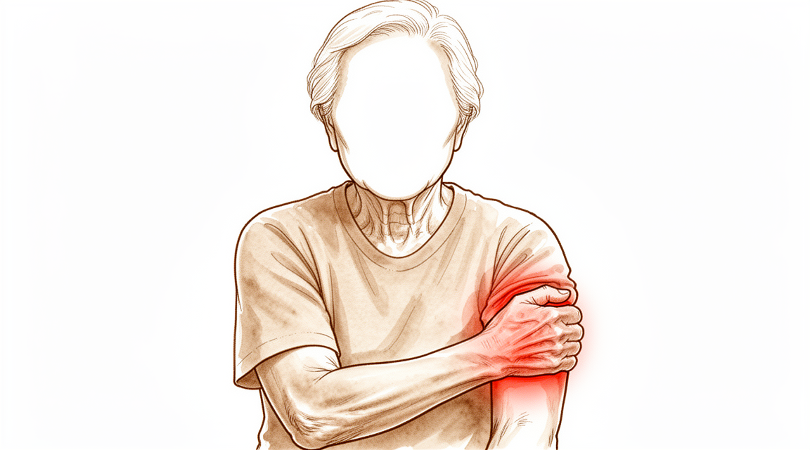
O que você está sentindo
A artrite do cotovelo geralmente se manifesta como rigidez e dor bem nas extremidades do movimento. Você pode perceber que não consegue esticar completamente o braço, ou que ele não dobra até o fim. A dor tende a ficar nesses limites, e não no meio do movimento. Muitas pessoas também notam sensação de “engate”, estalos ou travamentos, às vezes dolorosos, à medida que saliências ósseas e pequenos fragmentos soltos de cartilagem ou de osso ficam presos dentro da articulação.
As tarefas do dia a dia que exigem o braço esticado ou totalmente dobrado ficam difíceis. Alcançar uma prateleira alta, apoiar-se para se levantar de uma cadeira ou levar o garfo à boca: tudo isso pode se tornar complicado. Girar o antebraço, como ao girar uma chave ou uma chave de fenda, muitas vezes continua sem problemas no início e só fica rígido mais tarde. O quanto isso limita você depende muito do seu trabalho e das exigências do seu dia a dia. O trabalho manual pesado, o levantamento de peso ou o arremesso sobrecarregam muito mais a articulação do que as tarefas de escritório.
A dor noturna não é típica da artrite do cotovelo. Se o cotovelo acorda você à noite, isso aponta para outra causa e merece ser avaliado. A dor que não está melhorando, que está piorando ao longo de semanas ou que impede você de trabalhar ou de usar o braço deve ser avaliada pelo seu médico generalista, ou você pode nos pedir uma avaliação por um especialista.
Algumas pessoas com artrite do cotovelo também desenvolvem compressão do nervo ulnar, o nervo que passa por trás da parte interna do cotovelo. Isso pode causar formigamento ou dormência no dedo anelar e no dedo mínimo, ou fraqueza na mão. Se você notar esses sintomas, mencione-os na sua próxima consulta para que possamos avaliar o nervo.
O problema de base é o desgaste da superfície articular, somado à formação de osso extra ao redor das bordas do cotovelo. Esse osso extra, junto com um revestimento articular que vai ficando apertado e com os fragmentos soltos, bloqueia fisicamente o cotovelo nas extremidades da sua amplitude. Curiosamente, o espaço dentro da articulação muitas vezes ainda está preservado; por isso, em um exame de imagem, a articulação pode parecer melhor do que os seus sintomas sugerem.
O que está realmente acontecendo
Dentro do cotovelo, três pequenas articulações trabalham juntas. Uma permite dobrar e esticar o braço. Duas permitem girar o antebraço, para que você consiga virar a palma da mão para cima ou para baixo. Cada superfície articular é coberta por uma camada lisa e escorregadia de cartilagem, um pouco como o revestimento antiaderente de uma boa frigideira. Quando esse revestimento está intacto, as superfícies deslizam umas sobre as outras quase sem atrito.
Na artrite do cotovelo, esse revestimento fica fino em algumas áreas, e o corpo responde formando osso extra ao redor das bordas da articulação. Essas saliências ósseas, chamadas osteófitos ou esporões ósseos, se acumulam na frente e atrás do cotovelo. O revestimento da articulação, uma capa de tecido ao redor dela, também fica mais apertado e encurtado. Pequenos pedaços de cartilagem ou de osso podem se soltar e flutuar dentro da articulação como corpos soltos.
Juntas, essas alterações explicam o que você está sentindo. As saliências ósseas e o revestimento apertado funcionam como um batente físico nas extremidades do movimento, de modo que o cotovelo não estica nem dobra completamente. Os corpos soltos são o que causa a sensação de “engate”, os estalos ou os travamentos, quando ficam presos entre as superfícies em movimento. No início, a dor fica nesses limites das extremidades do movimento, porque é ali que o bloqueio acontece. À medida que mais do revestimento de cartilagem se desgasta, a dor pode se espalhar também para o meio do movimento.
Duas coisas tornam a artrite do cotovelo um pouco diferente da artrite no quadril ou no joelho. Primeiro, o espaço dentro da articulação muitas vezes continua amplo; por isso, uma radiografia pode parecer melhor do que os seus sintomas. Segundo, o desgaste tende a atingir uma parte da articulação mais do que o restante: a área onde a parte de cima de um dos ossos do antebraço encontra a extremidade do osso do braço logo acima. Esse ponto recebe muita carga quando você empurra ou levanta algo, e é por isso que o trabalho manual pesado, o levantamento de peso e o arremesso estão associados a essa condição.
Ela também pode surgir depois de uma lesão antiga. Uma fratura ou outro dano na articulação pode deixar as superfícies levemente irregulares, de modo que elas se desgastam de forma desigual depois disso.
O que podemos fazer a respeito
O Dr. Kieran Hirpara, cirurgião de membros superiores no Mater Private Hospital Rockhampton, começa com as opções menos invasivas adequadas ao seu quadro clínico. Em geral, os pacientes são encaminhados à nossa clínica pelo médico generalista; caso um fisioterapeuta tenha sugerido que você nos procure, ainda assim será necessário um encaminhamento do seu médico generalista para ter direito ao reembolso do Medicare. Na sua primeira consulta, colhemos o histórico clínico, examinamos o cotovelo e solicitamos radiografias, se forem necessárias. As radiografias mostram as saliências ósseas na frente e atrás da articulação. Uma tomografia computadorizada, que fornece uma imagem mais detalhada, pode nos ajudar a planejar a cirurgia, se for o caso.
Para um problema de longa data como este, geralmente tentamos primeiro o tratamento não cirúrgico. Mudar a forma como você trabalha e levanta peso tira a sobrecarga da articulação, e o repouso pode acalmar as crises. A terapia da mão busca manter o movimento que você tem e fortalecer a musculatura ao redor do cotovelo. Os comprimidos anti-inflamatórios, um grupo comum de medicamentos que aliviam a dor e o inchaço, podem ajudar com os sintomas. Damos a essas opções um período razoável de teste antes de falar sobre cirurgia.
Se essas medidas não trouxerem melhora suficiente, pode valer a pena considerar a cirurgia. Ela geralmente é oferecida quando a dor não melhora, quando a rigidez impede você de realizar as tarefas do dia a dia, ou quando o cotovelo trava ou “engata” de forma dolorosa. A principal operação é uma limpeza da articulação, chamada desbridamento. Ela remove as saliências ósseas e os fragmentos soltos que bloqueiam o movimento e libera o revestimento articular apertado, mantendo as suas próprias superfícies articulares no lugar. Isso pode ser feito por meio de pequenos cortes ou por uma incisão aberta, dependendo do que precisa ser alcançado. Conversaremos sobre qual opção é mais adequada para o seu cotovelo e decidiremos juntos o plano.
O que esperar
A artrite do cotovelo é uma condição de longo prazo. Ela geralmente não desaparece sozinha, mas costuma seguir um padrão: períodos mais tranquilos e, depois, crises quando o cotovelo é muito exigido. No início, a dor fica nas extremidades do movimento, e o espaço articular interno muitas vezes ainda está preservado. À medida que o desgaste avança, a dor pode se espalhar para o meio do movimento e a rigidez pode aumentar.
Sem tratamento, as saliências ósseas e o revestimento articular apertado continuam bloqueando as extremidades do movimento; por isso, a rigidez tende a aumentar aos poucos com o tempo. O tratamento não cirúrgico continua sendo o primeiro passo no manejo inicial e pode trazer alívio nas fases iniciais. Mudar a forma como você trabalha e levanta peso, a terapia da mão e os comprimidos anti-inflamatórios podem acalmar os sintomas o suficiente para você seguir em frente. Se essas medidas deixarem de funcionar, isso é um sinal de que a cirurgia pode ajudar.
A cirurgia para essa condição busca remover os bloqueios mecânicos, e não substituir a articulação. Uma limpeza das saliências ósseas e dos fragmentos soltos, com liberação do revestimento apertado, melhora a amplitude de movimento e a função na maioria das pessoas, com baixas taxas de complicações e de novas operações. A melhora é real, mas não é uma cura: ela não faz a cartilagem crescer de novo nem impede que a artrite avance. Algumas pessoas percebem que a amplitude de movimento volta a diminuir ao longo dos anos após a limpeza, e as alterações na radiografia tendem a avançar ao longo de uma década. A substituição completa do cotovelo raramente é oferecida para a artrite por desgaste e, em geral, não para pessoas com menos de 65 anos nem para pessoas fisicamente ativas, porque os implantes se desgastam sob carga pesada.
Se o cotovelo ficar quente, vermelho, inchado e dolorido, especialmente se houver febre, vá ao pronto-socorro no mesmo dia. Não é necessário encaminhamento do médico generalista. Se os seus sintomas não estiverem melhorando, estiverem piorando ao longo de semanas, acordarem você à noite ou impedirem você de trabalhar ou de usar o braço, procure o seu médico generalista ou nos peça uma avaliação por um especialista.
Quando procurar ajuda médica
A maior parte da artrite do cotovelo se desenvolve lentamente; por isso, você tem tempo para agir. Procure o seu médico generalista, ou nos peça uma avaliação por um especialista, se a rigidez e a dor não estiverem melhorando, estiverem piorando ao longo de semanas ou estiverem impedindo você de trabalhar ou de usar o braço. Travamentos ou a sensação de “engate” que continuam acontecendo também merecem ser avaliados, porque fragmentos soltos dentro da articulação podem bloquear o movimento. Formigamento ou dormência no dedo anelar e no dedo mínimo também devem ser mencionados, pois isso sugere que o nervo ulnar está sendo comprimido.
Um conjunto de sinais exige atendimento no mesmo dia. Se o cotovelo ficar quente, vermelho, inchado e dolorido, especialmente se houver febre, vá a um pronto-socorro. Não é necessário encaminhamento do médico generalista. Uma infecção dentro de uma articulação pode danificá-la rapidamente; por isso, essa situação não pode esperar por uma consulta de rotina.
Em maior profundidade
Advanced reading: the deeper science (optional)
Esta seção vai além do que você precisa saber para tomar decisões sobre o próprio tratamento. A osteoartrite do cotovelo merece uma leitura mais aprofundada, pois o procedimento que parece ser a solução definitiva — a substituição da articulação, realizada rotineiramente no quadril e no joelho — se comporta de maneira bem diferente no cotovelo. Esse único fato determina toda a sequência de opções terapêuticas disponíveis.
Por que a artroplastia de cotovelo não é o tratamento padrão
A artroplastia total do cotovelo é eficaz, porém não suporta cargas da mesma forma que as artroplastias de quadril ou joelho. Ao analisar 2.118 pacientes com artrite reumatoide, constatou-se que a artroplastia de cotovelo continuava a apresentar resultados satisfatórios; contudo, esteve associada a taxas de falha do implante e complicações substancialmente mais altas do que as artroplastias de quadril e joelho [1].
Isso resulta em uma restrição permanente ao levantamento de pesos, e não apenas uma medida de precaução durante a fase de recuperação. Como o cotovelo está situado na extremidade de um braço longo, pesos modestos seguros na mão geram forças consideráveis sobre o implante, as quais, com o tempo, provocam seu afrouxamento.
Por esse motivo, a hierarquia de tratamentos para o cotovelo é inversa àquela observada nos membros inferiores. No quadril, a artroplastia é o procedimento padrão para casos avançados de artrite; já no cotovelo, ela é indicada apenas para pacientes mais idosos, com menor demanda física, e para casos de artrite inflamatória, e não por desgaste articular.
A causa é tão importante quanto a gravidade
Duas pessoas com cotovelos igualmente desgastados podem ter prognósticos diferentes, dependendo do motivo do desgaste articular. Em um estudo com 679 pacientes, a etiologia da artrite influenciou os resultados, especialmente quanto aos tipos específicos de falha do implante; os pacientes com artrite reumatoide apresentaram melhores resultados funcionais do que aqueles submetidos à artroplastia devido a condições pós-traumáticas [2].
Isso merece destaque, pois é o oposto do que a maioria das pessoas espera: que uma articulação danificada por um único trauma apresente melhor desempenho do que uma afetada por doenças sistêmicas. A explicação está na demanda mecânica: a artrite pós-traumática costuma ocorrer em pessoas mais jovens, com estilos de vida fisicamente mais exigentes; nesses casos, o implante é submetido a forças para as quais não foi projetado.
O desbridamento é o procedimento que realiza a maior parte do trabalho
No caso da artrose primária, o tratamento principal não é a substituição articular, mas sim o desbridamento: a remoção dos osteófitos e corpos soltos que obstruem o movimento articular nos limites da amplitude de movimento, além da liberação da cápsula articular tensa, mantendo as superfícies articulares intactas.
As evidências são consistentes. Em 1.097 pacientes, o desbridamento gerou bons resultados funcionais a médio prazo, sem aumento das complicações quando utilizada a técnica artroscópica [3]. Em 871 pacientes, tanto o desbridamento osteocapsular aberto quanto o artroscópico melhoraram de forma confiável a flexão, a extensão e os índices funcionais, com baixas taxas de complicações [4]. Uma meta-análise envolvendo 586 pacientes constatou que o desbridamento é eficaz para os sintomas incapacitantes da artrose primária do cotovelo, apresentando uma taxa de complicações aceitável [5].
É importante compreender o que o desbridamento pode e não pode fazer. Ele não reestrutura as superfícies articulares nem interrompe o processo de artrose. Apenas remove os obstáculos mecânicos nos limites da amplitude de movimento. Por isso, é especialmente útil para pacientes cuja principal queixa é a incapacidade de estender ou dobrar completamente o cotovelo, sentindo dor ao atingir esses limites; já para pacientes cuja dor persiste ao longo de toda a amplitude de movimento, o benefício é menor.
A escolha entre cirurgia aberta ou artroscópica não é o fator decisivo
Assim como em várias cirurgias de cotovelo, a técnica cirúrgica gera mais debates do que as evidências clínicas justificam. As revisões mencionadas acima demonstraram que ambas as abordagens são seguras e eficazes; uma revisão narrativa envolvendo 639 pacientes concluiu que não foi possível determinar qual procedimento é superior [6].
O fator prático determinante é o que precisa ser alcançado durante a cirurgia. A artroscopia é adequada para procedimentos nos compartimentos anterior e posterior; porém, em casos de cotovelo rígido que exija liberação extensa da cápsula ou quando a anatomia está alterada e o nervo precisa ser visualizado e protegido, a cirurgia aberta pode ser mais segura.
Referências
[1] Chou TA, Ma H, Wang J, Tsai S, Chen C, Wu P, et al. Artroplastia total do cotovelo em pacientes com artrite reumatoide: uma revisão sistemática e meta-análise. Bone Joint J. 2020;102-B(8):967-80. https://doi.org/10.1302/0301-620X.102B8.BJJ-2019-1465.R1
[2] Wang J, Ma H, Chou TA, Tsai S, Chen C, Wu P, et al. Resultados após artroplastia total do cotovelo na artrite reumatoide em comparação com condições pós-traumáticas: uma revisão sistemática e meta-análise. Bone Joint J. 2019;101-B(12):1489-97. https://doi.org/10.1302/0301-620X.101B12.BJJ-2019-0799.R1
[3] White CHR, Ravi V, Watson J, Badhrinarayanan S, Phadnis J. Revisão sistemática sobre desbridamento artroscópico versus aberto no cotovelo artrítico. Arthroscopy. 2020;37(2):747-58. https://doi.org/10.1016/j.arthro.2020.09.005
[4] Guerrero EM, Bullock GS, Helmkamp JK, Madrid A, Ledbetter L, Richard MJ, et al. Impacto clínico do desbridamento osteocapsular artroscópico versus aberto na osteoartrite primária do cotovelo: uma revisão sistemática. J Shoulder Elbow Surg. 2020;29(4):689-98. https://doi.org/10.1016/j.jse.2019.12.003
[5] de Klerk HH, Welsink CL, Spaans AJ, Verweij LPE, van den Bekerom MPJ. Desbridamento artroscópico e aberto na osteoartrite primária do cotovelo: uma revisão sistemática e meta-análise. EFORT Open Rev. 2020;5(12):874-82. https://doi.org/10.1302/2058-5241.5.190095
[6] Poonit K, Zhou X, Zhao B, Sun C, Yao C, Zhang F, et al. Tratamento da osteoartrite do cotovelo mediante desbridamento aberto ou artroscópico: uma revisão narrativa. BMC Musculoskelet Disord. 2018;19(1). https://doi.org/10.1186/s12891-018-2318-x
Evidence & references
This is the clinical evidence summary written for health professionals. It is technical, and it lists the research this page was built from. You do not need to read it to understand your treatment or to make a decision about it.
Overview
Epidemiology and Demographics
- Primary osteoarthritis of the elbow is a relatively rare occurrence, seen in only 1% to 2% of patients with elbow arthrosis [3].
- Symptomatic primary osteoarthritis of the elbow affects 2% of the population [6].
- Primary osteoarthritis of the elbow is confined almost exclusively to men [3].
- Men are affected more often than women with a 4:1 ratio [6].
- The average age of presentation for primary elbow osteoarthritis is 50 years, with a range of 20 to 70 years [6].
- Hand dominance and strenuous manual labor are associated with primary osteoarthritis of the elbow [6].
- Secondary causes of elbow osteoarthritis include trauma, osteochondritis dissecans, and synovial osteochondromatosis [6].
Pathoanatomy
- Osteoarthritis of the elbow is characterized by osteophyte formation, capsular contracture, and loose bodies, often with relative preservation of the joint space [6].
- Periarticular hypertrophic osteophytes act as a mechanical block at the end ranges of flexion and extension [6].
- Advanced elbow osteoarthritis rarely presents with joint space narrowing [6].
- Osteoarthritis typically involves the radiocapitellar joint articular cartilage preferentially, with relative preservation of the ulnohumeral articular surfaces [6].
Clinical Presentation and Evaluation
- Patients typically present with loss of terminal extension and flexion and painful catching, clicking, or locking of the elbow [6].
- Pain in elbow osteoarthritis is typically noted at the end ranges of motion and not through the midrange [6].
- Night pain is not typical of elbow osteoarthritis; if present, an inflammatory cause of the arthritis should be considered [6].
- Ulnar neuropathy is present in up to 50% of patients with elbow osteoarthritis [6].
- Standard AP and lateral radiographs typically show osteophyte formation at the coronoid process (anterior and medial), coronoid fossa, radial fossa, radial head, olecranon tip, and olecranon fossa [6].
- Radiographs typically show preserved joint spaces at the ulnohumeral joint and mildly narrowed joint spaces at the radiocapitellar joint [6].
- Radiographs typically underestimate the number of loose bodies present in the elbow [6].
- CT may be useful for surgical planning, allowing a detailed assessment of osteophytes and the presence of loose bodies [6].
Treatment Principles
- Nonoperative treatment remains the first step in the early management of elbow osteoarthritis [4].
- The goal of treatment is to obtain a low level of pain with sufficient motion range to ensure good function, while preserving future surgical options and delaying elbow arthroplasty to the extent possible [1].
- The appropriate treatment for elbow arthritis depends on the etiology, severity, patient age, and functional demands [8].
- Treatment of elbow arthritis must be individualized based on etiology, severity, patient age, and functional demands [14].
- Nonsurgical management may provide relief in early stages of elbow arthritis [14].
Nonoperative Management
- Rest, NSAIDs, corticosteroid injections, and activity modification are the mainstays of nonsurgical treatment for elbow osteoarthritis [6].
Surgical Indications and Options
- Surgical indications for elbow osteoarthritis include failure to respond to nonsurgical interventions, loss of motion that interferes with activities of daily living, and painful locking or catching of the elbow [6].
- Joint-sparing procedures such as débridement, excision of osteophytes, capsular release, and removal of loose bodies are preferred surgical options [6].
- Total elbow arthroplasty is rarely indicated for elbow osteoarthritis and is not indicated for patients younger than 65 years or physically active patients because of concerns about implant longevity [6].
- Surgical options must be tailored to cartilage integrity and bone structure, with total elbow arthroplasty generally avoided in young, active patients due to poor durability [35].
- Total elbow arthroplasty is a treatment option for elbows severely affected by rheumatoid arthritis, with recent reports showing decreased prevalence of complications, maintenance of excellent pain relief, and functional restoration [32].
- Total elbow arthroplasty remains associated with substantial complication and reoperation rates [43].
Arthroscopic Procedures
- Elbow arthroscopic osteocapsular arthroplasty is a safe, efficacious treatment for patients with mild to moderate osteoarthritis [2].
- Arthroscopic treatment of elbow osteoarthritis provides good short-term outcomes in primary elbow osteoarthritis and is associated with a low complication rate [7].
- Arthroscopic debridement for primary degenerative osteoarthritis results in statistically significant and clinically relevant improvement in elbow range of motion and clinical outcomes with low complication and reoperation rates [15].
- Arthroscopic treatment of elbow osteoarthritis significantly improved 6-month clinical results for functional scores, pain, strength, and range of motion [30].
- Arthroscopic osteocapsular arthroplasty can be recommended for its favorable overall treatment outcomes for elbow osteoarthritis [29].
- Arthroscopic osteocapsular arthroplasty is comparable to the open procedure in managing primary osteoarthritis of the elbow [73].
- The open procedure shows better outcomes than arthroscopic osteocapsular arthroplasty in the improvement of flexion limitation [73].
- Contraindications for arthroscopic elbow procedures include severe contracture and periarticular heterotopic ossification [6].
- Relative contraindications for arthroscopic elbow procedures include prior ulnar nerve transposition and prior extensive open procedures [6].
- Osteocapsular arthroplasty refers to the arthroscopic technique for elbow joint débridement, involving capsular release, loose body removal, and excision of osteophytes [6].
- Miniopen ulnar nerve decompression is a safe and effective procedure that can be indicated for ulnar neuropathy in the setting of elbow osteoarthritis [117].
Open Procedures
- The Outerbridge-Kashiwagi arthroplasty is the classic open procedure, in which the olecranon fossa is trephinated and osteophytes are removed [6].
- Limitations of the Outerbridge-Kashiwagi procedure include incomplete anterior release and incomplete osteophyte removal anteriorly [6].
- The Outerbridge-Kashiwagi procedure is an effective and safe way of treating both posttraumatic arthritis and osteoarthritis of the elbow [13].
- Both open elbow debridement and the Outerbridge-Kashiwagi procedure had excellent survivorship until conversion to total elbow arthroplasty and are viable options in the treatment of primary elbow osteoarthritis and post-traumatic cases [22].
- A medial or lateral column approach can be used for open débridement, loose body removal, osteophyte resection, and capsulectomy depending upon the location of the pathology and concomitant procedures to be performed [6].
- Capsulectomy and debridement for primary osteoarthritis of the elbow through a medial trans-flexor approach is associated with a low rate of complications and is safe and effective [5].
Outcomes and Survivorship
- Patients with either posttraumatic or primary degenerative osteoarthritis can expect satisfactory elbow function and an improvement in pain with little chance of reoperation at the midterm of the follow-up duration [11].
- Open and arthroscopic debridement procedures seem to be safe and effective in the treatment of elbow osteoarthritis [61].
Complications
- Deep infections in the elbow are more common than other joints treated arthroscopically, with a rate of 0.8% to 2.2% [6].
- Transient nerve palsies complicate 1% to 3% of cases, with radial and ulnar nerves being the most common [6].
- Other complications of elbow osteoarthritis surgery include stiffness (heterotopic ossification), hematoma formation, and synovial ganglion formation [6].
Anatomy & Pathophysiology
Epidemiology & Demographics
- Primary osteoarthritis most commonly affects middle-aged males (approximately 50 years old), manual laborers, throwing athletes, and heavy weight lifters [70].
Etiology & Secondary Causes
- Posttraumatic arthritis is commonly seen after elbow injuries such as distal humerus fractures, radial head fractures, proximal ulna fractures, or elbow fracture-dislocations [12].
- Arthritis can develop in response to the initial cartilage insult or secondary to residual articular incongruities from injuries [12].
- Postinjury malunions, nonunions, or residual instability can alter kinematics and load across the elbow, predisposing the elbow to degenerative changes [12].
- Posttraumatic arthritis of the elbow is particularly challenging to treat due to associated bone loss and its frequent occurrence in younger populations with higher functional demands [12].
- Unlike primary osteoarthritis, rheumatoid arthritis frequently involves the elbow joint, can be bilateral, and often involves other joints [70].
- Rheumatoid arthritis causes an intense inflammatory synovitis that leads to progressive joint destruction, involving the ulnohumeral joint early in the disease process [70].
Pathoanatomy & Wear Patterns
- Rarely, advanced disease presents with joint space narrowing [6].
- Cadaveric studies have demonstrated that radiocapitellar degeneration is more common with increasing age [18].
- The posteromedial aspect of the radial head appears to be consistently involved with reciprocal changes seen on the capitellum in primary elbow osteoarthritis [18].
- Osteophytic change occurs predominantly in the ulnohumeral compartment of the elbow, whereas joint space narrowing more frequently affects the radiocapitellar articulation [53].
- Ulnohumeral osteophytes were found in 95% of patients with symptomatic osteoarthritis, while radiohumeral osteophytes were found in 59% [78].
- The primary pathology of elbow osteoarthritis is loss of articular cartilage with resulting osteophyte formation on the olecranon process, coronoid process, and their respective fossae [70].
- Secondary resultant changes in elbow osteoarthritis involve osteophyte formation along the margin of the radial head and formation of loose bodies that may result in mechanical block or crepitance [70].
- Three-dimensional computational models identified unique regions of bony impingement in elbow osteoarthritis, such as between the radial head and a posterior capitellar osteophyte in extension [54].
Clinical Presentation
- Pain is typically noted at the end ranges of motion and not through the midrange [6].
- Night pain is not typical; if present, an inflammatory cause of the arthritis should be considered [6].
- Forearm rotation is relatively preserved until later in the disease process [6].
- Most patients initially complain of pain at terminal limits of motion as a result of capsular stretch and osteophyte impingement at the extremes of motion [70].
- Later in the disease process, pain through the mid arc of motion develops as the extent of cartilage loss progresses [70].
Imaging Findings
- Radiographs typically show osteophyte formation at the coronoid process (anterior and medial), coronoid fossa, radial fossa, radial head, olecranon tip, and olecranon fossa [6].
- Joint spaces at the ulnohumeral joint are usually preserved, and those at the radiocapitellar joint are mildly narrowed [6].
- Loose bodies may be evident on radiographs, which typically underestimate the number present [6].
- The ulnohumeral articular joint space is preferentially preserved with osteoarthritis [71].
- Osteophytes are typically seen on the coronoid and olecranon tips, with loss of the concavity of the radial head, coronoid, and olecranon fossa [71].
- Ulnohumeral joint space loss suggests inflammatory or posttraumatic arthritis rather than primary osteoarthritis [71].
Classification
Radiographic Classification Systems
- The Broberg and Morrey classification system is based on osteophyte formation and joint space narrowing [18].
- The Hasting and Retting classification system focuses on radiocapitellar wear and subluxation [18].
- The Broberg and Morrey and Hasting and Retting classification systems are both X-ray based [18].
- Kwak et al. described a CT-based method of quantifying elbow arthritis [18].
- The CT-based classification demonstrated high correlation with the visual analog scale and the Mayo Elbow Performance Score [18].
- The CT-based classification demonstrated moderate correlation with range of motion [18].
- A new radiographic classification system was developed based on the lack of radiographic evidence for morphologic predisposition in primary elbow osteoarthritis [27].
- The extent of degenerative joint disease of the radiocapitellar joint had a negative effect on postoperative outcome [27].
- The developed radiographic classification system appears valid in predicting postoperative outcome [27].
- A bony landmarks classification system effectively delineated osteophyte distribution in elbow patients [58].
Reliability and Validity
- The Broberg and Morrey and Hasting and Retting classification systems demonstrated substantial intraobserver and interobserver reliability for evaluating radiographic severity of post-traumatic arthritis and primary osteoarthritis of the elbow [39].
- The CT-based staging system was highly reproducible and clinically feasible compared with previous plain radiograph-based staging systems for elbow osteoarthritis [28].
- The 4 grades of the Broberg and Morrey classification system have only fair interobserver reliability [118].
- Interobserver reliability for the Broberg and Morrey classification system is influenced by subspecialty and experience [118].
- Surgeons with more than 10 years of experience had greater agreement in arthrosis ratings than surgeons with less experience [118].
- Surgeons who treated more than 10 elbow fractures per year had better agreement in arthrosis ratings than those treating fewer fractures [118].
- Simplified binary rating systems for elbow arthrosis resulted in moderate agreement among observers [118].
- Interobserver agreement for radiographic assessment and classification in a cohort study of arthroscopic management yielded k values ranging from 0.81 to 0.85 [44].
Clinical Presentation
Epidemiology and Demographics
- The prevalence of primary elbow osteoarthritis in Japanese subjects aged 50-89 years was 25.2%, with most cases being asymptomatic [17].
- The prevalence of elbow osteoarthritis was 55.0% in respondents aged 40 years or older, with a symptomatic prevalence of 22.6% [25].
- Men are affected more often than women with primary elbow osteoarthritis at a 4:1 ratio [6].
- Older age, male sex, and a history of elbow trauma are identified as significant risk factors for elbow osteoarthritis [25].
Pathoanatomy
- Advanced disease rarely presents with joint space narrowing [6].
- Primary osteoarthritis of the elbow is unique due to relative preservation of articular cartilage and maintenance of joint space with hypertrophic osteophyte formation [21].
- Radiocapitellar degeneration is more common with increasing age [18].
- The posteromedial aspect of the radial head appears to be consistently involved in primary elbow osteoarthritis, with reciprocal changes seen on the capitellum [18].
History and Symptoms
- Night pain is not typical for elbow osteoarthritis; if present, an inflammatory cause of the arthritis should be considered [6].
- Most patients initially complain of pain at terminal limits of motion as a result of capsular stretch and osteophyte impingement at the extremes of motion, with or without mechanical symptoms [70].
- The degree of disability caused by osteoarthritis depends on the patient’s vocation and physical disability [6].
Physical Examination
- Inspection should check for prior surgical incisions and joint effusion at the lateral soft spot [6].
- Pain is usually felt at the end ranges of flexion and extension rather than throughout the arc during range of motion assessment [6].
Imaging
- Standard AP and lateral radiographs should be obtained for evaluation of elbow osteoarthritis [6].
- Joint spaces at the ulnohumeral joint are usually preserved on radiographs [6].
- Joint spaces at the radiocapitellar joint are mildly narrowed on radiographs [6].
- CT may be useful for surgical planning and allows a detailed assessment of osteophytes and the presence of loose bodies [6].
- CT scans with 3D reconstructions may be useful for evaluating the extent and location of disease and for surgical planning [101].
Investigations
History and Physical Examination
- Patients with elbow osteoarthritis typically present with loss of terminal extension and flexion, along with painful catching, clicking, or locking of the elbow [6].
- Pain in elbow osteoarthritis is typically noted at the end ranges of motion rather than through the midrange [6].
- The presence of night pain in a patient with elbow arthritis suggests an inflammatory cause rather than typical osteoarthritis [6].
- Physical examination for elbow osteoarthritis includes inspection for prior surgical incisions and joint effusion at the lateral soft spot [6].
- Forearm rotation is relatively preserved until later in the disease process of elbow osteoarthritis [6].
- The degree of disability caused by elbow osteoarthritis depends on the patient’s vocation and physical disability [6].
Imaging
- Standard AP and lateral radiographs are the initial imaging modality for evaluating elbow osteoarthritis [6].
- Radiographs of the elbow in osteoarthritis typically show osteophyte formation at the coronoid process, coronoid fossa, radial fossa, radial head, olecranon tip, and olecranon fossa [6].
- In elbow osteoarthritis, joint spaces at the ulnohumeral joint are usually preserved, while those at the radiocapitellar joint are mildly narrowed [6].
- CT imaging may be useful for surgical planning by allowing a detailed assessment of osteophytes and the presence of loose bodies [6].
- CT has greater sensitivity than radiographs for the detection of osteophytes and loose bodies in primary elbow osteoarthritis [139].
- A CT-based staging system for elbow osteoarthritis is highly reproducible and clinically feasible compared with plain radiograph-based staging systems [28].
- On imaging assessment for elbow arthritis, signs of impingement such as osteophytes and filling of fossae are more common than cartilage lesions like joint narrowing [145].
- Three-dimensional computational models can identify the locations and volumes of bony impingement in patients with elbow osteoarthritis [54].
- Pre-operative simulation results can be used as an index to determine which osteophytes to remove during arthroscopic surgery for elbow osteoarthritis [50].
- A bony landmarks classification system effectively delineates osteophyte distribution in patients with elbow osteoarthritis [58].
- The Broberg and Morrey classification for elbow arthritis is based on osteophyte formation and joint space narrowing [18].
- The Hasting and Retting classification system for elbow arthritis focuses on radiocapitellar wear and subluxation [18].
- Both the Broberg and Morrey and Hasting and Retting classification systems demonstrated substantial intraobserver and interobserver reliability for evaluating radiographic severity of elbow arthritis [39].
- A CT-based method of quantifying elbow arthritis described by Kwak et al. demonstrates high correlation with visual analog scale and the Mayo Elbow Performance Score, and moderate correlation with range of motion [18].
Treatment
Nonoperative Management
- Nonsurgical treatments can be effective for reducing symptoms but have limited effectiveness for improving range of motion limitations and pain related to impinging osteophytes [66].
Surgical Indications
- Surgical treatment for elbow arthritis is based on disease etiology, severity of degeneration, and patient age [10].
- Failure to respond to nonsurgical interventions is an indication for surgical treatment of elbow osteoarthritis [6].
- Loss of motion that interferes with activities of daily living is an indication for surgical treatment of elbow osteoarthritis [6].
- Painful locking or catching of the elbow is an indication for surgical treatment of elbow osteoarthritis [6].
- The goal of treatment for post-traumatic osteoarthritis of the elbow is to obtain a low level of pain with sufficient motion range to ensure good function, while preserving future surgical options and delaying elbow arthroplasty to the extent possible [1].
Joint-Sparing Procedures
- Joint-sparing procedures such as débridement, excision of osteophytes, capsular release, and removal of loose bodies are preferred for elbow osteoarthritis [6].
- Osteocapsular debridement is an effective surgical treatment option for patients with symptomatic primary elbow osteoarthritis who have failed conservative management [46].
- Surgical debridement is an effective treatment for the disabling symptoms of primary elbow OA with an acceptable complication rate [63].
- Elbow arthroscopic debridement for primary degenerative osteoarthritis results in statistically significant and clinically relevant improvement in elbow range of motion and clinical outcomes with low complication and reoperation rates [15].
- Arthroscopic debridement for elbow osteoarthritis provides satisfactory pain relief, improvement of elbow motion, and good functional outcome [64].
- Arthroscopic debridement for the elbow osteoarthritis provided satisfactory pain relief, improvement of elbow motion, and good functional outcome [68].
- Elbow AOA is a safe, efficacious treatment for patients with mild to moderate osteoarthritis [2].
- This minimally invasive technique provides good short-term outcomes in primary elbow osteoarthritis and is associated with a low complication rate [7].
- The study recommends arthroscopic debridement in the surgical management of patients with osteoarthritis of the elbow [9].
- This procedure is a valuable adjunct in the management of the arthritic elbow, serving as an intermediate step between nonoperative management and elbow replacement surgery [36].
- Both open elbow debridement and the OK procedure had excellent survivorship until conversion to total elbow arthroplasty and are viable options in the treatment of primary elbow osteoarthritis and post-traumatic cases [22].
- The OK procedure is an effective and safe way of treating both posttraumatic arthritis and osteoarthritis of the elbow [13].
- The modified Outerbridge-Kashiwagi procedure combining mini-open and arthroscopic techniques achieved short-term clinical success in pain relief and function restoration in patients with elbow osteoarthritis [105].
- Most patients had an uneventful postoperative course and have shown a painless elbow joint, with satisfactory functional recovery at short-term follow-up after open debridement and radiocapitellar replacement [34].
- The medial approach is effective for the treatment of advanced primary osteoarthritis of the elbow, especially in patients with ulnar nerve symptoms as well as medial osteophytes [19].
- Arthroscopic osteocapsular arthroplasty is comparable to the OPEN procedure in managing primary osteoarthritis of the elbow; however, the OPEN procedure shows the better outcome in improvement of flexion limitation [73].
- Open and arthroscopic debridement procedures seem to be safe and effective in the treatment of elbow OA [61].
- In subjects with slight or moderate OA and preserved elbow joint congruence, arthroscopy provides good-to-satisfactory results [44].
- The extent of degenerative joint disease of the radiocapitellar joint had a negative effect on postoperative outcome, and the developed classification system appears valid in predicting postoperative outcome [27].
- A reproducible triceps tendon-guided arthroscopic olecranon debridement technique can restore adequate joint space, mobility, and function to the patient with degenerative elbow osteoarthritis [23].
Arthroplasty and Salvage Procedures
- Total elbow arthroplasty is rarely indicated for elbow osteoarthritis, and it is not indicated for patients younger than 65 years or physically active patients because of concerns about implant longevity [6].
- Total elbow arthroplasty is best reserved for low demand, elderly patients who will be able to comply with the 5-lb weightlifting restriction imposed postoperatively to protect the implants from bearing wear, hardware loosening, or failure [100].
- Open or arthroscopic débridement may be effective in the treatment of early arthritis, whereas interposition arthroplasty or total elbow arthroplasty is best reserved for more advanced cases [100].
- Total elbow arthroplasty has become a reliable treatment option for elbows severely affected by rheumatoid arthritis, with recent reports showing decreased prevalence of complications, maintenance of excellent pain relief, and functional restoration [32].
- The study observed favorable midterm results with the capitellocondylar total elbow replacement, with good pain relief and improved function of the elbow [52].
- Although total elbow arthroplasty has become an accepted treatment for a variety of degenerative and traumatic elbow conditions, concerns regarding its durability in young and/or high-demand patients make elbow arthrodesis an acceptable alternative in this population [98].
- Elbow arthrodesis is reserved for patients with painful arthritis who are not candidates for total elbow arthroplasty, especially individuals who place high demands on the upper extremities, such as manual laborers [98].
- For unilateral arthrodesis of the elbow, a position of 90 to 100 degrees of flexion is desirable to provide the most powerful grip strength [98].
- Bilateral elbow arthrodesis rarely is indicated because of resultant functional limitations [98].
- If bilateral elbow arthrodesis is indicated, one elbow should be placed in 110 to 120 degrees of flexion to permit the patient to reach the mouth, and the other should be placed in 45 to 65 degrees to aid in personal hygiene [98].
- Interposition arthroplasty is considered for intrinsic contractures in young patients (20 to 50 years) with articular cartilage destruction in whom the anatomic architecture of the distal humerus and proximal ulna are relatively preserved [69].
Procedural Considerations and Contraindications
- Contraindications for arthroscopic procedures for elbow osteoarthritis include severe contracture and periarticular heterotopic ossification [6].
- Relative contraindications for arthroscopic procedures for elbow osteoarthritis include prior ulnar nerve transposition and prior extensive open procedures [6].
- Regardless of the type of procedure used, ulnar nerve transposition and release of the posterior bundle of the medial collateral ligament (MCL) should be considered for patients who have less than 90° to 100° of elbow flexion [6].
- Ulnar nerve decompression/transposition and release of the posterior bundle of the MCL should be considered for patients who have less than 90° to 100° of elbow flexion [66].
- During arthroscopic surgery, joint distention moves the capsule away from bone, but the distance between the neurovascular structures and the capsule remain unchanged; therefore, the nerves remain at risk with capsular work [6].
- Neurovascular structures at risk during portal placement, débridement, and capsular release include median nerve (anteromedial), the ulnar nerve (posteromedial), and the radial nerve (lies adjacent to the anterolateral capsule) [6].
- The brachialis muscle protects the median nerve and brachial artery during capsular procedures [6].
- The olecranon osteophytosis extends medially and laterally and not just at the tip, requiring resection to be extended along the medial and lateral aspects of the olecranon to allow maximal extension and prevent impingement [6].
- Coronoid osteophytosis extends medially and not just at the tip, requiring resection to be extended medially if necessary to maximize the restoration of flexion range of motion [6].
- Elbow arthroscopy is technically demanding, and several neurovascular structures that are at risk during the procedure include the radial, ulnar, and median nerves [66].
- Unlike other joints, arthroscopic débridement in the elbow can be very effective at reducing symptoms and improving range of motion [66].
Complications
- Deep infections in the elbow are more common than in other joints treated arthroscopically, with a rate of 0.8% to 2.2% [6].
- Infection can manifest as superficial minor wound complications or deep infection [6].
- Infection is associated with intraoperative corticosteroid injections [6].
- Transient nerve palsies complicate 1% to 3% of cases [6].
- Radial and ulnar nerves are the most commonly affected in transient nerve palsies [6].
- Heterotopic ossification is a complication associated with stiffness [6].
- Hematoma formation is a recognized complication [6].
- Synovial ganglion formation is a recognized complication [6].
- During arthroscopic surgery, joint distention moves the capsule away from bone, but the distance between neurovascular structures and the capsule remains unchanged, keeping nerves at risk during capsular work [6].
- The median nerve is at risk during portal placement, débridement, and capsular release due to its anteromedial location [6].
- The ulnar nerve is at risk during portal placement, débridement, and capsular release due to its posteromedial location [6].
- The radial nerve is at risk during portal placement, débridement, and capsular release as it lies adjacent to the anterolateral capsule [6].
- Total elbow arthroplasty is associated with substantial complication and reoperation rates [43].
- Total elbow replacement is associated with a high complication rate and may be warranted only for seriously disabled patients [113].
- Total elbow replacement for patients with rheumatoid arthritis has no more perioperative complications than for those who do not have rheumatoid arthritis [123].
Recovery
Nonoperative Management
- Rest, NSAIDs, corticosteroid injections, and activity modification are the mainstays of nonsurgical treatment [6].
Surgical Indications and Goals
- Surgical indications include failure to respond to nonsurgical interventions, loss of motion that interferes with activities of daily living, and painful locking or catching of the elbow [6].
Arthroscopic Outcomes
- Patients with primary or posttraumatic elbow osteoarthritis who underwent arthroscopic debridement can expect satisfactory elbow function and an improvement in pain with little chance of reoperation at the midterm of the follow-up duration [11].
- Clinical outcomes for patients with primary elbow osteoarthritis who underwent arthroscopic osteocapsular arthroplasty improved from preoperative assessment to short- and medium-term follow-up [31].
- Range of motion decreased between short- and medium-term follow-up after arthroscopic osteocapsular arthroplasty for primary elbow osteoarthritis [31].
Open Surgical Outcomes
- Most patients undergoing open debridement and radiocapitellar replacement for primary or post-traumatic arthritis of the elbow had an uneventful postoperative course and showed a painless elbow joint with satisfactory functional recovery at short-term follow-up [34].
- A tendency of advancing roentgenological osteoarthritic changes was recognized in most patients 10 years after the Outerbridge-Kashiwagi method [80].
Total Elbow Arthroplasty Outcomes
- Total elbow arthroplasty is rarely indicated and is not indicated for patients younger than 65 years or physically active patients because of concerns about implant longevity [6].
- The main objective of elbow joint replacement in arthritic diseases is to decrease pain, increase joint stability, and improve overall range of motion [51].
- In most cases, elbow function was maintained in the long-term without loosening of the implant following Kudo type-5 total elbow arthroplasty for rheumatoid arthritis [20].
- The capitellocondylar total elbow replacement provided significant long-term relief of pain and improvement in functional status and range of motion in patients with rheumatoid arthritis, with improvements not deteriorating over time [120].
- The mean Mayo elbow performance score significantly improved from 48 points preoperatively to 83 points at final follow-up for GSB III total elbow arthroplasty in patients with rheumatoid arthritis [75].
- In the course of total elbow arthroplasty, satisfactory supportive range of motion was restored to patients' elbow joints [76].
- Long-term follow-up showed acceptable results of the type-3 Kudo total elbow arthroplasty in patients with rheumatoid arthritis [72].
Complications
Key Evidence
- [L4] The goal of treatment is to obtain a low level of pain with sufficient motion range to ensure good function, while preserving future surgical options and delaying elbow arthroplasty to the extent possible. [1] (10.1016/j.otsr.2013.11.004)
- [L4] Elbow AOA is a safe, efficacious treatment for patients with mild to moderate osteoarthritis. [2] (10.1016/j.jhsa.2015.11.018)
- [L5] Primary osteoarthritis of the elbow is a relatively rare occurrence, seen in only 1% to 2% of patients with elbow arthrosis, and is confined almost exclusively to men. [3] (10.1016/s0894-1130(00)80037-x)
- [L5] Nonoperative treatment remains the first step in the early management of elbow osteoarthritis. [4] (10.2106/jbjs.e.00568)
- [L4] This approach is associated with a low rate of complications and is safe and effective for the treatment of primary osteoarthritis of the elbow. [5] (10.1016/j.jhsa.2011.07.018)
- [L4] This minimally invasive technique provides good short-term outcomes in primary elbow osteoarthritis and is associated with a low complication rate. [7] (10.1016/j.otsr.2019.09.003)
- [L5] The appropriate treatment for elbow arthritis depends on the etiology, severity, patient age, and functional demands. [8] (10.1016/j.jhsa.2009.02.019)
- [L4] The study recommends this technique in the surgical management of patients with osteoarthritis of the elbow. [9] (10.1302/0301-620x.96b2.30714)
- [L5] Surgical treatment for elbow arthritis is based on disease etiology, severity of degeneration, and patient age. [10] (10.1016/j.jhsa.2007.12.022)
- [L3] Patients with either pathology can expect satisfactory elbow function and an improvement in pain with little chance of reoperation at the midterm of the follow-up duration. [11] (10.1016/j.jseint.2021.07.018)
- [L5] [12] (10.1016/j.jhsa.2022.12.014)
- [L4] The OK procedure is an effective and safe way of treating both posttraumatic arthritis and osteoarthritis of the elbow. [13] (10.1016/j.jse.2015.11.052)
- [L5] Treatment of elbow arthritis must be individualized based on etiology, severity, patient age, and functional demands; nonsurgical management may provide relief in early stages, while surgical options range from arthroscopic debridement for pain at motion extremes to total elbow arthroplasty for pain throughout the arc of motion. [14] (10.1016/j.jhsa.2012.12.037)
- [L1] Elbow arthroscopic debridement for primary degenerative osteoarthritis results in statistically significant and clinically relevant improvement in elbow range of motion and clinical outcomes with low complication and reoperation rates. [15] (10.1016/j.arthro.2017.08.247)
- [L3] The prevalence of primary elbow osteoarthritis in Japanese subjects aged 50-89 years was 25.2%, with most cases being asymptomatic. [17] (10.1016/j.jse.2021.07.015)
- [L3] [18] (10.1177/17585732251327183)
- [L4] The medial approach is effective for the treatment of advanced primary osteoarthritis of the elbow, especially in patients with ulnar nerve symptoms as well as medial osteophytes. [19] (10.2106/jbjs.d.02684)
- [L3] In most cases, elbow function was maintained in the long-term without loosening of the implant. [20] (10.1302/0301-620x.99b6.bjj-2016-1033.r2)
- [L4] Primary osteoarthritis of the elbow is unique due to relative preservation of articular cartilage and maintenance of joint space with hypertrophic osteophyte formation. [21] (10.5435/00124635-200802000-00005)
- [L4] Both open elbow debridement and the OK procedure had excellent survivorship until conversion to total elbow arthroplasty and are viable options in the treatment of primary elbow osteoarthritis and post-traumatic cases. [22] (10.1016/j.jse.2022.01.138)
- [L5] In doing so, we present a reproducible technique that can restore adequate joint space, mobility, and function to the patient with degenerative elbow osteoarthritis. [23] (10.1016/j.eats.2024.103332)
- [L3] The prevalence of elbow OA was 55.0% in respondents aged 40 years or older, with a symptomatic prevalence of 22.6%; older age, male sex, and a history of elbow trauma were identified as significant risk factors. [25] (10.1016/j.jse.2018.02.049)
- [L4] The extent of degenerative joint disease of the radiocapitellar joint had a negative effect on postoperative outcome, and the developed classification system appears valid in predicting postoperative outcome. [27] (10.1016/j.jse.2007.03.014)
- [L4] CT-based staging system was highly reproducible and clinically feasible, compared with previous plain radiograph-based staging systems, for elbow osteoarthritis. [28] (10.1016/j.joca.2019.03.004)
- [L4] Arthroscopic osteocapsular arthroplasty can be recommended for its favorable overall treatment outcomes for elbow osteoarthritis. [29] (10.1016/j.jse.2019.09.036)
- [L3] Arthroscopic treatment of elbow osteoarthritis significantly improved 6-month clinical results for functional scores, pain, strength and range of motion. [30] (10.1016/j.otsr.2019.09.002)
- [L4] Serial assessment of patients with primary elbow OA who underwent arthroscopic OCA showed that the clinical outcomes improved from preoperative assessment to short- and medium-term follow-up, although ROM decreased between short- and medium-term follow-up. [31] (10.1177/23259671231162398)
- [L4] Total elbow arthroplasty has become a reliable treatment option for elbows severely affected by rheumatoid arthritis, with recent reports showing decreased prevalence of complications, maintenance of excellent pain relief, and functional restoration. [32] (10.2106/00004623-199805000-00008)
- [L4] Most patients had an uneventful postoperative course and have shown a painless elbow joint, with satisfactory functional recovery at short-term follow-up. [34] (10.1016/j.jse.2011.08.071)
- [L4] Surgical options must be tailored to cartilage integrity and bone structure, with total elbow arthroplasty generally avoided in young, active patients due to poor durability. [35] (10.1016/j.jhsg.2025.100736)
- [L4] This procedure is a valuable adjunct in the management of the arthritic elbow, serving as an intermediate step between nonoperative management and elbow replacement surgery. [36] (10.1016/s1058-2746(99)90131-3)
- [L3] Both the BM and HR classification systems demonstrated substantial intraobserver and interobserver reliability for evaluating radiographic severity of post-traumatic arthritis and primary osteoarthritis of the elbow. [39] (10.1016/j.jse.2014.10.015)
- [L4] Total elbow arthroplasty remains associated with substantial complication and reoperation rates. [43] (10.1016/j.jhsg.2026.100981)
- [L4] [44] (10.1007/s12306-015-0365-0)
- [L2] Osteocapsular debridement is an effective surgical treatment option for patients with symptomatic primary elbow osteoarthritis who have failed conservative management. [46] (10.1016/j.jse.2020.01.060)
- [L4] These results can be used as an index to determine the osteophytes to be removed during arthroscopic surgery for elbow osteoarthritis. [50] (10.1016/j.jseint.2026.101667)
- [L4] The main objective of elbow joint replacement in arthritic diseases is to decrease pain, increase joint stability and improve overall range of motion. [51] (10.1007/s12306-010-0070-y)
- [L4] The study observed favorable midterm results with the capitellocondylar total elbow replacement, with good pain relief and improved function of the elbow. [52] (10.1016/j.jse.2004.10.010)
- [L3] Osteophytic change occurs predominantly in the ulnohumeral compartment of the elbow, whereas joint space narrowing more frequently affects the radiocapitellar articulation. [53] (10.1016/j.jse.2006.08.005)
- [L4] Three-dimensional computational models identified the locations and volumes of bony impingement in patients with osteoarthritis of the elbow and highlighted unique regions of impingement, such as between the radial head and a posterior capitellar osteophyte in extension. [54] (10.1016/j.jhsa.2013.03.035)
- [L3] The bony landmarks classification system effectively delineated osteophyte distribution in elbow patients. [58] (10.1186/s13018-025-06145-9)
- [L5] However, from the data we obtained the open and arthroscopic debridement procedures seem to be safe and effective in the treatment of elbow OA. [61] (10.1186/s12891-018-2318-x)
- [L1] Surgical debridement is an effective treatment for the disabling symptoms of primary elbow OA with an acceptable complication rate. [63] (10.1302/2058-5241.5.190095)
- [L4] Arthroscopic debridement for elbow osteoarthritis provides satisfactory pain relief, improvement of elbow motion, and good functional outcome. [64] (10.1016/j.jse.2014.01.009)
- [L4] Arthroscopic debridement for the elbow osteoarthritis provided satisfactory pain relief, improvement of elbow motion, and good functional outcome. [68] (10.1016/s0363-5023(11)60056-7)
- [L3] This long-term follow-up study showed acceptable results of the type-3 Kudo total elbow arthroplasty in patients with rheumatoid arthritis. [72] (10.2106/00004623-200110000-00008)
- [L3] Arthroscopic osteocapsular arthroplasty is comparable to the OPEN procedure in managing primary osteoarthritis of the elbow; however, the OPEN procedure shows the better outcome in improvement of flexion limitation. [73] (10.1016/j.arthro.2018.11.057)
- [L4] The mean Mayo elbow performance score significantly improved from 48 points preoperatively to 83 points at final follow-up. [75] (10.1007/s10165-011-0509-5)
- [L4] In the course of total elbow arthroplasty, satisfactory supportive range of motion was restored to patients' elbow joints. [76] (10.1136/annrheumdis-2014-eular.2916)
- [L4] [78] (10.1016/j.jse.2008.03.012)
- [L4] However, a tendency of advancing roentgenological osteoarthritic changes was recognized in most patients 10 years after OKM. [80] (10.1007/bf01234111)
- [L4] The modified Outerbridge-Kashiwagi procedure combining mini-open and arthroscopic techniques achieved short-term clinical success in pain relief and function restoration in patients with elbow osteoarthritis. [105] (10.1016/j.jse.2020.05.007)
- [L3] Total elbow replacement is associated with a high complication rate and therefore may be warranted only for seriously disabled patients. [113] (10.2106/jbjs.d.02734)
- [L4] Miniopen ulnar nerve decompression was safe and effective procedure and can be indicated for ulnar neuropathy in the setting of elbow osteoarthritis. [117] (10.1016/j.arthro.2020.12.055)
- [L3] [118] (10.1016/j.jhsa.2011.12.043)
- [L3] The capitellocondylar total elbow replacement provided significant long-term relief of pain and improvement in functional status and range of motion in patients with rheumatoid arthritis, with improvements not deteriorating over time. [120] (10.2106/00004623-199304000-00004)
- [L3] Total elbow replacement for patients with RA has no more complications perioperatively than for those who do not have RA. [123] (10.1016/j.jse.2008.06.012)
- [L1] CT has greater sensitivity than radiographs for the detection of osteophytes and loose bodies in primary elbow osteoarthritis. [139] (10.1016/j.jse.2021.04.001)
- [L3] On the imaging assessment, signs of impingement (osteophytes and filling of fossae) are more common than cartilage lesions (joint narrowing). [145] (10.1016/j.otsr.2019.09.012)
References
[1] Post-traumatic osteoarthritis of the elbow. Orthopaedics & Traumatology: Surgery & Research. 2014. DOI: 10.1016/j.otsr.2013.11.004
[2] Outcomes of Elbow Arthroscopic Osteocapsular Arthroplasty. The Journal of Hand Surgery. 2016. DOI: 10.1016/j.jhsa.2015.11.018
[3] Osteoarthritis and traumatic arthritis of the elbow. Journal of Hand Therapy. 2000. DOI: 10.1016/s0894-1130(00)80037-x
[4] Management of Elbow Osteoarthritis. The Journal of Bone & Joint Surgery. 2006. DOI: 10.2106/jbjs.e.00568
[5] Capsulectomy and Debridement for Primary Osteoarthritis of the Elbow Through a Medial Trans-Flexor Approach. The Journal of Hand Surgery. 2011. DOI: 10.1016/j.jhsa.2011.07.018
[6] Aaos Comprehensive Orthopaedic Review 3. Arthritis and Arthroplasty of the Elbow > I. Osteoarthritis.
[7] Arthroscopic treatment of elbow osteoarthritis. Orthopaedics & Traumatology: Surgery & Research. 2019. DOI: 10.1016/j.otsr.2019.09.003
[8] Elbow Arthritis: Current Concepts. The Journal of Hand Surgery. 2009. DOI: 10.1016/j.jhsa.2009.02.019
[9] Arthroscopic debridement in the treatment of patients with osteoarthritis of the elbow, based on computer simulation. The Bone & Joint Journal. 2014. DOI: 10.1302/0301-620x.96b2.30714
[10] Surgical Options for the Arthritic Elbow. The Journal of Hand Surgery. 2008. DOI: 10.1016/j.jhsa.2007.12.022
[11] Midterm outcomes and survivorship of arthroscopic elbow debridement: a comparison of posttraumatic versus primary degenerative osteoarthritis. JSES International. 2022. DOI: 10.1016/j.jseint.2021.07.018
[12] Elbow Arthritis. The Journal of Hand Surgery. 2023. DOI: 10.1016/j.jhsa.2022.12.014
[13] The outerbridge-kashiwagi procedure for primary degenerative arthritis of the elbow vs. post traumatic arthritis. Journal of Shoulder and Elbow Surgery. 2016. DOI: 10.1016/j.jse.2015.11.052
[14] Elbow Arthritis: Current Concepts. The Journal of Hand Surgery. 2013. DOI: 10.1016/j.jhsa.2012.12.037
[15] Arthroscopic Debridement for Primary Degenerative Osteoarthritis of the Elbow Leads to Significant Improvement in Range of Motion and Clinical Outcomes: A Systematic Review. Arthroscopy. 2017. DOI: 10.1016/j.arthro.2017.08.247
[17] Prevalence and associated factors of primary elbow osteoarthritis in the Japanese general elderly population: a Japanese cohort survey randomly sampled from a basic resident registry. Journal of Shoulder and Elbow Surgery. 2022. DOI: 10.1016/j.jse.2021.07.015
[18] Sex-related differences in wear patterns in primary elbow osteoarthritis. Shoulder & Elbow. 2025. DOI: 10.1177/17585732251327183
[19] Débridement Arthroplasty for Primary Osteoarthritis of the Elbow. Journal of Bone and Joint Surgery. 2005. DOI: 10.2106/jbjs.d.02684
[20] Kudo type-5 total elbow arthroplasty for patients with rheumatoid arthritis. The Bone & Joint Journal. 2017. DOI: 10.1302/0301-620x.99b6.bjj-2016-1033.r2
[21] Primary Osteoarthritis of the Elbow: Current Treatment Options. Journal of the American Academy of Orthopaedic Surgeons. 2008. DOI: 10.5435/00124635-200802000-00005
[22] Long-term survivorship of open débridement and débridement arthroplasty for elbow arthritis: a retrospective chart review. Journal of Shoulder and Elbow Surgery. 2022. DOI: 10.1016/j.jse.2022.01.138
[23] Triceps Tendon‐Guided Arthroscopic Olecranon Debridement for Posterior Impingement Caused by Degenerative Elbow Osteoarthritis: A Technical Note. Arthroscopy Techniques. 2025. DOI: 10.1016/j.eats.2024.103332
[25] The prevalence of elbow osteoarthritis in Japanese middle-aged and elderly populations: the relationship between risk factors and function. Journal of Shoulder and Elbow Surgery. 2018. DOI: 10.1016/j.jse.2018.02.049
[27] Primary osteoarthritis of the elbow: Lack of radiographic evidence for morphologic predisposition, results of operative debridement at intermediate follow-up, and basis for a new radiographic classification system. Journal of Shoulder and Elbow Surgery. 2008. DOI: 10.1016/j.jse.2007.03.014
[28] Intraobserver and interobserver reliability of the computed tomography-based radiographic classification of primary elbow osteoarthritis: comparison with plain radiograph-based classification and clinical assessment. Osteoarthritis and Cartilage. 2019. DOI: 10.1016/j.joca.2019.03.004
[29] Arthroscopic osteocapsular arthroplasty for advanced-stage primary osteoarthritis of the elbow using a computed tomography–based classification. Journal of Shoulder and Elbow Surgery. 2020. DOI: 10.1016/j.jse.2019.09.036
[30] Arthroscopic debridement for osteoarthritis of the elbow: Results and analysis of predictive factors. Orthopaedics & Traumatology: Surgery & Research. 2019. DOI: 10.1016/j.otsr.2019.09.002
[31] Serial Changes in Clinical Outcomes After Arthroscopic Osteocapsular Arthroplasty for Primary Elbow Osteoarthritis: A Medium-term Follow-up Study. Orthopaedic Journal of Sports Medicine. 2023. DOI: 10.1177/23259671231162398
[32] Total Elbow Arthroplasty in Patients Who Have Juvenile Rheumatoid Arthritis. The Journal of Bone & Joint Surgery*. 1998. DOI: 10.2106/00004623-199805000-00008
[34] Open debridement and radiocapitellar replacement in primary and post-traumatic arthritis of the elbow: a multicenter study. Journal of Shoulder and Elbow Surgery. 2012. DOI: 10.1016/j.jse.2011.08.071
[35] Management of Severe Elbow Arthritis in a Young Patient. Journal of Hand Surgery Global Online. 2025. DOI: 10.1016/j.jhsg.2025.100736
[36] Arthroscopic management of the arthritic elbow: Indications, technique, and results. Journal of Shoulder and Elbow Surgery. 1999. DOI: 10.1016/s1058-2746(99)90131-3
[39] Reliability testing of two classification systems for osteoarthritis and post-traumatic arthritis of the elbow. Journal of Shoulder and Elbow Surgery. 2015. DOI: 10.1016/j.jse.2014.10.015
[43] Total Elbow Arthroplasty Complication, Reoperation, and Revision Rates: A Comparison Between Arthroplasty for Arthritis Versus Fracture. Journal of Hand Surgery Global Online. 2026. DOI: 10.1016/j.jhsg.2026.100981
[44] Arthroscopic joint debridement and capsular release in primary and post-traumatic elbow osteoarthritis: a retrospective blinded cohort study with minimum 24-month follow-up. MUSCULOSKELETAL SURGERY. 2015. DOI: 10.1007/s12306-015-0365-0
[46] The Clinical Impact of Different Approaches to Osteocapsular Debridement for Primary Osteoarthritis of the Elbow: A Systematic Review. Journal of Shoulder and Elbow Surgery. 2020. DOI: 10.1016/j.jse.2020.01.060
[50] Predicting impinging osteophytes based on pre-operative simulation results to guide arthroscopic débridement for elbow osteoarthritis. JSES International. 2026. DOI: 10.1016/j.jseint.2026.101667
[51] Linked semi-constrained total elbow prosthesis in chronic arthritis: results of 18 cases. MUSCULOSKELETAL SURGERY. 2010. DOI: 10.1007/s12306-010-0070-y
[52] Capitellocondylar total elbow replacement in late-stage rheumatoid arthritis. Journal of Shoulder and Elbow Surgery. 2005. DOI: 10.1016/j.jse.2004.10.010
[53] Radiographic changes at the elbow in primary osteoarthritis: A comparison with normal aging of the elbow joint. Journal of Shoulder and Elbow Surgery. 2007. DOI: 10.1016/j.jse.2006.08.005
[54] Identifying the Location and Volume of Bony Impingement in Elbow Osteoarthritis by 3-Dimensional Computational Modeling. The Journal of Hand Surgery. 2013. DOI: 10.1016/j.jhsa.2013.03.035
[58] Bony landmarks guided mapping of the osteophytes of the elbow osteoarthritis patients: a three dimensional computed tomograph based study. Journal of Orthopaedic Surgery and Research. 2025. DOI: 10.1186/s13018-025-06145-9
[61] Treatment of osteoarthritis of the elbow with open or arthroscopic debridement: a narrative review. BMC Musculoskeletal Disorders. 2018. DOI: 10.1186/s12891-018-2318-x
[63] Arthroscopic and open debridement in primary elbow osteoarthritis: a systematic review and meta-analysis. EFORT Open Reviews. 2020. DOI: 10.1302/2058-5241.5.190095
[64] Arthroscopic débridement for primary osteoarthritis of the elbow: analysis of preoperative factors affecting outcome. Journal of Shoulder and Elbow Surgery. 2014. DOI: 10.1016/j.jse.2014.01.009
[66] Aaos Comprehensive Orthopaedic Review 3. Arthritis and Arthroplasty of the Elbow > Osteoarthritis.
[68] Factors Affecting the Results of Arthroscopic Debridement for the Elbow Osteoarthritis. The Journal of Hand Surgery. 2011. DOI: 10.1016/s0363-5023(11)60056-7
[69] Aaos Comprehensive Orthopaedic Review 3. Elbow Stiffness* > VI. Treatment.
[70] Orthopaedic Knowledge Update 13 Ebook Without Multimedia. Elbow Degenerative Conditions and Nerve Disorders > Etiologies.
[71] Aaos Comprehensive Orthopaedic Review 3. Imaging of the Shoulder and Elbow > II. Elbow.
[72] Kudo Total Elbow Arthroplasty in Patients with Rheumatoid Arthritis. The Journal of Bone and Joint Surgery-American Volume. 2001. DOI: 10.2106/00004623-200110000-00008
[73] Clinical Outcome of Osteocapsular Arthroplasty for Primary Osteoarthritis of the Elbow: Comparison of Arthroscopic and Open Procedure. Arthroscopy: The Journal of Arthroscopic & Related Surgery. 2019. DOI: 10.1016/j.arthro.2018.11.057
[75] Clinical and radiological results of GSB III total elbow arthroplasty in patients with rheumatoid arthritis. Modern Rheumatology. 2011. DOI: 10.1007/s10165-011-0509-5
[76] AB0306 Does Total Elbow Arthroplasty Improve Disease Activity Scores and HAQ in Patients with Rheumatoid Arthritis Treated with Biologics?. Annals of the Rheumatic Diseases. 2014. DOI: 10.1136/annrheumdis-2014-eular.2916
[78] Pattern of osteophyte distribution in primary osteoarthritis of the elbow. Journal of Shoulder and Elbow Surgery. 2008. DOI: 10.1016/j.jse.2008.03.012
[80] Outerbridge-Kashiwagi's method for arthroplasty of osteoarthritis of the elbow — 44 elbows followed for 8–16 years. Journal of Orthopaedic Science. 1996. DOI: 10.1007/bf01234111
[98] Campbell S Operative Orthopaedics 4 Volume Set. ARTHROSCOPIC SHOULDER ARTHRODESIS FOR BRACHIAL PLEXUS INJURY > ELBOW ARTHRODESIS.
[100] Orthopaedic Knowledge Update 13 Ebook Without Multimedia. Elbow Degenerative Conditions and Nerve Disorders > Summary.
[101] Orthopaedic Knowledge Update 13 Ebook Without Multimedia. Elbow Degenerative Conditions and Nerve Disorders > Evaluation.
[105] Clinical outcomes of a combined arthroscopic and mini-open Outerbridge-Kashiwagi procedure for elbow osteoarthritis. Journal of Shoulder and Elbow Surgery. 2020. DOI: 10.1016/j.jse.2020.05.007
[113] Primary Souter-Strathclyde Total Elbow Prosthesis in Rheumatoid Arthritis. Journal of Bone and Joint Surgery. 2005. DOI: 10.2106/jbjs.d.02734
[117] Arthroscopic Osteocapsular Arthroplasty with Mini‐Open Ulnar Nerve Decompression for Elbow Arthritis. Arthroscopy. 2021. DOI: 10.1016/j.arthro.2020.12.055
[118] Radiographic Arthrosis After Elbow Trauma: Interobserver Reliability. The Journal of Hand Surgery. 2012. DOI: 10.1016/j.jhsa.2011.12.043
[120] Capitellocondylar total elbow replacement in rheumatoid arthritis. Long-term results.. The Journal of Bone & Joint Surgery. 1993. DOI: 10.2106/00004623-199304000-00004
[123] Comparison of perioperative complications in patients with and without rheumatoid arthritis who receive total elbow replacement. Journal of Shoulder and Elbow Surgery. 2009. DOI: 10.1016/j.jse.2008.06.012
[139] Effectiveness of radiographs and computed tomography in evaluating primary elbow osteoarthritis. Journal of Shoulder and Elbow Surgery. 2021. DOI: 10.1016/j.jse.2021.04.001
[145] Can preoperative imaging predict the outcomes after arthroscopic release for elbow arthritis?. Orthopaedics & Traumatology: Surgery & Research. 2019. DOI: 10.1016/j.otsr.2019.09.012