Patients › Hand
Infecções da Mão
Paronychia, deep-space and flexor sheath infections — recognition, antibiotics, drainage.
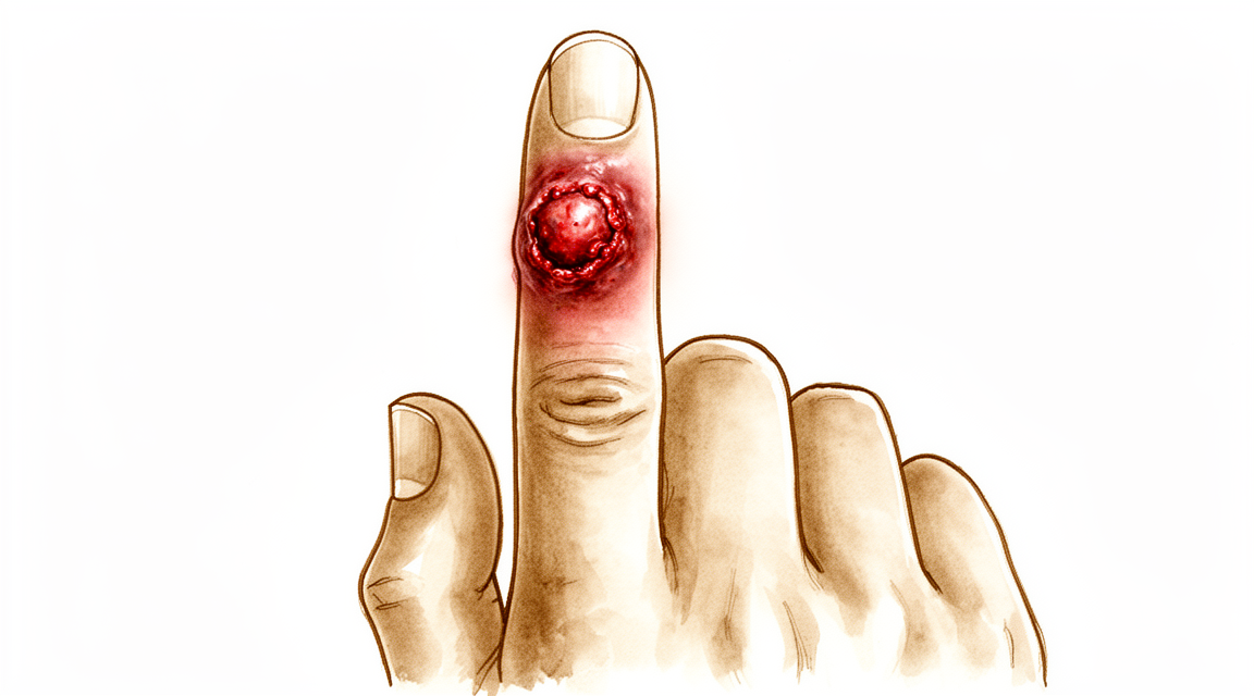
O que você está sentindo
Uma infecção na mão geralmente causa dor em um único ponto. A pele sobre essa área pode ficar vermelha, quente e inchada. A dor costuma piorar ao mover o dedo ou ao segurar algo. O repouso da mão alivia um pouco o desconforto, mas a dor não desaparece por conta própria.
Tarefas cotidianas tornam-se difíceis: abotoar roupas, girar uma chave, segurar uma xícara de chá ou digitar podem causar dor. Se a infecção atingir a bainha do tendão do dedo (o “túnel” por onde o tendão se move), todo o dedo pode ficar inchado e ligeiramente dobrado. Esticá-lo dói, e a dor persiste mesmo quando outra pessoa tenta fazê-lo com cuidado. Quando uma articulação está infectada, ela fica inchada, sensível ao toque e quente; mover a articulação em qualquer direção é bastante doloroso.
Algumas infecções se espalham pela pele na forma de faixas vermelhas e dolorosas; os gânglios na axila também podem ficar doloridos. O inchaço pode deixar o dorso da mão parecendo inchado, e fica difícil identificar onde exatamente o líquido se acumulou. À noite, o desconforto aumenta, pois o inchaço se intensifica quando a mão permanece imóvel.
A maioria das infecções se desenvolve em poucas horas ou dias, não semanas. Se a vermelhidão, o inchaço e a dor estiverem piorando em vez de melhorar, isso indica infecção e não apenas um entorse simples. Algumas condições têm aparência muito semelhante: gota, picada de inseto, reação a um espinho ou surto de artrite; por isso, a causa nem sempre fica clara de imediato.
Informe ao seu cirurgião se você tem diabetes, doença renal ou alguma condição ou medicamento que enfraqueça o sistema imunológico. Esses fatores alteram o comportamento da infecção e o tratamento necessário. O mesmo vale para qualquer infecção que não tenha melhorado após o uso de antibióticos ou após a limpeza da ferida; algumas infecções de crescimento lento exigem exames especiais para serem identificadas.
Com tratamento imediato, a maioria das infecções dos tecidos moles da mão é completamente curada. O risco de adiar o tratamento é o surgimento de rigidez, flexão permanente do dedo ou, em casos graves, até a perda do dedo.
O que realmente está acontecendo
A mão é um espaço bastante congestionado. Vinte e sete ossos ficam próximos uns dos outros sob uma pele fina na parte dorsal da mão e bem aderida à palma. Os tendões, que funcionam como “cordas” para mover os dedos, deslizam por túneis estreitos. Um desses túneis, a bainha flexora, envolve o tendão responsável pela flexão do dedo e possui uma camada lisa que nutre e lubrifica o tendão. Quando bactérias entram por meio de um corte, um espinho ou uma perfuração, esse espaço fechado não lhes permite se espalhar senão ao longo do túnel.
O inchaço é o verdadeiro problema. Dentro de um túnel assim, não há espaço para inchaço, então a pressão aumenta. Se essa pressão continuar a subir, pode interromper o fluxo sanguíneo para o próprio tendão. O tendão então pode formar cicatrizes aderindo à bainha ao seu redor, deixando o dedo rígido mesmo após a infecção ter desaparecido. É por isso que uma infecção da bainha tendínea precisa ser tratada rapidamente, em vez de se esperar alguns dias.
Outras regiões também têm suas “armadilhas”. A almofada de gordura na ponta do dedo é dividida em pequenas bolsas fechadas por fibras resistentes; assim, uma infecção nesse local (chamada felon) gera pressão rapidamente e pode se espalhar para o osso ou para a articulação vizinha. A pele ao lado da unha também pode abrigar uma infecção lenta e persistente (paroníquia crônica), geralmente causada por leveduras, na qual a umidade penetra sob a cutícula e mantém a inflamação. Em algumas pessoas, os túneis tendíneos do polegar e do dedo mínimo se conectam, permitindo que a infecção se espalhe de um lado da palma para o outro.
A maioria das infecções da mão é causada por bactérias que normalmente vivem na pele e entram por alguma lesão. Algumas surgem após mordidas, humanas ou de animais. Diabetes, medicamentos esteroides ou um sistema imunológico enfraquecido facilitam a instalação da infecção e sua propagação para estruturas mais profundas, como tendões, osso ou articulação. Por isso o cirurgião pergunta sobre seu histórico de saúde, e por isso o tratamento precoce é tão importante: com cuidados imediatos, a maioria das infecções dos tecidos moles resolve completamente; já o atraso no tratamento pode resultar em rigidez duradoura.
O que podemos fazer a respeito
O Dr. Kieran Hirpara, cirurgião de membros superiores no Mater Private Hospital Rockhampton, trata infecções da mão agindo rapidamente, pois o atraso é o que causa danos duradouros. Geralmente, os pacientes são encaminhados à nossa clínica pelo médico de família; caso um fisioterapeuta tenha sugerido que você nos procure, ainda assim será necessário um encaminhamento do seu médico de família para ter direito ao reembolso do Medicare. Na consulta, colhemos o histórico clínico, examinamos a sua mão e solicitamos os exames necessários para identificar qual microrganismo está causando a infecção e até que profundidade ela se espalhou.
Na maioria das infecções iniciais, o primeiro passo é não cirúrgico. Se uma infecção da bainha do tendão for detectada nas primeiras 48 horas após o início dos sintomas, antibióticos e uma tala podem impedir sua propagação. O paciente é então monitorado de perto; caso os sintomas não melhorem rapidamente, optamos pela cirurgia em vez de aguardar. Após qualquer tratamento, imobilizar a mão numa posição protetora e movimentar os dedos precocemente ajuda a evitar rigidez. A reabilitação é essencial: uma mão infectada que não é movimentada acaba ficando rígida; portanto, a fisioterapia faz parte do plano de tratamento, não é um item opcional.
Os antibióticos constituem a outra vertente do tratamento médico. A primeira prescrição visa abranger as bactérias mais prováveis de causar a infecção; uma vez que os resultados laboratoriais identifiquem exatamente o microrganismo, trocamos para o antibiótico mais adequado. Algumas infecções exigem antibióticos por via intravenosa, em vez de comprimidos; em casos graves, o tratamento pode durar de 4 a 6 semanas. Se o seu sistema imunológico estiver enfraquecido por doenças ou medicamentos como esteroides, o tratamento é mais rápido e agressivo, pois essas infecções podem evoluir para quadros graves mais rapidamente. Mordidas, sejam humanas ou de animais, também geralmente exigem antibióticos profiláticos.
A cirurgia é indicada quando há pus a ser drenado ou quando a infecção atinge tendões, ossos ou articulações. Um abscesso ou acúmulo de líquido sob a pele precisa ser liberado para aliviar a pressão; muitas vezes, o ferimento é tamponado inicialmente para manter a drenagem. Infecções na ponta do dedo (chamadas “felon”) ou ao redor da unha (“paroníquia”) são drenadas por meio de um pequeno corte, às vezes removendo parte da unha. Infecções na bainha do tendão exigem a lavagem do túnel tendíneo; já a osteomielite (infecção óssea) requer a remoção do tecido necrosado, além de um tratamento prolongado com antibióticos. Uma exceção importante: se uma bolha no dedo for na verdade uma infecção viral (herpes labial na mão), a cirurgia não é indicada; abri-la pode piorar a situação.
O que esperar
A maioria das infecções na mão detectadas precocemente resolve-se completamente com o tratamento. Com atendimento imediato, a maior parte das infecções dos tecidos moles desaparece por completo, e a mão volta ao normal. Quanto mais cedo você procurar atendimento, maiores são as chances de evitar rigidez ou danos permanentes.
O tempo de recuperação depende da profundidade da infecção. Uma infecção na pele ou nas unhas pode melhorar em poucos dias após a drenagem ou o uso de antibióticos. Já uma infecção na bainha do tendão, no osso ou na articulação leva mais tempo; algumas exigem semanas de tratamento antibiótico. Mesmo após a infecção desaparecer, o dedo pode permanecer rígido por algum tempo. Movimentar os dedos cedo, quando o cirurgião autorizar, ajuda a impedir que essa rigidez se torne permanente.
Se o tratamento for adiado, o prognóstico torna-se menos previsível. Uma infecção na bainha do tendão presente há dias pode causar cicatrização entre o tendão e seu canal, deixando o dedo rígido mesmo após a cura da infecção. Infecções graves podem ainda resultar em perda permanente de função ou, em alguns casos, na perda do dedo, mesmo com tratamento adequado e oportuno. Por isso, essas infecções são consideradas casos de urgência, em vez de serem monitoradas por alguns dias.
Algumas infecções têm comportamento diferente. Em pessoas com diabetes ou sistema imunológico enfraquecido, as infecções podem se disseminar mais rapidamente e atingir estruturas mais profundas; por isso, exigem tratamento mais agressivo e, às vezes, mais de uma cirurgia. Infecções de crescimento mais lento, como as causadas por micobactérias (um grupo de germes relacionado ao causador da tuberculose), precisam de longos ciclos de antibióticos combinados com cirurgia para serem erradicadas; a recuperação, nesses casos, leva meses, não dias.
Geralmente, você será reavaliado após o tratamento para verificar se a infecção está desaparecendo e se a mão está se movimentando bem. Mantenha essas consultas mesmo que tudo pareça melhor, pois alguns problemas só se tornam visíveis quando o inchaço diminui. Se dor, vermelhidão ou inchaço reaparecerem após a infecção parecer controlada, entre em contato com o seu cirurgião em vez de aguardar para ver como evolui.
Quando procurar ajuda médica
As infecções na mão são situações de urgência. O dano decorrente de qualquer atraso ocorre rapidamente; portanto, não espere para ver se a situação melhora por conta própria.
Procure o pronto-socorro se houver linhas vermelhas subindo pelo braço ou pela mão, se todo o dedo estiver inchado, dobrado e muito doloroso para ser esticado, ou se a pele estiver escurecendo rapidamente, formando bolhas ou se rompendo. O mesmo se aplica se você estiver com febre e se sentindo mal, além dos sintomas na mão; ou se a mão sofreu um ferimento e agora está muito inchada e tensa. Esses sinais podem indicar uma infecção profunda ou em propagação, que exige avaliação e tratamento imediatos.
Solicite avaliação urgente, preferencialmente no mesmo dia, se a dor for intensa e piorar, se uma articulação do dedo estiver quente, inchada e muito dolorida para ser movimentada, ou se houver acúmulo de pus sob a pele ou ao lado da unha. Uma infecção que não melhora após um ou dois dias de uso de antibióticos também requer avaliação, em vez de simplesmente aguardar o término do tratamento.
Procure seu médico de família assim que possível para qualquer infecção na mão, mesmo que seja leve. Isso é ainda mais importante se você tem diabetes, doença renal ou alguma condição ou medicação que enfraqueça o sistema imunológico, pois, nesses casos, as infecções podem se espalhar mais rápido e atingir camadas mais profundas dos tecidos. Informe ao médico sobre qualquer mordida recente de animal ou humana, ferimento por perfuração, ou histórico de trabalho ou viagens que possam indicar a presença de um patógeno incomum.
Se uma infecção persistir ou nunca se resolver completamente apesar do tratamento, peça uma avaliação por um especialista, pois algumas infecções de crescimento lento exigem exames específicos para serem identificadas.
Em maior profundidade
Advanced reading: the deeper science (optional)
Esta seção vai além do que você precisa saber para tomar decisões sobre o próprio tratamento. As infecções da mão merecem uma leitura mais aprofundada, pois os antibióticos prescritos inicialmente para a maioria dos pacientes são, neste contexto específico, inadequados; além disso, uma infecção na mão pode ser uma emergência cirúrgica frequentemente confundida com um caso leve.
O microrganismo mais comum resiste aos medicamentos mais comuns
Um estudo longitudinal de 10 anos envolvendo 815 infecções da mão em ambientes urbanos constatou que, embora a incidência anual de MRSA tenha diminuído no geral, esse microrganismo continua sendo o patógeno mais comum. Ao mesmo tempo, a resistência à clindamicina e à levofloxacina aumentou de forma constante. A conclusão dos autores é clara: a terapia antibiótica empírica para infecções da mão deve evitar penicilina, beta-lactâmicos, clindamicina e levofloxacina [1].
Essa lista de substâncias a serem evitadas é maior do que a maioria das pessoas imagina; ela inclui praticamente todos os medicamentos habitualmente prescritos para infecções cutâneas em outras partes do corpo. A implicação prática é que o fato de uma infecção da mão não responder ao tratamento com antibióticos de primeira linha é um acontecimento comum e previsível, e não algo incomum; por isso, é recomendável procurar avaliação médica em vez de persistir com o mesmo tratamento.
Os padrões de resistência variam conforme a região, portanto esse é um princípio geral, não uma prescrição rígida. Contudo, ele explica por que as infecções da mão frequentemente exigem intervenções mais agressivas do que o tamanho da lesão sugeriria.
Infecção da bainha do flexor: detalhes que influenciam o prognóstico
A tenossinovite flexora piogênica, ou seja, a infecção no espaço fechado por onde passa o tendão flexor, é a única infecção da mão que se comporta de maneira diferente das demais. A bainha do tendão é um espaço confinado com baixa irrigação sanguínea; por isso, o pus sob pressão nesse local pode destruir a superfície de deslizamento do tendão em apenas alguns dias.
Em um estudo com 763 pacientes, duas medidas melhoraram a amplitude de movimento: o uso de antibióticos como parte do tratamento e a irrigação por cateter em vez da lavagem cirúrgica aberta. As evidências corroboraram tanto a intervenção precoce quanto o uso sistêmico de antibióticos [2].
Vale ressaltar que o desfecho avaliado foi a amplitude de movimento, e não a erradicação total da infecção. É possível curar a infecção sem que o dedo recupere sua mobilidade plena, o que representa o verdadeiro custo de qualquer atraso no tratamento. Os quatro sinais clássicos – dedo inchado, com aspecto semelhante a uma “salsicha”, mantido ligeiramente flexionado; sensibilidade dolorosa ao longo de toda a bainha do tendão; e dor intensa ao ser esticado passivamente – merecem atenção, pois essa combinação exige avaliação imediata, e não apenas o uso de medicamentos por via oral.
O caso que realmente constitui uma emergência médica
A fasciíte necrotizante de origem na mão é rara e extremamente destrutiva. A revisão sistemática de 161 casos conclui que o diagnóstico precoce é de suma importância, e que a intervenção cirúrgica precoce e decisiva deve ser realizada com baixo limiar de indicação, especialmente quando existem fatores de risco [3].
O termo “baixo limiar de indicação” é utilizado intencionalmente. Trata-se de um diagnóstico em que a espera por uma confirmação pode custar o membro afetado. Os sinais que o diferenciam da celulite comum são: dor desproporcional em relação à aparência da lesão, progressão rápida ao longo de horas, e não dias; além de sintomas sistêmicos como febre, confusão mental e sensação de mal-estar intenso, tudo isso acompanhado de uma área de vermelhidão que pode parecer pequena.
Por que uma ferida pequena pode ser enganosa
Dois mecanismos podem provocar infecções muito mais profundas do que a ferida visível indica. Uma mordida na região do nó do dedo, ocorrida ao bater com a mão na boca, faz com que microrganismos orais penetrem na pele, no tendão extensor e na cápsula articular num único movimento; posteriormente, a pele se fecha sobre essa contaminação. Já uma perfuração na palma da mão pode permitir a entrada de bactérias na bainha do flexor, através de um ponto de entrada que se fecha em apenas um dia.
Nos dois casos, a ferida visível é um indicador pouco confiável da profundidade do problema. O padrão da dor — especialmente a dor ao mover o dedo, em vez da dor exatamente na área da ferida — é muito mais informativo do que a aparência da pele.
Referências
[1] Kistler JM, Thoder JJ, Ilyas AM. Incidência de MRSA e tendências no uso de antibióticos em infecções da mão em áreas urbanas: um estudo longitudinal de 10 anos. Hand (N Y). 2018;14(4):449-54. https://doi.org/10.1177/1558944717750921
[2] Giladi AM, Malay S, Chung KC. Revisão sistemática sobre o tratamento da tenossinovite flexora piogênica aguda. J Hand Surg Eur Vol. 2015;40(7):720-8. https://doi.org/10.1177/1753193415570248
[3] Christopoulos G, Khoury A, Johnson M, Sergentanis TN. Fascite necrotizante de origem na mão: revisão sistemática e meta-análise. Hand (N Y). 2022;19(4):568-74. https://doi.org/10.1177/15589447221141486
Evidence & references
This is the clinical evidence summary written for health professionals. It is technical, and it lists the research this page was built from. You do not need to read it to understand your treatment or to make a decision about it.
Overview
- Treatment of acute hand infections requires a combination of surgical intervention (incision and drainage) and appropriate antibiotic therapy tailored to the organism and infection severity [1].
- Fungal infections of the hand are most commonly cutaneous infections involving the skin and nails [2].
- Cutaneous fungal infections of the hand can be treated with topical or local therapy [2].
- Mycobacterium tuberculosis must be maintained in the differential diagnosis for subacute presentations of flexor tenosynovitis or carpal tunnel syndrome of the hand [3].
- Mycobacterium tuberculosis must be considered in the differential diagnosis of subacute flexor tenosynovitis or carpal tunnel syndrome even in nonendemic areas and in the absence of past tuberculosis infection or exposure [3].
- Stiffness and contracture are the most common sequela of pyogenic flexor tenosynovitis [4].
- Stiffness and contracture resulting from pyogenic flexor tenosynovitis benefit from early motion and hand therapy [4].
- Tuberculous tenosynovitis can be overlooked as a cause of chronic tenosynovitis [5].
- Tuberculous tenosynovitis is particularly easily overlooked in immunocompetent young people lacking risk factors [5].
- The majority of patients with fungal tenosynovitis of the hand and upper extremity were successfully treated with surgical debridement and antimicrobial therapy [6].
- Fungal tenosynovitis of the hand and upper extremity has a recurrence rate of 30% [6].
- A recurrence rate of 30% in fungal tenosynovitis highlights the need for close post-treatment follow-up [6].
- Management of fungal osteomyelitis and fungal septic arthritis is challenging, especially in immunocompromised patients [7].
- Outcomes for fungal osteomyelitis and fungal septic arthritis have historically been poor due to a lack of evidence-based treatment guidelines [7].
- Invasive infections may require a combination of serial surgical debridement and a multidrug antibiotic regimen [8].
Anatomy & Pathophysiology
General Principles & Diagnosis
- Chronic hand infections can be caused by viruses, bacteria, mycobacteria, fungi, Prototheca, protozoa, parasites, and insects [14].
- Hand infections may be superficial (skin or nails), subcutaneous, or deep (nerves, tendons/tenosynovium, joints/synovium, bone, and rarely muscles) [14].
- Chronic lesions of the hand have a nonspecific presentation, and early biopsy and cultures facilitate diagnosis [14].
- Infection must be included in the differential diagnosis for any chronic lesion of the hand, particularly in immunocompromised patients [14].
- An infection that does not respond to antibiotics, incision, drainage, or debridement is suspect for a chronic or atypical etiology [14].
- The adage "culture a tumor and biopsy an infection" is recommended when an unusual lesion is encountered to avoid diagnostic delay [14].
- Nontuberculous mycobacterial (NTM) infections of the hand are now more common than Mycobacterium tuberculosis infections [14].
- Tenosynovial infections are far more common than joint and bone infections in mycobacterial hand disease [14].
- Hansen disease is the most common chronic infection affecting the hand in developing countries and infects peripheral nerves [14].
- High suspicion for Hansen disease is warranted when peripheral neuropathy of the ulnar nerve, with or without nerve enlargement, is seen in an immigrant [14].
- Consultation with an infectious disease specialist, microbiology personnel, and a pathologist improves diagnostic accuracy for chronic hand infections where organisms are scarce or slow-growing [14].
Bacterial Pathophysiology
- Paronychia contains polymicrobial aerobic and anaerobic flora, with mixed aerobic/anaerobic flora isolated in half of the patients [9].
- Human bite injuries to the hand involve 42 different bacterial species identified in normal human mouth flora [48].
- The mechanism for introducing anaerobic bacteria into the joint during a clenched-fist injury involves the lacerated tendon gliding proximally when the finger is extended, creating a closed, anaerobic environment [48].
- Actinomycosis is caused by endogenous human flora, most commonly Actinomyces israelii, which is a normal inhabitant of the oral cavity [32].
- Actinomycosis spreads contiguously in a slow but progressive manner, ignoring tissue planes [32].
- The identification of "yellow sulfur granules" from a draining sinus or pus is diagnostic for actinomycosis [32].
- Actinomycosis cultures are positive in only 25% of cases, often because specimens were not sent in anaerobic media [32].
- Mycetoma presents with a clinical triad of tumification, draining sinuses, and granules in the discharging pus [33].
- Mycetoma grains are 0.2 to 5.0 mm in diameter and may be black, white, yellow, pink, or red depending on the causal organism [33].
- Mycetoma infection begins in the skin and subcutaneous tissues and tends to follow fascial planes as it spreads proximally and mediolaterally [33].
- With increasing depth, mycetoma progressively infects and destroys all connective tissues and eventually bone [33].
Fungal Pathophysiology
- Cutaneous fungal infections are caused by fungi that infect and metabolize keratin, preventing invasion beneath the skin [29].
- Candida albicans and dermatophytes (Trichophyton, Microsporum, and Epidermophyton) cause the majority of chronic cutaneous and nail infections [29].
- Chronic cutaneous C. albicans infection occurs in the moist palms and webs of a "clenched fist" in patients with spasticity [29].
- C. albicans involvement in chronic paronychia is characterized by mycelium in the outer epidermis with no involvement of the dermis [10].
- Fungal tenosynovitis has a recurrence rate of 30% despite treatment with surgical debridement and antimicrobial therapy [6].
- Management of fungal osteomyelitis and fungal septic arthritis is challenging, especially in immunocompromised patients, with historically poor outcomes due to a lack of evidence-based treatment guidelines [7].
Mycobacterial Pathophysiology
- Mycobacterium tuberculosis must be maintained in the differential diagnosis for subacute flexor tenosynovitis or carpal tunnel syndrome, even in nonendemic areas and without past exposure [3].
- Tuberculous tenosynovitis can be overlooked as a cause of chronic tenosynovitis, particularly in immunocompetent young people lacking risk factors [5].
- Extrapulmonary TB accounts for almost 20% of all TB cases in the general population and 72% of all cases in U.S. AIDS patients [30].
- Skeletal TB represents 1% of all TB and 10% of all extrapulmonary TB in the United States [30].
- Of skeletal TB cases, 10% occur in the upper extremity, with 4% specifically in the hand and fingers and an additional 3% in the hand accompanied by multifocal sites elsewhere [30].
- Tuberculous osteomyelitis of the hand typically occurs in the phalanges (tuberculous dactylitis) and metacarpals [30].
Anatomical Structures & Compartments
- The hand consists of 19 bones, 17 articulations, and 19 muscles situated entirely within the hand, plus approximately the same number of tendons activated by forearm muscles [11].
- The skeleton of the hand and wrist consists of 27 bones, of which 19 are long bones [19].
- The thumb ray is the shortest, composed of a metacarpal and two phalanges, and is more mobile and proximal than the other rays [19].
- The trapezium is angled out in front of the carpal plane, allowing the first metacarpal to make an angle of about 45 degrees with the second metacarpal in the sagittal plane [19].
- The transverse axis of the palm is oblique, forming an acute angle of approximately 75 degrees with the longitudinal axis [19].
- The extrinsic extensors run through six different fibroosseous retinacular compartments at the wrist level [18].
- The first extensor compartment contains the abductor pollicis longus and the extensor pollicis brevis [18].
- The second extensor compartment contains the extensor carpi radialis longus and the extensor carpi radialis brevis [18].
- The third extensor compartment contains the extensor pollicis longus, which turns abruptly radialward about Lister tubercle [18].
- The fourth extensor compartment contains the extensor indicis proprius lying deep to the four tendons of the extensor digitorum communis [18].
- The fifth extensor compartment contains the extensor digiti quinti [18].
- The sixth extensor compartment contains the extensor carpi ulnaris [18].
- The sagittal bands stabilize the digital extensor tendons over the midline of the metacarpophalangeal joint and transmit proximal extrinsic extensor tension to the proximal phalanx [18].
- Rupture or attenuation of the sagittal band fibers allows the extrinsic extensor tendon to sublux to the ulnar side of the metacarpal head, causing ulnar deviation of the finger [18].
- The flexor digitorum profundus inserts on the proximal volar aspect of the distal phalanx and flexes the distal, proximal interphalangeal, and metacarpophalangeal joints [18].
- The flexor digitorum superficialis inserts via radial and ulnar slips into the proximal metaphysis of the middle phalanx and primarily flexes the proximal interphalangeal joint [18].
- The tenosynovial sheath of the flexor pollicis longus is continuous with the radial bursa, and the sheath to the little finger is continuous with the ulnar digital bursa [28].
- In some patients, the radial and ulnar bursae communicate, allowing a horseshoe abscess to spread between the thumb and little finger if infection occurs in either flexor tendon sheath [28].
- The fibroosseous tunnel of the digital flexor sheath consists of annular pulleys for mechanical stability and cruciate pulleys for flexibility [28].
- The A2 and A4 annular pulleys are the most essential in maintaining the mechanical advantage of the flexor tendons [28].
- The tenosynovium lining the fibroosseous tunnel supplies nutrition and lubrication to the poorly vascularized flexor tendons [28].
- Within the flexor tendon sheath, tendon vascularity is supplied via the vincula system, specifically the vinculum longus and brevis [28].
- There are seven interosseous muscles in the hand: four dorsal and three volar [24].
- The dorsal interossei are abductors, while the volar interossei are adductors [24].
- The middle finger has two dorsal interossei (abductors) and no volar interossei (adductors) because the central axis of the hand lies within it [24].
- The deep head of each dorsal interosseous muscle forms a lateral band that flexes and weakly abducts the proximal phalanx while extending the middle and distal phalanges [24].
- The volar interossei form the ulnar lateral band of the index finger and the radial lateral band of the ring and little fingers [24].
- The abductor digiti quinti and flexor digiti quinti brevis are structurally and functionally similar to the superficial and deep heads of the dorsal interossei, respectively [24].
- The opponens digiti quinti arises from the pisohamate ligament and the hook of the hamate and inserts onto the ulnar side of the diaphysis of the fifth metacarpal [24].
- The flexor retinaculum maintains and restrains the tendons of the extrinsic flexors within the carpal canal, keeping them close to the axis of flexion–extension of the wrist [26].
- The metacarpal arch is adaptable due to the mobility of the peripheral metacarpals, which can deepen the palmar concavity as they approach each other [26].
- The index metacarpal is the most firmly fixed, while the fifth metacarpal is semi-independent with a range of flexion–extension of approximately 20 degrees [26].
- The metacarpophalangeal joints are the keystones of the longitudinal arches and are stabilized by collateral ligaments and the thick volar articular capsule (volar plate) [26].
- The volar plates are interconnected by the transverse interglenoid ligament, which ties together the anterior glenoid ligaments of the metacarpophalangeal articulations [26].
- The palmar skin is subdivided into two zones by the oppositional crease of the thumb: a radial mobile portion and an ulnar/distal fixed portion [23].
- The central triangular part of the palm has fixed, poorly vascularized skin covering almost directly the superficial palmar aponeurosis [23].
- Incisions made along the sides of the diamond-shaped cutaneous contact zones in flexed digits present a minimal chance of retraction [23].
- The dorsal slope of the web spaces has supple, non-adherent skin, while the palmar surface is flat and densely adherent to the commissural skeleton [23].
- The "princeps pollicis" artery is the terminal branch of the radial artery that crosses the first intermetacarpal space and runs along the ulnar side of the first metacarpal [27].
- Only 15% of anatomical dissections of the thumb palmar arteries fall into the classical "typical" category [27].
- In the second segment of the thumb, the ulnar collateral artery is often easier to dissect and larger than the radial collateral artery [27].
- The dorsal arteries of the thumb originate from palmar arteries (princeps, commissural, or anastomoses) at the level of the first metacarpal and head distally along the sides of the distal phalanges [27].
Classification
Flexor Tenosynovitis
- Kanavel identified four cardinal signs of suppurative flexor tenosynovitis: tenderness over the involved sheath, rigid positioning of the finger in flexion, pain on attempts to hyperextend the fingers, and swelling of the involved part [16].
- Of the four cardinal signs of suppurative flexor tenosynovitis, tenderness over the flexor sheath is considered the most significant [16].
- The Michon classification categorizes flexor tenosynovitis into three intraoperative stages based on characteristic findings [16].
- Michon stage I flexor tenosynovitis is characterized by increased fluid in the sheath, primarily serous exudate [16].
- Michon stage II flexor tenosynovitis is characterized by cloudy or purulent fluid and granulomatous synovium [16].
- Michon stage III flexor tenosynovitis is characterized by septic necrosis of the tendon, pulleys, or tendon sheath [16].
- The Michon classification has not been validated [16].
- Direct inoculation is the most common cause of infectious flexor tenosynovitis [16].
- Adjacent spread from a local infection or hematogenous spread are possible causes of infectious flexor tenosynovitis [16].
- While the flexor sheath is usually involved in infectious tenosynovitis, the radial and ulnar bursae may also be involved [16].
Subcutaneous Fungal Infections
- There are three major subcutaneous fungal infections of the hand: chronic paronychia, sporotrichosis, and phaeomycotic cysts [44].
- Chronic paronychia is usually initially misdiagnosed as a bacterial infection [44].
- A phaeomycotic cyst is a deep dermal or subcutaneous infection resulting from the traumatic implantation of a dematiaceous (pigmented) fungus such as Exophiala or Phialophora [44].
Mycetoma
- Mycetoma of the hand and upper extremity has been graded into five stages [47].
- Mycetoma stage I is the nodular stage, characterized by a small, firm, painless subcutaneous nodule or nodules under the skin [47].
- The duration of mycetoma stage I is 2 to 3 months [47].
- Mycetoma stage II is the sinusoidal stage, where nodules become abscesses and drain granules through sinuses to the surface of the skin or to the surface of an ulcerated nodule [47].
- The duration of mycetoma stage II is 4 to 12 months [47].
- Mycetoma stage III is the skeletal stage, where the infection progresses to osteomyelitis [47].
- Mycetoma stage IV is the skeletal stage, where limb deformity occurs over the course of a year or more [47].
- Mycetoma stage V is the metastatic stage, where lesions of the hand may metastasize to the axilla and the chest wall [47].
- Radiographic signs of mycetoma include thinning of the metacarpal, bone erosions and cavities, sclerosis, and periosteal reaction [47].
- A network of connected sinuses is present in the soft tissues from the bone to the skin in mycetoma [47].
- Constitutional symptoms are absent in mycetoma unless there is superimposed bacterial infection [47].
- Pain is not a predominant symptom in mycetoma [47].
- In 80% of cases, patients postpone seeking medical care for mycetoma until the second or third stage is reached due to lack of pain [47].
Clinical Presentation
General Principles and Diagnostic Approach
- Chronic lesions of the hand, both superficial and deep, have a nonspecific presentation [14].
- Early biopsy and cultures facilitate diagnosis of chronic hand infections [14].
- Infection must be included in the differential diagnosis for any chronic lesion of the hand [14].
- In an immunocompromised patient, infection must always be included in the differential diagnosis [14].
- An infection that does not respond to antibiotics, incision, drainage, or debridement is suspect [14].
- "Culture a tumor and biopsy an infection" is a useful adage when an unusual lesion is encountered [14].
- With a careful history and physical examination, the location of the infection, the extent of spread, and the presence of swelling, lymphangitis, lymphadenitis, and joint involvement can be determined [38].
- Fluctuance can be difficult to identify in the hand [38].
- Radiographs are helpful in revealing bone injury [38].
- Radionuclide scanning may show bone infection [38].
- MRI and ultrasound may localize an abscess [38].
- The specificity of all inflammation markers (WBC, C-reactive protein, ESR) was inadequate for diagnosis of finger infections [38].
- If any fluid or tissue is obtained, it is sent to the laboratory for Gram stain, crystals, culture, and antibiotic sensitivity determinations [38].
- Specific requests are usually made of the laboratory to culture for aerobic and anaerobic bacteria, mycobacteria, and fungi [38].
Specific Infection Types
- Tuberculous tenosynovitis can easily be overlooked as a cause of chronic tenosynovitis, particularly in immunocompetent young people lacking risk factors [5].
- M tuberculosis should be maintained in the differential diagnosis of a subacute presentation of flexor tenosynovitis or carpal tunnel syndrome of the hand, even in nonendemic areas and in the absence of past tuberculosis infection or exposure [3].
- In chronic paronychia lesions studied, C. albicans involvement was present with mycelium in the outer epidermis but no involvement of the dermis [10].
- Finger joint infections usually result from the spread of infection in adjacent structures, direct penetration of the joint, and less commonly, hematogenous spread [34].
- The involved joints in finger joint infections are usually swollen, tender, and warm, and the finger is usually held in slight flexion [34].
- Careful inspection and palpation may reveal a fluctuant joint effusion in finger joint infections [34].
- Active and passive motions are usually quite painful in finger joint infections [34].
- Fluid obtained from a septic joint is usually turbid, opaque, or grossly purulent [34].
- The joint fluid WBC is usually greater than 50,000/mm³ in septic joints [34].
- Lowering the cell count threshold to 17,500 increases the sensitivity of the diagnosis of septic arthritis to 83% [34].
- The polymorphonuclear count is usually greater than 75% in septic joint fluid [34].
- The synovial fluid glucose is 40 mg or less in septic joints [34].
- Blue nails and clubbing are both a sign of HIV infection [21].
- Red fingers syndrome has been reported in patients with HIV and hepatitis C infection [21].
- Fingers of patients with AIDS may be red with painless erythema and periungual telangiectasia [21].
- Nails in patients with AIDS may be blue with painless clubbing [21].
- Diagnosis of AIDS should be considered when there is an unusual hand infection [21].
- Diagnosis of AIDS should be taken into consideration for any patient who needs a repeat drainage or debridement procedure [21].
Differential Diagnosis Considerations
- Conditions that can be confused with hand infections include gout, acute calcium deposition, pseudogout, pyogenic granuloma, insect bites, pyoderma gangrenosum, foreign bodies, factitious lesions, herpetic gangrenes, metastatic lesions, silicone synovitis, granuloma annulare, rheumatoid arthritis, nonspecific tenosynovitis, reactions to intravenous medications (e.g., chemotherapeutic agents), and Sweet syndrome [38].
Investigations
- MRI, CT, and 3-phase bone scans may add to the radiographic diagnosis made on plain radiographs for hand osteomyelitis [42].
- MRI, CT, and 3-phase bone scans have poor sensitivity and specificity for the diagnosis of hand osteomyelitis [42].
- Deep tissue cultures are the most accurate method of diagnosis for hand osteomyelitis [42].
- The presence of an indolent course in hand osteomyelitis suggests a fungal or mycobacterial cause [42].
- Incisional biopsy is indicated for patients with lytic destructive lesions of the bones of the hand [46].
- Biopsy must be strongly considered before the initiation of treatment for presumed deep infection because misdiagnosis is the most common error in diagnosis [46].
- MRI is particularly useful in delineating the extent of bone and soft tissue involvement in Ewing’s sarcoma that may not be apparent on plain radiographs [46].
Treatment
General Principles
- Early suspicion, biopsy, and diagnosis of a chronic infection is the mainstay of all ensuing treatment principles [14].
- Pharmacologic treatment of a chronic hand infection requires close monitoring for serious side effects and drug resistance [14].
- Consultation with an infectious disease specialist, microbiology personnel, and a pathologist improves the accuracy of a diagnosis when organisms are scarce, slow to grow, and require special media and temperature to grow [14].
Fingertip Infections
- Early-stage paronychia can be managed with warm soaks and antistaphylococcal antibiotics [35].
- When paronychia has progressed to abscess formation, drainage, with or without removal of the involved portion of the nail plate from the fold, is required [35].
- Following drainage of paronychia, cultures should be taken whenever possible, and antibiotics as well as warm soapy soaks should be recommended [35].
- Routine oral antibiotics usually are not effective for chronic paronychia [35].
- Nail plate removal and eponychial marsupialization is the recommended treatment for chronic paronychia [35].
- The “Swiss roll” technique has also been described to treat chronic paronychia [35].
- Surgical drainage is the mainstay of treatment for felon [35].
- Drainage of a felon should be accomplished without violating the flexor sheath or DIP joint [35].
- A midaxial incision along the non–pressure-bearing side of the digit or a longitudinal incision over the volar pulp skin is preferred for felon drainage [35].
- The wound from a felon drainage is left open, and warm soapy soaks are initiated to allow drainage [35].
- Surgical drainage or débridement of herpetic whitlow lesions is contraindicated [35].
- When administered early, oral acyclovir may lessen symptom severity in herpetic whitlow [35].
- In children with herpetic whitlow, a 10-day course of a penicillinase-resistant oral antibiotic is required if cultures from blistering dactylitis reveal growth [35].
Cellulitis and Abscesses
- Empiric coverage for CA-MRSA should be provided if local prevalence rates exceed 10% to 15% [15].
- Ultrasonography has a 78.4% positive predictive value of identifying an abscess and a negative predictive value of 90% to rule out an abscess [15].
- Early antibiotic administration has not been shown to greatly reduce bacterial culture growth from hand abscess so long as decompression is performed within 24 hours [15].
- After surgical decompression, débridement, and irrigation of the abscess, packing is often used, at least initially, to allow for continued drainage [15].
- No difference has been shown between different soaks and daily dressing changes in clearing the infection postoperatively [15].
- Infections in immunosuppressed patients are more likely to involve deeper structures such as joints, bone, tendons, and muscle [15].
- Immunosuppressed patients with hand infections should be treated rapidly and aggressively as their potential for increased morbidity is high [15].
Flexor Tenosynovitis
- When early tenosynovitis is suspected, immediate treatment with antibiotics and splinting may abort the spread of infection if the patient’s symptoms have been present for less than 48 hours [16].
- If nonsurgical treatment is selected for early tenosynovitis, patients should be followed closely with a low threshold for hospital admission [16].
- Good results have been reported in patients with pyogenic flexor tenosynovitis treated with surgical drainage, followed by outpatient management with intravenous antibiotics, wound care, and rehabilitation [16].
- If gross pus is obtained from the aspiration of the digital flexor sheath, surgical drainage usually is indicated [16].
- Vancomycin is effective for infections caused by gram-positive bacteria [16].
- Ciprofloxacin is most effective for gram-negative organisms, including Pseudomonas [16].
- The Michon classification recommends minimally invasive drainage and catheter irrigation for stage I or II flexor tenosynovitis [16].
- The Michon classification recommends extensile open debridement and possible amputation for stage III flexor tenosynovitis [16].
- The use of a continuous postoperative irrigation catheter has not been shown to improve outcomes, but rather increases postoperative pain and adds difficulty to postoperative care [16].
- Stiffness and contracture are the most common sequela of pyogenic flexor tenosynovitis and benefit from early motion and hand therapy [4].
- Invasive infections may require a combination of a serial surgical debridement and a multidrug antibiotic regimen [8].
Fungal Infections
- Fungal infections of the hand are most commonly cutaneous infections involving the skin and nails and can be treated with topical or local therapy [2].
- Although the majority of patients with fungal tenosynovitis were successfully treated with surgical debridement and antimicrobial therapy, a recurrence rate of 30% highlights the need for close post-treatment follow-up [6].
- Fluconazole and AmB are the drugs of choice for use in patients with deep candidiasis [20].
- For periprosthetic fungal infection, radical synovectomy, tenosynovectomy, and removal of the implant are recommended in addition to chemotherapy combination of amphotericin B and 5-fluorouracil [20].
Mycobacterial Infections
- The case highlights the importance of maintaining M tuberculosis in the differential diagnosis of a subacute presentation of flexor tenosynovitis or carpal tunnel syndrome of the hand, even in nonendemic areas and in the absence of past tuberculosis infection or exposure [3].
Complications
Flexor Tenosynovitis
- Persistent tenosynovial infection can cause pressures within the flexor sheath to exceed 30 mm Hg, rendering tendons ischemic [16].
- Delay in treatment of flexor tenosynovitis may lead to damage to the flexor tendon, resulting in adhesion, loss of excursion, finger stiffness, and impaired function [16].
- The prognosis for function is poor if a flexor tenosynovitis infection produces pus that must be drained [16].
- If an open technique is used for flexor tenosynovitis drainage, healing and rehabilitation are prolonged and full motion may not be regained [16].
- Patients presenting with ischemic changes in flexor tenosynovitis had amputation rates of 59% [16].
- Patients presenting with subcutaneous purulence in flexor tenosynovitis had amputation rates of 8% [16].
- The use of a continuous postoperative irrigation catheter for flexor tenosynovitis has not been shown to improve outcomes, but rather increases postoperative pain and adds difficulty to postoperative care [16].
Fungal Infections
- Fungal infections following organ transplantation can have an associated mortality as high as 76% [40].
- Histoplasmosis necrotizing myofasciitis in severe burns of the upper extremity can result in fatality [40].
- Cutaneous histoplasmosis in a renal transplant patient with hand and forearm necrotizing myofasciitis required below-elbow amputation [40].
- Recurrent histoplasmosis infection after multiple antifungal medications and debridements may require above-elbow amputation to save the patient's life [40].
General Infection Complications
- Hand involvement is an independent risk factor for hospital admission for patients presenting to the emergency department with cellulitis [15].
- Patients with immunosuppression are at a higher risk for atypical infections [15].
- Patients with immunosuppression have a high potential for increased morbidity from hand infections [15].
- M tuberculosis must be maintained in the differential diagnosis of subacute flexor tenosynovitis or carpal tunnel syndrome, even in nonendemic areas and in the absence of past tuberculosis infection or exposure [3].
Recovery
- Stiffness and contracture following pyogenic flexor tenosynovitis benefit from early motion and hand therapy [4].
- A recurrence rate of 30% was observed in patients treated for fungal tenosynovitis of the hand and upper extremity [6].
- The 30% recurrence rate in fungal tenosynovitis highlights the need for close post-treatment follow-up [6].
- After surgical decompression, débridement, and irrigation of a hand abscess, packing is often used initially to allow for continued drainage [17].
- No difference has been shown between different soaks and daily dressing changes in clearing the infection postoperatively after hand abscess drainage [17].
- In a matched cohort study of upper extremity infections, the most frequent immunosuppressive medication was glucocorticoids [17].
- Infections in immunosuppressed patients were more likely to involve deeper structures such as joints, bone, tendons, and muscle [17].
- In a review of 74 HIV-seropositive patients treated for upper extremity infections, 26 infections (29%) required more than one operation [21].
- In a review of 74 HIV-seropositive patients treated for upper extremity infections, 11 infections (12%) resulted in amputation [21].
- Among 14 AIDS patients with hand infections, almost one-third needed multiple debridements and resulted in amputation of a finger or hand [21].
Key Evidence
- [L5] Treatment requires a combination of surgical intervention (incision and drainage) and appropriate antibiotic therapy tailored to the organism and infection severity. [1] (10.1016/j.jhsa.2014.03.031)
- [L5] Fungal infections of the hand are most commonly cutaneous infections involving the skin and nails and can be treated with topical or local therapy. [2] (10.1016/j.hcl.2020.03.009)
- [L5] The case highlights the importance of maintaining M tuberculosis in the differential diagnosis of a subacute presentation of flexor tenosynovitis or carpal tunnel syndrome of the hand, even in nonendemic areas and in the absence of past tuberculosis infection or exposure. [3] (10.5435/jaaosglobal-d-17-00083)
- [L5] Stiffness and contracture are the most common sequela and benefit from early motion and hand therapy. [4] (10.2106/jbjs.rvw.26.00015)
- [L5] Tuberculous tenosynovitis can easily be overlooked as a cause of chronic tenosynovitis, particularly in immunocompetent young people lacking risk factors. [5] (10.1007/s00402-012-1527-2)
- [L4] Although the majority of patients were successfully treated with surgical debridement and antimicrobial therapy, a recurrence rate of 30% highlights the need for close post-treatment follow-up. [6] (10.1016/j.jhsa.2016.11.014)
- [L5] Management of fungal osteomyelitis and fungal septic arthritis is challenging, especially in immunocompromised patients, and historically outcomes have been poor due to a lack of evidence-based treatment guidelines. [7] (10.5435/jaaos-22-06-390)
- [L5] Invasive infections may require a combination of a serial surgical debridement and a multidrug antibiotic regimen. [8] (10.5435/jaaosglobal-d-19-00024)
- [L4] Paronychia contains polymicrobial aerobic and anaerobic flora, with mixed aerobic/anaerobic flora isolated in half of the patients. [9] (10.1016/0266-7681(93)90063-l)
- [L4] Each lesion studied had C. albicans involvement with mycelium in the outer epidermis but no involvement of the dermis. [10] (10.1001/archderm.1962.01590090066015)
References
[1] Acute Hand Infections. The Journal of Hand Surgery. 2014. DOI: 10.1016/j.jhsa.2014.03.031
[2] Fungal Infections of the Hand. Hand Clinics. 2020. DOI: 10.1016/j.hcl.2020.03.009
[3] Flexor Tenosynovitis of the Hand Caused by Mycobacterium tuberculosis. JAAOS: Global Research and Reviews. 2018. DOI: 10.5435/jaaosglobal-d-17-00083
[4] Management of Pyogenic Flexor Tenosynovitis. JBJS Reviews. 2026. DOI: 10.2106/jbjs.rvw.26.00015
[5] Tuberculous extensor tenosynovitis of the hand. Archives of Orthopaedic and Trauma Surgery. 2012. DOI: 10.1007/s00402-012-1527-2
[6] A Rare Diagnosis: Recognizing and Managing Fungal Tenosynovitis of the Hand and Upper Extremity. The Journal of Hand Surgery. 2017. DOI: 10.1016/j.jhsa.2016.11.014
[7] Fungal Osteomyelitis and Septic Arthritis. Journal of the American Academy of Orthopaedic Surgeons. 2014. DOI: 10.5435/jaaos-22-06-390
[8] Finger Flexor Tenosynovitis From Stonefish Envenomation Injury. JAAOS: Global Research and Reviews. 2019. DOI: 10.5435/jaaosglobal-d-19-00024
[9] Paronychia: a Mixed Infection. Journal of Hand Surgery. 1993. DOI: 10.1016/0266-7681(93)90063-l
[10] Chronic Paronychia. Archives of Dermatology. 1962. DOI: 10.1001/archderm.1962.01590090066015
[11] Exam Of The Hand Wrist 2Ed. INTRODUCTION.
[14] Green S Operative Hand Surgery. GENERAL PRINCIPLES.
[15] Orthopaedic Knowledge Update 13 Ebook Without Multimedia. Bone and Soft-Tissue Infections of the Hand and Wrist > Soft-Tissue Infections > Hand Cellulitis and Abscesses.
[16] Campbell S Operative Orthopaedics 4 Volume Set. TUMORS AND TUMOROUS CONDITIONS OF THE HAND > TENOSYNOVITIS.
[17] Exam Of The Hand Wrist 2Ed. REFERENCES.
[18] A Lange Medical Book Current Diagnosis Treatment In Orthopedics Fifth Edition. 9Hand Surgery > Image DISORDERS OF THE MUSCULATURE OF THE HAND.
[19] Exam Of The Hand Wrist 2Ed. 1.1 SKELETON OF THE HAND > The osseous skeleton.
[20] Green S Operative Hand Surgery. Candidiasis.
[21] Green S Operative Hand Surgery. Clinical Manifestations of AIDS Infection in the Hands.
[23] Exam Of The Hand Wrist 2Ed. Functional cutaneous units.
[24] Green S Operative Hand Surgery. Interosseous and Hypothenar Muscles.
[26] Exam Of The Hand Wrist 2Ed. The arches of the hand > The metacarpal arch.
[27] Exam Of The Hand Wrist 2Ed. Techniques of investigation of the arterial supply by J P Melki > Vascularization of the thumb > Palmar aspect.
[28] A Lange Medical Book Current Diagnosis Treatment In Orthopedics Fifth Edition. 9Hand Surgery > FLEXOR TENDON INJURY.
[29] Green S Operative Hand Surgery. Cutaneous Fungal Infections > Etiology and Epidemiology.
[30] Green S Operative Hand Surgery. Mycobacterial Osteomyelitis.
[32] Green S Operative Hand Surgery. CHRONIC BACTERIAL INFECTIONS > Actinomycosis.
[33] Green S Operative Hand Surgery. Clinical Findings.
[34] Campbell S Operative Orthopaedics 4 Volume Set. TUMORS AND TUMOROUS CONDITIONS OF THE HAND > SEPTIC ARTHRITIS.
[35] Aaos Comprehensive Orthopaedic Review 3. Infections of the Hand* > I Fingertip Infections.
[38] Campbell S Operative Orthopaedics 4 Volume Set. TUMORS AND TUMOROUS CONDITIONS OF THE HAND > GENERAL APPROACH TO HAND INFECTIONS.
[40] Green S Operative Hand Surgery. Histoplasmosis.
[42] Orthopaedic Knowledge Update 13 Ebook Without Multimedia. Bone and Soft-Tissue Infections of the Hand and Wrist > Bone Infections.
[44] Green S Operative Hand Surgery. Subcutaneous Fungal Infections.
[46] Green S Operative Hand Surgery. BOX 59.1 Ganglions of the Hand and Wrist > Ewing's Sarcoma.
[47] Green S Operative Hand Surgery. Staging.
[48] Campbell S Operative Orthopaedics 4 Volume Set. TUMORS AND TUMOROUS CONDITIONS OF THE HAND > HUMAN BITE INJURIES.