Patients › Hand
Tumor de células gigantes da bainha do tendão (nódulo no dedo ou no polegar)
A giant cell tumour of tendon sheath is a common, benign (non-cancerous) lump on a finger or thumb. What causes it, how it is diagnosed and removed, and how often it comes back.
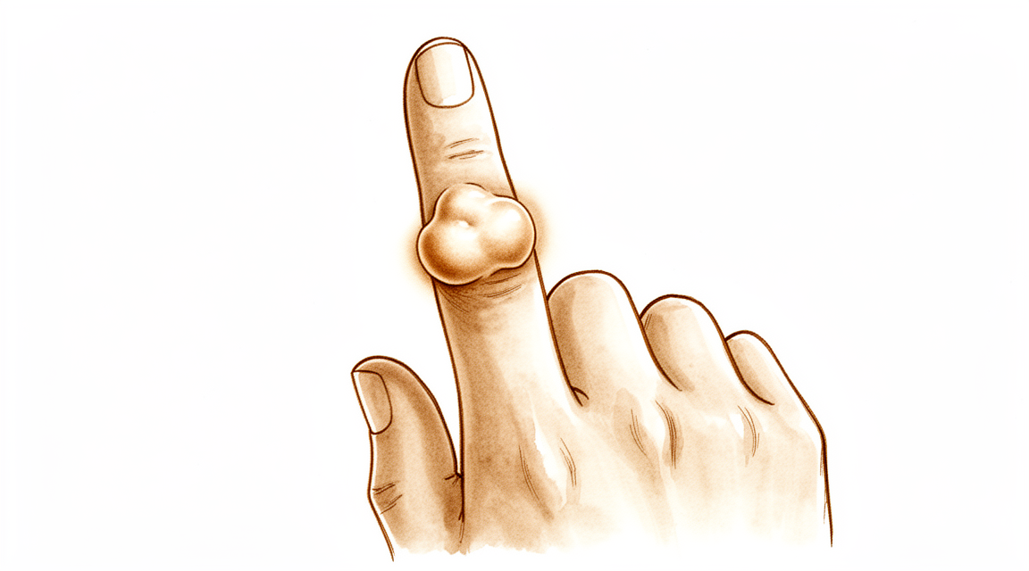
O que você está sentindo
A maioria das pessoas nota um nódulo em um dedo ou no polegar que não dói. Ele costuma ser firme e cresce lentamente. Alguns nódulos ficam bem ao lado de uma articulação, muitas vezes a articulação mais próxima da ponta do dedo, e podem aparecer no dorso, na lateral ou no lado da palma do dedo. O dedo indicador é o local mais comum.
O nódulo em si é indolor em quase todos os casos. Quando há desconforto, ele tende a vir da pressão que o nódulo exerce sobre as estruturas vizinhas à medida que cresce. Um pequeno número de pessoas nota formigamento ou dormência no dedo se o nódulo pressionar um nervo. Algumas pessoas percebem que o dedo prende ou estala ao se mover, um pouco como um dedo em gatilho, em que o dedo trava por um instante e depois se solta de repente. Se o nódulo estiver perto de uma articulação, dobrar esse dedo por completo pode ser difícil, porque o nódulo atrapalha.
As tarefas diárias que usam esse dedo podem se tornar desajeitadas. Pegar moedas pequenas, abotoar botões, segurar uma caneta ou agarrar a tampa de um pote pode parecer desajeitado se o nódulo ficar exatamente onde a sua mão faz a pegada. O nódulo também pode enroscar em luvas ou bolsos.
Muitas vezes o nódulo já está presente há muito tempo antes de incomodar o suficiente para você procurar orientação. Muitas pessoas esperam meses ou anos, observando-o mudar lentamente.
Vale a pena conhecer alguns pontos sobre essa condição. Ela é benigna, o que significa que não se espalha para outras partes do corpo. É um pouco mais comum em mulheres do que em homens e geralmente aparece entre os 32 e os 51 anos de idade. A maioria das pessoas tem um único nódulo, embora algumas tenham mais de um, em locais diferentes.
Fique atento a um dedo ou uma mão que fique quente, vermelho, inchado e dolorido, especialmente se houver febre. Se isso acontecer, vá a um pronto-socorro no mesmo dia. Se o nódulo continuar crescendo, começar a doer ou o dedo não estiver funcionando como deveria, procure o seu médico de família ou peça uma avaliação com um especialista.
O que está realmente acontecendo
Os seus tendões são os cordões fortes que ligam o músculo ao osso e permitem que os dedos se dobrem. Cada tendão corre dentro de uma fina capa de tecido chamada bainha do tendão, que funciona um pouco como uma junta de vedação ao redor de uma corda, mantendo o cordão escorregadio para que ele possa deslizar enquanto o dedo se move.
Nessa condição, as células dessa capa começam a crescer mais do que deveriam. O resultado é um nódulo de tecido mole que fica sobre o tendão ou ao lado dele, muitas vezes perto de uma articulação. Ele é benigno, o que significa que não se espalha, e cresce lentamente. A maioria dos nódulos é pequena, com cerca de 1,35 cm em média, embora alguns fiquem maiores.
O nódulo é formado por alguns elementos agrupados: células inflamatórias, células gigantes (que são simplesmente células com vários núcleos) e um pigmento escuro chamado hemossiderina, a mesma substância que dá cor a um hematoma antigo. Esse pigmento é o motivo pelo qual esses nódulos podem parecer acastanhados quando são vistos durante uma cirurgia.
Os sintomas que você notou na seção acima vêm do fato de o nódulo estar no caminho. À medida que cresce, ele pode pressionar o tendão, a articulação ou um nervo, o que explica o travamento, a rigidez e o formigamento ocasional. Na maioria das vezes ele não dói nada, e apenas cerca de 16 em cada 100 pessoas relatam dor.
Vale a pena conhecer alguns pontos. A maioria das pessoas tem um único nódulo, mas cerca de 21 em cada 100 têm dois ou mais nódulos separados. O nódulo geralmente fica ao lado de uma articulação e, em cerca de um terço dos casos nos dedos, está na articulação mais próxima da ponta do dedo. Muito raramente, o nódulo pode pressionar o próprio osso, o que aparece em uma radiografia.
Existe também uma forma mais rara e mais difusa da mesma condição, que se espalha ao longo do tendão em finas frondes, em vez de formar um único nódulo bem delimitado. Essa forma é muito menos comum em um dedo e tende a se comportar de maneira diferente; por isso, o seu cirurgião vai querer ter uma ideia clara de exatamente que tipo de nódulo é este antes de planejar qualquer coisa.
O que podemos fazer a respeito
O Dr. Kieran Hirpara, cirurgião de membros superiores no Mater Private Hospital Rockhampton, começa com as opções menos invasivas adequadas ao seu caso. Em geral, os pacientes são encaminhados à nossa clínica pelo médico de família; caso um fisioterapeuta tenha sugerido que você nos procure, ainda assim será necessário um encaminhamento do seu médico de família para ter direito ao reembolso do Medicare. Na clínica, colhemos o histórico clínico, examinamos a sua mão e, quando necessário, solicitamos exames de imagem para determinar exatamente que tipo de nódulo é este.
Como este nódulo é benigno e cresce lentamente, não fazer nada é uma opção real. No caso de um nódulo pequeno e indolor, simplesmente observá-lo pode ser a escolha certa. Se o nódulo estiver atrapalhando, a terapia da mão pode ajudar com a rigidez e o travamento. A terapia visa manter o dedo se movimentando e tornar as tarefas diárias mais fáceis. Geralmente sugerimos dar a ela uma chance razoável antes de pensar em cirurgia.
Não existe medicamento que faça esse nódulo diminuir; por isso, analgésicos e anti-inflamatórios apenas aliviam o desconforto causado pela pressão do nódulo sobre os tecidos vizinhos. Eles não fazem o nódulo ficar menor.
A cirurgia é o tratamento habitual quando o nódulo é grande, doloroso ou impede o dedo de funcionar adequadamente. O objetivo é remover o nódulo inteiro juntamente com a sua fixação na bainha do tendão, pois deixar essa fixação para trás é o que permite que ele volte a crescer. Planejamos com cuidado antes de operar, usamos ampliação durante a operação e removemos o nódulo por completo. A recorrência é o principal risco nessa condição e geralmente ocorre dentro de 36 meses após a excisão. Em um grupo de 605 pessoas que tiveram um nódulo removido de um dedo, 14,8% tiveram o retorno do nódulo. Dito de outra forma: a maioria das pessoas, cerca de 85 em cada 100, não tem retorno. Se ele voltar, removê-lo novamente com medidas adicionais para desencorajar um novo crescimento ainda pode resolver o problema.
O que esperar
A evolução deste nódulo é estável, sem grandes mudanças. Ele é benigno, portanto não se espalha para nenhuma outra parte do corpo. Se nada for feito, um nódulo pequeno e indolor geralmente continua crescendo lentamente, ou simplesmente permanece como está. Ele raramente desaparece sozinho e não se transforma em nada perigoso.
A principal coisa que essa condição faz ao longo do tempo é voltar após a remoção. A recorrência é o principal risco deste nódulo e geralmente ocorre dentro de 36 meses após a excisão. Alguns nódulos têm maior probabilidade de voltar do que outros: os formados por duas ou mais partes separadas que não estão unidas entre si, e os que cresceram para dentro do próprio tendão ou do revestimento da articulação. Quando o nódulo inteiro é removido juntamente com o ponto onde ele se fixa, esse risco cai bastante.
A função após o tratamento tende a ser boa. As pessoas que tiveram um desses nódulos removido de um dedo alcançam, em média, 92% da função normal do membro em uma medida padronizada de como o membro funciona. Nos casos relatados, as pessoas mantiveram o movimento completo do dedo, e o formato do osso do dedo na radiografia voltou ao normal na avaliação de dois anos.
Se o nódulo for do tipo mais raro e mais espalhado, que envolve o tendão em frondes, ele é mais difícil de remover por completo e tem maior probabilidade de voltar. Nesses casos, a radioterapia direcionada à região pode ajudar a controlar o nódulo, mantendo a sua mão funcionando.
Se você optar por simplesmente observar um nódulo pequeno e indolor, o quadro realista é o de um nódulo que cresce lentamente ou permanece igual, e que pode, aos poucos, tornar mais desajeitado dobrar o dedo ou usá-lo para tarefas pequenas. Se ele começar a doer, continuar crescendo ou o dedo deixar de funcionar como deveria, esse é o momento em que o tratamento geralmente passa a valer a pena.
Quando procurar ajuda médica
A maioria dos nódulos como este não é urgente, e o tratamento nunca é uma corrida contra o tempo. Procure o seu médico de família se um nódulo não estiver melhorando, estiver crescendo ao longo de semanas, começar a doer ou impedir que você use o dedo ou a mão normalmente. Peça uma avaliação com um especialista se o dedo prender ou estalar ao se mover, se você notar formigamento ou dormência, ou se dobrar o dedo por completo tiver se tornado difícil porque o nódulo atrapalha. Essas são as situações que vale a pena avaliar mais cedo do que tarde, porque um nódulo que cresceu para dentro do tendão ou do revestimento da articulação é mais difícil de remover por completo e tem maior probabilidade de voltar.
Um conjunto de sinais exige atendimento no mesmo dia. Se o seu dedo ou a sua mão ficar quente, vermelho, inchado e dolorido, especialmente se houver febre, vá a um pronto-socorro. Você não precisa de um encaminhamento do médico de família antes. Se não conseguir contato com a clínica fora do horário de atendimento ou no fim de semana, vá ao pronto-socorro mais próximo.
Em maior profundidade
Advanced reading: the deeper science (optional)
Esta seção vai além do que você precisa saber para tomar decisões sobre o próprio tratamento. O tumor de células gigantes da bainha do tendão merece uma leitura mais detalhada, pois seu principal problema é a recorrência; as evidências indicam que essa recorrência é influenciada mais pela biologia do próprio tumor do que por qualquer aspecto relacionado ao modo como ele é removido.
A recorrência é uma propriedade do tumor, não apenas da cirurgia
A explicação instintiva para o retorno de um nódulo é que parte dele teria sido deixada para trás. Contudo, uma revisão sistemática de 605 casos chegou a uma conclusão diferente: a biologia intrínseca do tumor parece desempenhar um papel mais fundamental na recorrência do que a localização do tumor ou sua invasividade local. Os autores defendem a realização de estudos prospectivos maiores para identificar quais tumores são propensos à recorrência [1].
Essa é uma informação realmente útil a ser conhecida antes de uma cirurgia. A recorrência após uma excisão bem realizada é um comportamento reconhecido desse tumor, e não evidência de que algo deu errado.
O tratamento é eletivo; não fazer nada também é uma opção válida
É fácil esquecer isso assim que se começa a planejar uma cirurgia. O princípio do tratamento de primeira linha é a ressecção completa, porém o tratamento nunca é urgente, e a indicação cirúrgica deve ser avaliada levando em conta os sintomas, a progressão da doença, a localização da lesão e as suas próprias circunstâncias [4].
No caso de um nódulo pequeno, indolor e de crescimento lento, optar por apenas acompanhamento é uma escolha legítima. O tumor é benigno e não se dissemina; portanto, os motivos para realizar a cirurgia dizem respeito à função, ao tamanho e ao incômodo causado — e não ao risco de complicações.
Porém, dois fatores cirúrgicos realmente importam
A biologia não explica tudo. Em 941 pacientes com tumor gigantocelular tenossinovial do tipo localizado, os fatores associados à recorrência após ressecção foram tamanho tumoral maior e tratamento inicial por artroscopia. Considerando as taxas de complicações relativamente baixas e os bons resultados funcionais, os autores recomendam uma abordagem aberta com ressecção completa, sempre que possível, para reduzir a recorrência em casos de alto risco [2].
Por outro lado, uma revisão envolvendo 1.448 pacientes demonstrou que a excisão artroscópica é eficaz para o tipo localizado em quatro articulações diferentes; já no tipo difuso, a sinovectomia artroscópica mostrou eficácia apenas no joelho [3].
Para conciliar esses dados: em lesões pequenas, localizadas e bem delimitadas, ambas as abordagens são viáveis. Contudo, à medida que o tamanho aumenta ou no caso do tipo difuso, a excisão aberta completa é a opção mais indicada. O motivo é mecânico: esse tumor se estende em “frondes” ao redor de tendões, nervos e articulações; as partes mais facilmente ignoradas são justamente aquelas situadas atrás de estruturas que precisam ser levantadas e examinadas diretamente.
Por que a nomenclatura é importante
Atualmente, essa condição é classificada como tumor de células gigantes tenossinoviais, abrangendo tanto a forma localizada na mão quanto a forma difusa intra-articular anteriormente denominada sinovite vilonodular pigmentada [4]. Trata-se da mesma entidade, apenas em locais e com padrões de crescimento diferentes.
É importante saber disso ao pesquisar sobre o tema, pois as buscas trarão informações referentes aos joelhos e quadris, que descrevem a forma difusa da doença. As taxas de recorrência citadas para essa forma difusa são consideravelmente mais altas do que as observadas em lesões digitais localizadas. Utilizar os dados referentes ao joelho para avaliar o risco de uma lesão no dedo resulta em uma superestimação do perigo real.
O papel da radioterapia
No caso de doença difusa que tenha recidivado ou que não possa ser completamente extirpada, a radioterapia adjuvante é por vezes considerada. Uma meta-análise constatou que a sinovectomia aberta, ou a sinovectomia combinada com radioterapia perioperatória, esteve associada a uma redução da taxa de recorrência na sinovite vilonodular pigmentada difusa; contudo, são necessários grandes estudos prospectivos de longo prazo para confirmar esse fato [5].
No caso de tumores digitais localizados – que representam a grande maioria dos casos na mão –, essa situação não se aplica. Esses tumores situam-se no extremo do espectro correspondente a lesões difusas, recorrentes e localizadas nas articulações; são mencionados aqui apenas porque uma busca pelo nome da doença pode trazer esses resultados.
O que não é
Apesar do nome, trata-se de um tumor benigno. Ele não se dissemina para outras partes do corpo. A palavra “tumor” tem uma conotação que não se aplica aqui; a preocupação com recorrência refere-se à necessidade de novas cirurgias locais, à rigidez articular e à proximidade com nervos, e não ao câncer.
Referências
[1] Fotiadis E, Papadopoulos A, Svarnas T, Akritopoulos P, Sachinis NP, Chalidis BE. Tumor de células gigantes da bainha tendinosa dos dedos: uma revisão sistemática. Hand (N Y). 2011;6(3):244-9. https://doi.org/10.1007/s11552-011-9341-9
[2] Mastboom M, Staals E, Verspoor F, Rueten-Budde A, Stacchiotti S, Palmerini E, et al. Tratamento cirúrgico dos tumores de células gigantes tenossinoviais de tipo localizado nas grandes articulações: estudo baseado em um banco de dados multicêntrico de 31 centros internacionais de sarcoma. J Bone Joint Surg Am. 2019;101(14):1309-18. https://doi.org/10.2106/JBJS.18.01147
[3] Noailles T, Brulefert K, Briand S, Longis P, Andrieu K, Chalopin A, et al. Tumor de células gigantes da bainha tendinosa: cirurgia aberta ou sinovectomia artroscópica? Uma revisão sistemática da literatura. Orthop Traumatol Surg Res. 2017;103(5):809-14. https://doi.org/10.1016/j.otsr.2017.03.016
[4] Gouin F, Noailles T. Formas localizadas e difusas do tumor de células gigantes tenossinoviais (anteriormente denominado tumor de células gigantes da bainha tendinosa e sinovite villonodular pigmentada). Orthop Traumatol Surg Res. 2017;103(1):S91-S97. https://doi.org/10.1016/j.otsr.2016.11.002
[5] Mollon B, Lee A, Busse JW, Griffin AM, Ferguson PC, Wunder JS, et al. Efeito da sinovectomia cirúrgica e da radioterapia na taxa de recorrência da sinovite villonodular pigmentada do joelho. Bone Joint J. 2015;97-B(4):550-7. https://doi.org/10.1302/0301-620X.97B4.34907
Evidence & references
This is the clinical evidence summary written for health professionals. It is technical, and it lists the research this page was built from. You do not need to read it to understand your treatment or to make a decision about it.
Overview
- The diagnosis and treatment of suspected hand neoplasms should proceed in an orderly fashion similar to accepted methods applied to tumors in other locations of the extremities [1].
- Giant cell tumors of the tendon sheaths in the hand are benign lesions where recurrence is the primary risk [3, 4].
- Recurrence of giant cell tumors of the tendon sheaths typically occurs within 36 months of excision [3].
- The fundamental consideration in the management of hand tumours is to reconcile adequate tumour clearance with the maintenance of function and appearance [20].
- Complete surgical resection remains the treatment of choice for most patients with tenosynovial giant cell tumors [15].
- Diffuse tenosynovial giant cell tumor disease presents challenges due to high recurrence rates [15].
- Curettage, cryosurgery, and cementation performed by experienced surgeons appears to be a safe, effective, and reliable method for treating selected giant cell tumors of the hand [5].
- In cases of infiltrative giant cell tumor of the tendon sheath, radiation therapy may provide local tumor control with preservation of hand function [12].
- En bloc resection of the tumour and reconstruction with a fibular graft where necessary should be considered as the treatment of choice in giant cell tumour of the hand [10].
- En bloc resection of the tumour and reconstruction with an autograft should be considered in the treatment of recurrent giant cell tumour of the hand [8].
- En bloc resection and matched nonvascularized toe phalangeal transfer for Campanacci Grade 2 or 3 giant cell tumor of the phalanges resulted in a functional tumor-free digit with a low complication rate and no recurrences [35].
- Well-designed studies combining the recurrence rates of several hand surgery centers implementing a standardized treatment are needed to better demonstrate the associated risk factors for recurrence [28].
Anatomy & Pathophysiology
Clinical Presentation and Demographics
- Giant cell tumour of tendon sheath (GCTTS) is the second most common benign proliferative tumour in the upper extremities after ganglion cysts [102].
- GCTTS are usually slow-growing, painless, benign soft tissue tumours [102].
- GCTTS are most commonly found in the fingers and among women in their fourth and fifth decades [102].
- The male-to-female ratio for GCTTS of the digits is 1:1.47 [23].
- The mean age range for GCTTS of the digits is 32 to 51 years [23].
- Pain was reported in 15.7% of GCTTS cases and sensory disturbances in 4.57% of cases [23].
- A definite history of trauma was recorded in 5% of GCTTS lesions [23].
- The most frequent tumour location for GCTTS of the digits is the index finger (29.7%) [23].
- In a series of 64 cases, the most frequent location for GCTTS was the long finger (23.5%), followed by the thumb, index finger, and hand (20.3% each) [49].
- Patients with GCTTS arising in the fingers complained of a painless mass in almost every instance [24].
- The duration of symptoms for finger GCTTS ranged from two weeks to fifteen years, with an average duration of thirty-eight months [24].
- GCTTS are usually firm, lobulated, and non-tender masses [24].
- As the tumour grows, patients may present with swelling, pain, and limitation of movement [102].
Anatomical Distribution and Morphology
- Fifty-three xanthomas (GCTTS) arose adjacent to joints in the fingers, whereas twenty-nine bore no relation to joints [24].
- Thirty-one of ninety-one finger GCTTS occurred at the distal joints [24].
- Of the thirty-one distal joint GCTTS, eighteen were on the dorsal aspect and thirteen were distributed evenly on the radial, ulnar, and volar aspects [24].
- Type I GCTTS (single lesions) were more frequently detected (78.7%) than Type II tumours (two or more distinct tumours that were not joined together) (21.3%) [23].
- Type 1 GCTTS includes a nodular or multinodular lesion surrounded by a capsule, while Type 2 describes tumours with no connective tissue membrane and satellite, diffuse, or multicentric nodules [102].
- The average size of GCTTS was 1.35 cm, with a range of 0.3 cm to 5 cm [49].
- Bone erosion was found in 3 patients (4.7%) in a series of 64 GCTTS cases [49].
- Tendon involvement with flexors/extensors ratio of 4:3 was found in 7 cases (10.9%) in a series of 64 GCTTS cases [49].
- Involvement of the neurovascular bundle was presented in 7 patients (10.9%) in a series of 64 GCTTS cases [49].
- A case report describes the first patient with three separate GCTTS developing from both flexor and extensor tendons in a single digit [7].
Histology and Pathogenesis
- Microscopically, all GCTTS contained multinucleated giant cells, histiocytes, and haemosiderin deposits [49].
- GCTTS originates from the synovial membrane, tendon sheath, or synovial bursa [102].
- Inflammation resulting from reactive or regenerative hyperplasia is the generally accepted theory of pathogenesis for GCTTS [102].
- Genetic factors have been observed in previous studies regarding GCTTS pathogenesis [102].
- Fibroma of tendon sheath is histologically distinct from giant cell tumor of tendon sheath, a lesion with which it is commonly confused [16].
Diagnostic Imaging
- Radiography can be helpful in evaluating cortical destruction but is not helpful in the definitive diagnosis of GCTTS [102].
- Magnetic resonance imaging (MRI) is the most useful examination for the diagnosis and treatment planning of GCTTS [102].
- Al-Qattan classified GCTTS with MRI into two types according to the risk of recurrence [102].
Classification
- Giant cell tumors of the synovial sheaths in the hand are benign lesions in which recurrence is the primary risk [4].
- Recurrence for giant cell tumors of the tendon sheaths typically occurs within 36 months of excision [3].
- The Al-Qattan classification divides giant cell tumours of tendon sheath into two main types based on whether the entire tumour is surrounded by one pseudocapsule as assessed by the surgeon during surgery [90].
- Type I tumours are defined as those where the entire tumour is surrounded by one pseudocapsule [90].
- Type II tumours are defined as those where the entire tumour is not surrounded by one pseudocapsule [90].
- Type I tumours are sub-classified according to the thickness of the capsule, lobulation of the tumour, the presence of satellite lesions, and the diffuse or multicenteric nature of the tumour [90].
- Type II tumours are sub-classified according to the thickness of the capsule, lobulation of the tumour, the presence of satellite lesions, and the diffuse or multicenteric nature of the tumour [90].
- In a prospective study of 43 consecutive cases, none of the type I tumours (n=30) recurred [90].
- In a prospective study of 43 consecutive cases, recurrence occurred in five out of 13 type II tumours [90].
- Second recurrences were seen with type II B and C tumours, but not type II A tumours [90].
- Type I tumours (single lesions) were more frequently detected (78.7%) than type II tumours (two or more distinct tumours that were not joined together) (21.3%) in a systematic review of 605 patients [23].
- Type II tumours were associated with higher recurrence rates compared to Type I tumours in a systematic review of 605 patients [23].
- Direct involvement of the extensor tendons, flexor tendons, or joint capsule puts patients in a high-risk category with respect to recurrence [43].
- Preoperative diagnosis and meticulous surgical technique were found to be the only predictive factor of recurrence in a series of 14 cases [34].
- The overall recurrence rate in a systematic review of 605 patients was 14.8% [23].
Clinical Presentation
- Patients with fibrous xanthomas arising in the fingers complained of a painless mass in almost every instance [24].
- The duration of symptoms for finger tumors ranged from two weeks to fifteen years, with an average duration of thirty-eight months [24].
- The tumors were usually firm, lobulated, and non-tender [24].
- Fifty-three xanthomas arose adjacent to joints in the fingers, whereas twenty-nine bore no relation to joints [24].
- Thirty-one of the ninety-one tumors in the fingers occurred at the distal joints [24].
- Eighteen of the thirty-one distal joint tumors were on the dorsal aspect of the joint, and thirteen were distributed about evenly on the radial, ulnar, and volar aspects of the joint [24].
- The most frequent tumour location for giant cell tumour of tendon sheath of the digits was the index finger (29.7%) [23].
- Pain was reported in 15.7% of cases and sensory disturbances in 4.57% of cases [23].
- A definite history of trauma was recorded in 5% of lesions [23].
- Type I tumours (single lesions) were more frequently detected (78.7%) than type II tumours (two or more distinct tumours that were not joined together) (21.3%) [23].
- The male-to-female ratio for giant cell tumour of tendon sheath of the digits was 1:1.47 [23].
- The mean age for giant cell tumour of tendon sheath of the digits ranged from 32 to 51 years [23].
- A tendon tumour can present as a trigger finger [18].
- Trigger wrist caused by a giant cell tumour of tendon sheath has been reported [26].
- The trigger phenomenon in trigger wrist caused by a giant cell tumour of tendon sheath probably occurred when the tumour entered and left the distal carpal tunnel [26].
- Fibroma of tendon sheath is a benign soft tissue tumor that has a predilection for the hand [16].
- Fibroma of tendon sheath may be more common than has been previously recognized [16].
- The differential diagnosis of a soft tissue tumor in the digits and hand is usually limited to a ganglion, an inclusion cyst, or a giant cell tumor of tendon sheath (xanthofibroma) [16].
- Fibroma of tendon sheath should be included in the differential diagnosis of a soft tissue tumor in the digits and hand [16].
- A case of fibroma of tendon sheath presented as a 6-year history of a slowly enlarging palmar mass of the left index finger [16].
- Examination of a fibroma of tendon sheath case demonstrated a soft, compressible, lobulated, nontender mass extending from the second web space to the tip of the finger [16].
- Flexion of the digit was limited by the bulge in a case of fibroma of tendon sheath [16].
- The unusual location of a fibroma of the tendon sheath and atypical nature of the triggering phenomenon can lead to it being reported as a rare cause of median nerve compression at the wrist [50].
- Giant cell tumor of tendon sheath and pigmented villonodular synovitis are benign, locally invasive tumors involving synovium, tendon sheaths, and bursae [47].
- Patients with giant cell tumor of tendon sheath or pigmented villonodular synovitis may present with a discrete mass or with joint swelling, pain, or locking or catching [47].
- The knee and digits of the hand are the most common locations of lesions for giant cell tumor of tendon sheath and pigmented villonodular synovitis [47].
- Multifocal lesions of giant cell tumor of tendon sheath and pigmented villonodular synovitis are rare [47].
- The authors report the uniqueness of a case of multicentric giant cell tumor in the upper extremity [9].
- Radiological changes in the form of bony indentation was seen in only 2 cases of giant cell tumor of tendon sheath in a series of 12 patients [34].
- The most common presentation for giant cell tumor of tendon sheath in a series of 12 patients was with a mass over the hand, with a predilection to the thumb [34].
- A 65-year-old man presented with a slowly enlarging mass on the dorsal aspect of the left thumb consistent with a recurrent giant cell tumor of the tendon sheath [33].
Investigations
- The differential diagnosis of a soft tissue tumor in the digits and hand is usually limited to a ganglion, an inclusion cyst, or a giant cell tumor of tendon sheath [16].
- Fibroma of tendon sheath should be included in the differential diagnosis as it may be more common than has been previously recognized [16].
- Fibro-osseous pseudotumor of the digit should be considered in the differential diagnosis of rapidly enlarging digital lesions [64].
- Although MRI findings and location might help in the diagnosis of tenosynovial giant cell tumors, careful assessment is mandatory, especially in unusual locations [129].
Treatment
General Principles
Surgical Excision
- Complete surgical resection remains the treatment of choice for most patients with tenosynovial giant cell tumors, though diffuse disease presents challenges due to high recurrence rates [15].
- En bloc resection of metacarpal giant cell tumors with reconstruction by iliac graft appears to be an effective treatment [14].
- En bloc resection and matched nonvascularized toe phalangeal transfer resulted in a functional tumor-free digit with a low complication rate and no recurrences for Campanacci Grade 2 or 3 giant cell tumors of the phalanges [35].
- Total surgical excision ensuring removal of the attachment site (flexor sheath/palmar fascia) resulted in no local recurrences at a mean follow-up of 3.2 years for fibroma of tendon sheath [11].
- Local excision of the tumour appears to be a satisfactory method of treatment for Nora’s lesion [109].
- Preoperative planning aided by a tissue diagnosis with fine needle aspiration cytology, wide surgical exposure, and meticulous dissection with help of magnification are imperative for a successful outcome in giant cell tumor of tendon sheath [34].
- In all cases of giant cell tumor of tendon sheath, magnifying loupe or operating microscope was used during surgical excision [49].
- In cases of infiltrative giant cell tumor of tendon sheath, radiation therapy may provide local tumor control with preservation of hand function [12].
Recurrence and Adjuvant Therapy
- Giant cell tumors of the tendon sheaths in the hand are benign lesions where recurrence is the primary risk, typically occurring within 36 months of excision [3].
- In a study of 64 cases of giant cell tumor of tendon sheath, the recurrence rate was 4.7% (n=3) [49].
- In a systematic review of 605 patients with giant cell tumor of tendon sheath of the digits, 14.8% of patients had tumour recurrence [23].
- Repeated curettage with adjuvants eventually resulted in the cure for all patients and is therefore a reasonable treatment for both primary and recurrent giant cell tumor of the small bones of the hands and feet [105].
- Intralesional excision with cautery and methylmethacrylate provides a reliable method of treatment of giant cell tumors with good long-term functional results [71].
- Intralesional excision with local adjuvant therapy is recommended for the treatment of giant cell tumor of bone because it results in a good functional outcome compared to extralesional excision [98].
- Intralesional excision remains a viable, and likely the standard, mode of treatment for most giant cell tumors of the distal radius unless there is extensive bone loss [29].
- Based on data obtained from the number of studies available, intralesional excision appears to be more appropriate for the treatment of local lesions (eg, Grades 1 and 2) than Grade 3 giant cell tumors of the distal radius [52].
- Well-designed studies combining the recurrence rates of several hand surgery centers implementing a standardized treatment are needed to better demonstrate the associated risk factors for recurrence in giant cell tumor of tendon sheath [28].
Reconstruction and Resection Options
- Both curettage and resection/amputation are acceptable treatment options for the rare condition of giant cell tumour of bone in the hand, with a need to individualize treatment decisions based on the site and extent of disease to minimize treatment morbidity while maximizing disease control [6].
- Aggressive and malignant bone tumors of the second through fifth metacarpals generally require en bloc bone excision [17].
- Ray excision is usually best for lesions of the second and fifth metacarpals [17].
- If bone graft has been used for reconstruction, carpometacarpal arthrodesis and MCP ligament reconstruction or silicone arthroplasty may be practical [17].
- Whatever level is chosen for reconstruction, it is essential that an adequate, safe margin of normal tissue first be excised en bloc with the tumor [17].
- Aggressive tumors of the second through fifth metacarpals that have invaded soft tissue or sustained pathologic fractures often require excision of not only the involved ray but also the contiguous ray or rays radial and ulnar to the involved digit [17].
- If a malignant tumor has broken into the midpalm and extends across the metacarpals, removal of all digital rays may be needed to gain an adequate soft tissue margin [17].
- Retention of a sensate and relatively mobile thumb may be considerably more esthetic and functionally satisfactory than a forearm- or wrist-level amputation [17].
- If a tumor extends proximally from the metacarpal level, a more proximal level of hand, wrist, or forearm amputation is required for safe tumor management [17].
- Malignant soft tissue tumors in the palm or carpal tunnel often require at least partial hand amputation [17].
- If treated by only local or limited excision, the chance of recurrence is extremely high particularly in the setting of a positive margin for malignant soft tissue tumors in the palm or carpal tunnel [17].
- Below-elbow amputation is necessary to treat larger tumors of the palm or carpal tunnel [17].
- Wide en bloc excision of soft tissue sarcomas with negative margins is required to achieve local control of the lesion [17].
- At a minimum, aggressive soft tissue tumors, such as bone tumors, require ray resection or removal of multiple rays [17].
- Central palmar lesions more likely require sacrifice of three rays; those on the border are more likely than those in the center to be salvageable by removing just two rays [17].
- In the presence of proximal, broader, and larger lesions, all four digits or the entire hand may have to be sacrificed to save the patient [17].
- The use of a massive biocompatible bipolar unconstrained prosthesis is a viable treatment option for distal radius reconstruction after en-bloc resection of a giant cell tumour, offering rapid functional improvement without donor-site morbidity [68].
- Reconstruction after wide excision by nonvascularized fibular graft is a viable alternative for giant cell tumors of the lower end of radius though it is a challenging procedure and may be accompanied by major complications [111].
- En bloc resection is recommended for aggressive giant cell tumours (GCT) of the bone involving the distal end of the radius to minimize the risk of recurrence [91].
- The distal ulna may be widely resected with or without stabilization of the residual ulnar stump, yielding satisfactory local disease control and functional outcome [48].
- This is a simple and effective modality of reconstruction after resection of distal radial tumors using centralization of the ulna [30].
Complications
- Recurrence is the primary risk associated with giant cell tumors of the tendon sheaths in the hand [3].
- The overall recurrence rate for giant cell tumour of tendon sheath of the digits is 14.8% [23].
- Type II tumours (two or more distinct tumours that were not joined together) are associated with a higher risk of recurrence compared to Type I tumours [23].
- Direct involvement of the extensor tendons, flexor tendons, or joint capsule places patients in a high-risk category for recurrence [43].
- Surgical treatment of pigmented villonodular synovitis led to good functional results with an average Enneking score of 92% of normal limb function [32].
- A tendon tumour arising from the superficialis tendon can present as a trigger finger [18].
- A giant cell tumour of the tendon sheath can cause a trigger wrist phenomenon when the tumour enters and leaves the distal carpal tunnel [26].
- A fibroma of the tendon sheath in an unusual location can cause a triggering phenomenon and median nerve compression at the wrist [50].
- Total surgical excision ensuring removal of the attachment site (flexor sheath/palmar fascia) for fibroma of tendon sheath resulted in no local recurrences at a mean follow-up of 3.2 years [11].
- Patients with giant cell tumor of bone in the hand who are at higher risk of recurrence should be clinically followed more closely [41].
- Both curettage and resection/amputation are acceptable treatment options for giant cell tumour of bone in the hand, with a need to individualize treatment decisions based on the site and extent of disease to minimize treatment morbidity while maximizing disease control [6].
- Below-elbow amputation is necessary to treat larger malignant soft tissue tumors in the palm or carpal tunnel [17].
- Patients who received preoperative radiotherapy for hand tumors had a decrease in functional outcomes and grip strength that averaged 66% compared with the contralateral hand [55].
- None of the patients who underwent single-ray amputation for tumors of the hand, including giant cell tumors of bone, had local recurrences [55].
- A 29-year-old patient with recurrent giant-cell tumor of the digit was treated by phalangeal excision and toe phalanx transplant [54].
Recovery
- Recurrence of giant cell tumors of the tendon sheaths in the hand typically occurs within 36 months of excision [3].
- Total surgical excision ensuring removal of the attachment site (flexor sheath/palmar fascia) resulted in no local recurrences at a mean follow-up of 3.2 years [11].
- In cases of infiltrative GCTTS, radiation therapy may provide local tumor control with preservation of hand function [12].
- At 2-year final follow-up examination, no recurrence was noted, finger range of motion was full, and the x-ray film contour of the phalanx had returned to normal [13].
- Surgical treatment led to good functional results with an average Enneking score of 92% of normal limb function [32].
- En bloc resection and matched nonvascularized toe phalangeal transfer resulted in a functional tumor-free digit with a low complication rate and no recurrences [35].
Key Evidence
- [L5] The diagnosis and treatment of suspected hand neoplasms should proceed in an orderly fashion similar to accepted methods applied to tumors in other locations of the extremities. [1] (10.1016/s0363-5023(83)80277-9)
- [L4] Giant cell tumors of the tendon sheaths in the hand are benign lesions where recurrence is the primary risk, typically occurring within 36 months of excision. [3] (10.1016/j.otsr.2013.03.008)
- [L4] Giant cell tumors of the synovial sheaths in the hand are benign lesions in which recurrence is the primary risk. [4] (10.1016/j.jhsa.2013.08.051)
- [L4] Curettage, cryosurgery, and cementation performed by experienced surgeons appears to be a safe, effective, and reliable method for treating selected giant cell tumors of the hand. [5] (10.1053/jhsu.2001.22525)
- [L4] Both curettage and resection/amputation are acceptable treatment options for the rare condition of giant cell tumour of bone in the hand, with a need to individualize treatment decisions based on the site and extent of disease to minimize treatment morbidity while maximizing disease control. [6] (10.1177/17531934211007820)
- [L5] We believe this case report describes the first patient with three separate GCTTS developing from both flexor and extensor tendons in a single digit. [7] (10.1142/s2424835518720189)
- [L4] En bloc resection of the tumour and reconstruction with an autograft should be considered in the treatment of recurrent giant cell tumour of the hand. [8] (10.1016/s0266-7681(98)80197-6)
- [L5] The authors report the uniqueness of this case of multicentric giant cell tumor in the upper extremity. [9] (10.1016/j.main.2011.11.006)
- [L4] En bloc resection of the tumour and reconstruction with a fibular graft where necessary should be considered as the treatment of choice in giant cell tumour of the hand. [10] (10.1016/s0266-7681(96)80161-6)
- [L4] Total surgical excision ensuring removal of the attachment site (flexor sheath/palmar fascia) resulted in no local recurrences at a mean follow-up of 3.2 years. [11] (10.1177/1753193412469146)
- [L4] In cases of infiltrative GCTTS, radiation therapy may provide local tumor control with preservation of hand function. [12] (10.1016/j.jhsa.2012.01.011)
- [L5] At 2-year final follow-up examination, no recurrence was noted, finger range of motion was full, and the x-ray film contour of the phalanx had returned to normal. [13] (10.1016/s0363-5023(05)80151-0)
- [L5] En bloc resection of metacarpal giant cell tumors with reconstruction by iliac graft appears to be an effective treatment. [14] (10.1016/s1297-3203(03)00042-8)
- [L5] Complete surgical resection remains the treatment of choice for most patients with tenosynovial giant cell tumors, though diffuse disease presents challenges due to high recurrence rates. [15] (10.5435/jaaos-d-24-01255)
- [L5] [16] (10.1016/s0363-5023(84)80031-3)
- [L5] At operation a tumour arising from the superficials tendon was found and removed. [18] (10.1016/0266-7681(86)90283-4)
- [L5] The fundamental consideration in the management of hand tumours is to reconcile adequate tumour clearance with the maintenance of function and appearance. [20] (10.1016/0266-7681(91)90159-l)
- [L1] [23] (10.1007/s11552-011-9341-9)
- [L4] [24] (10.2106/00004623-196951010-00005)
- [L5] [26] (10.1016/s0266-7681(85)80038-3)
- [L3] Well-designed studies combining the recurrence rates of several hand surgery centers implementing a standardized treatment are needed to better demonstrate the associated risk factors for recurrence. [28] (10.1186/s12891-019-2866-8)
- [L3] Intralesional excision remains a viable, and likely the standard, mode of treatment for most giant cell tumors of the distal radius unless there is extensive bone loss. [29] (10.1007/s11999-014-4054-3)
- [L4] This is a simple and effective modality of reconstruction after resection of distal radial tumors. [30] (10.1016/j.jhsa.2022.05.011)
- [L4] Surgical treatment led to good functional results with an average Enneking score of 92% of normal limb function. [32] (10.1097/01.blo.0000224051.01873.fb)
- [L4] [33] (10.1016/j.jhsa.2012.11.001)
- [L4] [34] (10.1007/s12593-010-0020-9)
- [L4] En bloc resection and matched nonvascularized toe phalangeal transfer resulted in a functional tumor-free digit with a low complication rate and no recurrences. [35] (10.1016/j.jhsa.2024.06.013)
- [L4] Our observations suggest there are subsets of patients with giant cell tumor of bone who are at higher risk of recurrence and should be clinically followed more closely. [41] (10.1007/s11999-011-2172-8)
- [L3] Direct involvement of the extensor tendons, flexor tendons, or joint capsule puts patients in a high-risk category with respect to recurrence. [43] (10.1016/j.jhsa.2009.12.004)
- [Case_report] [47] (10.1016/j.jhsa.2014.11.010)
- [L3] The distal ulna may be widely resected with or without stabilization of the residual ulnar stump, yielding satisfactory local disease control and functional outcome. [48] (10.1177/1558944717743598)
- [L4] [49] (10.11138/gchir/2013.34.5.149)
- [L5] The unusual location of the fibroma of the tendon sheath (FGT) and the atypical nature of the triggering phenomenon led to the reporting of this observation as a rare cause of median nerve compression at the wrist. [50] (10.1016/j.main.2006.03.001)
- [L3] Based on data obtained from the number of studies available, intralesional excision appears to be more appropriate for the treatment of local lesions (eg, Grades 1 and 2) than Grade 3 GCTs of the distal radius. [52] (10.1007/s11999-012-2464-7)
- [L5] [54] (10.1016/s0363-5023(79)80134-3)
- [Paper] Fibro-osseous pseudotumor of the digit should be considered in the differential diagnosis of rapidly enlarging digital lesions. [64] (10.1177/15589447261481209)
- [Case_report] The use of a massive biocompatible bipolar unconstrained prosthesis is a viable treatment option for distal radius reconstruction after en-bloc resection of a giant cell tumour, offering rapid functional improvement without donor-site morbidity. [68] (10.1016/j.otsr.2013.04.001)
- [L4] Intralesional excision with cautery and methylmethacrylate provides a reliable method of treatment of giant cell tumors with good long-term functional results. [71] (10.1097/01.blo.0000128280.59965.e3)
- [L3] [90] (10.1054/jhsb.2000.0522)
- [L4] [91] (10.1177/17531934211068622)
- [L3] Intralesional excision with local adjuvant therapy is recommended for the treatment of giant cell tumor of bone because it results in a good functional outcome compared to extralesional excision. [98] (10.1007/s004020100317)
- [L4] [102] (10.1177/17531934231222401)
- [L3] Repeated curettage with adjuvants eventually resulted in the cure for all patients and is therefore a reasonable treatment for both primary and recurrent GCT of the small bones of the hands and feet. [105] (10.1302/0301-620x.95b6.30876)
- [L5] Local excision of the tumour appears to be a satisfactory method of treatment. [109] (10.1016/s0266-7681(97)80269-0)
- [L4] Reconstruction after wide excision by nonvascularized fibular graft is a viable alternative for giant cell tumors of the lower end of radius though it is a challenging procedure and may be accompanied by major complications. [111] (10.1007/s00402-010-1059-6)
- [L4] Although MRI findings and location might help in the diagnosis of a T-GCT, careful assessment is mandatory, especially in unusual locations. [129] (10.1186/s12891-016-1050-7)
References
[1] Tumors in the hand. The Journal of Hand Surgery. 1983. DOI: 10.1016/s0363-5023(83)80277-9
[3] Giant cell tumors of the tendon sheaths in the hand: Review of 96 patients with an average follow-up of 12 years. Orthopaedics & Traumatology: Surgery & Research. 2013. DOI: 10.1016/j.otsr.2013.03.008
[4] Giant Cell Tumors of the Tendon Sheaths in the Hand: Review of 96 Patients With an Average Follow-Up of 12 Years. The Journal of Hand Surgery. 2013. DOI: 10.1016/j.jhsa.2013.08.051
[5] Giant cell tumor of the hand: Superior results with curettage, cryosurgery, and cementation. The Journal of Hand Surgery. 2001. DOI: 10.1053/jhsu.2001.22525
[6] Giant cell tumour of hand bones: outcomes of treatment. Journal of Hand Surgery (European Volume). 2021. DOI: 10.1177/17531934211007820
[7] Multiple Giant Cell Tumor of Tendon Sheath Involving Both Flexor and Extensor Tendons in a Single Digit: A Case Report and Review of the Literatures. The Journal of Hand Surgery (Asian-Pacific Volume). 2018. DOI: 10.1142/s2424835518720189
[8] Recurrent Metacarpal Giant Cell Tumour Treated by En Bloc Resection and Metatarsal Transfer. Journal of Hand Surgery. 1998. DOI: 10.1016/s0266-7681(98)80197-6
[9] Tumeurs à cellules géantes à localisations multiples du membre supérieur. Chirurgie de la Main. 2012. DOI: 10.1016/j.main.2011.11.006
[10] Giant Cell Tumours of the Hand. Journal of Hand Surgery. 1996. DOI: 10.1016/s0266-7681(96)80161-6
[11] Fibroma of tendon sheath of the hand: a series of 20 patients with 23 tumours. Journal of Hand Surgery (European Volume). 2012. DOI: 10.1177/1753193412469146
[12] Radiation Therapy for Infiltrative Giant Cell Tumor of the Tendon Sheath. The Journal of Hand Surgery. 2012. DOI: 10.1016/j.jhsa.2012.01.011
[13] Vascular leiomyoma of the finger causing bone erosion. The Journal of Hand Surgery. 1995. DOI: 10.1016/s0363-5023(05)80151-0
[14] Tumeur à cellules géantes du troisième métacarpien : à propos d’un cas. Chirurgie de la Main. 2003. DOI: 10.1016/s1297-3203(03)00042-8
[15] Tenosynovial Giant Cell Tumor and Pigmented Villonodular Synovitis. Journal of the American Academy of Orthopaedic Surgeons. 2025. DOI: 10.5435/jaaos-d-24-01255
[16] Fibroma of tendon sheath. The Journal of Hand Surgery. 1984. DOI: 10.1016/s0363-5023(84)80031-3
[17] Green S Operative Hand Surgery. BOX 59.1 Ganglions of the Hand and Wrist > Finger Metacarpals.
[18] A tendon tumour presenting as a trigger finger. The Journal of Hand Surgery: Journal of the British Society for Surgery of the Hand. 1986. DOI: 10.1016/0266-7681(86)90283-4
[20] Soft Tissue Tumours of the Hand. Journal of Hand Surgery. 1991. DOI: 10.1016/0266-7681(91)90159-l
[23] Giant Cell Tumour of Tendon Sheath of the Digits. A Systematic Review. HAND. 2011. DOI: 10.1007/s11552-011-9341-9
[24] Fibrous Xanthoma of Synovium (Giant-Cell Tumor of Tendon Sheath, Pigmented Nodular Synovitis). The Journal of Bone & Joint Surgery. 1969. DOI: 10.2106/00004623-196951010-00005
[26] Trigger Wrist Caused by a Giant Cell Tumour of Tendon Sheath. Journal of Hand Surgery. 1985. DOI: 10.1016/s0266-7681(85)80038-3
[28] Giant cell tumor of tendon sheath in the hand: analysis of risk factors for recurrence in 50 cases. BMC Musculoskeletal Disorders. 2019. DOI: 10.1186/s12891-019-2866-8
[29] Is Intralesional Treatment of Giant Cell Tumor of the Distal Radius Comparable to Resection With Respect to Local Control and Functional Outcome?. Clinical Orthopaedics & Related Research. 2015. DOI: 10.1007/s11999-014-4054-3
[30] Functional Outcomes of Centralization of the Ulna as a Method of Reconstruction Following Resection of Campanacci Grade 3 Giant Cell Tumor of the Distal Radius. The Journal of Hand Surgery. 2024. DOI: 10.1016/j.jhsa.2022.05.011
[32] What Affects the Recurrence and Clinical Outcome of Pigmented Villonodular Synovitis?. Clinical Orthopaedics and Related Research. 2006. DOI: 10.1097/01.blo.0000224051.01873.fb
[33] Giant Cell Tumor of the Tendon Sheath. The Journal of Hand Surgery. 2013. DOI: 10.1016/j.jhsa.2012.11.001
[34] Giant Cell Tumor of Tendon Sheath: Case Series and Review of Literature. Journal of Hand and Microsurgery. 2010. DOI: 10.1007/s12593-010-0020-9
[35] Campanacci Grade 2 or 3 Giant Cell Tumor of the Phalanges: En Bloc Resection and Matched Nonvascularized Toe Phalangeal Transfer. The Journal of Hand Surgery. 2025. DOI: 10.1016/j.jhsa.2024.06.013
[41] Giant Cell Tumor of Bone: Are We Stratifying Results Appropriately?. Clinical Orthopaedics & Related Research. 2012. DOI: 10.1007/s11999-011-2172-8
[43] Recurrence of Giant Cell Tumors in the Hand: A Prospective Study. The Journal of Hand Surgery. 2010. DOI: 10.1016/j.jhsa.2009.12.004
[47] Recurrent Pigmented Villonodular Synovitis and Multifocal Giant Cell Tumor of the Tendon Sheath: Case Report. The Journal of Hand Surgery. 2015. DOI: 10.1016/j.jhsa.2014.11.010
[48] Extensor Carpi Ulnaris Tenodesis Versus No Stabilization After Wide Resection of Distal Ulna Giant Cell Tumors. HAND. 2017. DOI: 10.1177/1558944717743598
[49] Giant cell tumor of tendon sheath: study of 64 cases and review of literature. Giornale di Chirurgia - Journal of Surgery. 2013. DOI: 10.11138/gchir/2013.34.5.149
[50] Fibrome de la gaine tendineuse occasionnant un « poignet à ressaut » et un syndrome du canal carpien. Chirurgie de la Main. 2006. DOI: 10.1016/j.main.2006.03.001
[52] Which Treatment is the Best for Giant Cell Tumors of the Distal Radius? A Meta-analysis. Clinical Orthopaedics & Related Research. 2012. DOI: 10.1007/s11999-012-2464-7
[54] Treatment of recurrent giant-cell tumor of the digit by phalangeal excision and toe phalanx transplant: A case report. The Journal of Hand Surgery. 1979. DOI: 10.1016/s0363-5023(79)80134-3
[55] Green S Operative Hand Surgery. Index Finger Ray Amputation.
[64] Recurrent Fibro-Osseous Pseudotumour of the Digit Mimicking Infection: A Narrative Review and Case Report. HAND. 2026. DOI: 10.1177/15589447261481209
[68] Massive wrist prosthesis for giant cell tumour of the distal radius: A case report with a 3-year follow-up. Orthopaedics & Traumatology: Surgery & Research. 2013. DOI: 10.1016/j.otsr.2013.04.001
[71] Results of Giant Cell Tumor of Bone Treated With Intralesional Excision. Clinical Orthopaedics & Related Research. 2004. DOI: 10.1097/01.blo.0000128280.59965.e3
[90] Giant Cell Tumours of Tendon Sheath: Classification and Recurrence Rate. Journal of Hand Surgery. 2001. DOI: 10.1054/jhsb.2000.0522
[91] En bloc resection and vascularized ulnar pedicle graft reconstruction with plate fixation for giant cell tumour of the distal radius. Journal of Hand Surgery (European Volume). 2022. DOI: 10.1177/17531934211068622
[98] Oncologic and functional results after treatment of giant cell tumors of bone. Archives of Orthopaedic and Trauma Surgery. 2001. DOI: 10.1007/s004020100317
[102] The effect of surgical factors on recurrence of tendon sheath giant cell tumours. Journal of Hand Surgery (European Volume). 2024. DOI: 10.1177/17531934231222401
[105] Giant cell tumours of the small bones of the hands and feet. The Bone & Joint Journal. 2013. DOI: 10.1302/0301-620x.95b6.30876
[109] Nora’s Lesion. Journal of Hand Surgery. 1997. DOI: 10.1016/s0266-7681(97)80269-0
[111] Autogenous non-vascularized fibula for treatment of giant cell tumor of distal end radius. Archives of Orthopaedic and Trauma Surgery. 2010. DOI: 10.1007/s00402-010-1059-6
[129] Tenosynovial giant cell tumors in unusual locations detected by positron emission tomography imaging confused with malignant tumors: report of two cases. BMC Musculoskeletal Disorders. 2016. DOI: 10.1186/s12891-016-1050-7