Patients › Hand
Deformidade em botoeira
Extensor central-slip injury at the middle finger joint; early splinting prevents the zigzag deformity, established cases need surgery.
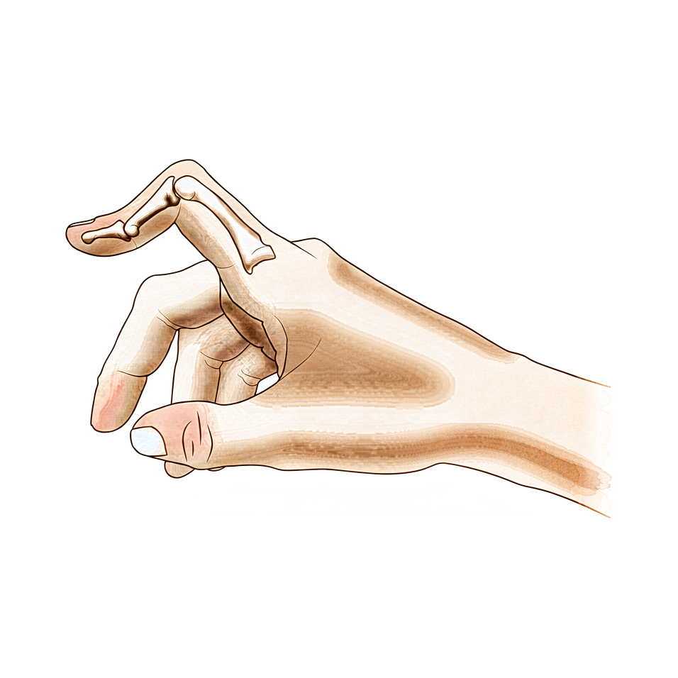
O que você está sentindo
Na deformidade em botoeira, a articulação do meio do dedo se dobra para baixo, em direção à palma da mão, e a ponta do dedo se dobra para trás, no sentido contrário. O dedo pode parecer inchado sobre a articulação do meio, e também pode doer nesse local. Esticar a articulação do meio fica difícil, e quanto mais você tenta, mais o dedo parece resistir.
O problema aparece principalmente em tarefas que exigem que o dedo se estique e depois volte a dobrar. Pegar uma moeda, segurar uma xícara pela alça, digitar, abotoar uma camisa ou girar uma chave podem se tornar desajeitados. A pegada em si muitas vezes continua funcionando, e é por isso que muitas pessoas convivem com o dedo assim por semanas antes de procurar ajuda.
Se a deformidade veio de uma lesão, a articulação do meio pode doer depois que você usa a mão, e a rigidez pode ser pior pela manhã, antes que o dedo se solte. Na artrite reumatoide, a mesma postura dobrada se desenvolve lentamente, à medida que o revestimento da articulação continua inflamado e inchado ao longo de meses.
Alguns sinais exigem atenção rápida. Se o seu dedo ou a sua mão ficar quente, vermelho, inchado e dolorido, principalmente com febre, vá ao pronto-socorro no mesmo dia. O mesmo vale se um dedo ficar pálido, frio, branco ou azulado, ou se você perder de repente a sensibilidade ou o movimento nele após uma lesão. Se, em vez disso, os sintomas não estiverem melhorando, estiverem piorando ao longo de semanas, acordarem você à noite ou impedirem você de trabalhar ou de usar a mão, consulte o seu médico de família ou peça uma avaliação por um especialista.
O que está realmente acontecendo
Ao longo da parte de trás de cada dedo passa um conjunto de tendões que esticam as articulações. Pense neles como cordas que correm sob uma capa na parte de trás do dedo. A corda que estica a articulação do meio se chama tendão central, e ela se prende na base do osso do meio. Outras duas cordas, chamadas bandas laterais, correm ao lado dele e ajudam a esticar tanto a articulação do meio quanto a ponta do dedo.
Na deformidade em botoeira, o tendão central se rompeu ou se esticou e se afastou do osso do meio. A articulação do meio já não consegue ser puxada até ficar reta, por isso cai em uma posição dobrada. As duas bandas laterais então escorregam de cima da articulação e deslizam em direção à palma da mão. Quando ficam abaixo do eixo da articulação, essas mesmas cordas passam a dobrar a articulação do meio em vez de esticá-la, e puxam a ponta do dedo para trás, no sentido contrário. É por isso que o dedo assume a articulação do meio dobrada e a ponta arqueada descritas acima, e por que cada tentativa de esticá-lo parece fazer as cordas puxarem com mais força contra você.
É preciso mais de uma estrutura danificada para produzir essa postura. O tendão central, o ligamento que une as duas cordas sobre a parte de trás do dedo e as fibras da capa que mantêm tudo no lugar precisam estar todos lesionados antes que a deformidade se desenvolva. Uma ruptura do tendão central sozinha pode deixar a articulação do meio fraca sem a deformidade completa, e esse é um dos motivos pelos quais o tratamento precoce é importante: ele pode impedir completamente que a deformidade se forme.
O mesmo resultado final pode vir de pontos de partida diferentes. Uma lesão, como uma pancada no dedo ou um corte sobre a parte de trás da articulação, pode causá-la, assim como a inflamação contínua da artrite reumatoide, que desgasta lentamente as mesmas estruturas. Um dedo também pode ficar com essa aparência sem nenhuma lesão de tendão, quando o problema está no lado da palma da articulação, e essa versão é tratada de forma diferente; por isso, descobrir qual delas você tem vem em primeiro lugar.
O que podemos fazer a respeito
O Dr. Kieran Hirpara, cirurgião de membros superiores no Mater Private Hospital Rockhampton, começa com as opções menos invasivas adequadas ao seu quadro clínico. Em geral, os pacientes são encaminhados à nossa clínica pelo médico de família; caso um fisioterapeuta tenha sugerido que você nos procure, ainda assim será necessário um encaminhamento do seu médico de família para ter direito ao reembolso do Medicare. Na clínica, colhemos o histórico clínico, examinamos a sua mão e solicitamos exames de imagem, se necessário, para entender o que está acontecendo. Como essa deformidade pode vir de vários pontos de partida diferentes, descobrir qual deles é o seu vem antes de qualquer tratamento.
O primeiro passo geralmente é o uso de talas e a terapia da mão. Em uma lesão recente, manter a articulação do meio reta em uma tala pode impedir completamente que a deformidade se forme, e quanto antes começar, melhor: as pessoas tratadas em até seis semanas após a lesão têm resultados melhores do que as tratadas mais tarde. A tala mantém reta apenas a articulação do meio. A ponta do dedo, o nó do dedo e os outros dedos continuam se movendo, porque manter o dedo inteiro imóvel faz a ponta do dedo enrijecer. Para uma deformidade que já existe há algum tempo, podemos usar gessos para endireitar a articulação do meio até onde for possível, seguidos de três meses de uma tala que permite dobrar o dedo enquanto ele trabalha. Vale a pena tentar essa abordagem antes de considerar a cirurgia.
Quando as talas e a terapia não trouxeram melhora suficiente, a cirurgia pode ser considerada. A operação reconstrói o tendão que estica a articulação do meio, às vezes usando um pequeno pedaço de tendão do seu próprio antebraço como enxerto. Em alguns casos, os tendões que escorregaram em direção à palma da mão são reposicionados de volta sobre a articulação, onde deveriam estar. O objetivo é o mesmo em todos os casos: permitir que você volte a esticar a articulação do meio e use o dedo sem que ele trave ou se dobre para baixo. A operação tem a sua própria página, e conversaremos sobre o que ela envolve para o seu dedo em particular antes de qualquer decisão.
O que esperar
O prognóstico depende muito do momento do tratamento. Identificada cedo, a deformidade em botoeira muitas vezes pode ser interrompida antes de se estabelecer. Manter a articulação do meio reta em uma tala logo após uma lesão pode impedir completamente que a deformidade se forme, e as pessoas tratadas em até seis semanas têm resultados melhores do que as tratadas mais tarde. Sem tratamento, a deformidade tende a piorar, em vez de permanecer igual, porque os tendões que saíram do lugar continuam puxando o dedo cada vez mais para a postura dobrada. Uma lesão que parecia leve pode resultar em um dedo rígido e fixo semanas depois.
Para uma deformidade que já existe há algum tempo, o tratamento não cirúrgico ainda tem um papel real. Gessos para endireitar a articulação do meio até onde for possível, seguidos de três meses de uma tala que permite dobrar o dedo enquanto você o usa, podem funcionar bem. O dedo responde a essa abordagem quando os gessos conseguem deixar a articulação do meio quase reta e você usa ativamente a mão com a tala durante esses três meses, com a flexão se recuperando ao longo do caminho. Nas lesões do polegar, manter o nó do dedo reto em uma tala pode ajudar se isso começar antes que a deformidade se desenvolva.
A cirurgia tem como objetivo permitir que você volte a esticar a articulação do meio. Em um grupo relatado de 18 pessoas que fizeram um enxerto de tendão para corrigir uma deformidade antiga, 16 tiveram resultados bons ou excelentes. Uma técnica mais recente, que permite movimentar o dedo cedo, foi relatada como eficaz em 11 casos. A cirurgia também pode ajudar na forma congênita dessa deformidade, quando ela está presente desde o nascimento.
É honesto dizer que nem toda correção dura. Em dedos afetados pela artrite reumatoide, a cirurgia apenas nos tecidos moles pode não se manter nas fases mais avançadas, e os resultados a longo prazo são menos previsíveis. Quando uma deformidade volta ou persiste, um procedimento diferente pode ser a melhor resposta. Qualquer que seja o ponto de partida, o objetivo realista é um dedo que você consiga esticar e usar, e não a promessa de um dedo perfeitamente normal.
Quando procurar ajuda médica
A deformidade em botoeira responde bem à atenção precoce, por isso vale a pena saber quando agir. Se você machucou o dedo e a articulação do meio não se estica, ou se o dedo fica dobrado e inchado dias depois de uma lesão, consulte o seu médico de família e peça uma avaliação por um especialista. Quanto antes o uso da tala começar, maior a chance de impedir que a deformidade se estabeleça.
Alguns sinais exigem atendimento mais rápido. Vá a um pronto-socorro no mesmo dia se o seu dedo ou a sua mão ficar quente, vermelho, inchado e dolorido, principalmente com febre. O mesmo vale se um dedo ficar pálido, frio, branco ou azulado, ou se você perder de repente a sensibilidade ou o movimento nele após uma lesão. Se houver pele rompida sobre uma articulação lesionada, ou se o dedo parecer claramente deformado, vá também.
Se, em vez disso, os sintomas não estiverem melhorando, estiverem piorando ao longo de semanas, acordarem você à noite ou impedirem você de trabalhar ou de usar a mão, consulte o seu médico de família ou peça uma avaliação por um especialista. Se não conseguir contato com a clínica fora do horário de atendimento ou no fim de semana, vá ao pronto-socorro mais próximo.
Em maior profundidade
Advanced reading: the deeper science (optional)
Esta seção aborda tópicos além do necessário para as suas próprias decisões de tratamento. A deformidade em botoeira merece uma leitura mais aprofundada, pois o tratamento instintivo para um dedo dolorido, inchado e curvado — deixá-lo em repouso e mantê-lo imóvel — é justamente aquilo que as evidências especificamente não apoiam.
Mobilização precoce, não imobilização prolongada
Uma revisão sistemática sobre os tratamentos para lesões do tendão extensor central constatou que a base de evidências é limitada; os efeitos das diversas estratégias terapêuticas tanto para lesões abertas quanto fechadas não estão bem comprovados [1]. Mesmo dentro dessa limitação, as evidências provenientes de estudos individuais apoiam, de forma preliminar, a mobilização precoce e não a imobilização prolongada [1].
Isso merece ser afirmado claramente, pois vai contra o senso comum. A distinção reside em proteger o tendão extensor central em processo de cicatrização – o que exige manter a articulação média do dedo esticada – e na imobilização de todo o dedo, que pode levar ao enrijecimento da articulação distal e à rigidez do membro inteiro. A tala tem como objetivo manter uma articulação imóvel enquanto as demais continuam a se mover.
Por que a deformidade piora em vez de permanecer estável
A deformidade em botoeira é progressiva, o que é incomum e merece ser compreendido. O tendão central liga-se à base do osso médio, sendo responsável por esticar a articulação média. Quando esse tendão falha, a articulação cai em flexão, e as duas bandas laterais, que normalmente ficam acima do eixo da articulação, deslocam-se para abaixo dele.
Uma vez abaixo do eixo, os mesmos tendões que antes ajudavam a esticar a articulação média passam a dobrá-la, ao mesmo tempo em que puxam a ponta do dedo para uma posição de hiperextensão. Assim, a deformidade se auto-reforça: toda tentativa de endireitar o dedo acaba tensionando ainda mais as estruturas que o mantêm dobrado. É por isso que uma lesão aparentemente leve pode resultar em uma deformidade fixa semanas depois, e por que o período adequado para o uso de talas simples é bem inicial.
A deformidade que não é botoeira
Um dedo pode assumir a mesma postura mesmo na ausência de qualquer lesão no tendão extensor central; nesse caso, os tratamentos são completamente diferentes. As deformidades em botoeira e em pseudobotoeira diferem quanto à patoanatomia, diagnóstico e manejo [2]. A forma pseudobotoeira surge de uma lesão na placa volar da articulação média; nesse caso, a articulação distal do dedo mantém a flexão passiva normal, em vez de ficar em hiperextensão.
A distinção entre elas no exame clínico baseia-se na ponta do dedo: na verdadeira botoeira, a ponta permanece hiperextendida e resiste à flexão, especialmente quando a articulação média está esticada; na pseudobotoeira, ela flexiona livremente. Imobilizar uma pseudobotoeira da mesma forma que se faria com a verdadeira lesão acaba atingindo a articulação errada.
Trata-se de um tipo de lesão definida pelo local da ruptura do tendão
A lesão em botoeira está classificada, juntamente com a lesão do dedo em “martelo” e a lesão da banda sagital, como uma das três lesões do mecanismo extensor fechado, categorizadas conforme a zona em que ocorre a ruptura [3]. Todas as três podem se manifestar como um dedo inchado e mantido numa posição anormal poucos dias após a lesão; cada uma exige uma posição específica da tala, razão pela qual é importante obter um diagnóstico preciso antes de se comprometer com semanas de imobilização.
Referências
[1] Geoghegan L, Wormald JCR, Adami RZ, Rodrigues JN. Lesões do tendão extensor da falange central: uma revisão sistemática dos tratamentos. J Hand Surg Eur Vol. 2019;44(8):825-32. https://doi.org/10.1177/1753193419845311
[2] Hanson ZC, Thompson RG, Andrews JR, Lourie GM. Deformidades em botoeira versus pseudobotoeira: patoanatomia, diagnóstico e tratamento. J Hand Surg Am. 2023;48(5):489-97. https://doi.org/10.1016/j.jhsa.2022.10.019
[3] Lin JD, Strauch RJ. Lesões fechadas do mecanismo extensor de partes moles (dedo em martelo, deformidade em botoeira e lesão da banda sagital). J Hand Surg Am. 2014;39(5):1005-11. https://doi.org/10.1016/j.jhsa.2013.11.018
Evidence & references
This is the clinical evidence summary written for health professionals. It is technical, and it lists the research this page was built from. You do not need to read it to understand your treatment or to make a decision about it.
Overview
- A staged technique for the repair of traumatic boutonniere deformity allows the surgeon to divide the deformity into various categories and use the operative procedure indicated for each particular problem, often avoiding unnecessary surgery [1].
- Differentiating a true boutonniere deformity from a pseudoboutonniere injury is critical in determining clinical management [2].
- A retrospective study of fifty-four longstanding post-burn boutonnière deformities was carried out to evaluate the problems associated with the surgical procedures [3].
- An understanding of the anatomy, clinical presentation, treatment options, and expected outcomes is crucial for optimal treatment of posttraumatic boutonnière and swan neck deformities [4].
- Early treatment of central slip injuries can prevent the deformity [5].
- Transection of the terminal extensor tendon is a useful procedure for salvage of the boutonniere deformity [6].
- No boutonniere deformities were produced in a series of 20 patients who had chronic mallet finger deformity treated with central slip tenotomy [7].
- Reconstruction of the central slip by the transverse retinacular ligament is not indicated for boutonnière deformities with severe contracture of the lateral band [8].
- The best indication for reconstruction of the central slip by the transverse retinacular ligament is in cases in which the central slip is defective and conservative treatment is not effective [8].
- Similar results occurred for chronic boutonniere deformity using serial casting for adequate extension followed by 3 months of relative motion flexion (RMF) orthotic use, which should be attempted prior to surgical intervention [9].
- A new surgical technique with early mobilization is an effective and simplified treatment of Boutonniere deformity [10].
- The natural history of the boutonnière deformity in rheumatoid arthritis is outlined, and a simple method of repair is described [11].
- Chronic boutonniere deformity will respond to relative motion flexion splinting if serial casting can place the proximal interphalangeal joint in less than -20° extension, and the patient actively uses the hand in a relative motion flexion orthosis for 3 months, recovering flexion [12].
- The Y-shaped tendon graft can be a useful procedure for the correction of chronic boutonniere deformity [13].
- In a patient series of 18 patients, the Y-shaped tendon graft provided good or excellent results in 16 patients [13].
- Extension splinting of the metacarpophalangeal (MP) joint may be effective if traumatic thumb injuries are treated before development of a boutonnière deformity [14].
- Congenital boutonniere deformity is understood as various embryonic developmental failures [15].
- A reliable surgical option for congenital boutonniere deformity involves central slip reconstruction [15].
- Extensor tenotomy to correct a boutonnière deformity of long standing markedly lessened disability with restoration of flexion of the distal joint [16].
- In chronic boutonniere deformity, when the lateral bands are deficient or damaged, the cross-lateral band reconstruction technique using palmaris longus autograft is a treatment option with satisfactory results [17].
- Extension splinting may be effective if traumatic thumb injuries are treated before deformity development [18].
- Surgical treatment with advancement of the extensor pollicis brevis tendon and imbrication of the dorsoradial capsule is necessary once a traumatic thumb boutonnière deformity is established [18].
- The prevalence of boutonnière deformity without rheumatoid arthritis or trauma is approximately 13% [19].
- Current concepts relating to the presentation and treatment of closed soft tissue injuries of the extensor mechanism in zones I, III, and V, commonly known as mallet finger, boutonniere deformities, and sagittal band injuries, are summarized [20].
- Long-term results following soft tissue reconstruction for boutonniere finger deformity in rheumatoid arthritis are unreliable [24].
- Recurrent or persistent deformity in rheumatoid arthritis is best treated with a salvage procedure [24].
Anatomy & Pathophysiology
Extensor Mechanism Anatomy
- The extensor mechanism of the finger at the level of the proximal interphalangeal (PIP) joint consists of both intrinsic and extrinsic contributions [28].
- The extensor tendon entering the digit is the continuation of the extensor digitorum communis (EDC), with contributions from the extensor indicis proprius in the index finger and the extensor digiti quinti in the small finger [28].
- In 30% of cases, the EDC has a separate insertion into the base of the proximal phalanx, but extension of the metacarpophalangeal (MP) joint typically occurs through the pull of the sagittal bands [28].
- The principal bony insertion of the extrinsic digital extensors is on the dorsal proximal aspect of the middle phalanx [32].
- Metacarpophalangeal joint extension is provided by extrinsic extensor force transmitted through the sagittal bands [32].
- Distal interphalangeal joint extension is achieved through conjoined lateral bands composed of tendinous slips from both extrinsic and intrinsic tendons [32].
- The deep head of each dorsal interosseous muscle forms a lateral tendon, or lateral band, at the level of the MP joint [41].
- At the level of the middle of the proximal phalanx, transverse fibers arch dorsally from each lateral band to join over the dorsum of the finger, flexing the proximal phalanx [41].
- Oblique fibers (spiral fibers) from the lateral bands sweep over the distal third of the proximal phalanx to insert onto the lateral tubercles at the base of the middle phalanx, extending the middle phalanx [41].
- The lateral bands are joined by the lateral slips of the extensor tendon to form the conjoined lateral band [41].
- The two conjoined lateral bands to each finger unite at the distal third of the middle phalanx to form the terminal tendon, which inserts at the base of the distal phalanx to extend it [41].
- The volar interossei form the ulnar lateral band of the index finger and the radial lateral band of the ring and little fingers [41].
- The flexor digiti quinti brevis is structurally and functionally similar to the deep head of the dorsal interossei, forming the ulnar lateral band of the little finger [41].
Pathophysiology & Biomechanics
- A boutonniere deformity requires subluxation of the lateral bands volar to the axis of rotation of the PIP joint [22].
- Damage to the central slip alone does not cause a boutonniere deformity [22].
- A boutonniere deformity occurs only when the central slip, triangular ligament, and transverse and oblique fibers of the interosseous hood are all damaged [22].
- Detachment of the central slip from the middle phalanx produces a decrease in extension of the PIP joint [22].
- When the transverse and oblique fibers of the interosseous hood are divided in addition to the central slip, extension at the PIP joint is further decreased [22].
- Early treatment of central slip injuries can prevent the development of the deformity [5].
- The extensor mechanism is a complex structure that demands a delicate balance from intrinsic and extrinsic tendons of the hand [28].
- Injury to the extensor mechanism can disrupt this delicate balance, leading to significant sequelae [28].
- Understanding the anatomy, clinical presentation, treatment options, and expected outcomes is crucial for optimal treatment of posttraumatic boutonnière and swan neck deformities [4].
Classification
- A staged management plan allows the surgeon to divide traumatic boutonniere deformity into various categories and use the operative procedure indicated for each particular problem [1].
- The choice of surgical procedure for rheumatoid boutonniere deformity is dependent on accurate staging of the deformity, which is based on the flexibility of the proximal interphalangeal joint and the state of the articular cartilage [23].
- In a cadaveric model, damage to the central slip alone does not cause a boutonniere deformity [22].
- The boutonniere deformity requires subluxation of the lateral bands volar to the axis of rotation of the PIP joint [22].
- In a cadaveric model, the central slip, triangular ligament, and oblique fibers need to all be damaged to cause subluxation of the lateral bands which leads to an acute boutonniere deformity [29].
- Anterior dislocation of the proximal interphalangeal joint is associated with ruptures of the central slip of the extensor mechanism and results in boutonnière deformity unless repaired [27].
- The incidence of uncorrectable swan-neck and boutonniere deformities is estimated to be between 8% and 16% during the first 2 years after the onset of systemic disease in rheumatoid arthritis [23].
- The prevalence of boutonniere deformities in patients with established rheumatoid arthritis is approximately 36% [23].
Clinical Presentation
Diagnostic Differentiation and Classification
- The surgeon can divide the boutonniere deformity into various categories to use the operative procedure indicated for each particular problem, often avoiding unnecessary surgery [1].
Etiology and Pathomechanics
- The cause of boutonniere deformity in rheumatoid arthritis is chronic synovitis of the proximal interphalangeal joint [23].
- Combined injury of the central slip, triangular ligament, and transverse and oblique fibers of the interosseous hood causes a boutonniere deformity [22].
Prevalence and Natural History
- A swan-neck or boutonniere deformity occurs in approximately half of patients with rheumatoid arthritis [23].
- The incidence of uncorrectable swan-neck and boutonniere deformities is estimated to be between 8% and 16% during the first 2 years after the onset of systemic disease [23].
- The prevalence of finger deformities in patients with established rheumatoid arthritis is approximately 36% for boutonniere deformities [23].
- The natural history of the boutonnière deformity in rheumatoid arthritis is outlined in the literature [11].
Clinical Assessment and Testing
- The central slip tenodesis test detects disruption of the central slip before the boutonniere deformity becomes established [34].
- The central slip tenodesis test is applicable in uncooperative children and unconscious patients [34].
- Treating swan neck and boutonniere deformities of the PIP joint requires understanding the cause, biomechanical changes, and articular status [26].
Investigations
Clinical Examination and Diagnostic Tests
- A careful physical examination is essential to direct care and future testing if indicated [31].
- Diagnostic tests such as imaging and serum laboratory studies are useful in the determination of pathology but can be expensive, time consuming, and often nonspecific [31].
Biomechanical and Pathomechanical Assessment
Imaging and Advanced Modalities
- A computational 3D model validation reassures the reliability of research in understanding the pathomechanics of boutonniere deformity [21].
Treatment
Non-Operative
- Early treatment of central slip injuries can prevent the development of boutonniere deformity [5].
- Extension splinting of the metacarpophalangeal joint may be effective for traumatic thumb boutonniere deformity if treated before the deformity develops [14].
- Extension splinting may be effective for traumatic thumb boutonniere deformity if treated before deformity development [18].
- Chronic boutonniere deformity responds to relative motion flexion splinting if serial casting places the proximal interphalangeal joint in less than -20° extension and the patient actively uses a relative motion flexion orthosis for 3 months [12].
- Serial casting for adequate extension followed by 3 months of relative motion flexion orthotic use should be attempted prior to surgical intervention for chronic boutonniere deformity [9].
- The use of relative motion flexion orthoses is effective in increasing active distal interphalangeal joint flexion and improving proximal interphalangeal joint extension in patients with Burton stage 1 chronic boutonniere deformity [30].
- Conservative treatment of closed central slip injuries involves immobilization of the proximal interphalangeal, distal interphalangeal, and metacarpophalangeal joints in full extension for four weeks followed by a Capener spring splint for two further weeks [92].
- In a review of 115 central slip injuries, 17 of 24 patients treated conservatively achieved an outcome of less than a 20° extension deficit at the proximal interphalangeal joint and more than 80% return of proximal interphalangeal joint flexion [92].
- Patients treated for central slip injuries within six weeks of injury had better outcomes than those treated later [92].
Operative
- A staged technique allows the surgeon to divide traumatic boutonniere deformity into various categories and use the operative procedure indicated for each particular problem, often avoiding unnecessary surgery [1].
- Reconstruction of the central slip by the transverse retinacular ligament is best indicated in cases in which the central slip is defective and conservative treatment is not effective [8].
- A simple method of repair for the rheumatoid boutonnière deformity is described in the literature [11].
- In a patient series, the Y-shaped tendon graft provided good or excellent results in 16 of 18 patients with chronic boutonniere deformity [13].
- Surgical treatment with advancement of the extensor pollicis brevis tendon and imbrication of the dorsoradial capsule is necessary once a traumatic boutonnière deformity of the thumb is established [18].
- The cross-lateral band reconstruction technique using palmaris longus autograft is a treatment option with satisfactory results for chronic boutonniere deformity when the lateral bands are deficient or damaged [17].
- A reliable surgical option involving central slip reconstruction is suggested for the congenital form of boutonniere deformity [15].
- A simple staged procedure using a central slip facilitation device is a valid alternative in the management of severe Dupuytren's proximal interphalangeal joint contracture, demonstrating reliable, reproducible correction of the deformity and acceptable patient outcomes [38].
Complications
- In a patient series of 18 patients, the Y-shaped tendon graft provided good or excellent results in 16 of 18 patients for the correction of chronic boutonniere deformity [13].
- Extension splinting of the MP joint may be effective if traumatic thumb injuries are treated before development of a boutonnière deformity [14].
- The case extends understanding of congenital boutonniere deformity as various embryonic developmental failures and suggests a reliable surgical option involving central slip reconstruction [15].
- Surgical treatment with advancement of the extensor pollicis brevis tendon and imbrication of the dorsoradial capsule is necessary once a boutonnière deformity is established in the thumb [18].
- This article summarizes current concepts relating to the presentation and treatment of closed soft tissue injuries of the extensor mechanism in zones I, III, and V, commonly known as mallet finger, boutonniere deformities, and sagittal band injuries [20].
- This validation reassures the reliability of the research in understanding the pathomechanics of boutonniere deformity [21].
- In a cadaver model, detachment of the central slip from the middle phalanx produced a decrease in extension of the PIP joint [22].
- When the transverse and oblique fibers of the interosseous hood were also divided after central slip detachment, extension at the PIP joint was further decreased [22].
- A boutonniere deformity occurred only when all 3 structures (central slip, transverse and oblique fibers of the interosseous hood, and triangular ligament) were damaged [22].
- Division of the central slip leads to loss of extension at the PIP joint [22].
- The prevalence of finger deformities in patients with established rheumatoid arthritis is approximately 14% for swan-neck deformities and 36% for boutonniere deformities [23].
- In the later stages of both swan-neck and boutonniere deformities, soft-tissue procedures alone may not result in lasting operative correction [23].
- Long-term results following soft tissue reconstruction are unreliable [24].
- Recurrent or persistent deformity is best treated with a salvage procedure [24].
- Treating swan neck and boutonniere deformities of the PIP joint is a difficult challenge that requires understanding the cause, biomechanical changes, and articular status [26].
- Anterior dislocation of the proximal interphalangeal joint is associated with ruptures of the central slip of the extensor mechanism [27].
- Anterior dislocation of the proximal interphalangeal joint results in boutonnière deformity unless repaired [27].
Recovery
Non-Operative Management
- Serial casting for adequate extension followed by 3 months of relative motion flexion (RMF) orthotic use should be attempted prior to surgical intervention for chronic boutonniere deformity [9].
- Chronic boutonniere deformity responds to relative motion flexion splinting if serial casting places the proximal interphalangeal joint in less than -20° extension and the patient actively uses the hand in a relative motion flexion orthosis for 3 months, recovering flexion [12].
- Extension splinting of the metacarpophalangeal joint may be effective for traumatic thumb boutonniere injuries if treated before the development of a boutonnière deformity [14].
Operative Management
- A staged technique allows the surgeon to divide the traumatic boutonniere deformity into various categories and use the operative procedure indicated for each particular problem, often avoiding unnecessary surgery [1].
- The Y-shaped tendon graft provided good or excellent results in 16 of 18 patients for the correction of chronic boutonniere deformity [13].
- Reconstruction of the central slip by the transverse retinacular ligament is indicated in cases in which the central slip is defective and conservative treatment is not effective, but is not indicated for boutonnière deformities with severe contracture of the lateral band [8].
- A simple method of repair for the correction of the rheumatoid boutonnière deformity has been described [11].
- A new surgical technique with early mobilization for chronic boutonniere deformity was reported to be an effective and simplified treatment in 11 cases [10].
- Central slip reconstruction is a reliable surgical option for the congenital form of boutonniere deformity [15].
Outcomes and Prognosis
- Early treatment of central slip injuries can prevent the boutonniere deformity [5].
- Long-term results following soft tissue reconstruction for boutonniere finger deformity in rheumatoid arthritis are unreliable, and recurrent or persistent deformity is best treated with a salvage procedure [24].
- In the later stages of rheumatoid boutonniere deformities, soft-tissue procedures alone may not result in lasting operative correction [23].
- A patient with boutonniere deformity of the second toe after planter dislocation of the proximal interphalangeal joint reported neither deformity nor difficulty when last seen 22 months after the operation [95].
Key Evidence
- [L4] With this plan of management, the surgeon is able to divide the boutonniere deformity into various categories and then use the operative procedure indicated for each particular problem, often avoiding unnecessary surgery. [1] (10.1016/s0363-5023(83)80009-4)
- [L5] Differentiating a true boutonniere deformity from a pseudoboutonniere injury is critical in determining clinical management. [2] (10.1016/j.jhsa.2022.10.019)
- [L4] A retrospective study of fifty-four longstanding post-burn boutonnière deformities was carried out to evaluate the problems associated with the surgical procedures. [3] (10.1016/0266-7681(86)90006-9)
- [L5] An understanding of the anatomy, clinical presentation, treatment options, and expected outcomes is crucial for optimal treatment of posttraumatic boutonnière and swan neck deformities. [4] (10.5435/jaaos-d-14-00272)
- [L5] The paper outlines the anatomy, pathoanatomy, and treatment concepts for boutonniere deformity, emphasizing that early treatment of central slip injuries can prevent the deformity. [5] (10.1016/s0749-0712(21)00060-3)
- [L4] Transection of the terminal extensor tendon is a useful procedure for salvage of the boutonniere deformity. [6] (10.1016/s0749-0712(21)00065-2)
- [L4] No boutonniere deformities were produced in this series of 20 patients who had chronic mallet finger deformity. [7] (10.1016/s0363-5023(87)80205-8)
- [L4] This procedure is not indicated for boutonnière deformities with severe contracture of the lateral band; its best indication is in cases in which the central slip is defective and conservative treatment is not effective. [8] (10.1016/0266-7681(90)90080-n)
- [L4] Similar results occurred for chronic boutonniere deformity using serial casting for adequate extension followed by 3 months of RMF orthotic use, which should be attempted prior to surgical intervention. [9] (10.1016/j.jht.2023.02.005)
- [L4] Our technique is an effective and simplified treatment of Boutonniere deformity. [10] (10.1097/bth.0000000000000152)
- [L4] The natural history of the boutonnière deformity in rheumatoid arthritis is outlined, and a simple method of repair is described. [11] (10.2106/00004623-196951070-00009)
- [L4] Chronic boutonniere deformity will respond to relative motion flexion splinting if serial casting can place the proximal interphalangeal joint in less than -20° extension, and the patient actively uses the hand in a relative motion flexion orthosis for 3 months, recovering flexion. [12] (10.1097/sap.0000000000002307)
- [L4] The Y-shaped tendon graft can be a useful procedure for the correction of chronic boutonniere deformity; in our patient series, this provided good or excellent results in 16 of 18 patients. [13] (10.1016/j.jhsa.2021.01.003)
- [L4] Extension splinting of the MP joint may be effective if these injuries are treated before development of a boutonnière deformity. [14] (10.1016/s0266-7681(00)80024-8)
- [Case_report] The case extends understanding of congenital boutonniere deformity as various embryonic developmental failures and suggests a reliable surgical option involving central slip reconstruction. [15] (10.1016/j.jhsa.2014.05.030)
- [L4] In chronic boutonniere deformity, when the lateral bands are deficient or damaged, the cross-lateral band reconstruction technique using palmaris longus autograft is a treatment option with satisfactory results. [17] (10.1016/j.jhsa.2017.04.010)
- [L4] Extension splinting may be effective if treated before deformity development, but surgical treatment with advancement of the extensor pollicis brevis tendon and imbrication of the dorsoradial capsule is necessary once a boutonnière deformity is established. [18] (10.1054/jhsb.1999.0303)
- [L3] The prevalence of boutonnière deformity without rheumatoid arthritis or trauma is approximately 13%. [19] (10.1177/1753193417704610)
- [L5] This article summarizes current concepts relating to the presentation and treatment of closed soft tissue injuries of the extensor mechanism in zones I, III, and V, commonly known as mallet finger, boutonniere deformities, and sagittal band injuries. [20] (10.1016/j.jhsa.2013.11.018)
- [L5] This validation reassures the reliability of the research in understanding the pathomechanics of boutonniere deformity. [21] (10.1186/s13018-025-06329-3)
- [L5] [22] (10.1016/j.jhsa.2017.07.011)
- [L5] [23] (10.5435/00124635-199903000-00002)
- [L5] Long-term results following soft tissue reconstruction are unreliable, and recurrent or persistent deformity is best treated with a salvage procedure. [24] (10.1016/j.jhsa.2011.05.029)
- [L4] Treating swan neck and boutonniere deformities of the PIP joint is a difficult challenge that requires understanding the cause, biomechanical changes, and articular status. [26] (10.1016/j.hcl.2017.12.006)
- [Paper] [28] (10.1016/j.hcl.2012.05.044)
- [Paper] In a cadaveric model, the central slip, triangular ligament, and oblique fibers need to all be damaged to cause subluxation of the lateral bands which leads to an acute boutonniere deformity. [29] (10.1016/s0363-5023(12)60014-8)
- [L4] The use of RMFO is effective in increasing active distal interphalangeal joint flexion and improving PIP extension in patients with Burton stage 1 chronic boutonniere deformity. [30] (10.1016/j.jhsa.2022.08.007)
- [Paper] The test detects disruption of the central slip before the boutonniere deformity becomes established and is applicable in uncooperative children and unconscious patients. [34] (10.1016/0266-7681(94)90057-4)
- [L4] The simple staged procedure is a valid alternative in the management of severe Dupuytren's PIPJ contracture, demonstrating reliable, reproducible correction of the deformity and acceptable patient outcomes. [38] (10.1177/1753193412439673)
- [L4] [92] (10.1177/1758998318822663)
- [Case_report] The patient reported neither deformity nor difficulty when last seen 22 months after the operation. [95] (10.1007/s00402-009-0816-x)
References
[1] A staged technique for the repair of the traumatic boutonniere deformity. The Journal of Hand Surgery. 1983. DOI: 10.1016/s0363-5023(83)80009-4
[2] Boutonniere Versus Pseudoboutonniere Deformities: Pathoanatomy, Diagnosis, and Treatment. The Journal of Hand Surgery. 2023. DOI: 10.1016/j.jhsa.2022.10.019
[3] Reconstructive surgery of the post-burn boutonnière deformity. The Journal of Hand Surgery: Journal of the British Society for Surgery of the Hand. 1986. DOI: 10.1016/0266-7681(86)90006-9
[4] Posttraumatic Boutonnière and Swan Neck Deformities. Journal of the American Academy of Orthopaedic Surgeons. 2015. DOI: 10.5435/jaaos-d-14-00272
[5] BOUTONNIERE DEFORMITY. Hand Clinics. 1995. DOI: 10.1016/s0749-0712(21)00060-3
[6] TREATMENT OF THE CHRONIC BOUTONNIERE DEFORMITY BY EXTENSOR TENOTOMY. Hand Clinics. 1995. DOI: 10.1016/s0749-0712(21)00065-2
[7] Central slip tenotomy for chronic mallet finger deformity. The Journal of Hand Surgery. 1987. DOI: 10.1016/s0363-5023(87)80205-8
[8] Reconstruction of the Central Slip by the Transverse Retinacular Ligament for Boutonnière Deformity. Journal of Hand Surgery. 1990. DOI: 10.1016/0266-7681(90)90080-n
[9] The relative motion concept in acute and chronic boutonniere deformity: Invited commentary. Journal of Hand Therapy. 2023. DOI: 10.1016/j.jht.2023.02.005
[10] Chronic Boutonniere Deformities, Supple, or Stiff: A New Surgical Technique With Early Mobilization in 11 Cases. Techniques in Hand & Upper Extremity Surgery. 2017. DOI: 10.1097/bth.0000000000000152
[11] Correction of the Rheumatoid Boutonnière Deformity. The Journal of Bone & Joint Surgery. 1969. DOI: 10.2106/00004623-196951070-00009
[12] A Paradigm Shift in Managing Acute and Chronic Boutonniere Deformity. Annals of Plastic Surgery. 2020. DOI: 10.1097/sap.0000000000002307
[13] Y-Shaped Tendon Graft—A Technique in the Reconstruction of Posttraumatic Chronic Boutonniere Deformity. The Journal of Hand Surgery. 2021. DOI: 10.1016/j.jhsa.2021.01.003
[14] Traumatic Boutonnière Deformity of the Thumb. Journal of Hand Surgery. 2000. DOI: 10.1016/s0266-7681(00)80024-8
[15] Restoration of the Central Slip in Congenital Form of Boutonniere Deformity: Case Report. The Journal of Hand Surgery. 2014. DOI: 10.1016/j.jhsa.2014.05.030
[16] Extensor Tenotomy for Chronic Boutonniere Deformity of the Finger: REPORT OF TWO CASES.. The Journal of Bone and Joint Surgery. American Volume. 1965.
[17] Chronic Boutonniere Deformity: Cross-Lateral Band Technique Using Palmaris Longus Autograft. The Journal of Hand Surgery. 2017. DOI: 10.1016/j.jhsa.2017.04.010
[18] TRAUMATIC BOUTONNIÈRE DEFORMITY OF THE THUMB. The Journal of Hand Surgery: Journal of the British Society for Surgery of the Hand. 2000. DOI: 10.1054/jhsb.1999.0303
[19] Thumb boutonnière deformity without rheumatoid arthritis or trauma. Journal of Hand Surgery (European Volume). 2017. DOI: 10.1177/1753193417704610
[20] Closed Soft Tissue Extensor Mechanism Injuries (Mallet, Boutonniere, and Sagittal Band). The Journal of Hand Surgery. 2014. DOI: 10.1016/j.jhsa.2013.11.018
[21] Assessment of the ligamentous stress distribution in the pathomechanics of the boutonniere deformity through a computational 3D model. Journal of Orthopaedic Surgery and Research. 2025. DOI: 10.1186/s13018-025-06329-3
[22] Biomechanics of the Acute Boutonniere Deformity. The Journal of Hand Surgery. 2018. DOI: 10.1016/j.jhsa.2017.07.011
[23] Operative Correction of Swan-Neck and Boutonniere Deformities in the Rheumatoid Hand. Journal of the American Academy of Orthopaedic Surgeons. 1999. DOI: 10.5435/00124635-199903000-00002
[24] Treatment of Boutonniere Finger Deformity in Rheumatoid Arthritis. The Journal of Hand Surgery. 2011. DOI: 10.1016/j.jhsa.2011.05.029
[26] Treating the Proximal Interphalangeal Joint in Swan Neck and Boutonniere Deformities. Hand Clinics. 2018. DOI: 10.1016/j.hcl.2017.12.006
[27] Anterior dislocation of the proximal interphalangeal joint. A cause of rupture of the central slip of the extensor mechanism.. The Journal of bone and joint surgery. American volume. 1970.
[28] Boutonnière and Pulley Rupture in Elite Athletes. Hand Clinics. 2012. DOI: 10.1016/j.hcl.2012.05.044
[29] Biomechanics of the Acute Boutonniere Deformity. The Journal of Hand Surgery. 2012. DOI: 10.1016/s0363-5023(12)60014-8
[30] The Use of Relative Motion Flexion Orthoses for Chronic Boutonniere Deformity. The Journal of Hand Surgery. 2024. DOI: 10.1016/j.jhsa.2022.08.007
[31] Orthopaedic Knowledge Update 13 Ebook Without Multimedia. Anatomy, Evaluation, Clinical Examination, and Imaging > Evaluation and Clinical Examination: Current Concepts.
[32] A Lange Medical Book Current Diagnosis Treatment In Orthopedics Fifth Edition. 9Hand Surgery > Image DISORDERS OF THE MUSCULATURE OF THE HAND.
[34] The Central Slip Tenodesis Test for Early Diagnosis of Potential Boutonnière Deformities. Journal of Hand Surgery. 1994. DOI: 10.1016/0266-7681(94)90057-4
[38] Management of severe Dupuytren’s contracture of the proximal interphalangeal joint with use of a central slip facilitation device. Journal of Hand Surgery (European Volume). 2012. DOI: 10.1177/1753193412439673
[41] Green S Operative Hand Surgery. Interosseous and Hypothenar Muscles.
[92] Therapeutic management of closed central slip injuries: Outcome of a service evaluation. Hand Therapy. 2019. DOI: 10.1177/1758998318822663
[95] Boutonniere deformity of the second toe after planter dislocation of proximal interphalangeal joint: a case report. Archives of Orthopaedic and Trauma Surgery. 2009. DOI: 10.1007/s00402-009-0816-x