Patients › Elbow
Ruptura do bíceps distal
Distal biceps rupture causes sudden elbow pain, bruising, and weakness—often needing surgical repair.
O que você está sentindo
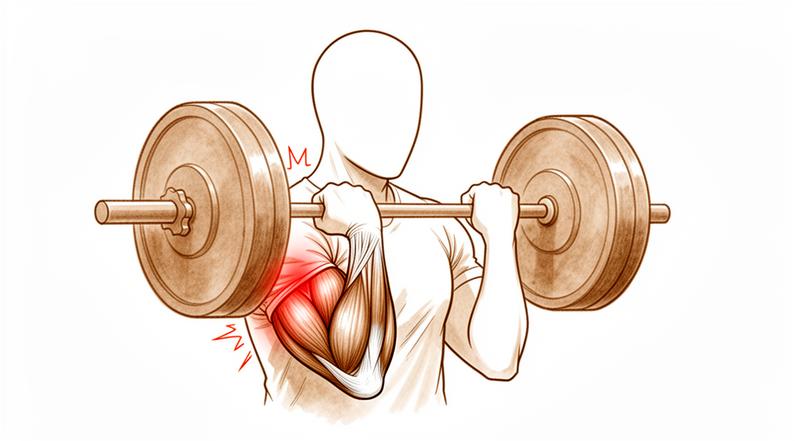
A ruptura do bíceps distal acontece num único instante, e não ao longo de meses. Geralmente resulta de uma força súbita e inesperada que estica um braço dobrado a 90 graus, como ao segurar algo pesado que está caindo ou ao segurar algo com força enquanto o braço é puxado e esticado. Muitas pessoas sentem ou ouvem um estalo na parte da frente do cotovelo no momento da lesão. Normalmente, o tendão se desprende do seu ponto de fixação em um dos ossos do antebraço. Você pode notar que o formato do músculo muda, porque ele se retrai para cima no braço e se acumula perto do meio do braço.
Logo de início, a parte da frente do cotovelo tende a ficar dolorida e inchada. O hematoma é comum e pode aparecer ao redor do cotovelo e do antebraço ao longo dos próximos um ou dois dias. Dobrar e girar o braço dói, por isso você pode perceber que está evitando levantar a chaleira, girar uma maçaneta ou segurar o volante com firmeza. Virar a palma da mão para cima contra resistência, como ao usar uma chave de fenda, costuma ser o movimento mais difícil. Algumas pessoas ainda conseguem dobrar o cotovelo, porque outros músculos ajudam nesse movimento, o que pode fazer a lesão parecer menos grave do que realmente é.
Nos primeiros dias e semanas, a dor costuma piorar com o movimento e pode incomodar à noite. Ela melhora aos poucos, à medida que o corpo começa a cicatrizar ao redor do tendão rompido. O inchaço e o hematoma diminuem ao longo de algumas semanas, mas a perda de força não se recupera sozinha, porque o tendão se desprendeu do osso e não consegue se fixar novamente por conta própria no lugar certo.
Alguns sinais de alerta exigem ação rápida. Vá ao pronto-socorro no mesmo dia se a mão ou os dedos ficarem pálidos, frios, brancos ou azulados, ou se você perder subitamente a sensibilidade ou o movimento da mão após a lesão. Consulte o seu médico de família ou peça uma avaliação com especialista se a dor não estiver melhorando, estiver piorando ao longo de semanas ou acordar você à noite.
O que realmente acontece
O músculo bíceps percorre a parte da frente do braço e, pouco antes do cotovelo, transforma-se em um cordão resistente, o tendão. Esse tendão se fixa em uma pequena protuberância de um dos ossos do antebraço, o rádio. A função dele é girar o antebraço para que a palma da mão fique voltada para cima e ajudar a dobrar o cotovelo.
Quando o tendão se rompe, ele se desprende dessa protuberância do osso e se retrai para cima no braço. O músculo continua saudável, mas não está mais conectado ao osso que movimenta, por isso a sua força de tração não produz nenhum movimento. Virar a palma da mão para cima é o movimento mais prejudicado, porque nenhum outro músculo consegue assumir essa função. A capacidade de dobrar o cotovelo geralmente se recupera melhor, porque um segundo músculo, situado sob o bíceps, ajuda nesse movimento.
Pense em uma corda presa a um guincho. Se a corda se solta do tambor, o motor do guincho continua funcionando, mas nada gira. A corda não volta sozinha para se amarrar de novo. Ela precisa ser fixada novamente.
Se nada for feito, a extremidade rompida não volta a se unir ao osso no lugar certo. Em vez disso, o espaço é preenchido por tecido cicatricial, que mantém as estruturas unidas, mas não restaura a força. A cirurgia fixa novamente a extremidade do tendão no seu ponto de fixação original, para que a conexão possa cicatrizar de forma sólida outra vez.
A ruptura geralmente acontece no braço dominante de homens entre os quarenta e os sessenta anos, na maioria das vezes durante um único esforço pesado com o braço dobrado e a palma da mão voltada para cima, como ao levantar pesos. O tendão quase sempre se arranca por completo do osso, em vez de se partir ao longo do seu comprimento, embora também possam ocorrer rupturas parciais, em que parte do tendão permanece fixada.
O que podemos fazer a respeito
O Dr. Kieran Hirpara, cirurgião de membros superiores no Mater Private Hospital Rockhampton, adapta o tratamento à sua lesão específica. Algumas rupturas do bíceps distal podem ser tratadas sem cirurgia, enquanto outras precisam de cirurgia logo após a lesão, por isso uma avaliação rápida é importante. Em geral, os pacientes são encaminhados à nossa clínica pelo médico de família; caso um fisioterapeuta tenha sugerido que nos procure, ainda assim será necessário um encaminhamento do seu médico de família para que você tenha direito ao reembolso do Medicare. Na clínica, colhemos seu histórico clínico, examinamos os dois cotovelos e solicitamos exames de imagem quando eles ajudam. A radiografia costuma ser o primeiro exame, e a ressonância magnética pode confirmar se o tendão está rompido e qual é a gravidade da ruptura.
A cirurgia é recomendada desde o início em caso de ruptura completa, quando o tendão se soltou totalmente do osso. Geralmente é a escolha certa para pessoas cujo trabalho ou esporte exige girar o antebraço com força, como trabalhadores braçais e atletas. A operação fixa novamente o tendão no seu ponto de fixação original, para que a força e a função possam voltar. A reparação com fixação por um pequeno botão restaura força e função quase normais, e a cirurgia tem uma baixa taxa de complicações graves. A maioria dos problemas menores após a reparação são áreas de dormência que melhoram com o tempo. A reparação funciona melhor logo após a lesão, idealmente em até cerca de 3 semanas, porque o tendão se retrai para cima no braço e o espaço de onde ele saiu é preenchido por cicatriz. A reparação tardia às vezes é possível, mas é muito mais difícil e pode exigir um enxerto de tendão para restaurar o comprimento.
Em caso de ruptura parcial, quando parte do tendão ainda está fixada, o tratamento não cirúrgico às vezes é uma escolha razoável. Ele envolve deixar o braço em repouso numa tipoia ou tala, proteger a ruptura enquanto ela cicatriza e retomar os movimentos em etapas com a sua terapeuta da mão. Esse caminho abre mão de um pouco de força para evitar a cirurgia, e essa troca é uma decisão realmente compartilhada entre você e nós. Se a fraqueza para virar a palma da mão para cima não for aceitável para o seu trabalho ou para o seu dia a dia, a cirurgia pode ser mais adequada para você.
Seja qual for o caminho escolhido, as primeiras semanas seguem os mesmos princípios básicos: controle da dor, proteção do braço enquanto ele cicatriza e terapia da mão no momento certo. Após a cirurgia, a sua terapeuta da mão conduz a sua recuperação e confecciona qualquer tala de que você precise; a reabilitação com Ruby Doolan, da Extend Rehabilitation, começa logo após a operação, e movimentos suaves desde cedo são bem tolerados.
O que esperar
A recuperação após uma ruptura do bíceps distal é um processo lento e gradual, não uma solução rápida. O tendão precisa de tempo para se unir novamente ao osso, e a força volta aos poucos, ao longo de meses e não de dias. Nas primeiras semanas, o foco é proteger o braço enquanto ele cicatriza, com a dor e o inchaço melhorando de forma bem parecida com o que aconteceu nos primeiros dias após a lesão.
Com a cirurgia, a maioria das pessoas volta ao trabalho sem nenhuma alteração em suas funções, e o tempo médio até o retorno ao trabalho é de cerca de 14 semanas. O retorno aos esportes leva mais tempo. Cerca de 95% dos atletas voltam a praticar esportes, e 82% retornam ao nível de competição que tinham antes da lesão, em cerca de 40 semanas, dependendo do esporte. A força para dobrar e girar o braço pode voltar a níveis próximos do normal. Algumas pessoas notam uma pequena diferença em comparação com o braço antes da lesão, principalmente em movimentos de rotação repetidos.
Se a sua ruptura for tratada sem cirurgia, o braço fica em repouso numa tipoia ou tala enquanto cicatriza, e você recupera os movimentos em etapas com a sua terapeuta da mão. Esse caminho deixa alguma fraqueza permanente para virar a palma da mão para cima, que é a troca discutida anteriormente nesta página.
A maioria das pessoas se recupera bem, mas é justo que você saiba o que pode dar errado. Os principais riscos após a reparação são lesão nervosa, na maioria das vezes uma área de dormência que melhora com o tempo, rigidez no cotovelo e uma nova ruptura da reparação. A chance de a reparação se romper novamente é baixa, de 1,5%, e, quando isso acontece, costuma ser nas primeiras 3 semanas após a cirurgia. Às vezes pode se formar osso novo no antebraço, limitando a rotação, embora a força possa ser mantida sem diferença mensurável quando isso acontece.
Alguns sinais exigem ação rápida. Vá ao pronto-socorro no mesmo dia se a mão ou os dedos ficarem pálidos, frios, brancos ou azulados, ou se você perder subitamente a sensibilidade ou o movimento da mão após a lesão. Consulte o seu médico de família ou peça uma avaliação com especialista se a dor ou a rigidez não estiverem melhorando, estiverem piorando ao longo de semanas ou impedirem você de usar o braço.
Quando procurar ajuda médica
Procure atendimento urgente se o seu braço estiver visivelmente deformado, se houver uma ferida aberta sobre a lesão ou se você não conseguir usar o membro de forma alguma. Vá ao pronto-socorro no mesmo dia se a mão ou os dedos ficarem pálidos, frios, brancos ou azulados, ou se você perder subitamente a sensibilidade ou o movimento da mão após a lesão. Esses sinais foram descritos anteriormente nesta página.
Para todo o resto, comece pelo seu médico de família. Consulte o seu médico de família ou peça uma avaliação com especialista se a dor não estiver melhorando, ou se o inchaço, o movimento ou a força não estiverem melhorando semana após semana à medida que a cicatrização avança. Um estalo súbito na parte da frente do cotovelo, com alteração no formato do músculo, merece avaliação rápida, porque a reparação funciona melhor em até cerca de 3 semanas após a lesão.
Em maior profundidade
Advanced reading: the deeper science (optional)
Esta seção vai além do que você precisa saber para tomar decisões sobre o próprio tratamento. A ruptura do bíceps distal merece uma leitura mais aprofundada, pois é uma das poucas lesões deste site em que a cirurgia comprovadamente restaura algo que não seria recuperado sem ela; isso torna o equilíbrio entre benefícios e riscos excepcionalmente concreto, e os dados sobre complicações especialmente relevantes.
Neste caso, a cirurgia realmente altera a função
Na maioria das comparações apresentadas neste site, o resultado é “sem diferença significativa”. Neste caso, não é assim. A análise conjunta de 2.481 pacientes revelou que o tratamento cirúrgico proporcionou maior força e resistência do cotovelo e do antebraço, além de melhores índices nos questionários DASH e Mayo Elbow Performance, em comparação ao tratamento não cirúrgico [1].
O motivo é de natureza anatômica, não estatística. O bíceps distal é o principal músculo supinador do antebraço, ou seja, aquele responsável por virar a palma da mão para cima contra resistência. Quando o tendão se desprende do rádio, ele retrai ao longo do braço, impedindo que o músculo gere esse movimento de rotação, independentemente do que mais cicatrize ao seu redor. A flexão do cotovelo é parcialmente realizada pelo músculo braquial; por isso, a capacidade de dobrar o cotovelo se recupera razoavelmente bem. Contudo, não existe outro músculo que assuma a função de supinação.
É por isso que a perda funcional fica mais evidente na resistência do que em um único esforço máximo. Movimentos repetitivos, como girar uma chave de fenda, manusear uma maçaneta ou dirigir por longos períodos, são situações em que essa diferença se torna perceptível.
A taxa de complicações é o outro fator decisivo
Uma cirurgia capaz de restaurar de forma confiável a força ainda é uma cirurgia, e esta possui um perfil de complicações bem definido. Na maior análise realizada, envolvendo 3.091 pacientes, a taxa de complicações graves foi de 4,6% [2].
A maioria dos eventos adversos é leve e temporária; a dormência na parte posterior do antebraço, causada pela tração sobre um pequeno nervo sensorial, é o mais comum e geralmente desaparece com o tempo. As complicações graves, porém, são as que influenciam diretamente a técnica cirúrgica: lesão nervosa permanente, ossificação heterotópica (formação de novo osso no antebraço, limitando sua rotação) e nova ruptura.
Duas abordagens, com um verdadeiro trade-off
A reparação pode ser feita seja por meio de uma única incisão na parte anterior do cotovelo, seja por duas incisões menores, uma na frente e outra atrás. Ambas alcançam o mesmo osso, porém a partir de direções diferentes; cada uma evita um risco específico.
Em 2.622 pacientes, não houve diferenças significativas nos escores funcionais. A abordagem com incisão única proporcionou maior amplitude de flexão e pronação, além de menor risco de ossificação heterotópica e de reoperação; por outro lado, a abordagem com duas incisões apresentou vantagens em outros aspectos [3]. Uma meta-análise de 2025, envolvendo 2.833 pacientes, revelou o mesmo padrão: a incisão única está associada a uma função objetiva mais favorável e menos complicações estruturais, enquanto a incisão dupla reduz o risco de determinados tipos de lesões nos nervos sensoriais [4].
Em conjunto, a escolha se dá entre o nervo na parte anterior e o osso na parte posterior. Nenhuma das abordagens é incorreta; ambos os grupos de autores defendem a realização de ensaios randomizados de maior qualidade antes de se considerar a questão resolvida.
Por que o tempo é um fator ainda mais crítico neste caso
Como o tendão se retrai e o espaço que ocupava acaba cicatrizando, qualquer atraso dificulta a reinserção do tendão. Quando o reparo direto já não é viável, torna-se necessária a reconstrução com enxerto. Este é o motivo prático pelo qual uma ruptura do bíceps distal deve ser tratada com maior urgência do que a maioria das lesões tendinosas. É também por isso que um estalo doloroso e súbito na parte anterior do cotovelo, acompanhado de alteração visível na forma do músculo, justifica uma avaliação imediata, em vez de se optar por apenas observação por algum tempo.
O tratamento não cirúrgico continua sendo uma opção válida, especialmente para pessoas que não exigem grande esforço físico dos braços ou que não precisam realizar movimentos de supinação repetidos e fortes. Trata-se de uma decisão consciente de aceitar um déficit funcional mensurável; não se trata de uma versão inferior do mesmo resultado.
Referências
[1] Looney AM, Day J, Bodendorfer BM, Wang D, Fryar CM, Murphy JP, et al. Tratamento cirúrgico versus não cirúrgico das rupturas do bíceps distal: uma revisão sistemática e meta-análise. J Shoulder Elbow Surg. 2022;31(4):e169-e189. https://doi.org/10.1016/j.jse.2021.12.001
[2] Amarasooriya M, Bain GI, Roper T, Bryant K, Iqbal K, Phadnis J. Complicações após a reparação do tendão do bíceps distal: uma revisão sistemática. Am J Sports Med. 2020;48(12):3103-11. https://doi.org/10.1177/0363546519899933
[3] Castioni D, Mercurio M, Fanelli D, Cosentino O, Gasparini G, Galasso O. Técnica de incisão única versus dupla no tratamento da ruptura do tendão do bíceps distal: uma revisão sistemática e meta-análise de estudos comparativos. Bone Joint J. 2020;102-B(12):1608-17. https://doi.org/10.1302/0301-620X.102B12.BJJ-2020-0822.R2
[4] Awad G, Boutros M, Hajj Youssef M, Elhassan B. Comparação entre as técnicas de incisão única e dupla na reparação do tendão do bíceps distal: uma revisão sistemática e meta-análise. Shoulder Elbow. 2025. https://doi.org/10.1177/17585732251399838
Evidence & references
This is the clinical evidence summary written for health professionals. It is technical, and it lists the research this page was built from. You do not need to read it to understand your treatment or to make a decision about it.
Overview
- Distal biceps tendon injuries typically occur in middle-aged, higher-demand patients who often require surgical reconstruction to optimize outcome and performance [24].
- Surgical repair of distal biceps tendon ruptures has an overall low rate of serious complications regardless of approach or technique [8, 9, 11, 12].
- Distal biceps repair is associated with a 7.5% major complication rate and a 4.5% reoperation rate [7].
- A systematic review indicates a major complication rate of 4.6% after distal biceps repair [31].
- Patients should be counseled that 1 in 5 patients will have a minor complication and 1 in 20 patients will have a major complication after surgery on the distal biceps tendon [14].
- The overall frequency of reported complications is higher for single-incision distal biceps repair than for double-incision repair [1].
- Nerve injury is the most common complication following distal biceps tendon repair [38].
- The early complication rate following partial distal biceps tendon repair was 20.5% [23].
- Surgical treatment of chronic distal biceps injuries yields favorable objective and subjective outcomes [3].
- Although there may be a slightly higher immediate complication rate for chronic distal biceps ruptures, functional outcomes remain comparable with those seen in the patient population with acute distal biceps [6].
- Patients who underwent distal biceps reconstruction with a graft had similar failure rates, reoperation rates, and final range of motion compared to primary repair [18].
- Even in the setting of a distal biceps reconstruction with graft augmentation, patients can expect low complications and good functional results [10].
- Surgical repair of the distal biceps insertion reliably regains both supination and flexion strength in appropriate patients [55].
- Athletes sustaining distal biceps tendon rupture have a high postoperative return to sport rate, independently of selected surgical technique or rehabilitation program [50].
- Surgical management of distal biceps tears was a predictor of improved patient-reported outcomes only for patients with complete tears [16].
Anatomy & Pathophysiology
Anatomy
- The biceps brachii is the primary supinator and a secondary flexor of the elbow [52].
- The short head of the biceps muscle arises from the coracoid process of the scapula [52].
- The long head of the biceps originates from the supraglenoid tubercle of the scapula and the glenoid labrum [52].
- The long head merges with the short head to form the distal biceps tendon and its medial extension, the lacertus fibrosus [52].
- The distal biceps tendon rotates 90 degrees such that the medial side of the tendon attaches to the distal portion of the radial tuberosity [52].
- The distal biceps tendon is a paratenon-covered extrasynovial tendon with three zones of blood supply [120].
- Zone 1 of the distal biceps tendon blood supply receives vessels from the brachial artery via branches extending past the musculotendinous junction to supply the bulk of the tendon [120].
- Zone 3 of the distal biceps tendon blood supply receives contributions from the posterior interosseous recurrent artery and is limited to the tendon enthesis on the radial tuberosity [120].
- Zone 2 of the distal biceps tendon is covered in a significantly thinner paratenon and receives vessels from both Zone 1 and Zone 3 sources through its extratendinous paratenon cover [120].
- The junction among the three zones of blood supply in the distal biceps tendon may be a transition area where tendon repair mechanisms are limited [120].
- The narrow passage between the lateral ulnar border and the radial tuberosity decreases by roughly 50% in pronation as compared with supination [120].
- The brachialis tendon is an intramuscular tendon at the level of the antecubital fossa and cannot be hooked away from an underlying muscle belly [37].
- The normal lacertus fibrosus often remains intact even with biceps rupture and must be discriminated from the biceps tendon during examination [34].
Pathophysiology
- Distal biceps injuries frequently occur in middle-aged males after an eccentric load to the elbow in flexion [17].
- Distal biceps tendon ruptures typically occur in the dominant arm of males during the fifth to sixth decade of life [37].
- The incidence of distal biceps tendon ruptures is 1.2 per 100,000 persons per year [37].
- Distal biceps tendon ruptures are associated with a traumatic event causing a sudden, eccentric load to a flexed and supinated forearm [37].
- Common risk factors for distal biceps tendon rupture include anabolic steroid use, body building, and smoking [37].
- Distal biceps tendon ruptures were most commonly observed in weightlifting with a slightly flexed or isometrically extended elbow and forearm supination [98].
- In a series of competitive strength athletes, the most common strength sport activities leading to distal biceps tendon rupture were deadlifting (20%) and biceps curls (13%) [119].
- Outside of sporting activities, the most common mechanisms of injury for distal biceps tendon rupture were lifting (34%) or carrying (11%) a heavy object and falling onto the outstretched arm (13%) [119].
- A hypovascular zone near the tendon insertion on the tuberosity can limit repair mechanisms in this area [52].
- A 50% narrowing of the space available for the tendon between the radius and ulna occurs in pronation as opposed to supination [52].
- Preexisting inflammatory or degenerative changes involving the distal biceps tendon predispose to rupture from the radial tuberosity [120].
- The junction among the three zones of blood supply in the distal biceps tendon is more prone to attritional injury and rupture [120].
- Rupture of the distal insertion of the biceps tendon necessarily completely eliminates the function of the muscle [52].
- Distal biceps tendon rupture has been found to result in a 30–50% decrease in supination endurance and strength [52].
- Distal biceps tendon rupture has been found to result in an approximate 20% decrease in flexion strength [52].
- Partial distal biceps tendon tears encompass a spectrum of disease from tendinopathic micro-tears to near-full-thickness rupture from the radial tuberosity [2].
- The mechanism of full-thickness distal biceps tendon tears is well reported with a defined episode of injury, deformity, pain, and weakness [2].
- Partial distal biceps tendon tears may have a variable presentation with either an insidious or acute traumatic history, pain, mechanical symptoms, and subtle examination findings [2].
- The condition of partial distal biceps tendon tears may remain undiagnosed or mismanaged, leading to a delay in treatment [2].
- The insertion site of the distal biceps tendon on the radial tuberosity undergoes degenerative changes that begin with partial tearing of the tendon [40].
- Degenerative changes at the distal biceps insertion can be painful but also clearly can be asymptomatic [40].
- Patients with distal biceps ruptures often show signs of significant chronic degeneration with partial tearing before the acute rupture [40].
- Many patients with distal biceps ruptures deny having any symptoms referable to their distal biceps insertion before the acute event that caused them to present for treatment [40].
- Partial biceps tendon tears may result from an acute event or tearing in conjunction with chronic degeneration [40].
Classification
- Partial distal biceps tendon tears encompass a spectrum of disease ranging from tendinopathic micro-tears to near-full-thickness rupture from the radial tuberosity [2].
- Full-thickness distal biceps tendon tear patients tend to present acutely [2].
- Recommended management algorithms for partial distal biceps tendon tears typically focus on patient factors and a tear percentage of 50% as a guide for conservative treatment versus repair [2].
- The '50%-rule' on a 2-dimensional MRI measurement is likely an over-simplified parameter for a complex three-dimensional structure with known anatomical variants [2].
- Isolated short head ruptures are a rare and distinct form of distal biceps tear that present with consistent clinical findings that can aid in diagnosis [19].
- The incidence of women sustaining a distal biceps tendon tear is 3.2% [20].
- Partial tears are statistically more common than complete ruptures in women sustaining a distal biceps tendon tear [20].
- Injury to the distal biceps typically occurs in middle aged, higher demand patients who often require surgical reconstruction to optimize outcome and performance [24].
- Distal biceps tendon ruptures occur at a rate of 1.2 ruptures per 100,000 persons per year [47].
- Distal biceps tendon ruptures account for approximately 3% of all biceps tendon injuries [47].
- Distal biceps tendon ruptures remain a predominately male injury, possibly due to the larger cross-sectional area of biceps in men [47].
- Distal biceps tendon injuries are most likely traumatic events arising when sudden extension load is applied to a flexed, supinated arm [47].
- The aetiology of biceps tendon ruptures is most likely multifactorial, involving a variety of mechanical, degenerative and hypovascular factors [47].
- Seiler et al. discovered three vascular zones in the tendon and reported an approximately 2 cm zone of hypovascularity between the proximal and distal zones [47].
- A more than 50% decrease in the cross sectional area available for the tendon from full supination to full pronation leads to an increased risk for rupture [47].
- Kannus and colleagues reported degenerative tendinopathy in ruptured tendons, based on histopathologic findings [47].
- Smokers have a 7.5 times higher risk of a total rupture compared with non smokers [47].
- Smoking along with anabolic steroids may be related to tendon degeneration and rupture [47].
- There was a 7-fold increase in the incidence of distal biceps tendon rupture surgery in Finnish men during 1997 to 2016 [25].
- There was a 28-fold increase in the incidence of distal biceps tendon rupture surgery in Swedish men during 1997 to 2016 [25].
Clinical Presentation
Epidemiology and Risk Factors
- Distal biceps tendon ruptures typically occur in the dominant arm of males during the fifth to sixth decade of life at a rate of 1.2 per 100,000 persons per year [37].
- The vast majority of complete distal biceps tendon ruptures occurs in men between 40 and 60 years of age [46].
- The dominant limb is involved in 52% of complete distal biceps tendon rupture cases [46].
- An 8% cumulative incidence of bilateral biceps tendon ruptures has been reported [46].
- The incidence of women sustaining a distal biceps tendon tear is 3.2%, with partial tears being statistically more common than complete ruptures in women [20].
- In women, distal biceps tendon ruptures are described as having a more gradual onset of symptoms and a higher incidence of partial tears [46].
- There was a 7-fold increase in the incidence of distal biceps tendon rupture surgery in Finnish men between 1997 and 2016 [25].
- There was a 28-fold increase in the incidence of distal biceps tendon rupture surgery in Swedish men between 1997 and 2016 [25].
History and Mechanism
- Patients with full-thickness distal biceps tendon tears tend to present acutely [2].
- Distal biceps tendon ruptures are associated with a traumatic event which causes a sudden, eccentric load to a flexed and supinated forearm [37].
- A patient typically relates a history of lifting an object or a forced extension of a flexed and loaded forearm [40].
- The patient may recall an audible pop over the anterior elbow [37].
- Patients with chronic and slowly degenerative partial tears often complain of nonspecific elbow pain that is worsened with daily activities [40].
Physical Examination Findings
- A patient with biceps pathologic conditions exhibits pain and weakness in flexion and supination [34].
- Ecchymoses in the antecubital fossa may be present in patients with biceps pathologic conditions [34].
- An abnormal muscle contour of the biceps may be observed in patients with biceps pathologic conditions [34].
- Inspection of the soft tissues tends to demonstrate swelling of the soft tissues in the antecubital fossa [37].
- Retraction of the muscle belly with a visible bulge, known as a reverse Popeye deformity, is a clinical finding supporting the diagnosis of distal biceps rupture [37].
- Weakness in flexion and supination is a clinical finding supporting the diagnosis of distal biceps rupture [37].
- Bruising is a clinical finding supporting the diagnosis of distal biceps rupture [37].
- If seen a few days after the injury episode, ecchymosis is commonly present over the elbow and proximal forearm [40].
- Patients often point out the asymmetry and the "Pop-eye" deformity in the biceps muscle belly [40].
- In partial tears, the biceps may still be palpable [40].
- Patients with chronic partial tears often point to the anterior middle aspect of the antecubital fossa but do not specifically point to the posterolateral aspect of the elbow or the area of the radial tuberosity [40].
- Pain elicited by deep palpation of the radial tuberosity from the posterolateral aspect while passively pronating the forearm is a good indication of tendinopathy involving the insertion of the distal biceps [40].
- Isolated short head ruptures present with consistent clinical findings that can aid in diagnosis [19].
Diagnostic Tests
- The hook test involves having the patient's elbow in 90° of flexion and full supination, then hooking a finger around the lateral edge of the distal biceps tendon as it spans the antecubital fossa [37].
- The hook test has proven 100% sensitivity and specificity for making a prompt diagnosis of distal biceps rupture [37].
- The hook test allows for the distinction between complete avulsion, which results in an abnormal hook test, and partial avulsion, which yields a painful hook test [37].
- O'Driscoll hook test sensitivity is not 100% but remains a useful clinical test to assess patients with suspected distal biceps tendon tears [81].
- A negative O'Driscoll hook test does not exclude rupture [81].
- The evidence-based diagnostic algorithm using the combination of the Flexion Initiation Test (FIT) and hook test demonstrates high accuracy for the diagnosis of both complete and high-grade partial distal biceps tendon tears [33].
- Missed injuries have been attributed to the examiner palpating an intact brachialis tendon, which is an intramuscular tendon at the level of the antecubital fossa and cannot be hooked away from an underlying muscle belly [37].
- Occasionally, some fibers or a fascial sleeve may remain intact, giving an appearance of an intact tendon [34].
- The examiner must discriminate between the normal lacertus fibrosus, which often remains intact even with biceps rupture, and the biceps tendon [34].
Imaging
- Plain film radiographs are most commonly normal and do not show pathologic changes in distal biceps ruptures [34].
- Biceps ruptures typically do not involve bony ruptures, and minimal changes are seen on standard radiographs of the elbow [34].
- MRI may show rupture but is unnecessary in most cases and occasionally may be read as falsely negative [34].
- Some authors suggest a 100% correlation between MRI findings and intraoperative findings for distal biceps ruptures [34].
- MRI did not affect the operative plan in the setting of rupture of a previously repaired ruptured distal biceps tendon [34].
- MRI is useful to exclude alternative diagnoses and to evaluate the extent of suspected partial rupture [34].
- MRI is useful in cases where the history suggests a biceps rupture but the clinical examination is unclear [34].
- MRI tells the surgeon the expected location of the distal tendon end and whether it has retracted proximally [34].
- MRI findings suggestive of complete rupture include absence of the tendon insertion or a fluid-filled sheath [34].
- Partial ruptures on MRI are typically seen as high signal intensity, fluid within the tendon sheath, or thinning or thickening of the tendon distally [34].
- The FABS view (shoulder abducted over the head, elbow in 90 degrees of flexion, forearm in supination) has been suggested to improve visualization of the distal biceps insertion into the radial tuberosity [34].
- Ultrasound is a lower-cost diagnostic tool that is usually unnecessary but has been shown to be accurate in the diagnosis of complete or partial tears [34].
- Ultrasound findings suggestive of complete rupture include tendon absence, fluid, and mass in the antecubital fossa [34].
- Incomplete rupture on ultrasound may be represented by a focal hypoechogenic area or thinning of the tendon [34].
- Advanced imaging with MRI or ultrasonography is helpful in cases of partial avulsions as it can assess the biceps insertion [37].
- The finding that distal biceps MRI signal changes consistent with tendinopathy are common even in asymptomatic elbows reduces the probability that symptoms correlate with pathology on imaging [42].
- The prevalence of distal biceps tendon signal changes on MRI in asymptomatic patients is very low [43].
Investigations
Clinical Examination
- The hook test has proven 100% sensitivity and specificity for making a prompt diagnosis of distal biceps tendon rupture [37].
- Missed injuries have been attributed to the examiner palpating an intact brachialis tendon, which is an intramuscular tendon at the level of the antecubital fossa and cannot be hooked away from an underlying muscle belly as in the case of an intact distal biceps tendon [37].
- On clinical examination, a patient with biceps pathologic conditions exhibits pain and weakness in flexion and supination and sometimes ecchymoses in the antecubital fossa or an abnormal muscle contour of the biceps [34].
- With the elbow at 90 degrees of flexion, the examiner may ask the patient to rotate the pronated forearm into supination; normally, the examiner can hook the tip of his or her thumb around the biceps tendon in the cubital fossa, but with distal avulsion of the tendon, this may be impossible [34].
- Deep palpation of the radial tuberosity from the posterolateral aspect while passively pronating the forearm sometimes elicits pain, which is a very good indication of tendinopathy involving the insertion of the distal biceps [40].
Imaging
- Plain film radiographs should be obtained but are most commonly normal and do not show pathologic changes, as biceps ruptures typically do not involve bony ruptures [34].
- MRI may show rupture, but it is unnecessary in most cases and occasionally may be read as falsely negative [34].
- Some authors suggest a 100% correlation between MRI findings and intraoperative findings for distal biceps rupture [34].
- If obtained, the surgeon should view the MRI images themselves, as the radiologic interpretation is just an interpretation [34].
- In the setting of rupture of a previously repaired ruptured distal biceps tendon, MRI did not affect the operative plan [34].
- MRI may be useful to exclude alternative diagnoses and to evaluate the extent of suspected partial rupture [34].
- MRI may be useful in cases in which the history suggests a biceps rupture but the clinical examination is unclear [34].
- Positioning the prone patient with the shoulder abducted over the head and the elbow in 90 degrees of flexion and the forearm in supination (the FABS view) has been suggested to improve visualization of the distal biceps insertion into the radial tuberosity [34].
- Obtaining an MRI view of the elbow during flexion, abduction, and supination allows for optimal evaluation of the tendon along its long axis and minimizes error by reducing volume averaging [37].
Treatment
Non-Operative Management
- Nonoperative management of complete distal biceps tendon ruptures does not restore function to the elbow [45].
- Conservative treatment is a viable option for very low-demand patients who have concerns about problems connected to surgery [86].
- For partial distal biceps tears, nonoperative treatment is recommended initially if less than 50% of the tendon is involved [86].
- Patients with partial distal biceps tears should be treated nonoperatively for the first 3 to 6 months [87].
- Patients with partial distal biceps tears are encouraged to avoid heavy lifting and use the arm primarily for activities of daily living only [87].
- Strengthening exercises and physical therapy are unnecessary for patients with partial biceps tendon ruptures [87].
- In a cohort study, 82% of participants with partial distal biceps tears had failed non-surgical treatment [86].
- In a study by Bauer et al., 55.7% of patients who tried a nonoperative course for partial distal biceps tears ultimately underwent surgery [36].
- High-need patients, as defined by occupation, were more likely to report ideal recovery if they underwent surgery compared to those who did not [36].
- An MRI-diagnosed tear of greater than 50% was a predictor of needing surgery for partial distal biceps tears [36].
- A meta-analysis showed the superiority of surgical management over the nonoperative approach for distal biceps tendon detachment, with superior flexion and supination strength and better patient-reported outcomes [73].
- In two case reports of nonoperative management for distal biceps tendon rupture, flexion and supination strength returned to normal limits when compared with the opposite upper extremity [22].
Operative Management
- Surgical reinsertion of the biceps tendon to the radial tuberosity is considered the treatment of choice for most complete ruptures [112].
- Although there may be a slightly higher immediate complication rate for chronic distal biceps repairs, functional outcomes remain comparable with those seen in the patient population with acute distal biceps [6].
- Patients undergoing distal biceps reconstruction with a graft had similar failure rates, reoperation rates, and final range of motion compared to primary repair [18].
- Patients can be consulted that direct repair in the acute setting is preferred; however, even in the setting of a distal biceps reconstruction with graft augmentation, they can expect low complications and good functional results [10].
- Distal biceps tendon repair surgery can be safely and successfully executed with either a single- or double-incision approach [48].
- Single anterior incision with suture anchor repair may be utilized for repair of ruptures of the distal biceps tendon with good clinical and functional outcomes and minimal loss of range of motion and strength [80].
- Bioabsorbable interference screw fixation of distal biceps ruptures through a single anterior incision is a safe and successful technique [15].
- Anatomic reattachment of the distal biceps tendon is a successful operative treatment option [29].
- In chronic biceps tendon rupture, anatomical reinsertion is the desired surgical approach due to a high unsatisfactory results rate of 40% in non-anatomical insertion [86].
- Morrey et al. suggest performing primary distal biceps tendon repairs in up to 90 degrees of flexion [86].
- Operative treatment for significant partial distal biceps tears that do not improve after 6 months of nonoperative treatment involves taking down the remaining fibers of the distal biceps and repair to the insertion site [87].
- Operative management for significant partial distal biceps tears has been found to be satisfactory with restoration of function and resolution of symptoms through a single posterior incision [87].
- Patients who present with a classic history of a “pop” in the arm with resisted motion and sudden onset of pain should be treated as having complete biceps tears regardless of MRI findings suggestive of “partial tear” [87].
- The average time to surgery for the operative group in a study of distal biceps tendon ruptures was 9 days, ranging from 3 days to 4 weeks [69].
Complications and Outcomes
- Complication rates after distal biceps repair are low [5].
- Distal biceps repair is associated with a 7.5% major complication rate and 4.5% reoperation rate [7].
- Workers' compensation patients who underwent distal biceps tendon repair took longer to return to work and had worse DASH scores than non-workers' compensation patients [56].
Complications
- The short-term clinical influence of biceps complications on shoulder outcome is very limited [4].
- Although there may be a slightly higher immediate complication rate, the functional outcomes remain comparable with those seen in the patient population with acute distal biceps [6].
- The study examined clinical and surgical outcomes for distal biceps tendon repairs in a large cohort, finding that anterior single incision techniques were used in the majority of cases with varying fixation methods [13].
- This is a safe and successful technique for the management of distal biceps tendon ruptures [15].
- Patients that underwent distal biceps reconstruction with a graft had similar failure rates, reoperation rates, and final range of motion compared to primary repair [18].
- Minor complications were common after distal biceps tendon repair; however, most were sensory nerve injuries that resolved with time [26].
- This is the largest analysis of complications after distal biceps repair, indicating a major complication rate of 4.6% [31].
- Complication rates after distal biceps tendon repair performed by newly trained surgeons were similar to those previously reported in large cohort studies, with nerve injury as the most common complication [38].
Complication Rates and Outcomes
Specific Complications and Risk Factors
- Timing significantly impacts surgical technique selection in distal biceps tendon ruptures, with direct repair favored for acute injuries and high flexion angle repair or allograft reconstruction for chronic cases [39].
Functional Recovery and Strength
- Distal biceps repair/reconstruction can restore near-normal flexion and supination strength [108].
Key Evidence
- [L1] The overall frequency of reported complications is higher for single-incision distal biceps repair than for double-incision repair. [1] (10.1177/2325967116668137)
- [L5] [2] (10.1177/17585732241245054)
- [L1] Surgical treatment of chronic distal biceps injuries yields favorable objective and subjective outcomes. [3] (10.1016/j.xrrt.2022.02.007)
- [L3] Nevertheless, the short-term clinical influence of biceps complications on shoulder outcome is very limited. [4] (10.1177/2325967121s00362)
- [L3] Complication rates after distal biceps repair are low. [5] (10.5397/cise.2021.00472)
- [L4] Although there may be a slightly higher immediate complication rate, the functional outcomes remain comparable with those seen in the patient population with acute distal biceps. [6] (10.1177/23259671211065772)
- [L3] Distal biceps repair is associated with a 7.5% major complication rate and 4.5% reoperation rate. [7] (10.1016/j.jse.2018.06.028)
- [L3] Surgical repair of distal biceps tendon ruptures has an overall low rate of serious complications regardless of approach or technique. [8] (10.1016/j.jse.2017.06.021)
- [L3] Surgical repair of distal biceps tendon ruptures has an overall low rate of serious complications regardless of approach or technique. [9] (10.1016/j.jse.2017.06.020)
- [L3] Patients can be consulted that direct repair in the acute setting is preferred; however, even in the setting of a distal biceps reconstruction with graft augmentation, they can expect low complications and good functional results. [10] (10.1016/j.jseint.2020.10.023)
- [L3] The surgical repair of distal biceps tendon ruptures has an overall low rate of serious complications, regardless of approach or technique. [11] (10.1177/0363546517720200)
- [L3] The surgical repair of distal biceps tendon ruptures has an overall low rate of serious complications, regardless of approach or technique. [12] (10.1016/j.arthro.2017.08.065)
- [L3] The study examined clinical and surgical outcomes for distal biceps tendon repairs in a large cohort, finding that anterior single incision techniques were used in the majority of cases with varying fixation methods. [13] (10.1016/j.jse.2016.12.053)
- [L4] Patients should be counseled that 1 in 5 patients will have a minor complication and 1 in 20 patients will have a major complication after surgery on the distal biceps tendon. [14] (10.1016/j.jse.2016.02.032)
- [Paper] This is a safe and successful technique for the management of distal biceps tendon ruptures. [15] (10.1007/s00402-009-0974-x)
- [L2] Surgical management of distal biceps tears was a predictor of improved patient-reported outcomes only for patients with complete tears. [16] (10.1016/j.jhsa.2025.12.027)
- [Paper] Distal biceps injuries frequently occur in middle-aged males after an eccentric load to the elbow in flexion. [17] (10.1016/j.injury.2025.112556)
- [L3] Patients that underwent distal biceps reconstruction with a graft had similar failure rates, reoperation rates, and final range of motion compared to primary repair. [18] (10.1016/j.jse.2020.01.062)
- [L4] Isolated short head ruptures are a rare and distinct form of distal biceps tear that present with consistent clinical findings that can aid in diagnosis. [19] (10.1016/j.jse.2020.04.038)
- [L4] The incidence of women sustaining a distal biceps tendon tear is 3.2%, with partial tears being statistically more common than complete ruptures. [20] (10.1016/j.jse.2014.02.006)
- [L5] In both patients, flexion and supination strength returned to normal limits when compared with the opposite upper extremity. [22] (10.1016/j.jht.2020.03.013)
- [L4] The early complication rate following partial distal biceps tendon repair was 20.5%. [23] (10.1016/j.jhsa.2025.04.012)
- [Paper] Injury to the distal biceps typically occurs in middle aged, higher demand patients who often require surgical reconstruction to optimize outcome and performance. [24] (10.1016/j.csm.2020.02.004)
- [L4] There was a 7-fold and a 28-fold increase in the incidence of distal biceps tendon rupture surgery in Finnish and Swedish men during 1997 to 2016. [25] (10.1016/j.jhsa.2020.07.024)
- [L4] Minor complications were common after distal biceps tendon repair; however, most were sensory nerve injuries that resolved with time. [26] (10.1016/j.jhsa.2012.06.022)
- [Case_report] Anatomic reattachment of the distal biceps tendon is a successful operative treatment option. [29] (10.1186/s12891-020-03304-3)
- [L2] This is the largest analysis of complications after distal biceps repair, indicating a major complication rate of 4.6%. [31] (10.1177/0363546519899933)
- [L4] The evidence-based diagnostic algorithm using the combination of the FIT and hook test demonstrates high accuracy for the diagnosis of both complete and high-grade partial distal biceps tendon tears. [33] (10.1016/j.asmr.2021.01.010)
- [L4] [36] (10.1016/j.jse.2017.12.010)
- [L4] Complication rates after distal biceps tendon repair performed by newly trained surgeons were similar to those previously reported in large cohort studies, with nerve injury as the most common complication. [38] (10.1016/j.jse.2022.09.014)
- [L3] Timing significantly impacts surgical technique selection in distal biceps tendon ruptures, with direct repair favored for acute injuries and high flexion angle repair or allograft reconstruction for chronic cases. [39] (10.1016/j.jse.2025.07.027)
- [L4] The finding that distal biceps MRI signal changes consistent with tendinopathy are common even in asymptomatic elbows reduces the probability that symptoms correlate with pathology on imaging. [42] (10.5397/cise.2023.00164)
- [L3] The prevalence of distal biceps tendon signal changes on MRI in asymptomatic patients is very low. [43] (10.1016/j.jhsa.2022.01.020)
- [L5] Nonoperative management does not restore function to the elbow. [45] (10.1007/s11552-013-9551-4)
- [L4] [46] (10.1302/2058-5241.6.200145)
- [Paper] [47] (10.1016/j.injury.2013.01.003)
- [L4] Distal biceps tendon repair surgery can be safely and successfully executed with either a single- or double-incision approach. [48] (10.1016/j.jse.2010.11.009)
- [L4] Athletes sustaining distal biceps tendon rupture have a high postoperative return to sport rate, independently of selected surgical technique or rehabilitation program. [50] (10.1016/j.jse.2022.02.027)
- [L4] [52] (10.1097/00130911-199812000-00005)
- [L4] Surgical repair of the distal biceps insertion reliably regains both supination and flexion strength in appropriate patients. [55] (10.5435/00124635-201003000-00003)
- [L3] WC patients who underwent distal biceps tendon repair took longer to return to work and had worse DASH scores than non-WC patients. [56] (10.1016/j.jse.2012.11.011)
- [L4] [69] (10.1016/s1058-2746(00)90059-4)
- [L1] The results of this meta-analysis showed the superiority of surgical management over the nonoperative approach for distal biceps tendon detachment, with superior flexion and supination strength and better patient-reported outcomes. [73] (10.1177/23259671211037311)
- [L4] Single anterior incision with suture anchor repair may be utilized for repair of ruptures of the distal biceps tendon with good clinical and functional outcomes and minimal loss of range of motion and strength. [80] (10.1097/bco.0000000000000529)
- [L3] O'Driscoll hook test sensitivity is not 100% but remains a useful clinical test to assess patients with suspected distal biceps tendon tears; a negative test does not exclude rupture. [81] (10.1177/1758573219847146)
- [L5] [86] (10.1530/eor-23-0035)
- [L4] Distal biceps tendon ruptures were most commonly observed in weightlifting with a slightly flexed or isometrically extended elbow and forearm supination. [98] (10.1016/j.jse.2023.02.061)
- [L5] Distal biceps repair/reconstruction can restore near-normal flexion and supination strength. [108] (10.1016/j.jhsa.2019.09.014)
- [Paper] Surgical reinsertion of the biceps tendon to the radial tuberosity is considered the treatment of choice for most complete ruptures. [112] (10.1097/bte.0000000000000011)
- [L4] [119] (10.1177/23259671251322700)
- [L5] [120] (10.1016/s1058-2746(05)80044-8)
References
[1] Complications of Distal Biceps Tendon Repair. Orthopaedic Journal of Sports Medicine. 2016. DOI: 10.1177/2325967116668137
[2] Current concepts in the aetiology, assessment and management of partial distal biceps tendon tears. Shoulder & Elbow. 2024. DOI: 10.1177/17585732241245054
[3] Outcomes and complications after different surgical techniques for the treatment of chronic distal biceps tendon ruptures: a systematic review and quantitative synthesis. JSES Reviews, Reports, and Techniques. 2022. DOI: 10.1016/j.xrrt.2022.02.007
[4] Biceps-related complications after tenotomy associated with arthroscopic rotator cuff repair: risk factors and clinical impact. Orthopaedic Journal of Sports Medicine. 2022. DOI: 10.1177/2325967121s00362
[5] Determining the incidence and risk factors for short-term complications following distal biceps tendon repair. Clinics in Shoulder and Elbow. 2022. DOI: 10.5397/cise.2021.00472
[6] Surgical Results of Chronic Distal Biceps Ruptures: A Systematic Review. Orthopaedic Journal of Sports Medicine. 2022. DOI: 10.1177/23259671211065772
[7] Major complications after distal biceps tendon repairs: retrospective cohort analysis of 970 cases. Journal of Shoulder and Elbow Surgery. 2018. DOI: 10.1016/j.jse.2018.06.028
[8] Analysis of 784 surgically treated distal biceps tendon ruptures. Journal of Shoulder and Elbow Surgery. 2017. DOI: 10.1016/j.jse.2017.06.021
[9] The distal biceps deficient arm, “the short arc syndrome”. Journal of Shoulder and Elbow Surgery. 2017. DOI: 10.1016/j.jse.2017.06.020
[10] Outcomes of chronic distal biceps reconstruction with tendon grafting: a matched comparison with primary repair. JSES International. 2021. DOI: 10.1016/j.jseint.2020.10.023
[11] Surgical Treatment of Distal Biceps Tendon Ruptures: An Analysis of Complications in 784 Surgical Repairs. The American Journal of Sports Medicine. 2017. DOI: 10.1177/0363546517720200
[12] Paper #76: All‐Endoscopic Distal Biceps Repair: Cadaveric Portal Safety Analysis and Technical Feasibility Using 2 Fixation Techniques. Arthroscopy. 2017. DOI: 10.1016/j.arthro.2017.08.065
[13] The distal biceps deficient arm, “the short arc syndrome”. Journal of Shoulder and Elbow Surgery. 2017. DOI: 10.1016/j.jse.2016.12.053
[14] Factors associated with adverse events after distal biceps tendon repair or reconstruction. Journal of Shoulder and Elbow Surgery. 2016. DOI: 10.1016/j.jse.2016.02.032
[15] Bioabsorbable interference screw fixation of distal biceps ruptures through a single anterior incision: a single-surgeon case series and review of the literature. Archives of Orthopaedic and Trauma Surgery. 2009. DOI: 10.1007/s00402-009-0974-x
[16] Thirty-Six Percent Conversion to Surgery Rate for Nonsurgical Treatment of Complete and Partial Distal Biceps Tears. The Journal of Hand Surgery. 2026. DOI: 10.1016/j.jhsa.2025.12.027
[17] Distal biceps injuries: an overview. Injury. 2025. DOI: 10.1016/j.injury.2025.112556
[18] Chronic Distal Biceps Repair with Tendon Grafting: A Matched Comparison to Primary Repair. Journal of Shoulder and Elbow Surgery. 2020. DOI: 10.1016/j.jse.2020.01.062
[19] Distal biceps short head tears: repair, reconstruction, and systematic review. Journal of Shoulder and Elbow Surgery. 2020. DOI: 10.1016/j.jse.2020.04.038
[20] Re-rupture rate of primarily repaired distal biceps tendon injuries. Journal of Shoulder and Elbow Surgery. 2014. DOI: 10.1016/j.jse.2014.02.006
[22] Distal biceps tendon rupture: Is surgery the best course of treatment? Two case reports. Journal of Hand Therapy. 2021. DOI: 10.1016/j.jht.2020.03.013
[23] Early Postoperative Complications Following Partial Distal Biceps Tendon Surgical Repair. The Journal of Hand Surgery. 2026. DOI: 10.1016/j.jhsa.2025.04.012
[24] Distal Biceps Injuries. Clinics in Sports Medicine. 2020. DOI: 10.1016/j.csm.2020.02.004
[25] Distal Biceps Tendon Rupture Surgery: Changing Incidence in Finnish and Swedish Men Between 1997 and 2016. The Journal of Hand Surgery. 2020. DOI: 10.1016/j.jhsa.2020.07.024
[26] Complications Following Distal Biceps Repair. The Journal of Hand Surgery. 2012. DOI: 10.1016/j.jhsa.2012.06.022
[29] No functional differences in anatomic reconstruction with one vs. two suture anchors after non-simultaneous bilateral distal biceps brachii tendon rupture: a case report and review of the literature. BMC Musculoskeletal Disorders. 2020. DOI: 10.1186/s12891-020-03304-3
[31] Complications After Distal Biceps Tendon Repair: A Systematic Review. The American Journal of Sports Medicine. 2020. DOI: 10.1177/0363546519899933
[33] The Flexion Initiation Test and an Evidence‐Based Diagnostic Algorithm for Distal Biceps Tendon Tears. Arthroscopy, Sports Medicine, and Rehabilitation. 2021. DOI: 10.1016/j.asmr.2021.01.010
[34] Green S Operative Hand Surgery. DISTAL BICEPS RUPTURE > Diagnosis.
[36] Is nonoperative management of partial distal biceps tears really successful?. Journal of Shoulder and Elbow Surgery. 2018. DOI: 10.1016/j.jse.2017.12.010
[37] Orthopaedic Knowledge Update 13 Ebook Without Multimedia. Tendinopathy, Elbow Ligament Reconstruction, and Throwing Injuries > Tendinopathy > Distal Biceps Injury.
[38] Trends and complications of distal biceps tendon repair among American Board of Orthopaedic Surgery part II oral examination candidates. Journal of Shoulder and Elbow Surgery. 2023. DOI: 10.1016/j.jse.2022.09.014
[39] Effect of time from injury to surgery on surgical technique and complication rate in distal biceps tendon repair. Journal of Shoulder and Elbow Surgery. 2026. DOI: 10.1016/j.jse.2025.07.027
[40] Green S Operative Hand Surgery. DISTAL BICEPS RUPTURE.
[42] Prevalence of incidental distal biceps signal changes on magnetic resonance imaging. Clinics in Shoulder and Elbow. 2023. DOI: 10.5397/cise.2023.00164
[43] Evaluation of MRI Signal Changes of the Distal Biceps Tendon in Asymptomatic Patients. The Journal of Hand Surgery. 2022. DOI: 10.1016/j.jhsa.2022.01.020
[45] Treatment of Chronic Biceps Tendon Ruptures. HAND. 2013. DOI: 10.1007/s11552-013-9551-4
[46] Acute complete and partial distal biceps tendon ruptures: what have we learned? A review. EFORT Open Reviews. 2021. DOI: 10.1302/2058-5241.6.200145
[47] Distal biceps and triceps ruptures. Injury. 2013. DOI: 10.1016/j.injury.2013.01.003
[48] Controversies in the surgical treatment of distal biceps tendon ruptures: single versus double-incision repairs. Journal of Shoulder and Elbow Surgery. 2011. DOI: 10.1016/j.jse.2010.11.009
[50] Systematic review of distal biceps tendon rupture in athletes: treatment and rehabilitation. Journal of Shoulder and Elbow Surgery. 2022. DOI: 10.1016/j.jse.2022.02.027
[52] Single Incision Repair of Distal Biceps Tendon Rupture. Techniques in Hand & Upper Extremity Surgery. 1998. DOI: 10.1097/00130911-199812000-00005
[55] Surgical Treatment of Distal Biceps Rupture. American Academy of Orthopaedic Surgeon. 2010. DOI: 10.5435/00124635-201003000-00003
[56] Outcomes after distal biceps repair in patients with workers' compensation claims. Journal of Shoulder and Elbow Surgery. 2013. DOI: 10.1016/j.jse.2012.11.011
[69] Repair of distal biceps brachii tendon ruptures. Journal of Shoulder and Elbow Surgery. 2000. DOI: 10.1016/s1058-2746(00)90059-4
[73] Operative Versus Nonoperative Management for Distal Biceps Brachii Tendon Lesions: A Systematic Review and Meta-analysis. Orthopaedic Journal of Sports Medicine. 2021. DOI: 10.1177/23259671211037311
[80] Clinical outcomes of single-incision suture anchor repair of distal biceps tendon rupture. Current Orthopaedic Practice. 2017. DOI: 10.1097/bco.0000000000000529
[81] Distal biceps hook test – Sensitivity in acute and chronic tears and ability to predict the need for graft reconstruction. Shoulder & Elbow. 2019. DOI: 10.1177/1758573219847146
[86] Distal biceps tendon rupture: a comprehensive overview. EFORT Open Reviews. 2023. DOI: 10.1530/eor-23-0035
[87] Green S Operative Hand Surgery. AUTHORS’ PREFERRED METHOD OF TREATMENT: PARTIAL BICEPS TEARS.
[98] Distal Biceps Tendon Ruptures Occur With The Almost Extended Elbow And Supinated Forearm – An Online Video Analytic Study. Journal of Shoulder and Elbow Surgery. 2023. DOI: 10.1016/j.jse.2023.02.061
[108] Distal Biceps Tendon Repair and Reconstruction. The Journal of Hand Surgery. 2020. DOI: 10.1016/j.jhsa.2019.09.014
[112] Repair and Rehabilitation of Distal Biceps Ruptures. Techniques in Shoulder & Elbow Surgery. 2014. DOI: 10.1097/bte.0000000000000011
[119] Distal Biceps Tendon Repair in Competitive Strength Athletes: A Retrospective Series of 183 Athletes. Orthopaedic Journal of Sports Medicine. 2025. DOI: 10.1177/23259671251322700
[120] The distal biceps tendon. Journal of Shoulder and Elbow Surgery. 1995. DOI: 10.1016/s1058-2746(05)80044-8