Patients › Wrist
Scaphoid Fracture
Scaphoid fractures — recognition, the high non-union risk, casting and percutaneous/open fixation.
What you're feeling
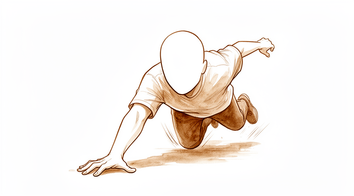
A scaphoid fracture usually happens in one moment. You fall onto an outstretched hand, or you land hard during sport, and your wrist is bent back further than it should go. Most people remember exactly when it happened. The scaphoid is a small bone on the thumb side of your wrist, near the base of your thumb.
Straight away, you feel pain on the thumb side of your wrist. There may be some swelling and bruising. The spot just below the base of your thumb, in the hollow between the wrist tendons, is often tender to press. There can be slight puffiness there too. Moving your wrist, especially bending it back or towards your little finger, tends to hurt. You may also feel pain when you press on the palm side of your wrist near the thumb, or when you push along the length of your thumb. Pinching with your thumb and index finger can be uncomfortable.
In the first days, the pain is there when you move and can be worse at night. It often settles gradually over the following weeks as healing starts. Everyday tasks that load the wrist, like pushing up from a chair, carrying shopping, or getting dressed, may be uncomfortable early on.
The tricky part is that this injury can feel mild. Pain and swelling are sometimes subtle, so some people keep using the wrist and only come in later. That matters, because a scaphoid fracture does not always show up on the first X-rays. Up to 30% to 40% of these fractures are not seen on the first set of images, and the fracture is only confirmed at a repeat check and further imaging, most often 10 to 14 days after the injury. So if your wrist is still sore in that hollow near your thumb after a fall, it is worth having it looked at, even if the first X-ray was clear.
What's actually happening
The scaphoid is a small, curved bone on the thumb side of your wrist. It sits inside the wrist joint itself, and it links the two rows of small bones that make up your wrist. Think of it as a strut or tie-rod that joins the two halves together and keeps them moving as one. When it breaks, that link is disrupted, so the wrist cannot load and move normally.
Most of these breaks happen through the middle of the bone, called the waist. The waist takes the biggest bending force when you land on an outstretched hand, which is why it is the part that usually gives way. The break can also happen near the end closest to your forearm, or near the end closest to your thumb.
Bone heals by knitting back together, the same as any other broken bone. But this bone is a difficult one to heal. Almost its whole surface is covered in smooth joint cartilage, which leaves little room for the healing tissue that normally helps a bone knit. Its blood supply is also sparse, and it flows in the reverse direction to most bones, entering near the thumb end and travelling back towards the forearm. So the further up the bone the break sits, the more likely it is that the broken piece loses its blood supply. A break near the forearm end can cut that piece off from its blood flow entirely, and those breaks take longer to heal.
Where the break sits matters. Breaks through the waist and towards the forearm end are the ones that tend to need closer watching or surgery. If the broken pieces have not shifted out of line, the bone can often heal in a cast without an operation. If the pieces have shifted or rotated, they are much less likely to knit on their own, and surgery to hold them together is usually recommended.
What we can do about it
Dr Kieran Hirpara, an upper-limb surgeon at Mater Private Hospital Rockhampton, matches the treatment to your specific injury. Patients are generally referred to our clinic by their GP; if a physiotherapist has suggested you see us, you will still need a referral from your GP in order to be eligible for the Medicare rebate. At that first visit we take a history, examine your wrist, and arrange imaging where it is needed. The plan depends on where the break sits, whether the pieces have shifted out of line, and what your wrist needs to do.
If the break is stable and the pieces have not shifted, a cast is usually the right choice. Most of these fractures heal in a cast without an operation. The cast holds your wrist still while the bone knits, and we check healing with repeat imaging along the way. A scan around week 4 can predict whether the break is likely to unite without surgery. Including your thumb in the cast has not been shown to help the bone heal any faster, so the cast may not need to cover it. Once healing is under way, movement is built back up in stages with physiotherapy or hand therapy.
Surgery is recommended from the outset when the broken pieces have shifted or rotated, because those breaks rarely knit on their own. It is also offered when your wrist needs to get back to work or sport sooner, since a cast can mean a long stretch of no loading. The operation holds the bone in the right position while it heals, usually with a small screw through the middle of the bone. Sometimes two screws are used, and sometimes a bone graft is added to help the break knit. The operation has its own page with more detail. Where both paths are possible, the choice is genuinely shared. A cast avoids an operation but means longer immobilisation, and surgery shortens that but carries the usual risks of any operation.
Either way, the first weeks are similar. Pain settles with simple measures and by protecting the wrist. You keep the injury still while it heals, following the plan we set together. Physiotherapy starts at the right stage, once the bone is strong enough to load. Getting treatment started early matters: healing rates are better when treatment begins within 4 weeks of the injury.
What to expect
Healing starts slowly and builds over weeks. If your break is in a cast, the bone knits while you keep the wrist still, and we check progress with repeat imaging along the way. Signs of healing can show on a scan as early as 3 weeks after surgery, and most breaks that are going to unite show it by around 6 weeks. Breaks where the pieces have not shifted heal faster than breaks where they have. Treatment started within 4 weeks of the injury gives the bone its best chance to knit.
Once the bone is strong enough, you build movement and strength back in stages. Everyday tasks like dressing and carrying shopping come back first, then heavier loading. How quickly you return to work or sport depends on your job, your wrist, and which treatment path you took. Surgery usually means less time in a cast, which is one reason it is offered when you need your wrist back sooner. From the longer view, people with breaks near the thumb end of the bone report normal hand function, good wrist motion and good strength years down the track.
Most breaks heal and most people get on with their lives. But it is honest to say this bone can be slow. More than 10% of breaks treated with surgery do not knit, and a break left untreated for a long time is even less likely to heal in a useful position. If the bone does not knit, that is called a non-union. An unhealed break can lead over time to wear-and-tear arthritis in the wrist: among people with an unhealed break left alone for five years or more, arthritis had developed in 97%. That is why we keep checking your wrist until we are sure it has healed, rather than letting follow-up lapse.
If healing is slow or fails, surgery with a bone graft can still be done later, and results can be good provided arthritis has not set in. Stiffness is the other common hiccup, and staged physiotherapy is there to work on it.
When to see someone
Seek urgent care if your wrist is badly deformed, if there is an open wound, if you have numbness or tingling, or if you cannot use the arm at all. Otherwise, see your GP if pain in that hollow near the base of your thumb is not settling after a fall. Ask for a specialist review if swelling, movement or grip are not improving week on week as healing progresses. This injury can feel mild, and the first X-rays do not always show it, so a wrist that stays sore deserves a closer look. Getting assessed early matters, because treatment started within 4 weeks of the injury gives the bone its best chance to knit. If a break is missed and left alone, it can lead to lasting problems, including wear-and-tear arthritis in the wrist.
In more depth
Advanced reading: the deeper science (optional)
This section goes further than you need for your own treatment decisions. Scaphoid fracture is worth the extra reading because where the break sits along the bone matters more than almost any other feature, and because the argument for operating is not the one most people assume.
Position on the bone changes the risk sevenfold
The scaphoid receives most of its blood supply through vessels entering near its far end, so blood travels backwards along the bone towards the proximal pole. A fracture across the waist interrupts that supply to everything beyond it.
The consequence is quantified. Amalgamating published series, 34% of acute proximal scaphoid fractures progress to nonunion when managed non-operatively, and the relative risk of nonunion for these fractures is 7.5 compared with more distal fractures treated the same way [1].
That is why two fractures which look similar on a film can carry entirely different recommendations. A distal fracture in a cast has a strong chance of healing. A proximal pole fracture treated identically fails to unite in about a third of cases.
Surgery lowers the nonunion rate but does not improve the destination
For the common waist fracture, comparing surgery with cast treatment produces a result worth reading carefully. Surgery gave a nonunion rate three times lower, a quicker return to function, and transiently better grip strength and range of movement, but was associated with more complications. There were no significant differences in pain, tenderness, cost or functional outcome [2].
So the operation buys reliability of union and speed of recovery, at the cost of surgical complications, and the two groups converge on the same place. That makes it a decision about how much a faster, more certain path is worth to a particular person, a manual worker or an athlete weighs those months differently from someone who can accommodate a cast.
Diagnosis is where most of the harm is avoided
Because a missed scaphoid fracture is the one that becomes a nonunion, the diagnostic pathway matters. Anatomical snuffbox tenderness was the most sensitive clinical test, and combining tests improved the post-test probability of fracture, which can be used to limit unnecessary immobilisation, hospital visits and imaging [3].
That last clause is the useful one. Combining examination findings is not only about catching fractures; it also identifies the people who can safely stop wearing a cast, which is why a structured reassessment is worth attending even when the wrist feels better.
For an established nonunion, the fancier graft is not the better graft
Where a fracture has failed to unite, the standard treatment is bone grafting, and a vascularised graft, one brought with its own blood supply, is intuitively superior.
The evidence does not support that. Current evidence suggests vascularised bone grafting does not yield significantly superior results to non-vascularised grafting in scaphoid nonunion, though the authors note potential selection bias reduces certainty [4]. The union figures are close: 84% for vascularised and 80% for non-vascularised grafts, with considerable variation between reports attributable to patient, fracture, treatment and study-design factors [5].
Since vascularised grafting is the longer and more technically demanding operation, that near-parity is worth knowing. Selection bias runs in a particular direction here, vascularised grafts tend to be used for the harder cases, so the comparison likely understates them. But it does not support choosing one as routinely better.
References for the advanced reading
- Eastley N, Singh H, Dias JJ, Taub N. Union rates after proximal scaphoid fractures; meta-analyses and review of available evidence. J Hand Surg Eur Vol. 2012;38(8):888-97.
- Symes TH, Stothard J. A systematic review of the treatment of acute fractures of the scaphoid. J Hand Surg Eur Vol. 2011;36(9):802-10.
- Mallee WH, Henny EP, van Dijk CN, Kamminga SP, van Enst WA, Kloen P. Clinical diagnostic evaluation for scaphoid fractures: a systematic review and meta-analysis. J Hand Surg Am. 2014;39(9):1683-1691.e2.
- Duncumb JW, Robinson PG, Williamson TR, Murray IR, Campbell D, Molyneux SG, et al. Bone grafting for scaphoid nonunion surgery: a systematic review and meta-analysis. Bone Joint J. 2022;104-B(5):549-58.
- Ferguson DO, Shanbhag V, Hedley H, Reichert I, Lipscombe S, Davis TRC. Scaphoid fracture non-union: a systematic review of surgical treatment using bone graft. J Hand Surg Eur Vol. 2015;41(5):492-500.
Evidence & references
This is the clinical evidence summary written for health professionals. It is technical, and it lists the research this page was built from. You do not need to read it to understand your treatment or to make a decision about it.
Overview
- Pediatric scaphoid fractures have excellent outcomes [1].
- Even some well-established and widely used principles of scaphoid fracture management are supported by an insufficient amount of evidence, with many decisions based on small case series [2].
- The clinical outcomes of malunited scaphoids after reconstruction for scaphoid fractures nonunion did not differ significantly from well-united scaphoids at a minimum 5-year follow-up [7].
- A study did not demonstrate a true long-term benefit of internal fixation, compared with nonoperative treatment, for acute nondisplaced or minimally displaced scaphoid fractures [8].
- For all indications, the scaphoid staple has a high union rate and a low complication rate [9].
- The definition of instability of scaphoid fractures and the indications for conservative treatment must be considered carefully [15].
- Internal fixation of scaphoid fractures is indicated in certain acute situations and in chronic nonunion cases [16].
- Appropriately performed acute percutaneous internal fixation is now a standard treatment option for a selected group of patients with acute scaphoid fracture [17].
- Nondisplaced scaphoid fractures can be effectively treated nonoperatively with union rates approaching or exceeding those of operative intervention [32].
- Operative intervention is recommended for displaced scaphoid fractures [32].
- Patients with recent scaphoid fractures that failed treatment may also be treated with distal scaphoid resection [34].
- Despite improvements in diagnosis and surgical techniques, nonunion rates remain high for scaphoid fractures [44].
- Early internal fixation is increasingly favored even for nondisplaced scaphoid fractures [44].
Anatomy & Pathophysiology
Bony Anatomy
- The scaphoid is a small, irregular S-shaped tubular bone located in the proximal carpal row on the radial aspect of the wrist [51].
- The scaphoid lies entirely within the wrist joint at a 45-degree plane to the longitudinal and horizontal axis of the wrist [51].
- The scaphoid articulates with the trapezium and trapezoid on its distal surface, the radius on its proximal and lateral surface, and the capitate and lunate on its medial surface [51].
- The proximal articular surface of the scaphoid is convex and articulates with the radius [51].
- The capitate head articulates with a sulcus on the radial articular surface of the scaphoid, providing a socket-like fit [51].
- The distal pole of the scaphoid sits ulnarly angulated relative to the proximal pole due to gentle pronation and distal flexion [51].
- The distal articular surface of the scaphoid features two distinct articular facets for the trapezium and trapezoid, forming the STT joint [51].
- Over 80% of the scaphoid surface is covered with articular cartilage [51].
- The extensive articular cartilage coverage of the scaphoid results in a reduced capacity for periosteal healing and an increased tendency for delayed union and nonunion [51].
- The scaphoid is ridged across its nonarticular dorsoradial surface, which serves as the insertion point for the dorsal component of the scapholunate and intercarpal ligaments [51].
- The scaphoid acts as a midcarpal joint "bridge" linking and synchronizing the motions of the proximal and distal carpal rows as part of the key intercalated segment [51].
- Motion of the scaphoid includes rotation proximally and gliding distally while providing stability to the midcarpal joint [51].
- The scaphoid is critical to the coordination of normal carpal kinematics [50].
- The scaphoid moves with nearly all carpal motions, especially volar flexion [59].
Vascular Supply
- The blood supply of the scaphoid arises from two vascular pedicles originating from the scaphoid branches of the radial artery [51].
- The dorsal branch of the radial artery enters via small foramina along the spiral groove and dorsal ridge of the scaphoid [51].
- The dorsal branch supplies 70% to 80% of the scaphoid proximally, including the proximal pole [51].
- The volar branch of the radial artery enters via the scaphoid tubercle [51].
- The volar branch supplies the remaining 20% to 30% of the distal scaphoid [51].
- The waist of the scaphoid has minimal or no perforating vasculature [51].
- No vessels perforate the proximal dorsal cartilaginous area or through the scapholunate ligament [51].
- Only 67% of scaphoid bones have arterial foramina throughout their length, including the distal, middle, and proximal thirds [59].
- 13% of scaphoid bones have blood supply predominantly in the distal third [59].
- 20% of scaphoid bones have most arterial foramina in the waist area with no more than a single foramen near the proximal third [59].
- One third of scaphoid fractures occurring in the proximal third may be without adequate blood supply [59].
- The prevalence of osteonecrosis can be 35% in fractures at the proximal pole level [59].
- Vessels enter the scaphoid from the radial artery laterovolarly, dorsally, and distally [59].
- The laterovolar and dorsal systems share in the blood supply to the proximal two thirds of the scaphoid [59].
- The vascularity of the proximal pole and 70% to 80% of the interosseous circulation are provided through branches of the radial artery entering through the dorsal ridge [59].
- In the distal tuberosity region, 20% to 30% of the bone receives its blood supply from volar branches of the radial artery [59].
Ligamentous Anatomy
- Ligamentous attachments of the scaphoid are predominantly found on the nonarticular dorsoradial surface [51].
- Short intrinsic ligaments provide stability to the scaphoid through attachments to other carpal bones, particularly the lunate [51].
- The short intrinsic ligaments merge with the extrinsic ligaments and capsule of the wrist [51].
- The radioscapocapitate ligament does not attach to the bone itself but crosses the waist, acting as a sling across it to allow rotation [51].
- There are no tendon attachments to the scaphoid [51].
- The RSC ligament acts as a fulcrum over which the scaphoid waist fractures [10].
Mechanisms of Injury
- Acute scaphoid fractures account for 2% to 3% of all fractures, approximately 10% of all hand fractures, and between 60% and 80% of all carpal fractures [37].
- The incidence of scaphoid fractures in the literature ranges from 1.5 to 121 fractures per 100,000 persons per year [37].
- A prospective dataset from Edinburgh documented an annual incidence of true radiographically confirmed acute scaphoid fractures of 29 per 100,000 per year [37].
- Previous studies from Scandinavia quoted an annual incidence of 26 to 39 per 100,000 per year [37].
- The mean age for scaphoid fractures in the literature ranges from 25 to 35 years [37].
- Males are significantly younger at the time of injury compared to females [37].
- Scaphoid fractures fit a type B fracture distribution curve [37].
- There is a male predominance with a male to female ratio of approximately 2.5:1 [37].
- Two studies documented male sex as a risk factor associated with a true scaphoid fracture [37].
- Scaphoid fractures usually occur after a fall onto the outstretched hand or during sports [37].
- Two studies reported that sports injuries are associated with a true scaphoid fracture [37].
- Low-energy falls from standing height occur more frequently in females, while males are more likely to sustain fractures after high-energy injuries such as sports or motor-vehicle collisions [37].
- Sports noted to cause increased risk include football, basketball, cycling, and skateboarding [37].
- Scaphoid fractures are increasingly documented after punching or assault-related injuries [37].
- The usual mechanism of injury is forced hyperextension of the wrist [11].
- Almost 90% of patients recall a hyperextension injury [33].
- The fracture is caused by a fall on the outstretched palm, resulting in severe hyperextension and slight radial deviation of the wrist [59].
- The scaphoid usually fractures on tension at the radial-palmar side [59].
- During the injury mechanism, the proximal pole locks in the scaphoid fossa of the radius while the distal pole moves excessively dorsal [59].
- Hyperextension past 95 degrees is the usual position of injury [71].
- Other mechanisms such as axial loading and hyperflexion of the wrist have been postulated to produce scaphoid fractures [71].
- With the hyperextension mechanism, a fracture of the scaphoid usually begins at the volar waist with a tensile failure [71].
- Forces propagate to the dorsal surface with compression loading until failure occurs [71].
- In cadaveric studies, wrists placed in extreme dorsiflexion and ulnar deviation produced fractures through the scaphoid waist as the scaphoid impinged on the dorsal rim of the radius [71].
- Proximal scaphoid fractures resulted from dorsal subluxation during forced hyperextension in cadaveric studies [71].
- Carpal dislocations and scapholunate ligament tears were reproduced with wrist extension and ulnar deviation combined with intercarpal supination [71].
Fracture Epidemiology and Location
- Scaphoid fractures account for almost 75% of all carpal fractures [11].
- Scaphoid fractures are rare in children and in the elderly [11].
- Approximately 70% to 80% of scaphoid fractures occur in the midportion or waist area [60].
- 10% to 20% of scaphoid fractures involve the proximal third [60].
- A smattering of fractures occur in the distal scaphoid, involving the tuberosity or articular surface [60].
- The vast majority of distal scaphoid fractures occur in children [60].
- 60% to 80% of scaphoid fractures occur at the scaphoid waist or midportion [59].
- Snuffbox tenderness applies predominantly to waist fractures, which represent 70% of scaphoid fractures [10].
- Proximal pole fractures are the second most common type, at 20% [10].
- Distal pole fractures are the least common, at 10% [10].
- Fractures tend to occur at the waist partly because the RSC ligament acts as a fulcrum over which the scaphoid waist fractures [10].
- Some fractures, especially distal oblique and waist fractures, are unstable, predisposing to non-union or malunion [11].
- 17% of patients with scaphoid fractures have other fractures of the carpus and forearm [59].
- Associated injuries include transscaphoid perilunar dislocations, fractures of the trapezium, Bennett fractures, fractures of the radial head, dislocations of the lunate, and fractures at the distal end of the radius [59].
- In the pediatric population, scaphoid fractures are the most common carpal injury [39].
- Scaphoid fractures account for approximately 3% of hand and carpal fractures and 0.34% of all fractures in children [39].
- Traditional thinking held that scaphoid fractures involved the distal pole in children with excellent healing rates [39].
- Increasing incidence and fracture patterns similar to adults are now seen in children, with the majority of fractures occurring at the waist [39].
- An estimated incidence rate for the U.S. population is 1.47 fractures per 100,000 person-years [47].
- Scaphoid fractures made up 2.36% of wrist fractures overall in the U.S. population [47].
- 66.4% of scaphoid fractures occurred in males in the U.S. population [47].
- The incidence rate ratio for gender, using females as the referent group, was 2.04 [47].
- Peak incidence of scaphoid fracture occurred in the second and third decades [47].
- The incidence rate of scaphoid injury for those aged 10 to 19 years was 3.38 per 100,000 [47].
- The incidence rate of scaphoid injury for those aged 20 to 29 years was 2.34 per 100,000 person-years [47].
Clinical Presentation and Examination
- Patients usually present with pain on the radial side of the wrist [10].
- There may be swelling on the radial side of the wrist [10].
- There is usually a history of trauma, such as falling on an outstretched hand, collision of the wrist against a person or heavy obstacle, or a direct blow against an object [10].
- Limited range of motion and pain when applying extended wrist loading or positioning the wrist in extreme positions of flexion or extension may be present [10].
- Wrists with acute fractures may have swelling and bruising in the radial aspect of the wrist [10].
- Wrists with chronic injury may have swelling in the dorsoradial wrist [10].
- "Snuffbox tenderness" has become synonymous with scaphoid fracture [10].
- The full physical examination of the scaphoid bone should include the waist, distal pole, and proximal pole [10].
- To palpate the anatomic snuffbox for the waist examination, palpate just distal to the radial styloid in the "soft spot" [10].
- The distal pole should be palpated at the scaphoid tubercle on the palmar aspect of the wrist [10].
- To palpate the distal pole, place the index finger in the anatomic snuffbox and place the thumb on the palmar aspect just distal to the anatomic snuffbox [10].
- With radial deviation of the wrist, the prominence of the distal pole should move palmarly toward the examiner’s thumb [10].
- The proximal pole is palpated dorsally in line with the second ray just distal to the dorsal radius lip [10].
- The scapholunate ligament is in line between the second and third rays just distal to the dorsal radius lip and corresponds to the 3-4 wrist arthroscopy portal [10].
- The proximal pole is just radial to the scapholunate ligament/3-4 portal area [10].
- Pain on longitudinal compression of the thumb (scaphoid axial compression test) is a sign of scaphoid fracture [10].
- If anatomic snuffbox tenderness, scaphoid tubercle tenderness, and the scaphoid axial compression test are all positive, there is 87% to 100% sensitivity and 74% specificity for scaphoid fracture [10].
- There may be slight fullness in the anatomical snuffbox [11].
- Precisely localized tenderness in the anatomical snuffbox is an important diagnostic sign [11].
- Examination must include pressure backwards over the scaphoid tubercle, palpation over the proximal pole, and telescoping of the thumb base [11].
- If any of these examination findings are positive, the suspicion for a scaphoid fracture should be high [11].
- Patients classically present with wrist pain following a fall onto the outstretched hand [33].
- Pain, swelling, ecchymosis, and tenderness around the region of the scaphoid may be present in the acute phase [33].
- The main complaint is radial-sided wrist pain with localized tenderness over the scaphoid in the region of the anatomic snuffbox [33].
- No single clinical sign has been found to be adequately sensitive or specific for scaphoid fracture [33].
- Anatomical snuffbox tenderness has a sensitivity of 87–100% and specificity of 3–98% [33].
- Axial compression of the thumb has a sensitivity of 48–100% and specificity of 22–97% [33].
- Scaphoid tubercle tenderness has a sensitivity of 82–100% and specificity of 17–57% [33].
- Pain on ulnar deviation has a sensitivity of 67–100% and specificity of 17–60% [33].
- Pain on radial deviation has a sensitivity of 67–90% and specificity of 31–42% [33].
- Reduced range of movement of the thumb has a sensitivity of 65–66% and specificity of 38–59% [33].
- Thumb–index finger pinch has a sensitivity of 75–79% and specificity of 44–76% [33].
- ASB tenderness is oversensitive and has poor specificity [33].
- In a study of 246 patients with a suspected fracture of the scaphoid, ASB tenderness had a sensitivity of 90% and a specificity of 40% [33].
- In the same study, scaphoid tubercle tenderness had a sensitivity of 87% and a specificity of 57% [33].
- In a prospective analysis of 73 patients with a suspected scaphoid fracture, ASB pain on ulnar deviation of the pronated wrist had a negative predictive value (NPV) of 100% [33].
- Patients with a negative test for ASB pain on ulnar deviation of the pronated wrist could be safely discharged at presentation as they did not have a scaphoid fracture [33].
- The use of one clinical sign in isolation is insufficient for the diagnosis of a scaphoid fracture [33].
- A combination of ASB tenderness, scaphoid tubercle tenderness, and ASB pain on longitudinal compression of the thumb generated a sensitivity of 100% and a specificity of 74% [33].
- These combined findings were valid only for the first 24 hours after injury [33].
- Pain on thumb–index finger pinch and ASB pain on pronation of the forearm were most suggestive of a true scaphoid fracture [33].
- The best predictors of fracture within 72 hours of injury were the absence of pain on ulnar deviation of the wrist and pain on thumb–index finger pinch [33].
- Scaphoid tubercle tenderness was most predictive at week 2 [33].
- A clinical scaphoid score (CSS) uses three clinical tests: tenderness in the ASB with the wrist in ulnar deviation (3 points), tenderness over the scaphoid
Classification
- Scaphoid fractures are managed largely on the basis of anecdotal evidence and traditional remedies [3].
- There is no consensus regarding the imaging modality and measurements to use to define a scaphoid fracture as 'nondisplaced' [13].
- Scaphoid nonunions are described by their anatomic location or with clinically specific terms such as stable, fibrous, sclerotic, unstable, humpback, synovial, cystic, pseudarthrosis, or avascular [117].
- Scaphoid nonunions can be divided roughly into two groups: early nonunions without substantial bone resorption, and older nonunions with substantial bone resorption [117].
- Complicating efforts to treat scaphoid nonunions are perfusion, deformity, and instability (bony or ligamentous) [117].
- There is a need for a validated prognostic classification system for scaphoid nonunions that can allow comparisons between outcome studies [73].
- Several classification schemes have been proposed for scaphoid nonunion, generally based on factors such as time since injury, mobility of the fragments, cystic or flexion deformity, and degenerative change [118].
- Type I scaphoid nonunion is defined as delayed or fibrous union with no deformity [118].
- Scaphoid nonunions without substantial bone loss require only rigid fixation to heal if there is adequate perfusion [118].
- Scaphoid nonunions that have minimal bone resorption of the anterior cortical bone and minimal fracture sclerosis (<2 mm confirmed by CT scan) still have the potential for healing in the early stages with screw fixation alone [118].
- The scaphoid experiences a period of maturation from age 6 to 15 years, altering its physical properties during this time [122].
- Patient age, degree of ossification, and fracture location are interrelated factors important for determining fracture type, classification, and treatment in pediatric scaphoid fractures [122].
- D’Arienzo proposed a three-part classification system for pediatric scaphoid fractures based on the age of the child and the presumed degree of ossification [122].
- Type 1 pediatric scaphoid lesions occur in children younger than age 8 years [122].
- Type 1 pediatric scaphoid lesions may be purely chondral or may involve part of the ossific nucleus [122].
- Type 2 pediatric scaphoid lesions are osteochondral fractures and occur in patients aged 8 to 11 years [122].
- Type 3 pediatric scaphoid lesions are the most common fractures and occur in adolescents aged ≥12 years [122].
- In Type 3 pediatric scaphoid lesions, the scaphoid is almost completely ossified and these fractures behave similarly to those in the adult population [122].
- Pediatric scaphoid fractures may be classified according to anatomic location: tuberosity, transverse distal pole, avulsion distal pole, waist, and proximal pole [122].
- In children, fractures of the distal third of the scaphoid are the most common [122].
Clinical Presentation
Epidemiology and Mechanism
- Scaphoid fractures account for 2% of all fractures and are the most commonly injured carpal bone [14].
- The annual incidence of true radiographically confirmed acute scaphoid fractures is approximately 29 per 100,000 persons per year [37].
- The mean age for scaphoid fracture in the literature ranges from 25 to 35 years [37].
- A male predominance is seen with a male-to-female ratio of approximately 2.5:1 [37].
- Male gender and sports injuries are risk factors associated with a true scaphoid fracture [46].
- Almost 90% of patients recall a hyperextension injury to the wrist [33].
- Males are more likely to sustain scaphoid fractures after high-energy injuries such as sports or motor-vehicle collisions, while low-energy falls from standing height occur more frequently in females [37].
- Scaphoid fractures are being increasingly documented after punching or assault-related injuries [37].
- In the pediatric population, scaphoid fractures account for approximately 3% of hand and carpal fractures and 0.34% of all fractures in children [39].
- Pediatric scaphoid fracture patterns are shifting toward the waist due to increased participation in high-energy extreme sports and increasing body mass indices [39].
Clinical Signs and Symptoms
- Patients classically present with pain on the radial side of the wrist following a fall onto an outstretched hand [10].
- Swelling, ecchymosis, and tenderness around the region of the scaphoid may be present in the acute phase [33].
- Limited range of motion and pain when applying extended wrist loading or positioning the wrist in extreme positions of flexion or extension are common presentations [10].
- Anatomical snuffbox tenderness has a sensitivity range of 87% to 100% and a specificity range of 3% to 98% for scaphoid fracture [33].
- Axial compression of the thumb has a sensitivity range of 48% to 100% and a specificity range of 22% to 97% for scaphoid fracture [33].
- Scaphoid tubercle tenderness has a sensitivity range of 82% to 100% and a specificity range of 17% to 57% for scaphoid fracture [33].
- Pain on ulnar deviation has a sensitivity range of 67% to 100% and a specificity range of 17% to 60% for scaphoid fracture [33].
- Pain on radial deviation has a sensitivity range of 67% to 90% and a specificity range of 31% to 42% for scaphoid fracture [33].
- Reduced range of movement of the thumb has a sensitivity range of 65% to 66% and a specificity range of 38% to 59% for scaphoid fracture [33].
- Thumb–index finger pinch has a sensitivity range of 75% to 79% and a specificity range of 44% to 76% for scaphoid fracture [33].
- No single clinical sign has been found to be adequately sensitive or specific for the diagnosis of scaphoid fracture [33].
- A combination of anatomical snuffbox tenderness, scaphoid tubercle tenderness, and anatomical snuffbox pain on longitudinal compression of the thumb generates a sensitivity of 100% and a specificity of 74% [33].
- The combination of anatomical snuffbox tenderness, scaphoid tubercle tenderness, and scaphoid axial compression test has a sensitivity of 87% to 100% and a specificity of 74% for scaphoid fracture [10].
- Anatomical snuffbox tenderness is oversensitive and has poor specificity [33].
- In a study of 246 patients with suspected scaphoid fracture, anatomical snuffbox tenderness had a sensitivity of 90% and a specificity of 40% [33].
- In the same study of 246 patients, scaphoid tubercle tenderness had a sensitivity of 87% and a specificity of 57% [33].
- The absence of pain on ulnar deviation of the wrist and pain on thumb–index finger pinch were the best predictors of fracture within 72 hours of injury [33].
- Scaphoid tubercle tenderness was the most predictive sign at week 2 post-injury [33].
- A clinical scaphoid score of 4 or higher, based on anatomical snuffbox tenderness with ulnar deviation, scaphoid tubercle tenderness, and pain upon longitudinal compression of the thumb, indicates the need for MRI [33].
- Most missed scaphoid fractures were due to failure to consider the possibility of the injury and search for clinical signs [23].
Diagnostic Challenges and Imaging Limitations
- Up to 30% to 40% of scaphoid fractures are not identified on initial assessment with standard four-view radiographs [33].
- The combination of conventional radiographs and two clinical examinations does not provide adequate diagnostic certainty, identifying a true fracture in only about 40% of patients [5].
- The combination of conventional radiographs and clinical reassessment does not increase diagnostic accuracy compared with conventional radiographs alone [12].
- Conventional radiography has a sensitivity of up to 64% and a negative predictive value with a weighted average of 74% for scaphoid fractures [65].
- The incidence of true scaphoid fractures in patients with suspected scaphoid fractures is typically low, within the 10%-20% range [65].
- Six-week radiographs are not adequate for evaluating suspected scaphoid fractures due to low observer agreement and poor diagnostic performance [28].
- There is no consensus reference standard to assess the diagnostic performance characteristics of imaging strategies for scaphoid fractures [26].
- MRI-detected scaphoid fractures are not universally benign, with delayed or nonunion seen in over 6% despite appropriate initial immobilization [29].
- Most patients with MRI-detected scaphoid nonunion require surgery to achieve union [29].
- Early MRI in patients with clinically suspected scaphoid fracture results in the accurate and reliable identification of a significant number of radiological occult injuries [30].
- Clinical examination along with early MRI scan should form the basis of diagnosing a suspected scaphoid fracture [24].
- If there is strong clinical suspicion of a scaphoid fracture that cannot be confirmed by conventional radiology, bone scintigraphy is a valuable diagnostic tool [6].
- Ultrasonic assessment has a sensitivity of only 50% and is not recommended for the early diagnosis of acute scaphoid fractures [67].
- The diagnosis of scaphoid fractures is reliable when using high-resolution peripheral quantitative CT in patients with a clinically suspected fracture [70].
- True scaphoid waist fractures are uncommon among patients with suspected scaphoid fractures [64].
- Even well-established principles of scaphoid fracture management are supported by insufficient evidence, with many decisions based on small case series [2].
Investigations
Clinical Examination
- The patient usually presents with pain on the radial side of the wrist, which may be accompanied by swelling [10].
- Limited range of motion and pain when applying extended wrist loading or positioning the wrist in extreme positions of flexion or extension are common clinical features [10].
- Acute fractures may present with swelling and bruising in the radial aspect of the wrist, while chronic injury may present with swelling in the dorsoradial wrist [10].
- Proximal pole fractures are the second most common type of scaphoid fracture, accounting for 20% of cases [10].
- Distal pole fractures are the least common type of scaphoid fracture, accounting for 10% of cases [10].
- The full physical examination of the scaphoid bone should include palpation of the waist, distal pole, and proximal pole [10].
- The anatomic snuffbox for the waist examination is palpated just distal to the radial styloid in the “soft spot” [10].
- The distal pole is palpated at the scaphoid tubercle on the palmar aspect of the wrist by placing the index finger in the anatomic snuffbox and the thumb on the palmar aspect just distal to it [10].
- The scapholunate ligament is located in line between the second and third rays just distal to the dorsal radius lip and corresponds to the 3-4 wrist arthroscopy portal [10].
- The proximal pole is located just radial to the scapholunate ligament/3-4 portal area [10].
- Slight fullness in the anatomical snuffbox with precisely localized tenderness is an important diagnostic sign [11].
- Physical examination must include pressure backwards over the scaphoid tubercle, palpation over the proximal pole, and telescoping of the thumb base [11].
- If any of the clinical tests (snuffbox tenderness, tubercle pressure, proximal pole palpation, or thumb telescoping) are positive, the suspicion for a scaphoid fracture should be high [11].
- Most scaphoid fractures were missed due to failure to consider the possibility of the injury and search for clinical signs [23].
Radiography
- X-rays should include AP, lateral, and two oblique views [11].
- The fracture may not be seen on X-ray in the first few days after the injury [11].
- Two weeks later, the break is usually much clearer on X-ray due to bone resorption at the fracture site and slight displacement of fragments [11].
- The crack is usually transverse through the narrowest part of the bone (the waist), but it may be more proximal or more distal [11].
- Signs of associated carpal displacement should always be looked for on X-ray [11].
- If the X-ray looks normal but the clinical features are suggestive of a fracture, the patient must not be discharged [11].
- The usual advice is to return for a second X-ray 2 weeks later while immobilizing the wrist in a cast [11].
- Plain radiography is approximately 50% sensitive for the detection of a scaphoid fracture [66].
- If a scaphoid fracture is suggested but radiographs are negative, the wrist should be immobilized and reevaluated in 2 weeks because up to 30% of patients may have positive follow-up radiographs [66].
- The combination of conventional radiographs and two clinical examinations does not provide adequate diagnostic certainty for scaphoid fractures, as a true fracture was identified in only about 40% of patients [5].
- The combination of conventional radiographs and clinical reassessment does not increase the accuracy of these diagnostic tests compared with the accuracy of conventional radiographs alone [12].
- Normal radiographs do not preclude the presence of a scaphoid fracture [66].
- Normal radiographs do not exclude a scaphoid fracture [26].
Advanced Imaging (MRI, CT, Scintigraphy)
- A CT scan is more sensitive for diagnosing a scaphoid fracture than X-ray [11].
- CT is particularly useful in confirming the alignment of the bone fragments if surgery is planned, or to confirm whether the fracture has united or not [11].
- MRI is the definitive way to confirm or exclude a diagnosis of scaphoid fracture if the technique is available [11].
- MRI, which is more sensitive than CT, is useful in making the diagnosis, and a normal study as early as 2 days after injury has a negative predictive value of 100% [66].
- Early magnetic resonance imaging (MRI) provides an immediate diagnosis for suspected scaphoid fractures when initial radiographs are inconclusive, which is cost-effective and minimizes complications [87].
- The use of early MRI in patients with clinically suspected scaphoid fracture results in the accurate and reliable identification of a significant number of radiological occult injuries and early identification of patients without acute injuries [30].
- MRI is the optimal second test for assessing a possible scaphoid fracture after a negative radiograph [90].
- CT is preferred when the fracture is visible for further assessment and surgical planning [90].
- If there is a strong clinical suspicion of a scaphoid fracture which cannot be confirmed by conventional radiology, bone scintigraphy is a valuable diagnostic tool [6].
- The authors argue that bone scintigraphy is inappropriate for evaluating specificity and sensitivity against clinical examination, and that MRI is the recommended examination of choice for diagnosing occult scaphoid fractures [93].
- According to the existing literature, MRI is the best diagnostic radiological test for triage of suspected scaphoid fractures, but bone scanning, CT, and ultrasound may also be useful, particularly when MRI is not readily available [96].
- Although MRI remains the best diagnostic tool after radiography for detecting occult scaphoid fractures, MDCT sensitivity was 86% and specificity was 100% in this study [89].
- CT is a good way to screen occult fractures but may not be any better than MRI or bone scanning in detecting scaphoid fractures without some over treatment [88].
- MRI is not 100% specific for diagnosing an occult scaphoid fracture, with a specificity of 96% in healthy volunteers [110].
- Routine MRI of suspected scaphoid fractures carries a notable risk of overdiagnosis and potential overtreatment, with nearly 70% of MRI findings categorized as distracting and potentially misleading [102].
- Since MRI detects any change in water content, misinterpretations of bone bruises and other variations as fractures may be common [26].
- The authors argue that better standardization of MRI definitions for scaphoid fractures is required, but acknowledge that a definition may not exist to solve the potentially unsolvable issue of diagnostic uncertainty [109].
Treatment
Non-Operative Management
- Subacute scaphoid fractures presenting within 6 months from injury can be expected to successfully heal with casting alone, even if the initial diagnosis is delayed [4].
- A restricted period of cast immobilisation is recommended for the initial treatment of non-displaced scaphoid fractures [18].
- Nondisplaced fractures of the scaphoid heal with cast immobilization in most cases [36].
- Non- and minimally displaced scaphoid waist fractures are best treated conservatively [57].
- The authors recommend considering nonoperative management for asymptomatic scaphoid nonunion in children [85].
- Among patients with nonoperatively managed scaphoid fractures, those prescribed NSAIDs within 1 month of diagnosis demonstrated an increased risk of nonunion and subsequent salvage procedures [84].
- Tubercle fractures are treated with a short-arm cast for 6–8 weeks [43].
- Distal third fractures or incomplete fractures are treated with a short-arm cast for 6–8 weeks [43].
- Nondisplaced waist fractures in pediatric patients and sedentary or low-demand patients are treated with a short-arm cast until healed [43].
- Distal pole fractures are almost always treated nonoperatively in a short-arm cast unless a person's occupation or special needs requires earlier and unrestricted mobility [43].
- Nonoperative management is routinely employed for suspected scaphoid fractures and tubercle fractures [46].
Operative Management
- Minimally invasive fixation has been demonstrated to have a higher union rate than cast treatment and has relatively few complications [40].
- Percutaneous fixation for undisplaced or minimally displaced waist fractures may reduce the time in cast, increase the rate of return to function, and increase the rate of union [46].
- Surgical management is recommended for displaced scaphoid fractures, proximal pole fractures, comminuted fractures, and fractures that are part of a greater perilunate injury [46].
- Nondisplaced proximal pole fractures are treated with mini-open internal fixation via a dorsal approach [43].
- Displacement of more than 1 mm is treated with open reduction and internal fixation, with or without bone graft [43].
- A lateral intrascaphoid angle of more than 35 degrees is treated with open reduction and internal fixation, with or without bone graft [43].
- Bone loss or comminution is treated with open reduction and internal fixation, with or without bone graft [43].
- Perilunate fracture-dislocation is treated with open reduction and internal fixation, with or without bone graft [43].
- Dorsal intercalated segmental instability alignment (DISI) with a radiolunate angle >15 degrees is treated with open reduction and internal fixation, with or without bone graft [43].
- The use of 2 headless compression screws for the treatment of scaphoid nonunions is safe and effective [76].
Outcomes and Evidence Quality
- This study did not demonstrate a true long-term benefit of internal fixation, compared with nonoperative treatment, for acute nondisplaced or minimally displaced scaphoid fractures [8].
- We found no difference in functional outcome at 12 months for fractures of the waist of the scaphoid with ≤ 2 mm displacement treated operatively or nonoperatively [19].
- Surgical treatment for non-displaced and minimally displaced acute scaphoid fractures may be slightly favourable compared to conservative treatment for standardised functional outcome on the short term (within 2 years), with a significantly faster return to work (SMD of 7 weeks) [25].
- The frequency of non-union after surgical management for closed scaphoid fractures exceeds 10% and remained consistent during the study period [27].
- Currently, there is insufficient evidence to support the most effective treatment for acute scaphoid fractures [81].
- Early treatment of acute scaphoid fractures is important, with union rates significantly greater when treatment is instituted prior to 4 weeks from injury [35].
- The optimal protocol for postoperative immobilization following operative treatment of scaphoid fractures remains controversial [92].
- Neglected scaphoid fractures seldom heal with acceptable alignment, which often leads to complications in their management [3].
- In cases of non-union or malunion, reconstruction of the scaphoid is exceedingly difficult, and this increases the chances of morbidity already associated with an extended period of hand immobilisation [3].
Complications
Nonunion and Malunion
- Neglected scaphoid fractures seldom heal with acceptable alignment due to the bone's unique anatomical structure [3].
- Scaphoid non-unions are unlikely to remain aligned or free of arthritis after 10 years [108].
- Problem fractures and non-unions of the scaphoid are associated with major alterations in wrist kinematics [31].
- Problem fractures and non-unions of the scaphoid are associated with a higher incidence of premature carpal collapse and degenerative arthritis than previously appreciated [31].
- Persistent nonunion is common after surgery for scaphoid non-union [42].
- Surgeries for persistent scaphoid nonunion are less successful than initial surgeries [42].
Diagnostic and Treatment Delays
- The diagnosis of scaphoid fracture can be complicated and the fracture can be easily overlooked in an acute injury [3].
- Delayed presentation of scaphoid fractures 21 days or more after injury predicts a greater risk of casting failure [45].
- Increased likelihood for nonunion was found when the fracture was treated greater than 31 days from injury [48].
- Increased likelihood for nonunion was found when fracture volume was less than 38% of the entire scaphoid [48].
Pediatric Complications
- Delayed presentation of scaphoid fractures 21 days or more after injury in children predicts a greater risk of casting failure, however, the union rate remains high with comparable time in cast [45].
Recovery
Pediatric Outcomes
- Delayed presentation of scaphoid fractures 21 days or more after injury predicts a greater risk of casting failure in pediatric patients [45].
- The union rate remains high with comparable time in cast for pediatric patients with delayed presentation of 21 days or more [45].
Acute Fracture Healing and Treatment Timing
- Union rates are significantly greater when treatment is instituted prior to 4 weeks from injury for acute scaphoid fractures [35].
- Neglected scaphoid fractures seldom heal with acceptable alignment [3].
- Patients with comorbid psychiatric conditions experienced increased rates of delayed scaphoid union [123].
Operative vs. Nonoperative Outcomes
- There was no difference in functional outcome at 12 months for fractures of the waist of the scaphoid with ≤ 2 mm displacement treated operatively or nonoperatively [19].
- Surgical treatment for non-displaced and minimally displaced acute scaphoid fractures may be slightly favourable compared to conservative treatment for standardised functional outcome on the short term (within 2 years) [25].
- Surgical treatment for non-displaced and minimally displaced acute scaphoid fractures is associated with a significantly faster return to work (SMD of 7 weeks) compared to conservative treatment [25].
- The scaphoid staple has a high union rate and a low complication rate for all indications [9].
Long-Term Outcomes
- Patients with distal scaphoid fractures report normal self-assessed hand function as well as good wrist motion and strength from an 8- to 11-year perspective [20].
- Patients treated nonoperatively or with salvage procedures had similar long-term outcomes as those treated with a corrective scaphoid osteotomy for scaphoid malunion [94].
- Nearly half of all patients with malunited acute scaphoid fractures demonstrated radiographic findings of early arthritis on CT imaging but overall good clinical results on midterm follow-up [121].
Nonunion and Complications
- Persistent nonunion is common after surgery for scaphoid non-union, and surgeries for persistent nonunion are even less successful [42].
- Increased likelihood for nonunion was found when the fracture was treated greater than 31 days from injury and when fracture volume was less than 38% of the entire scaphoid [48].
- Scaphoid nonunions demonstrate findings indicative of progression to union on CT at a mean of 6 weeks and as early as 3 weeks postoperatively [127].
- The fractures of the carpal scaphoid were all united within the usual time limits and there was no residual disability in any of the elbows in cases associated with radial head fracture [38].
Key Evidence
- [L1] Pediatric scaphoid fractures have excellent outcomes. [1] (10.1177/1558944717735948)
- [L5] Even some well-established and widely used principles of scaphoid fracture management are supported by an insufficient amount of evidence, with many decisions based on small case series. [2] (10.1177/1753193420977241)
- [L4] [3] (10.1016/j.injury.2009.07.078)
- [L4] Subacute scaphoid fractures presenting within 6 months from injury can be expected to successfully heal with casting alone, even if the initial diagnosis is delayed. [4] (10.1055/s-0035-1564983)
- [L5] The combination of conventional radiographs and two clinical examinations does not provide adequate diagnostic certainty for scaphoid fractures, as a true fracture was identified in only about 40% of patients. [5] (10.1097/corr.0000000000002413)
- [Paper] If there is a strong clinical suspicion of a scaphoid fracture which cannot be confirmed by conventional radiology, bone scintigraphy is a valuable diagnostic tool. [6] (10.1016/j.injury.2005.02.009)
- [L4] The clinical outcomes of malunited scaphoids after reconstruction for scaphoid fractures nonunion did not differ significantly from well-united scaphoids at a minimum 5-year follow-up. [7] (10.1016/j.otsr.2014.09.026)
- [L1] This study did not demonstrate a true long-term benefit of internal fixation, compared with nonoperative treatment, for acute nondisplaced or minimally displaced scaphoid fractures. [8] (10.2106/jbjs.g.00673)
- [L4] For all indications, the scaphoid staple has a high union rate and a low complication rate. [9] (10.1177/1558944716658747)
- [L2] The combination of conventional radiographs and clinical reassessment does not increase the accuracy of these diagnostic tests compared with the accuracy of conventional radiographs alone and is therefore also limited in diagnosing scaphoid fractures. [12] (10.1097/corr.0000000000002310)
- [L5] There is no consensus regarding the imaging modality and measurements to use to define a scaphoid fracture as 'nondisplaced.' [13] (10.1016/j.jhsa.2012.10.025)
- [L5] Scaphoid fractures account for 2% of all fractures and are the most commonly injured carpal bone. [14] (10.1016/j.hcl.2017.04.003)
- [L5] The definition of instability of scaphoid fractures and the indications for conservative treatment must be considered carefully. [15] (10.1142/s0218810415400018)
- [L5] Internal fixation of scaphoid fractures is indicated in certain acute situations and in chronic nonunion cases. [16] (10.1016/s0749-0712(21)00118-9)
- [L4] Appropriately performed acute percutaneous internal fixation is now a standard treatment option for a selected group of patients with acute scaphoid fracture. [17] (10.5435/00124635-200708000-00004)
- [L4] [18] (10.1016/j.injury.2008.10.028)
- [L1] We found no difference in functional outcome at 12 months for fractures of the waist of the scaphoid with ≤ 2 mm displacement treated operatively or nonoperatively. [19] (10.1302/0301-620x.104b8.bjj-2022-0085.r2)
- [L2] From an 8- to 11-year perspective, patients with distal scaphoid fractures report normal self-assessed hand function as well as good wrist motion and strength. [20] (10.1016/j.jhsa.2017.06.016)
- [L4] Most scaphoid fractures were missed due to failure to consider the possibility of the injury and search for clinical signs. [23] (10.1016/j.injury.2019.05.009)
- [L3] Clinical examination along with early MRI scan should form the basis of diagnosing a suspected scaphoid fracture. [24] (10.1177/1753193420979465)
- [L1] Surgical treatment for non-displaced and minimally displaced acute scaphoid fractures may be slightly favourable compared to conservative treatment for standardised functional outcome on the short term (within 2 years), with a significantly faster return to work (SMD of 7 weeks). [25] (10.1136/jisakos-2015-000024)
- [L3] [26] (10.1177/17531934251367541)
- [L3] The frequency of non-union after surgical management for closed scaphoid fractures exceeds 10% and remained consistent during the study period. [27] (10.1016/j.jhsa.2015.06.019)
- [L2] Due to low agreement between observers for the recognition of scaphoid fractures and poor diagnostic performance, 6-week radiographs are not adequate for evaluating suspected scaphoid fractures. [28] (10.1007/s00402-016-2438-4)
- [L3] MRI-detected scaphoid fractures are not universally benign, with delayed or nonunion seen in over 6% despite appropriate initial immobilization, with most of these patients with nonunion requiring surgery to achieve union. [29] (10.1302/0301-620x.106b4.bjj-2023-1171.r1)
- [L2] The use of early MRI in patients with clinically suspected scaphoid fracture results in the accurate and reliable identification of a significant number of radiological occult injuries and early identification of patients without acute injuries. [30] (10.1177/1753193412471008)
- [L5] Problem fractures and non-unions of the scaphoid are associated with major alterations in wrist kinematics and a higher incidence of premature carpal collapse and degenerative arthritis than previously appreciated. [31] (10.2106/00004623-199274030-00014)
- [L1] Nondisplaced scaphoid fractures can be effectively treated nonoperatively with union rates approaching or exceeding those of operative intervention, while operative intervention is recommended for displaced fractures. [32] (10.2106/jbjs.rvw.15.00073)
- [L4] Patients with recent scaphoid fractures that failed treatment may also be treated with distal scaphoid resection. [34] (10.1016/j.jhsg.2024.03.013)
- [L5] Early treatment of acute scaphoid fractures is important, with union rates significantly greater when treatment is instituted prior to 4 weeks from injury. [35] (10.1016/s0749-0712(21)00580-1)
- [L5] Nondisplaced fractures of the scaphoid heal with cast immobilization in most cases, but operative treatment is being offered with greater frequency to active patients to reduce the period of cast immobilization. [36] (10.5435/00124635-200007000-00003)
- [L4] The fractures of the carpal scaphoid were all united within the usual time limits and there was no residual disability in any of the elbows. [38] (10.1016/s0020-1383(73)80017-8)
- [L4] Persistent nonunion is common after surgery for scaphoid non-union, and surgeries for persistent nonunion are even less successful. [42] (10.1016/j.jhsa.2015.06.022)
- [L5] This article reviews current concepts regarding the treatment of scaphoid fractures and nonunions, highlighting that despite improvements in diagnosis and surgical techniques, nonunion rates remain high and early internal fixation is increasingly favored even for nondisplaced fractures. [44] (10.1016/j.jhsa.2008.04.026)
- [L4] Delayed presentation of scaphoid fractures 21 days or more after injury predicts a greater risk of casting failure; however, the union rate remains high with comparable time in cast. [45] (10.1016/j.jhsa.2023.10.020)
- [L4] [47] (10.1016/j.jhsa.2010.05.017)
- [L3] Increased likelihood for nonunion was found when the fracture was treated greater than 31 days from injury and when fracture volume was less than 38% of the entire scaphoid. [48] (10.1055/s-0039-3402769)
- [L5] The scaphoid is critical to the coordination of normal carpal kinematics, and its fracture has significant biomechanical consequences to the wrist. [50] (10.1016/s0749-0712(21)01439-6)
- [L2] Non- and minimally displaced scaphoid waist fractures are best treated conservatively. [57] (10.1016/j.jhsa.2015.03.007)
- [L5] [60] (10.1016/s0749-0712(21)01437-2)
- [L2] True scaphoid waist fractures are uncommon among patients with suspected scaphoid fractures. [64] (10.1007/s11552-007-9077-8)
- [L1] [65] (10.1177/1753193417742553)
- [L4] With a sensitivity of only 50% and five missed scaphoid fractures in this small series, we can not recommend ultrasonic assessment for the early diagnosis of acute scaphoid fractures. [67] (10.1054/jhsb.2000.0432)
- [L4] The diagnosis of scaphoid and other fractures is reliable when using HRpQCT in patients with a clinically-suspected fracture. [70] (10.1302/0301-620x.102b4.bjj-2019-0632.r3)
- [L4] There is a need for a validated prognostic classification system for scaphoid nonunions that can allow comparisons between outcome studies. [73] (10.1177/1753193417739510)
- [L4] The use of 2 headless compression screws for the treatment of scaphoid nonunions is safe and effective. [76] (10.1016/j.jhsa.2014.02.030)
- [L1] Currently, there is insufficient evidence to support the most effective treatment for acute scaphoid fractures. [81] (10.1007/s11552-010-9276-6)
- [L2] Among patients with nonoperatively managed scaphoid fractures, those prescribed NSAIDs within 1 month of diagnosis demonstrated an increased risk of nonunion and subsequent salvage procedures. [84] (10.1016/j.jhsg.2026.100958)
- [L4] The authors recommend considering nonoperative management for asymptomatic scaphoid nonunion in children. [85] (10.1055/s-0037-1602799)
- [L5] Early magnetic resonance imaging (MRI) provides an immediate diagnosis for suspected scaphoid fractures when initial radiographs are inconclusive, which is cost-effective and minimizes complications. [87] (10.1016/j.jhsa.2013.03.055)
- [Commentary] CT is a good way to screen occult fractures but may not be any better than MRI or bone scanning in detecting scaphoid fractures without some over treatment. [88] (10.1177/1753193412446273)
- [L2] Although MRI remains the best diagnostic tool after radiography for detecting occult scaphoid fractures, MDCT sensitivity was 86% and specificity was 100% in this study. [89] (10.1007/s11604-010-0520-3)
- [Paper] MRI is the optimal second test for assessing a possible scaphoid fracture after a negative radiograph, while CT is preferred when the fracture is visible for further assessment and surgical planning. [90] (10.1016/j.hcl.2019.03.001)
- [L4] The optimal protocol for postoperative immobilization following operative treatment of scaphoid fractures remains controversial. [92] (10.1177/15589447221093675)
- [L5] The authors argue that bone scintigraphy is inappropriate for evaluating specificity and sensitivity against clinical examination, and that MRI is the recommended examination of choice for diagnosing occult scaphoid fractures. [93] (10.1016/j.injury.2007.12.013)
- [L4] Patients treated nonoperatively or with salvage procedures had similar long-term outcomes as those treated with a corrective scaphoid osteotomy. [94] (10.1177/1558944716643295)
- [L5] According to the existing literature, MRI is the best diagnostic radiological test for triage of suspected scaphoid fractures, but bone scanning, CT, and ultrasound may also be useful, particularly when MRI is not readily available. [96] (10.1016/j.jhsa.2008.04.016)
- [L5] Routine MRI of suspected scaphoid fractures carries a notable risk of overdiagnosis and potential overtreatment, with nearly 70% of MRI findings categorized as distracting and potentially misleading, suggesting that stopping the pursuit of occult fractures may prevent unnecessary treatment. [102] (10.1097/corr.0000000000002914)
- [L4] [108] (10.1016/j.jhsa.2014.08.030)
- [L5] The authors argue that better standardization of MRI definitions for scaphoid fractures is required, but acknowledge that a definition may not exist to solve the potentially unsolvable issue of diagnostic uncertainty, suggesting patients should participate in decisions regarding diagnostic and treatment strategies. [109] (10.1177/17531934251394819)
- [Paper] MRI is not 100% specific for diagnosing an occult scaphoid fracture, with a specificity of 96% in healthy volunteers. [110] (10.1016/s0363-5023(10)60085-8)
- [L5] [117] (10.1097/01.blo.0000205886.66081.9d)
- [L4] Nearly half of all patients with malunited acute scaphoid fractures demonstrated radiographic findings of early arthritis on CT imaging but overall good clinical results on midterm follow-up. [121] (10.1016/j.jhsa.2020.04.002)
- [L5] [122] (10.5435/00124635-200902000-00004)
- [L3] Patients with comorbid psychiatric conditions experienced increased rates of delayed scaphoid union. [123] (10.1177/15589447221142894)
- [L4] Scaphoid nonunions demonstrate findings indicative of progression to union on CT at a mean of 6 weeks and as early as 3 weeks postoperatively. [127] (10.1016/j.jhsa.2016.07.051)
References
[1] Management Modalities and Outcomes Following Acute Scaphoid Fractures in Children: A Quantitative Review and Meta-Analysis. HAND. 2017. DOI: 10.1177/1558944717735948
[2] Questions regarding the evidence guiding treatment of displaced scaphoid fractures. Journal of Hand Surgery (European Volume). 2020. DOI: 10.1177/1753193420977241
[3] Management of late-diagnosed scaphoid fractures. Injury. 2010. DOI: 10.1016/j.injury.2009.07.078
[4] The Missed Scaphoid Fracture–Outcomes of Delayed Cast Treatment. Journal of Wrist Surgery. 2015. DOI: 10.1055/s-0035-1564983
[5] CORR Insights®: What Is the Diagnostic Performance of Conventional Radiographs and Clinical Reassessment Compared With HR-pQCT Scaphoid Fracture Diagnosis?. Clinical Orthopaedics & Related Research. 2022. DOI: 10.1097/corr.0000000000002413
[6] Outcome of routine bone scintigraphy in suspected scaphoid fractures. Injury. 2005. DOI: 10.1016/j.injury.2005.02.009
[7] Clinical outcome of scaphoid malunion as a result of scaphoid fracture nonunion surgical treatment: A 5-year minimum follow-up study. Orthopaedics & Traumatology: Surgery & Research. 2015. DOI: 10.1016/j.otsr.2014.09.026
[8] Nonoperative Compared with Operative Treatment of Acute Scaphoid Fractures. The Journal of Bone & Joint Surgery. 2008. DOI: 10.2106/jbjs.g.00673
[9] The Scaphoid Staple: A Systematic Review. HAND. 2016. DOI: 10.1177/1558944716658747
[10] Green S Operative Hand Surgery. Examination and Imaging of the Scaphoid.
[11] Apley And Solomon S Concise System Of Orthopaedics And Trauma. FRACTURES OF THE DISTAL RADIUS IN CHILDREN > FRACTURE OF THE SCAPHOID.
[12] What Is the Diagnostic Performance of Conventional Radiographs and Clinical Reassessment Compared With HR-pQCT Scaphoid Fracture Diagnosis?. Clinical Orthopaedics & Related Research. 2022. DOI: 10.1097/corr.0000000000002310
[13] Diagnosis of Scaphoid Fracture Displacement. The Journal of Hand Surgery. 2013. DOI: 10.1016/j.jhsa.2012.10.025
[14] Treatment of Scaphoid Fractures. Hand Clinics. 2017. DOI: 10.1016/j.hcl.2017.04.003
[15] Scaphoid Fracture - Overview and Conservative Treatment. Hand Surgery. 2015. DOI: 10.1142/s0218810415400018
[16] INTERNAL FIXATION OF SCAPHOID FRACTURES. Hand Clinics. 1997. DOI: 10.1016/s0749-0712(21)00118-9
[17] Percutaneous Fixation of Scaphoid Fractures. Journal of the American Academy of Orthopaedic Surgeons. 2007. DOI: 10.5435/00124635-200708000-00004
[18] Non-operative treatment of non-displaced scaphoid fractures may be preferred. Injury. 2009. DOI: 10.1016/j.injury.2008.10.028
[19] One-year outcome of surgery compared with immobilization in a cast for adults with an undisplaced or minimally displaced scaphoid fracture. The Bone & Joint Journal. 2022. DOI: 10.1302/0301-620x.104b8.bjj-2022-0085.r2
[20] Long-Term Outcomes After Distal Scaphoid Fractures: A 10-Year Follow-Up. The Journal of Hand Surgery. 2017. DOI: 10.1016/j.jhsa.2017.06.016
[23] Why scaphoid fractures are missed. A review of 52 medical negligence cases. Injury. 2019. DOI: 10.1016/j.injury.2019.05.009
[24] Reliability of clinical tests for prediction of occult scaphoid fractures and cost benefit analysis of a dedicated scaphoid pathway. Journal of Hand Surgery (European Volume). 2020. DOI: 10.1177/1753193420979465
[25] Surgical treatment of non- and minimally-displaced acute scaphoid fractures favours over-conservative treatment but only in the short term: an updated meta-analysis. Journal of ISAKOS. 2016. DOI: 10.1136/jisakos-2015-000024
[26] Variation in definitions of scaphoid fracture on MRI scans for suspected fracture: a systematic review. Journal of Hand Surgery (European Volume). 2025. DOI: 10.1177/17531934251367541
[27] An Epidemiologic Perspective on Scaphoid Fracture Treatment and Frequency of Nonunion. The Journal of Hand Surgery. 2015. DOI: 10.1016/j.jhsa.2015.06.019
[28] 6-week radiographs unsuitable for diagnosis of suspected scaphoid fractures. Archives of Orthopaedic and Trauma Surgery. 2016. DOI: 10.1007/s00402-016-2438-4
[29] The rate of nonunion in the MRI-detected occult scaphoid fracture. The Bone & Joint Journal. 2024. DOI: 10.1302/0301-620x.106b4.bjj-2023-1171.r1
[30] Early magnetic resonance imaging in patients with a clinically suspected scaphoid fracture may identify occult wrist injuries. Journal of Hand Surgery (European Volume). 2012. DOI: 10.1177/1753193412471008
[31] Compression-staple fixation for fractures, non-unions, and delayed unions of the carpal scaphoid.. The Journal of Bone & Joint Surgery. 1992. DOI: 10.2106/00004623-199274030-00014
[32] Acute Scaphoid Fractures. JBJS Reviews. 2016. DOI: 10.2106/jbjs.rvw.15.00073
[33] Rockwood And Green S Fractures In Adults. 42: Fractures of the Distal Radius and Ulna > Signs and Symptoms of Scaphoid Fractures.
[34] Distal Scaphoid Excision for Chronic and Nonchronic Scaphoid Fracture Nonunion. Journal of Hand Surgery Global Online. 2024. DOI: 10.1016/j.jhsg.2024.03.013
[35] MANAGEMENT OF ACUTE SCAPHOID FRACTURES. Hand Clinics. 2000. DOI: 10.1016/s0749-0712(21)00580-1
[36] Acute Fractures of the Scaphoid. Journal of the American Academy of Orthopaedic Surgeons. 2000. DOI: 10.5435/00124635-200007000-00003
[37] Rockwood And Green S Fractures In Adults. 42: Fractures of the Distal Radius and Ulna > Assessment of Scaphoid Fractures.
[38] Carpal scaphoid fracture associated with fracture of the radial head. Injury. 1973. DOI: 10.1016/s0020-1383(73)80017-8
[39] Orthopaedic Knowledge Update 13 Ebook Without Multimedia. Pediatric Forearm, Wrist, and Hand Trauma > Scaphoid Fractures.
[40] Green S Operative Hand Surgery. SCAPHOID FRACTURES AND NONUNION.
[42] Importance of Computed Tomography in Determining Displacement of Scaphoid Fractures. The Journal of Hand Surgery. 2015. DOI: 10.1016/j.jhsa.2015.06.022
[43] Green S Operative Hand Surgery. Special Considerations > Managing Scaphoid Fractures in Athletes.
[44] Treatment of Scaphoid Fractures and Nonunions. The Journal of Hand Surgery. 2008. DOI: 10.1016/j.jhsa.2008.04.026
[45] Clinically Significant Treatment Delay in Pediatric Scaphoid Fractures. The Journal of Hand Surgery. 2024. DOI: 10.1016/j.jhsa.2023.10.020
[46] Rockwood And Green S Fractures In Adults. 42: Fractures of the Distal Radius and Ulna > Future Directions Related to Scaphoid Fractures.
[47] Incidence Estimates and Demographics of Scaphoid Fracture in the U.S. Population. The Journal of Hand Surgery. 2010. DOI: 10.1016/j.jhsa.2010.05.017
[48] Factors Associated with Scaphoid Nonunion following Early Open Reduction and Internal Fixation. Journal of Wrist Surgery. 2020. DOI: 10.1055/s-0039-3402769
[50] EFFECTS OF SCAPHOID FRACTURES ON THE BIOMECHANICS OF THE WRIST. Hand Clinics. 2001. DOI: 10.1016/s0749-0712(21)01439-6
[51] Rockwood And Green S Fractures In Adults. 42: Fractures of the Distal Radius and Ulna > Pathoanatomy and Applied Anatomy Related to Scaphoid Fractures.
[57] Conservative Treatment Versus Arthroscopic-Assisted Screw Fixation of Scaphoid Waist Fractures—A Randomized Trial With Minimum 4-Year Follow-Up. The Journal of Hand Surgery. 2015. DOI: 10.1016/j.jhsa.2015.03.007
[59] Campbell S Operative Orthopaedics 4 Volume Set. NERVE INJURIES AT THE LEVEL OF THE HAND AND WRIST > FRACTURES OF THE SCAPHOID.
[60] INCIDENCE, MECHANISM, AND NATURAL HISTORY OF SCAPHOID FRACTURES. Hand Clinics. 2001. DOI: 10.1016/s0749-0712(21)01437-2
[64] Computed Tomography for Triage of Suspected Scaphoid Fracturesw. HAND. 2007. DOI: 10.1007/s11552-007-9077-8
[65] The economic evidence for advanced imaging in the diagnosis of suspected scaphoid fractures: systematic review of evidence. Journal of Hand Surgery (European Volume). 2017. DOI: 10.1177/1753193417742553
[66] Campbell S Operative Orthopaedics 4 Volume Set. OVERCORRECTION OSTEOTOMY AND LIGAMENTOUS REPAIR OR RECONSTRUCTION > SCAPHOID AND CARPAL FRACTURES.
[67] Ultrasound for Diagnosis of Scaphoid Fractures. Journal of Hand Surgery. 2000. DOI: 10.1054/jhsb.2000.0432
[70] The interobserver reliability of the diagnosis and classification of scaphoid fractures using high-resolution peripheral quantitative CT. The Bone & Joint Journal. 2020. DOI: 10.1302/0301-620x.102b4.bjj-2019-0632.r3
[71] Green S Operative Hand Surgery. Biomechanics of Scaphoid Fractures and Implications of Nonunion.
[73] Scaphoid nonunion: what is the role of the Zaidemberg 1,2 intercompartmental supraretinacular arterial flap?. Journal of Hand Surgery (European Volume). 2017. DOI: 10.1177/1753193417739510
[76] Scaphoid Nonunions Treated With 2 Headless Compression Screws and Bone Grafting. The Journal of Hand Surgery. 2014. DOI: 10.1016/j.jhsa.2014.02.030
[81] Treatment of Acute Scaphoid Fractures: A Systematic Review and Meta-Analysis. HAND. 2010. DOI: 10.1007/s11552-010-9276-6
[84] Early Nonsteroidal Anti-Inflammatory Drug Prescriptions and Nonunion After Scaphoid Fractures: A TriNetX Matched Cohort Study. Journal of Hand Surgery Global Online. 2026. DOI: 10.1016/j.jhsg.2026.100958
[85] Spontaneous Healing of a Pediatric Scaphoid Proximal Pole Fracture Nonunion. Journal of Wrist Surgery. 2017. DOI: 10.1055/s-0037-1602799
[87] The Role of Magnetic Resonance Imaging in Scaphoid Fractures. The Journal of Hand Surgery. 2013. DOI: 10.1016/j.jhsa.2013.03.055
[88] Commentary on ‘Early CT for suspected occult scaphoid fractures’ by Stevenson et al. J Hand Surg Eur. 2012, 37: 447-51. Journal of Hand Surgery (European Volume). 2012. DOI: 10.1177/1753193412446273
[89] Diagnostic accuracy of multidetector computed tomography for patients with suspected scaphoid fractures and negative radiographic examinations. Japanese Journal of Radiology. 2011. DOI: 10.1007/s11604-010-0520-3
[90] Imaging for Acute and Chronic Scaphoid Fractures. Hand Clinics. 2019. DOI: 10.1016/j.hcl.2019.03.001
[92] Postoperative Immobilization of Scaphoid Fractures: A Comprehensive Review of the Literature. HAND. 2022. DOI: 10.1177/15589447221093675
[93] A prospective comparison for suspected scaphoid fractures: Bone scintigraphy versus clinical outcome. Injury. 2008. DOI: 10.1016/j.injury.2007.12.013
[94] Long-Term Outcomes of Scaphoid Malunion. HAND. 2016. DOI: 10.1177/1558944716643295
[96] Imaging for Suspected Scaphoid Fracture. The Journal of Hand Surgery. 2008. DOI: 10.1016/j.jhsa.2008.04.016
[102] Editor’s Spotlight/Take 5: Routine MRI Among Patients With a Suspected Scaphoid Fracture Risks Overdiagnosis. Clinical Orthopaedics & Related Research. 2023. DOI: 10.1097/corr.0000000000002914
[108] Persistent Fracture Line After Scaphoid Fracture Fixation. The Journal of Hand Surgery. 2014. DOI: 10.1016/j.jhsa.2014.08.030
[109] Re: van Boxel et al. Variation in definitions of scaphoid fracture on MRI scans for suspected fracture: a systematic review. Journal of Hand Surgery (European Volume). 2025. DOI: 10.1177/17531934251394819
[110] False Positive MRI's for Scaphoid Fracture in Healthy Volunteers. The Journal of Hand Surgery. 2010. DOI: 10.1016/s0363-5023(10)60085-8
[117] Minimally Invasive Management of Scaphoid Nonunions. Clinical Orthopaedics and Related Research. 2006. DOI: 10.1097/01.blo.0000205886.66081.9d
[118] Scaphoid Fractures and Nonunion. 2021.
[121] Scaphoid Malunion Clinical and Radiographic Outcomes at a Minimum of 4 Years Follow-Up. The Journal of Hand Surgery. 2020. DOI: 10.1016/j.jhsa.2020.04.002
[122] Pediatric Scaphoid Fractures. Journal of the American Academy of Orthopaedic Surgeons. 2009. DOI: 10.5435/00124635-200902000-00004
[123] Delayed Scaphoid Fracture Union in Patients With Comorbid Psychiatric Diagnoses: A Retrospective Analysis of 20 340 Patients. HAND. 2022. DOI: 10.1177/15589447221142894
[127] Early Detection of Healing of Scaphoid Fracture Nonunions Using Computed Tomography. The Journal of Hand Surgery. 2016. DOI: 10.1016/j.jhsa.2016.07.051