您的感受
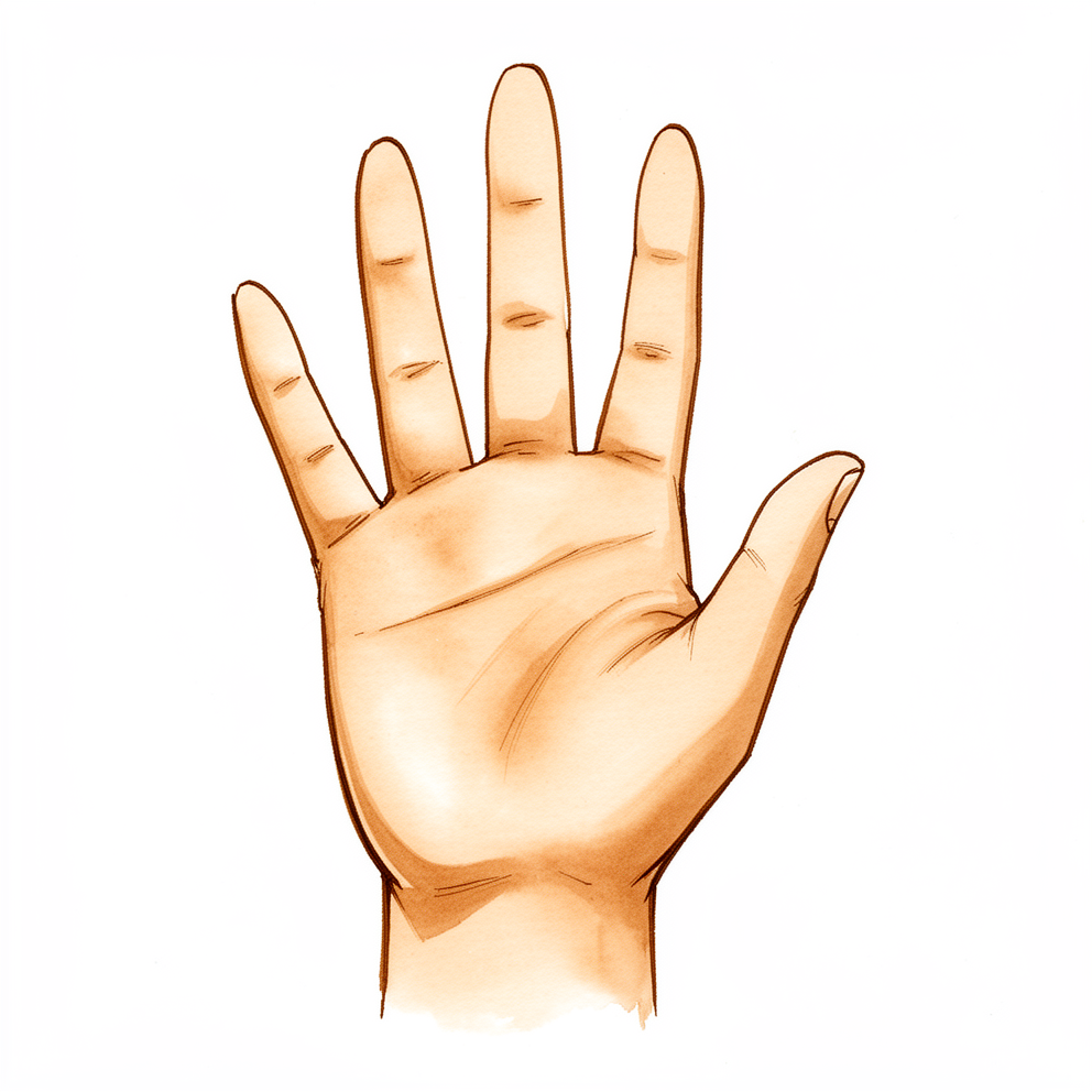
STT关节位于手腕的舟骨与两块名为大多角骨和小多角骨的小骨头相接的地方,就在拇指根部关节的后方。这里的关节炎会引起拇指根部以及手腕拇指一侧的疼痛或酸痛。抓握和捏东西时疼痛会加重。
日常动作都可能诱发疼痛:转动钥匙、拧开罐盖、写字、提购物袋或端一杯茶。
这个关节的磨损在X线片上很常见,尤其是在年长女性中。许多有这些X线改变的人根本没有疼痛。X线片也很难判断这个关节是否就是引起您疼痛的那个关节:它可能低估或高估磨损的程度。这就是为什么您的外科医生可能会建议在X线或超声引导下,向该关节内注射局部麻醉药和类固醇。如果注射后疼痛缓解,就说明STT关节是疼痛的来源。
STT关节炎常常与拇指根部关节的关节炎同时出现,两者可能很难区分。当两个关节都发生磨损时,称为全大多角骨关节炎(pantrapezial arthritis)。了解哪些关节受累很重要,因为这决定了哪些手术是合理的。
在一些STT关节炎患者中,腕骨会向后倾斜,这种模式称为DISI(dorsal intercalated segment instability,背侧嵌入段不稳定的缩写)。在选择手术之前,您的外科医生会在X线片上检查是否存在这种情况,因为它可能会改变手术方案。
磨损的关节紧邻一块名为桡侧腕屈肌的手腕肌肉的肌腱。这条肌腱可能会发炎和疼痛。极少数情况下,它可能会断裂。
如果您的症状没有缓解、在数周内逐渐加重、在夜间把您痛醒,或使您无法工作或无法使用这只手,请去看您的全科医生。如果您的手或手臂变得发热、发红、肿胀和疼痛,尤其是伴有发烧时,请当天前往急诊科,因为这可能是需要当天处理的感染。
实际发生了什么
关节炎是关节衬层的磨损。在这种病症中,磨损的关节是STT关节:即舟骨(手腕深处的一块小骨头)与大多角骨和小多角骨(帮助构成拇指根部的两块小骨头)相接的部位。可以把这些骨头想象成一小串积木,它们让您的拇指能够摆动、抓握和捏东西。在积木互相摩擦的地方,光滑的表面可能会磨薄。一旦磨薄,下面裸露的骨头在活动时就会相互摩擦,这种摩擦就是您在拇指根部和手腕拇指一侧感到的酸痛。
这个关节由一组称为韧带的“绑带”固定在一起。如果这些绑带被拉长或受损,舟骨可能会偏离其正常位置。这会改变骨头的受力方式,使磨损加重。在一些人中,这会导致腕骨向后倾斜的模式,即您的外科医生在X线片上查找的DISI模式。磨损的关节还紧邻一条手腕肌腱,这条肌腱本身也可能发炎和疼痛。
这里的磨损常常与拇指最根部关节的磨损同时出现。当两者都发生磨损时,称为全大多角骨关节炎。这一点很重要,因为这两个关节靠得非常近,一个关节的疼痛可能会被感觉成是另一个关节的疼痛,这就是为什么您的外科医生可能会使用引导下注射来判断究竟是哪个关节引起了您的症状。
我们如何处理该问题
Mater Private Hospital Rockhampton 的上肢外科医生 Kieran Hirpara 医生会从适合您病情的最微创方案入手。治疗通常从非手术方法开始。固定拇指和手腕的夹板可以缓解酸痛,尤其是在您双手需要大量活动的时候。改变做事的方式也有帮助:减少抓握和捏东西的动作,或者找到其他方法来打开罐子和搬运物品。手部治疗旨在保持拇指和手腕的活动,并增强它们周围的肌肉。我们通常会请您坚持数周、认真尝试这些措施,然后再讨论进一步的方案。
简单的止痛药和抗炎药(能消肿的那类药物)可以减轻疼痛。在X线或超声引导下向关节内注射类固醇,可以帮助判断STT关节是否就是您疼痛的来源。如果注射后疼痛缓解,就说明问题出在这个关节。这种缓解可能会持续一段时间,如果效果消退,还可以重复注射。
当这些措施未能给您带来足够的改善、疼痛仍然限制您的活动时,就会考虑手术。哪种手术合理取决于哪些关节发生了磨损。如果只有STT关节磨损,我们可以切除舟骨磨损的末端,或在关节内放置一个小的热解碳间隔物来替代磨损的关节面。如果拇指根部的关节也有磨损,通常的手术是切除大多角骨以及部分小多角骨。在做出选择之前,我们会在X线片上检查您的腕骨是否已经倾斜,因为这可能会改变手术方案。
预期情况
STT关节炎是一种长期病症,而不是一时的损伤。拇指根部的酸痛往往时轻时重。它常在用力抓握或捏东西时加剧,让手休息后又会缓解。如果不治疗,疼痛通常会持续存在或反复出现,并可能逐渐限制您觉得这只手能做的事情。
大多数人通过非手术措施就能得到改善。夹板、改变做事的方式、简单的止痛药以及类固醇注射,可以在一段时间内缓解疼痛。注射带来的缓解可能会逐渐消退,但可以重复注射。如果经过数周这些措施仍未给您带来足够的改善,就值得讨论手术了。
如果您确实接受手术,术后的手部治疗是整个方案的一部分。您的治疗由 Extend Rehabilitation 的 Ruby Doolan 负责。Ruby 是一名手部治疗师:她将指导您的康复锻炼,并在整个过程中为您制作所需的夹板。
恢复是循序渐进的。预计酸痛会在数周而非数天内逐渐减轻,手部的力量和信心则需要数月才能逐步建立。有些人需要的时间比其他人更长,目标是让您的手以更少的疼痛完成您需要它做的事情,而不是让手感觉焕然一新。
何时就医
大多数拇指根部的酸痛通过休息、夹板和简单的止痛药就能缓解。如果您的疼痛在数周内没有缓解、逐渐加重、在夜间把您痛醒,或使您无法工作或无法使用这只手,请去看您的全科医生。如果抓握和捏东西时疼痛反复出现,或者您比以前更常掉东西,请要求专科评估。由于这个关节的磨损常常与拇指根部的磨损同时出现,您的外科医生可能会建议进行引导下注射,以判断是哪个关节引起了疼痛。
如果您的手或手臂变得发热、发红、肿胀和疼痛,尤其是伴有发烧时,请当天前往急诊科。这可能是感染,需要当天处理。您无需转诊即可就诊。
深入探讨
Advanced reading: the deeper science (optional)
本节内容超出了您自身治疗决策所需的范围。STT关节炎值得额外阅读,原因有二:X线片很难判断这个关节是否就是疼痛的那个关节;而且针对它的每一种手术,都是在缓解疼痛与改变手腕小骨头的排列方式之间进行权衡。
它有多常见,以及X线片为何会误导
STT关节磨损是X线片上的常见发现。在因任何不适而拍摄X线片的700名连续手外科门诊患者中,有111例(16%)显示STT关节炎;年龄越大、舟骨与月骨之间存在间隙、以及伴有拇指根部关节炎时,出现的可能性越大 [1]。在一项针对50至89岁日本居民的随机抽样中,5.3%的手腕显示有STT关节炎,同样与拇指根部关节炎、女性和年龄相关 [2]。这些人中的大多数并没有来自STT关节的症状。
X线片在两个方向上都不可靠。当外科医生在拇指根部手术中直接观察该关节时,62%的拇指在舟骨与小多角骨之间存在关节炎,但X线片只检测出了其中的44% [3]。一项纳入257名患者的较大系列研究发现,31%的患者在手术时存在终末期磨损,X线片的敏感性为47%,特异性为94% [4]。在一项长期系列研究中,拇指根部手术时未作处理的中度STT磨损,在平均8年的随访中并未进展或引起症状 [5]。这就是为什么决策取决于压痛的部位,并常常依据引导下注射,而不是仅凭X线片。
腕骨的倾斜
STT关节炎与一种特定的腕骨倾斜有关,称为背侧嵌入段不稳定(DISI):月骨向后倾斜,而舟骨则更为直立。在135个患有STT关节炎的手腕中,中度病例有5%存在DISI,重度病例则有60%;与舟月韧带撕裂后出现的倾斜不同,舟骨与月骨之间的间隙保持正常,主要的腕关节也没有发生磨损 [6]。一些作者现在将由STT关节炎导致的塌陷模式(SOAC腕)描述为一种有别于更广为人知的SLAC腕的类型 [7]。
这对手术很重要,因为切除骨头的手术可能会加重倾斜。如果手腕的中排骨头本来就已经不稳定,那么在单纯切除术后可能会发生塌陷,正如一例报道中的病例,后来需要进行融合术 [8]。因此,外科医生在选择手术之前会检查是否存在倾斜以及腕中关节不稳定。
舟骨远端切除术:缓解疼痛,代价是倾斜
在大多数系列研究中,切除舟骨末端能够缓解疼痛,而术后腕骨通常会发生倾斜。在最早的系列研究之一中,21个中有12个手腕出现了DISI,没有一个出现进一步的关节退变,满意度与未发生倾斜的手腕相同 [9]。之后的一项系列研究发现21个中有19个手腕(90%;该论文报告为95%)出现DISI,月骨角度平均增加了13°,在平均3年时,倾斜与功能评分之间没有关联 [10]。
切除多少骨头很重要。在17个手腕接受关节镜下切除后,疼痛评分从6.1降至1.7(满分10分),捏力从2.5 kg增加到4.4 kg;当切除的骨头超过3 mm时,更容易出现倾斜 [11]。在尸体手腕中,切除舟骨的四分之一几乎不改变骨头的活动方式,切除一半会导致明显的倾斜,切除四分之三则会使手腕变得不稳定 [12]。一项解剖学研究发现,舟大多角韧带的深层束附着在距舟骨末端约3.3 mm处,这为将切除范围控制在约3 mm提供了解剖学依据 [13]。一项针对同一手术的尸体研究测得头状骨向后旋转约14°,月骨向后旋转约8° [14]。罕见情况下,舟骨上残留的锐利边缘会磨断一条屈肌腱 [15]。
当拇指根部也有磨损时
当拇指根部和STT关节都发生磨损时,通常的手术是切除大多角骨以及小多角骨磨损的末端。在37个拇指中,加做部分小多角骨切除与单纯切除大多角骨相比,并未带来额外的问题 [3]。切除多少小多角骨是有争议的焦点。在79只中位随访92个月的手中,切除部分或全部小多角骨会使月骨向后倾斜15°或以上的可能性增加(部分切除后为29%,完全切除后为26%),而存在这种倾斜的手腕评分更差;出现症状的示指掌骨下沉,在完全切除后发生率为18%,部分切除后为10%,作者建议不要完全切除 [16]。一项尸体研究发现,需要切除4至6 mm的小多角骨,舟骨与小多角骨之间的压力在屈伸和左右活动中才会下降 [17]。在一项平均随访13.5年的150个拇指的系列研究中,大多角骨切除加部分小多角骨切除及肌腱重建,平均DASH功能障碍评分为8.7,且无一例翻修 [18]。
热解碳植入物
热解碳间隔物旨在缓解疼痛的同时保持舟骨的高度,从而限制倾斜。使用 Pyrocardan 盘的20名患者(22个植入物)平均随访3.8年,疼痛减轻、评分改善、捏力增强,手腕背伸有一定丧失,且无脱位;头月角得到改善而非恶化 [19]。一项平均随访5年的系列研究报告,95%的植入物无需再次手术,有一例无痛性脱位,4个手腕关节周围出现钙化,倾斜在3个手腕中持续存在,在1个手腕中新出现 [20]。在一项非随机研究中与STT融合术相比,除植入物组的手腕背伸更好(58°对38°)外,结果相似;22%的植入物手腕出现了倾斜,而9%的融合术未能愈合 [21]。这些系列研究大多来自少数几家法国中心。
其他设计的结果则不太一致。使用另一种形状为替代舟骨末端的热解碳植入物(STPI),29个中有7个植入物进行了翻修,主要是因为疼痛,在中位68个月的随访期间,出现倾斜的手腕数量从6个翻倍到12个 [22]。一份病例报告描述了一名网球运动员的 Pyrocardan 植入物两次断裂,这就是为什么术前会讨论手腕在背伸位承受重负荷的问题 [23]。
作为对照的STT融合术
融合术将三块骨头连接成一块。在一项系统综述纳入的1,429例手术中,6.3%的融合未能愈合,4.2%转为全腕关节融合,握力平均为另一只手的76% [24]。在随访10年的58个手腕中,10%需要再次手术,7%出现有疼痛的骨不连 [25]。融合术通常能保留约四分之三的手腕活动度,而桡骨茎突撞击融合后的舟骨所引起的疼痛相当常见,以至于一些外科医生会同时修整桡骨茎突 [26]。
尚不清楚的问题
目前没有比较这些手术的随机试验。一项针对欧洲手外科医生的调查发现,对于同一问题,医生们偏好的手术方式差异很大 [27],近期的综述得出结论,没有哪种方案有明确证据表明更优;选择取决于是否只有STT关节磨损、拇指根部是否也有磨损,以及腕骨是否已经倾斜 [28] [29]。大多数系列研究随访的患者少于30名、随访时间短于10年,因此切除术后倾斜的长期影响,以及热解碳植入物的长期存留情况,仍是悬而未决的问题。
参考文献
[1] Scordino LE, Bernstein J, Nakashian M, et al. Radiographic prevalence of scaphotrapeziotrapezoid osteoarthrosis. J Hand Surg Am. 2014;39(9):1677-82. https://doi.org/10.1016/j.jhsa.2014.05.033
[2] Kitamura Y, Kato H, Hayashi M, et al. Prevalence and associated factors for primary osteoarthritis of the scaphotrapeziotrapezoid, radiocarpal, and distal radioulnar joints in the Japanese general elderly population. J Hand Surg Am. 2025;50(1):103.e1-103.e10. Epub 2023. https://doi.org/10.1016/j.jhsa.2023.05.009
[3] Tomaino MM, Vogt M, Weiser R. Scaphotrapezoid arthritis: prevalence in thumbs undergoing trapezium excision arthroplasty and efficacy of proximal trapezoid excision. J Hand Surg Am. 1999;24(6):1220-4. https://doi.org/10.1053/jhsu.1999.1220
[4] Akoon A, Grandizio LC, Mookerjee GG, et al. Comparison of radiographic and intraoperative visual assessment of scaphotrapezoid joint arthritis in patients with end-stage carpometacarpal arthritis of the thumb base. Hand (N Y). 2019;14(5):609-13. Epub 2018. https://doi.org/10.1177/1558944718765246
[5] Glickel SZ, Kornstein AN, Eaton RG. Long-term follow-up of trapeziometacarpal arthroplasty with coexisting scaphotrapezial disease. J Hand Surg Am. 1992;17(4):612-20. https://doi.org/10.1016/0363-5023(92)90303-7
[6] Hattori Y, Sasaki J, Sakamoto S, et al. Radiographic characteristics of carpal instability associated with scaphotrapeziotrapezoid osteoarthritis. J Hand Surg Am. 2026;51(3):259-67. https://doi.org/10.1016/j.jhsa.2025.11.018
[7] Rodriguez Fontan F, Netscher DT. Scaphotrapeziotrapezoid joint osteoarthritis: differentiating SOAC wrist from SLAC wrist. J Am Acad Orthop Surg. 2026. https://doi.org/10.5435/jaaos-d-25-01408
[8] Corbin C, Warwick D. Midcarpal instability after excision arthroplasty for scapho-trapezial-trapezoid (STT) arthritis. J Hand Surg Eur Vol. 2009;34(4):537-8. https://doi.org/10.1177/1753193408098903
[9] Garcia-Elias M, Lluch AL, Farreres A, et al. Resection of the distal scaphoid for scaphotrapeziotrapezoid osteoarthritis. J Hand Surg Br. 1999;24(4):448-52. https://doi.org/10.1054/jhsb.1999.0169
[10] Brouwers D, Decramer A, Cornu P, Le Viet D. Does the DISI matter after distal scaphoidectomy with tendon interposition for STT osteoarthritis? Hand Surg Rehabil. 2020;39(4):284-90. https://doi.org/10.1016/j.hansur.2020.03.005
[11] Iida A, Omokawa S, Kawamura K, et al. Arthroscopic distal scaphoid resection for isolated scaphotrapeziotrapezoid osteoarthritis. J Hand Surg Am. 2019;44(4):337.e1-337.e5. https://doi.org/10.1016/j.jhsa.2018.06.021
[12] Kadar A, Grewal R, Padmore CE, et al. How much scaphoid can be safely resected? A biomechanical analysis of the effects of distal scaphoid resection. Hand (N Y). 2022;17(5):885-92. Epub 2020. https://doi.org/10.1177/1558944720966717
[13] Yoshimura Y, Yokota A, Fujino K, et al. Anatomy of the scaphotrapezial ligament complex and its implications for distal scaphoid resection. J Hand Surg Am. 2026;51(1):25.e1-25.e8. https://doi.org/10.1016/j.jhsa.2025.09.010
[14] Paeffgen L, Riederer J, Adler T, et al. Carpal instability after partial trapeziectomy, total trapeziectomy and the resection of the distal scaphoid pole: a cadaveric study. Arch Orthop Trauma Surg. 2024;144(3):1443-51. Epub 2023. https://doi.org/10.1007/s00402-023-05134-z
[15] Deren ME, Mitchell CH, Weiss AC. Flexor tendon ruptures after distal scaphoid excision for scaphotrapeziotrapezoid osteoarthritis. Hand (N Y). 2017;12(5):NP152-6. https://doi.org/10.1177/1558944717708028
[16] Renfree KJ, Odgers R, Tillinghast C, Zhang N. Effect of partial and complete trapezoid excision on radiographic and functional results after abductor pollicis longus suspensionplasty. J Hand Surg Am. 2020;45(4):364.e1-364.e9. https://doi.org/10.1016/j.jhsa.2019.10.006
[17] Anz H, Roylance J, Ismaily S, et al. Correlation of scaphotrapezoid contact pressures with degree of trapezoid resection: does eliminating contact matter? J Hand Surg Glob Online. 2026;8(4):101049. https://doi.org/10.1016/j.jhsg.2026.101049
[18] Melone CP, Lovy AJ, Ciaglia M, Qin B. Trapeziectomy, partial trapezoidectomy, and abductor pollicis longus dual loop ligament reconstruction for advanced basal joint arthritis: long-term outcome of 150 cases. Hand (N Y). 2023;18(3):491-500. Epub 2022. https://doi.org/10.1177/15589447221092060
[19] Gauthier E, Truffandier M, Gaisne E, Bellemère P. Treatment of scaphotrapeziotrapezoid osteoarthritis with the Pyrocardan implant: results with a minimum follow-up of 2 years. Hand Surg Rehabil. 2017;36(2):113-21. https://doi.org/10.1016/j.hansur.2017.01.003
[20] Cholley-Roulleau M, Bouju Y, Lecoq FA, et al. Pyrocardan scaphotrapeziotrapezoid joint arthroplasty for isolated osteoarthritis: results after a mean follow-up of 5 years. J Wrist Surg. 2022;11(3):262-8. Epub 2021. https://doi.org/10.1055/s-0041-1733877
[21] Cholley-Roulleau M, Dautel G, Dap F, et al. Comparison between scaphotrapeziotrapezoid arthrodesis and Pyrocardan implant for isolated scaphotrapeziotrapezoid osteoarthritis. Orthop Traumatol Surg Res. 2025;111(5):103867. https://doi.org/10.1016/j.otsr.2024.103867
[22] Zander M, Ibsen-Sörensen A, Nilsson A, Björkman A. Retrospective analysis of scaphoid trapezium pyrocarbon implant intervention in STT arthritis: a 3-year follow-up study. J Plast Surg Hand Surg. 2024;59:40-5. https://doi.org/10.2340/jphs.v59.34985
[23] Sarigiannis CN, Axelsson P. Game, set… revision! A case report of a tennis player who smashed his scaphotrapeziotrapezoid-joint Pyrocardan implant twice. J Hand Surg Glob Online. 2026;8(3):100964. https://doi.org/10.1016/j.jhsg.2026.100964
[24] Stephens AR, Garcia BN, Rogers MJ, et al. Scaphotrapeziotrapezoid arthrodesis: systematic review. J Hand Surg Am. 2022;47(3):218-227.e2. https://doi.org/10.1016/j.jhsa.2021.09.029
[25] Rogers MJ, Lu C, Stephens AR, et al. Scaphotrapeziotrapezoid arthrodesis: a 10-year follow-up study of complications in 58 wrists. Hand (N Y). 2022;17(5):919-25. Epub 2020. https://doi.org/10.1177/1558944720964972
[26] Deans VM, Naqui Z, Muir LT. Scaphotrapeziotrapezoidal joint osteoarthritis: a systematic review of surgical treatment. J Hand Surg Asian Pac Vol. 2017;22(1):1-9. https://doi.org/10.1142/s0218810417300017
[27] Berkhout MJL, Yin Q, Ritt MJPF. Current trends in operative treatment of scaphotrapeziotrapezoid osteoarthritis: a survey among European hand surgeons. J Wrist Surg. 2020;9(2):94-9. https://doi.org/10.1055/s-0039-3402796
[28] Bellemère P, Ardouin L. Surgical options and outcomes for treatment of osteoarthritis of the scaphotrapeziotrapezoidal joint. J Hand Surg Eur Vol. 2025;50(1):58-67. https://doi.org/10.1177/17531934241295345
[29] Alder KD, Feroe AG, Karim KE. Management of scaphotrapeziotrapezoid osteoarthritis: a critical analysis review. JBJS Rev. 2023;11(10):e23.00093. https://doi.org/10.2106/JBJS.RVW.23.00093
Evidence & references
This is the clinical evidence summary written for health professionals. It is technical, and it lists the research this page was built from. You do not need to read it to understand your treatment or to make a decision about it.
Anatomy & Pathophysiology
Osseous Skeleton
- The skeleton of the hand and wrist consists of 27 bones, of which 19 are long bones [3].
- The skeleton is divided into five rays, each comprising a polyarticulated chain of metacarpals and phalanges [3].
- The base of each metacarpal articulates with the distal row of the carpus [3].
- The carpus articulates with the skeleton of the forearm through its proximal row [3].
- The radioulnocarpal articulation has two axes of movement, to which a third axis of pronation and supination from the forearm is added [3].
- The wrist has three axes of movement, permitting the hand to be positioned in any spatial configuration [3].
- The radial ray (first ray) is the shortest, made up of a metacarpal and two phalanges [3].
- The first ray continues the external column of the carpus formed by the scaphoid and trapezium [3].
- The trapezium is angled out in front of the carpal plane so that the first metacarpal makes an angle of about 45 degrees with the second metacarpal in the sagittal plane [3].
- The thumb metacarpal is the shortest, while the index metacarpal is by far the longest [3].
- The proximal and middle phalanges of the long and ring fingers are longer than those of the index finger [3].
- The epiphyseal plates are located at the proximal ends of the phalanges and the first metacarpal [3].
- The epiphyseal plates are located at the distal ends of the other metacarpals [3].
- The hand presents a longitudinal and transverse concavity, giving it the shape of a cup with a palmar concavity when the thumb is placed next to the index finger [3].
- The transverse axis of the palm corresponds to the metacarpophalangeal articulations and is not perpendicular to the longitudinal axis [3].
- The transverse axis forms an acute angle of approximately 75 degrees with the longitudinal axis [3].
Musculature
- Extrinsic muscles have their origin outside of the hand and their insertion on the hand or carpus [2].
- Intrinsic muscles have both origin and insertion within the hand [2].
- The extrinsic extensors run through six different fibroosseous retinacular compartments at the wrist level [2].
- The first extensor compartment contains the abductor pollicis longus and the extensor pollicis brevis [2].
- The abductor pollicis longus inserts at the base of the thumb metacarpal and radially abducts the thumb [2].
- The extensor pollicis brevis inserts on the dorsum of the proximal aspect of the proximal phalanx of the thumb and actively extends the metacarpophalangeal joint of the thumb [2].
- The second extensor compartment contains the extensor carpi radialis longus and the extensor carpi radialis brevis [2].
- The extensor carpi radialis longus inserts on the index metacarpal, dorsiflexes and radially deviates the wrist [2].
- The extensor carpi radialis brevis inserts into the base of the middle metacarpal and provides balanced wrist dorsiflexion [2].
- The third compartment contains the extensor pollicis longus, which turns abruptly radialward about Lister tubercle [2].
- The extensor pollicis longus inserts on the distal phalanx and provides forceful extension of the thumb interphalangeal joint [2].
- The fourth extensor compartment contains the extensor indicis proprius lying deep to the four tendons of the extensor digitorum communis [2].
- The fifth compartment contains the extensor digiti quinti [2].
- The principal bony insertion of the extrinsic digital extensors is on the dorsal proximal aspect of the middle phalanx [2].
- Metacarpophalangeal joint extension is provided by extrinsic extensor force transmitted through the sagittal bands [2].
- Distal interphalangeal joint extension is achieved through the conjoined lateral bands composed of tendinous slips from the extrinsic and intrinsic tendons [2].
- The extensor carpi ulnaris tendon runs through the sixth compartment and inserts at the base of the little finger metacarpal [2].
- The extensor carpi ulnaris provides wrist extension and ulnar deviation [2].
- The extensor digitorum communis tendons of the middle, ring, and little fingers are tethered together by juncturae tendinum over the dorsum of the hand proximal to the metacarpophalangeal joint [2].
- The digital extensor tendons are stabilized over the mid-line of the metacarpophalangeal joint by their attachment to sagittal band fibers [2].
- The sagittal band fibers insert onto the volar proximal phalanx and onto the lateral borders of the volar plate [2].
- The sagittal bands keep the extrinsic extensor as far as possible away from the center of rotation of the metacarpophalangeal joint, giving it the greatest mechanical efficiency [2].
- With rupture or attenuation of the sagittal band fibers, the extrinsic extensor tendon can sublux to the ulnar side of the metacarpal head causing ulnar deviation of the finger [2].
- The extrinsic finger flexors are the flexor digitorum profundus and the flexor digitorum superficialis [2].
- The flexor digitorum profundus inserts on the proximal volar aspect of the distal phalanx [2].
- The flexor digitorum profundus flexes the distal interphalangeal joint as well as the proximal interphalangeal and metacarpophalangeal joints [2].
- The flexor digitorum superficialis acts as a flexor of the proximal interphalangeal and metacarpophalangeal joints [2].
- There are seven interosseous muscles, four dorsal and three volar [6].
- The dorsal interossei are abductors [6].
- The dorsal interossei lie to the radial side of the index and middle fingers and the ulnar side of the middle and ring fingers [6].
- The little finger is abducted by the abductor digiti quinti [6].
- The volar interossei are adductors and lie to the ulnar side of the index finger and the radial side of the ring and little fingers [6].
- The middle finger has two dorsal interossei (abductors) and no volar interossei (adductors) because the central axis of the hand lies within it [6].
- Each dorsal interosseous muscle, with the exception of the third, has two muscle heads [6].
- The superficial head of the dorsal interosseous muscles abducts and weakly flexes the proximal phalanx [6].
- The deep head of each dorsal interosseous muscle forms a lateral tendon, or lateral band, at the level of the MP joint [6].
- The deep head flexes and weakly abducts the proximal phalanx while extending the middle and distal phalanges [6].
- Transverse fibers arch dorsally from each lateral band to join each other over the dorsum of the finger, flexing the proximal phalanx [6].
- Oblique fibers (spiral fibers) from the lateral bands insert onto the lateral tubercles at the base of the middle phalanx and extend the middle phalanx (PIP joint) [6].
- The lateral bands are joined by the lateral slips of the extensor tendon to form the conjoined lateral band [6].
- The two conjoined lateral bands to each finger unite at the distal third of the middle phalanx to form the terminal tendon [6].
- The terminal tendon inserts at the base of the distal phalanx to extend it [6].
- The flexor digiti quinti brevis is structurally and functionally similar to the deep head of the dorsal interossei, forming the ulnar lateral band of the little finger [6].
- Each volar interosseous muscle has only one muscle head and none of them insert onto the proximal phalanx [6].
- The volar interossei form the ulnar lateral band of the index finger and the radial lateral band of the ring and little fingers [6].
- The abductor digiti quinti inserts onto the ulnar lateral tubercle at the base of the proximal phalanx of the little finger [6].
- The flexor digiti quinti forms the ulnar lateral band [6].
- The opponens digiti quinti arises from the pisohamate ligament and the hook of the hamate and inserts onto the ulnar side of the diaphysis of the fifth metacarpal [6].
- The opponens digiti quinti flexes and supinates the fifth metacarpal [6].
- The flexor digitorum profundus originates from the proximal ulna and the interosseous membrane [13].
- The flexor digitorum profundus and the flexor pollicis longus muscles form the deep compartment of the volar forearm [13].
- As the flexor digitorum profundus and flexor pollicis longus tendons travel through the carpal tunnel, they occupy the floor of the carpal tunnel [13].
- The tenosynovial sheath of the flexor pollicis longus is continuous with the radial bursa [13].
- The tenosynovial sheath to the little finger is continuous with the ulnar digital bursa [13].
- In some patients, the radial and ulnar bursae communicate, allowing a horseshoe abscess to spread between the thumb and little finger if infection occurs in the flexor tendon sheath of either one of these digits [13].
- The lumbricals originate from the radial side of the index, middle, ring, and little fingers in the palm [13].
- The profundus tendon passes through the bifurcation of the flexor digitorum superficialis before inserting into the proximal palmar base of the distal phalanx [13].
- The innervation of the flexor digitorum profundus of the index and middle fingers is through the anterior interosseous branch of the median nerve [13].
- The profundus of the ring and little fingers is innervated by the ulnar nerve [13].
- The flexor digitorum superficialis has two heads: a radial head originating from the proximal shaft of the radius, and a humeral ulnar head originating from the medial humeral epicondyle and coronoid process of the ulna [13].
- As the superficialis tendons pass through the carpal tunnel, the tendons of the middle and ring fingers are more superficial and central than those of the index and little fingers [13].
- In the proximal aspect of the finger, the flexor digitorum superficialis tendon bifurcates around the flexor digitorum profundus at the beginning of the A2 pulley [13].
- The flexor digitorum superficialis tendon slips reunite distally at the Camper chiasm, with approximately half of the fibers staying on the ipsilateral side and half crossing to the contralateral side of the finger [13].
- The flexor digitorum superficialis inserts via radial and ulnar slips into the proximal metaphysis of the middle phalanx [13].
- The entire flexor digitorum superficialis muscle receives innervation from the median nerve [13].
- The primary function of the superficialis is digital flexion at the proximal interphalangeal joint [13].
- The flexor pollicis longus originates from two heads: a radial head from the proximal radius and interosseous membrane, and an accessory head from the coronoid process of the ulna and medial epicondyle of the humerus [13].
- In the palm, the flexor pollicis longus tendon transverses between the abductor pollicis brevis and the flexor pollicis brevis [13].
- The flexor pollicis longus inserts into the proximal base of the thumb distal phalanx [13].
- The flexor pollicis longus is innervated by the anterior interosseous branch of the median nerve [13].
- The flexor pollicis longus flexes both the interphalangeal and metacarpophalangeal joints of the thumb [13].
Ligaments and Fascia
- The second to fifth metacarpals are all bound together by various fibrous structures, the most distal of which is the deep transverse intermetacarpal ligament [9].
- The deep transverse intermetacarpal ligament is better named the interglenoid ligament because it ties together the anterior "glenoid ligaments" of the metacarpophalangeal articulations, known as the "volar plates" [9].
- The volar plates are interconnected by the transverse interglenoid ligament [9].
- The stability of the metacarpophalangeal joints is essential to the support of the longitudinal arch as well as of the transverse metacarpal arch [9].
- The superficial palmar fascia lies in a coronal plane beneath the palmar subcutaneous tissue [16].
- The superficial palmar fascia covers a triangular area of the central palm, with the proximal corner facing directly proximal [16].
- The palmaris longus tendon, when present, terminates in continuity with the fibers of this proximal corner [16].
- From this common point, four central bands of fascia extend distally toward each of the fingers [16].
- There is no central band for the thumb [16].
- At the distal palmar crease level, the central bands are bridged transversely by the superficial transverse palmar ligament [16].
- Fibers from the central bands pass above, below, and through the superficial transverse palmar ligament [16].
- Superficial fibers of the central bands stay superficial and merge with vertical retinacular fibers at the dermis’s undersurface in the distal palm in areas between skin flexion creases [16].
- Intermediate fibers split transversely into two sections, which extend toward the lateral border of the digit base [16].
- These intermediate fibers are the spiral band that tracks around the neurovascular bundle [16].
- Proximally, the spiral band is central and superficial to the neurovascular bundle; distally, it is lateral and deep beneath it [16].
- Spiral neurovascular bundles develop when spiral bands shorten [16].
- Deep fibers continue dorsally to merge with sagittal interosseous fascia fibers and pierce the transverse deep intermetacarpal ligament to merge with fibers of the sagittal bands of the extensor mechanism [16].
- The digital neurovascular structures are surrounded by a diffuse network of thin transverse oblique fibers [16].
- Fibers dorsal to the neurovascular bundle are collectively called Cleland ligament [16].
- Fibers palmar to the neurovascular bundle are called Grayson ligament [16].
- Undisturbed, these "ligaments" are a mesh of nonparallel individual fibers [16].
- The appearance of a ligament is a dissection artifact from releasing skin attachments [16].
- These fibers have a common origin in continuity with the fingers’ retinacular ligaments, the floor of the flexor tendon sheath, and retinacular fibers attaching the palmar digital skin to deeper structures [16].
- On the small finger’s ulnar border, lateral fascial attachments are in continuity with the abductor digiti minimi fascia and tendon [16].
- A subdermal fascial layer borders the periphery of the web spaces from roughly the radial thumb sesamoid to the ulnar side of the small finger’s proximal flexion crease [16].
- These border fibers form the distal first web space ligament between thumb and index in continuity with the natatory ligament that spans the remaining web spaces [16].
- Fibers from the natatory ligament extend distally at each finger’s lateral base in continuity with Grayson ligament fibers and the lateral digit dermis [16].
Vascular Anatomy
- The "princeps pollicis" artery, the terminal branch of the radial artery, crosses the first intermetacarpal space [10].
- The "princeps pollicis" artery runs along the ulnar side of the first metacarpal bone and along the volar surface of the adductor muscle [10].
- The "princeps pollicis" artery emerges onto the subcutaneous palmar tissue at the level of the cutaneous flexion crease of the metacarpophalangeal joint [10].
- At the metacarpophalangeal joint level, the "princeps pollicis" divides into two terminal rami, namely the collateral palmar arteries of the thumb [10].
- The collateral palmar arteries run along the digital tunnel symmetrically and are of equal caliber [10].
- The collateral palmar arteries head distally to finally unite in the pulp arcade [10].
- During their transit in the digital tunnel, the collateral palmar arteries break off into numerous collateral branches, either cutaneous, articular or osseous [10].
- An arcade located deep in the flexor tendon joins together the two arteries at the level of the distal metaphysis of the first phalanx [10].
- From this arcade, vessels originate which enter the "vincula" and irrigate the flexor tendon [10].
- In anatomical studies, only 15% of dissections fall into the classical category of palmar artery layout [10].
- In
Clinical Presentation
- Inflammatory arthritis is a collection of different diseases that cause inflammation in the joints [20].
- The diagnosis of inflammatory arthritis requires a complete review of the disease process profile [20].
- There are no simple clinical tests that can be used to diagnose inflammatory arthritis diseases [20].
- The rheumatologic history and physical examination play a critical role in the diagnosis and treatment of inflammatory arthritis [20].
- The location of the problems is an important disease feature for diagnosing inflammatory arthritis [20].
- The symmetry of presentation is an important disease feature for diagnosing inflammatory arthritis [20].
- Rheumatoid arthritis has a predilection for the wrists and the proximal joints of the hands and feet [20].
- Psoriatic arthritis involves the distal interphalangeal joint of the hands [20].
- The physical findings with rheumatoid arthritis tend to be symmetrical [20].
- Other inflammatory arthritis conditions do not tend to present with symmetrical physical findings [20].
- The onset of the disease is an important feature for diagnosing inflammatory arthritis [20].
- The chronology of the disease is an important feature for diagnosing inflammatory arthritis [20].
- The onset of rheumatoid arthritis tends to present in a subacute manner [20].
- Septic arthritis has a rapid onset in several hours [20].
- Age can aid in the diagnosis of inflammatory arthritis [20].
- Gender can aid in the diagnosis of inflammatory arthritis [20].
- Precipitating factors can aid in the diagnosis of inflammatory arthritis [20].
Investigations
- Clinical evaluation of the hand and wrist requires combining patient history with a careful physical examination to narrow the scope of possible pathologic processes [1].
- Diagnostic tests such as imaging and serum laboratory studies are useful in determining pathology but can be expensive, time-consuming, and often nonspecific [1].
- A careful physical examination is essential to direct care and future testing if indicated [1].
- The radioulnocarpal articulation has two axes of movement, to which a third axis—pronation and supination from the forearm—is added [3].
- The transverse axis of the palm forms an acute angle of approximately 75 degrees with the longitudinal axis [3].
- An 8-MHz Doppler tone assessment may be used to identify superficially displaced neurovascular bundles when Dupuytren cords lie beneath soft fleshy prominences [19].
- False-negatives are possible with 8-MHz Doppler tone assessment for identifying neurovascular bundles [19].
- MR assessment of Dupuytren’s disease is hindered by the resolution of current equipment, orientation issues due to multiplanar deformities of the fingers, and lack of intraoperative availability [19].
- MRI is probably most useful in identifying additional pathology such as flexor tendon bowstringing [19].
- MRI may be helpful in providing a quantitative noninvasive measure of cellularity of affected areas, which is an index of biologic activity [19].
References
[1] Orthopaedic Knowledge Update 13 Ebook Without Multimedia. Anatomy, Evaluation, Clinical Examination, and Imaging > Evaluation and Clinical Examination: Current Concepts.
[2] A Lange Medical Book Current Diagnosis Treatment In Orthopedics Fifth Edition. 9Hand Surgery > Image DISORDERS OF THE MUSCULATURE OF THE HAND.
[3] Exam Of The Hand Wrist 2Ed. 1.1 SKELETON OF THE HAND > The osseous skeleton.
[6] Green S Operative Hand Surgery. Interosseous and Hypothenar Muscles.
[9] Exam Of The Hand Wrist 2Ed. The arches of the hand > The metacarpal arch.
[10] Exam Of The Hand Wrist 2Ed. Techniques of investigation of the arterial supply by J P Melki > Vascularization of the thumb > Palmar aspect.
[13] A Lange Medical Book Current Diagnosis Treatment In Orthopedics Fifth Edition. 9Hand Surgery > FLEXOR TENDON INJURY.
[16] Green S Operative Hand Surgery. PERTINENT ANATOMY.
[19] Dupuytren S Disease And Related Hyperproliferative Disorders. 54. The Future of Dupuytren’s Research and Treatment > 54.4 Mechanical Measurements and Procedures > 54.4.4 Imaging.
[20] Orthopaedic Knowledge Update 13 Ebook Without Multimedia. Degenerative Conditions of the Knee > Inflammatory Arthritis.