O que você está sentindo
Essas lesões geralmente acontecem em uma queda sobre a mão estendida, em um golpe direto no punho ou em uma carga súbita e pesada através da mão. Algumas pessoas sentem ou ouvem um estalo ou um clique no momento da lesão. Impactos de alta energia, como uma queda de altura ou um acidente de trânsito, podem quebrar mais de um dos pequenos ossos do punho ao mesmo tempo.
O punho dói imediatamente. Geralmente há inchaço e manchas roxas ao redor da área lesionada. A dor costuma ficar em um ponto que você consegue indicar com o dedo, e apertar esse ponto é o sinal mais confiável de que algo está errado. Mexer o punho, segurar algo com força ou fazer força apoiando-se na mão piora a dor. Se a lesão for mais grave, o punho pode parecer claramente deformado, e talvez você não queira usar a mão de forma alguma.
Nos primeiros dias a semanas, a dor muitas vezes está presente mesmo em repouso e pode acordar você à noite. As tarefas do dia a dia que colocam carga sobre o punho, como apoiar-se para levantar de uma cadeira, carregar compras ou girar uma maçaneta, doem. A dor geralmente diminui aos poucos à medida que o osso consolida, embora o punho possa continuar dolorido ao segurar ou torcer objetos por algum tempo.
Vale a pena saber uma coisa: essas fraturas passam despercebidas com facilidade. Elas são pouco comuns, e os pequenos ossos se sobrepõem uns aos outros nas radiografias simples, por isso uma radiografia normal nem sempre descarta uma fratura. Algumas dessas fraturas, como as do trapézio ou do trapezoide, são difíceis de ver nas imagens convencionais. Se o seu punho continuar dolorido depois de uma lesão, o seu cirurgião pode solicitar um exame como ressonância magnética ou tomografia computadorizada para examinar com mais detalhe.
Alguns sinais de alerta exigem atendimento urgente. Vá ao pronto-socorro no mesmo dia se a sua mão ou o seu braço ficarem quentes, vermelhos, inchados e doloridos, especialmente com febre, ou se os dedos ou a mão ficarem pálidos, frios, brancos ou azulados, ou se você perder subitamente a sensibilidade ou o movimento após a lesão. Se os sintomas não estiverem melhorando, estiverem piorando ao longo de semanas, acordarem você à noite ou impedirem você de trabalhar ou de usar a mão, consulte o seu médico de família ou peça uma avaliação com um especialista.
O que está realmente acontecendo
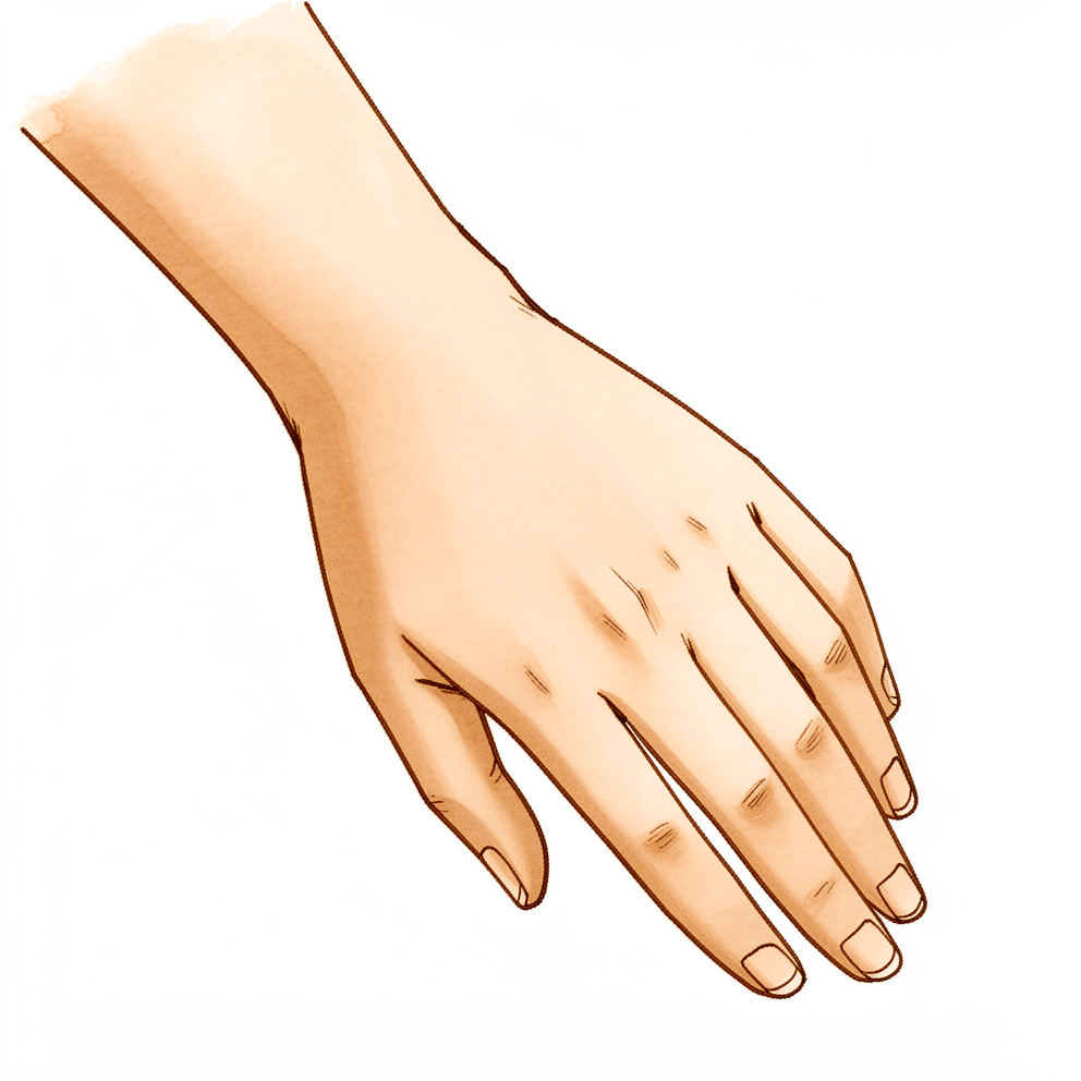
O seu punho não é uma única articulação. É um pequeno conjunto de oito ossos organizados em duas fileiras, entre o antebraço e a mão. Os ossos de cada fileira são unidos por faixas fortes de tecido chamadas ligamentos, e todo o conjunto se move como um sistema interligado quando você dobra, estica ou gira o punho. Quando um desses ossos quebra, ou os ligamentos que mantêm as fileiras unidas se rompem, esse sistema interligado deixa de funcionar de forma suave. O punho perde o seu ritmo normal, por isso colocar carga sobre ele dói e a preensão parece fraca ou pouco confiável.
A maioria dessas fraturas acontece em uma queda sobre a mão estendida. A força desce pela mão e se concentra em um pequeno osso, e esse osso trinca. Às vezes, a força rompe ligamentos em vez de quebrar o osso, ou além de quebrá-lo. O piramidal, no lado do dedo mínimo do punho, é o que quebra com mais frequência. Os outros, o semilunar, o capitato, o trapézio, o trapezoide e o pisiforme, quebram com muito menos frequência.
Pense no punho como uma pilha de blocos de montar presos com elásticos. Se um bloco trinca mas permanece no lugar, a pilha ainda consegue suportar peso enquanto a trinca se solda. O osso cicatriza assim: as duas bordas formam osso novo através da falha, que vai ficando mais firme ao longo de semanas. Mas, se um bloco sai do alinhamento, ou se os elásticos que mantêm a pilha unida se rompem, a pilha não consegue mais se alinhar sozinha. Os ossos podem deslizar de maneiras para as quais nunca foram feitos, e é isso que causa problemas duradouros se não for corrigido.
É por isso que o padrão da fratura é importante. Uma fratura limpa que não se deslocou muitas vezes pode melhorar em um gesso enquanto consolida. Uma fratura que saiu do lugar, ou que tem ligamentos rompidos ao lado, pode precisar de cirurgia para manter os fragmentos imóveis enquanto se soldam. Ter o diagnóstico cedo também é importante, porque algumas dessas lesões passam despercebidas com facilidade no início e causam mais problemas depois se não forem tratadas.
O que podemos fazer a respeito
O Dr Kieran Hirpara, cirurgião de membro superior no Mater Private Hospital Rockhampton, adapta o tratamento à sua lesão específica. Algumas dessas fraturas consolidam sem operação, e outras precisam de cirurgia logo após a lesão, por isso uma avaliação rápida é importante. Em geral, os pacientes são encaminhados à nossa clínica pelo médico de família; caso um fisioterapeuta tenha sugerido que você nos procure, ainda assim será necessário um encaminhamento do seu médico de família para que você tenha direito ao reembolso do Medicare. Na clínica, levantamos o seu histórico, examinamos o seu punho e solicitamos exames de imagem quando necessário para descobrir qual osso quebrou e se ele saiu do lugar.
Se a fratura for estável e os fragmentos não tiverem saído do lugar, geralmente a tratamos sem cirurgia. Isso significa uma tala ou um gesso para manter o punho imóvel enquanto o osso se solda, muitas vezes por cerca de 4 semanas em algumas dessas lesões. Podemos verificar a consolidação com novos exames de imagem ao longo do caminho. Conforme o conforto permitir, você começa alongamentos e movimentos leves para evitar que o punho fique rígido. Um terapeuta de mão orienta esse retorno ao movimento em etapas e confecciona qualquer tala de que você precise.
A cirurgia é recomendada desde o início quando os fragmentos quebrados se deslocaram, quando os ossos ou os ligamentos do punho estão instáveis, ou quando o padrão da lesão exige que os ossos sejam mantidos com precisão enquanto consolidam. Lesões de alta energia que quebram vários ossos ou rompem ligamentos muitas vezes se enquadram nesse grupo. O objetivo é realinhar os ossos e mantê-los imóveis para que possam consolidar na posição correta. Às vezes existe uma escolha real: uma fratura pode consolidar em um gesso, mas a dor ou a posição final do osso podem não ser adequadas para o seu trabalho ou esporte, e conversaremos sobre isso com você.
Seja qual for o caminho, as primeiras semanas são dedicadas ao conforto e à proteção. O alívio da dor mantém você confortável enquanto o inchaço diminui. Você segue com a tala ou o gesso e com o plano até confirmarmos na radiografia que o osso consolidou, mesmo que o punho comece a melhorar antes disso. A terapia da mão no momento certo ajuda você a recuperar o movimento e a força sem colocar carga sobre a lesão cedo demais.
O que esperar
A consolidação é medida em semanas e meses, e não em dias. Em algumas dessas fraturas, o punho pode melhorar e funcionar bem muito antes de a radiografia mostrar que o osso está totalmente soldado. Siga com a tala ou o gesso e com o plano até confirmarmos na radiografia que o osso consolidou, mesmo que o seu punho pareça bem.
Se a sua fratura estiver consolidando em um gesso ou tala, o osso geralmente se solda ao longo de várias semanas. Um caso relatado de fratura do escafoide e do trapézio ao mesmo tempo mostrou os ossos unidos após seis semanas, com movimento completo do punho e do polegar e força de preensão normal às dezesseis semanas. Com o passar das semanas, primeiro a dor diminui; depois, o movimento e a força voltam com a terapia da mão. A maioria das pessoas volta às tarefas do dia a dia conforme o conforto permite, e o trabalho mais pesado ou o esporte vêm mais tarde, quando o osso estiver firme.
Se você fizer cirurgia, o osso ainda precisa de tempo para se soldar, mas os fragmentos ficam imóveis enquanto isso acontece. A recuperação se estende por meses. Em um caso de fratura combinada do escafoide e do semilunar, os dois ossos consolidaram, com o movimento do punho e a força de preensão preservados 10 meses após a cirurgia. Outra lesão complexa envolvendo vários ossos do punho teve um resultado razoável, com pontuação de 75 em 100 em uma escala de função do punho aos quatro anos, enquanto uma lesão semelhante teve pontuação de 85 em 100 aos dois anos. Uma fratura do trapezoide tratada sem cirurgia deixou o paciente sem dor e com movimento completo do punho aos 12 meses.
A maioria das fraturas isoladas como essas consolida bem. Entre as coisas que podem dar errado estão o osso não se soldar, soldar-se em uma posição ruim ou o punho acabar ficando rígido. Esses riscos aumentam quando vários ossos ou ligamentos são lesionados ao mesmo tempo, e é por isso que o padrão da sua fratura define o plano. Se uma fratura passar despercebida ou o tratamento for adiado, o osso pode não consolidar, pode perder o suprimento de sangue, ou os ossos do punho podem ficar instáveis. É por isso que verificamos a consolidação ao longo do caminho e pedimos que você volte se os sintomas não estiverem melhorando, estiverem piorando ao longo de semanas, acordarem você à noite ou impedirem você de trabalhar ou de usar a mão.
Quando procurar ajuda médica
Procure atendimento urgente se o seu punho parecer claramente deformado, se houver uma ferida na pele sobre a lesão, ou se você perder subitamente a sensibilidade ou o movimento da mão após uma lesão. Essas lesões também podem vir acompanhadas de problemas nos nervos ou nos vasos sanguíneos, por isso dormência, formigamento ou uma mão que fica pálida ou fria precisam de avaliação no mesmo dia.
Consulte o seu médico de família ou peça uma avaliação com um especialista se a dor não estiver melhorando, ou se o inchaço, o movimento ou a força de preensão não estiverem melhorando semana a semana à medida que o osso consolida. Como essas fraturas passam despercebidas com facilidade em uma primeira radiografia, um punho que continua dolorido após uma lesão merece um exame mais detalhado. Exames feitos cedo podem detectar o que as imagens simples não mostram, e uma avaliação rápida ajuda a evitar os problemas de consolidação mais lenta e de instabilidade que acompanham um diagnóstico tardio.
Evidence & references
This is the clinical evidence summary written for health professionals. It is technical, and it lists the research this page was built from. You do not need to read it to understand your treatment or to make a decision about it.
Overview
- Carpal fractures other than the scaphoid account for 15% to 41% of carpal fractures [1].
- Triquetral fractures are the most common carpal fracture other than the scaphoid [1].
- Fractures of the carpal bones excluding the scaphoid represent 30% to 40% of all osseous injuries to the wrist [3].
- Non-scaphoid carpal fractures are a clinically relevant and underdiagnosed category of wrist trauma [2].
- Non-scaphoid carpal fractures are particularly underdiagnosed in high-risk, young male populations [2].
- Fractures of the carpus other than the scaphoid are frequently missed on initial presentation [4].
- Diagnosis of carpal fractures other than the scaphoid requires a high index of suspicion with tailored imaging [4].
- Carpal fractures other than the scaphoid are exceedingly rare and comprise approximately 1.1% of all fractures [5].
- Management of carpal fractures other than the scaphoid depends on the specific fracture pattern, displacement, and associated ligamentous injuries [5].
- Outcomes for isolated carpal fractures other than the scaphoid are generally good [5].
- Outcomes for carpal fractures other than the scaphoid are complicated by nonunion and instability in cases with concomitant injuries [5].
- Fractures of the carpal bones excluding the scaphoid are fairly uncommon [6].
- Fractures of the carpal bones excluding the scaphoid can cause a disproportionate amount of morbidity from missed diagnosis due to their subtlety [6].
- Fractures of the carpal bones excluding the scaphoid are frequently associated with significant ligamentous disruption or other carpal bone fractures [6].
- Early magnetic resonance (MR) imaging can exclude carpal fracture when initial radiographs yield negative results [7].
- If initial radiographs are negative, a fracture of the trapezium is more likely to be present than one of the scaphoid despite high levels of clinical suspicion for scaphoid injuries [8].
- Early recognition of uncommon carpal disruptions may guide appropriate surgical treatment and improve long-term functional outcomes [9].
- Timely recognition and thoughtful management of capitate fractures, including the use of advanced imaging and individualized surgical strategies, may help reduce the risk of long-term complications and support favorable outcomes in select cases [10].
- When a trapezium fracture is identified, additional evaluation for ulnar-sided injuries of the carpus is recommended [11].
- Carpal fractures other than the scaphoid are rare but can be devastating if missed [12].
- Prompt diagnosis and early treatment of carpal fractures other than the scaphoid are crucial to prevent complications such as nonunion, avascular necrosis, and carpal instability [12].
- Physicians must discriminate between carpal injuries in athletes that are manageable with early return to sport and those requiring aggressive treatment to prevent late disability [13].
- A lunate fracture has been associated with an unstable fracture of the scaphoid [14].
- Isolated trapezoid fractures may be appropriately treated without surgical intervention [15].
- Isolated scaphoid and other carpal fractures exhibit different demographics in terms of age and gender [18].
- Differences in demographics for isolated scaphoid and other carpal fractures may be related to differences in the mechanism of injury [18].
- A high-energy mechanism of injury was associated with multiple carpal fractures [18].
- The capitate is covered mainly by cartilage [20].
- The blood supply to the capitate is tenuous and can be compromised by transverse fractures [20].
- Scaphocapitate syndrome refers to a greater arc injury pattern in which force passes from the scaphoid to the capitate neck, resulting in both scaphoid and capitate fractures [20].
- In scaphocapitate syndrome, the capitate head may be rotated 180° [20].
- Scaphocapitate syndrome with a rotated capitate head requires open reduction and internal fixation (ORIF) through a dorsal approach [20].
- Fracture-dislocation of the ring and little metacarpal joints often result in fracture of the hamate [20].
- Hamate fractures resulting from ring and little metacarpal joint fracture-dislocations involve a shearing fracture of the dorsal part of the hamate with or without central articular impaction [20].
- Small hamate fractures without articular impaction can be treated with closed reduction of the carpometacarpal joint and 4 weeks of Kirschner wire (K-wire) immobilization [20].
- Larger hamate fractures and fractures with articular impaction are treated with open reduction and screw fixation [20].
- Fracture of the hook of the hamate usually results from a direct blow from a golf club, baseball bat, or racket [20].
- Most hamate hook fractures are diagnosed months after injury as a nonunion causing tenderness with direct pressure [20].
- Acute hamate hook fractures tend to heal but often go undiagnosed [20].
- Surgery for hamate hook fractures is elective and usually consists of excision of the hook of the hamate [20].
- If a stable anatomic situation is not obtained with closed reduction for transscapho-transcapitate fracture dislocation, surgical reduction and internal fixation of the scaphoid and capitate bones are recommended to prevent loss of function [27].
- Two-stage management may be considered in carefully selected severe trapezial bone defect cases when conventional fixation or bone grafting is unlikely to restore joint congruity and thumb motion [70].
- Acute proximal row carpectomy is an option for some patients with complex carpal fracture dislocations [117].
- Acute proximal row carpectomy is particularly an option for patients with complex carpal fracture dislocations involving fracture of the lunate, concomitant scaphoid fracture and scapholunate ligament injury, or preexisting wrist arthritis [117].
Anatomy & Pathophysiology
Bony Anatomy
- The carpus consists of two rows of eight bones that serve as a bridge between the forearm and the hand [44].
- The proximal carpal row includes the scaphoid, lunate, and triquetrum [44].
- The distal carpal row includes the trapezium, trapezoid, capitate, and hamate [44].
- The pisiform is a sesamoid bone enclosed within the sheath of the flexor carpi ulnaris tendon and is not theoretically considered part of the proximal carpal row [44].
- The distal carpal row articulates with the proximal carpal row and distally with the five metacarpals by forming a transverse arch [44].
- The trapezium articulates with the first metacarpal, the trapezoid with the second, the capitate with the third, and the hamate with the fourth and fifth [44].
- The capitate and trapezoid are tightly connected to the metacarpals, whereas there is 30 to 40 degrees of flexion–extension and rotation at the metacarpotrapezial joint [44].
- The proximal carpal row has no direct tendinous attachments, and its movement results from bone shape, interaction with other bones, and ligament attachments [44].
- The distal carpal row is rigid with little motion between its bones due to stout intercarpal ligaments [42].
- The proximal row of carpal bones forms an intercalated segment between the distal carpal row and the distal radius, bound into a functional unit by the scapholunate and lunotriquetral interosseous ligaments [42].
- The scaphoid bridges both carpal rows [42].
- The capitate and hamate have only 2 degrees of intercarpal rotation and less than 2 mm of proximal-distal translation [75].
- There are no ligaments, palmar or dorsal, between the lunate and capitate [75].
Ligamentous Anatomy
- Extrinsic carpal ligaments connect the radius or ulna to the carpus [42].
- In general, volar ligaments are stronger than dorsal ligaments [42].
- The scapholunate interosseous ligament is the major stabilizer of the wrist and the most commonly injured wrist ligament [42].
- The scapholunate interosseous ligament is C-shaped, consisting of dorsal, palmar, and interosseous portions, with the dorsal portion being the strongest and thickest [42].
- The lunotriquetral interosseous ligament is C-shaped, with the volar portion being the thickest and strongest [42].
- The capitohamate ligament is a thick ligament measuring 5 × 5 mm in cross section, with extensions to the third or fourth metacarpals [42].
- The dorsal intercarpal ligament passes from the dorsal tubercle of the triquetrum to the distal pole of the scaphoid [42].
- The dorsal intercarpal ligament reinforces the elastic dorsal wrist capsule and helps stabilize the scapholunate articulation [42].
- The space of Poirier is an area adjacent to the proximal capitate without ligamentous attachment, situated ulnar to the radioscaphocapitate ligament and radial to the long radiolunate ligament in the floor of the carpal tunnel [42].
- The space of Poirier is a weak area vulnerable to instability, through which the distal carpal row separates from the lunate during a perilunate dislocation [42].
- The dorsal scaphotriquetral ligament spans the dorsal-distal rims of the scaphoid, lunate, and triquetrum [75].
- The dorsal scaphotriquetral ligament represents both a robust attachment of the transverse fibers of the dorsal intercarpal ligament to each bone of the proximal carpal row and a labral extension of the interosseous scapholunate and lunotriquetral ligaments across the dorsal base of the capitate [75].
- The palmar scaphotriquetral ligament spans transversely from the waist of the scaphoid to insert just palmar and distal to the lunotriquetral ligament on the triquetrum [75].
- The dorsal and palmar scaphotriquetral ligaments complete a near-acetabular shaped articulation for the capitate [75].
- The dorsal capsuloligamentous scapholunate septum is a fibrous capsular structure that originates from the deep surface of the intracapsular dorsal intercarpal ligament and inserts on the dorsal scapholunate interosseous ligament [75].
- The dorsal intercarpal ligament is a critical stabilizer of the proximal carpal row through its connections to the dorsal scapholunate and lunotriquetral interosseous ligaments [75].
- The dorsal intercarpal ligament has a distal component that inserts on the trapezoid and trapezium, crossing the capitate neck to add stability to the midcarpal joint [75].
- The palmar triquetrohamate and triquetrocapitate ligaments are thick structures that play an important role in the stabilization of the midcarpal joint [75].
- The scaphoid tuberosity is linked to the distal row by the anteromedial scaphocapitate ligament and the stout radiopalmar scaphotrapeziotrapezoid ligament [75].
- Disruption of the strong ligamentous attachments of the scaphoid to the distal carpal row has been associated with both dissociative and nondissociative instability patterns of the proximal carpal row [75].
- The distal carpal row bones are strongly bound to each other by stout transverse intercarpal ligaments, which are essential to ensure the rigidity of the transverse carpal arch and protect the carpal tunnel contents [75].
Vascular Anatomy
- Circulation to the carpus is comprised of an extraosseous and intraosseous vasculature via both dorsal and palmar vascular systems, which are branches of the radial, ulnar, anterior interosseous, and deep palmar arch arteries [76].
- The extraosseous arterial supply is formed by an anastomotic network of dorsal and palmar transverse arches connected longitudinally by the radial, ulnar, and anterior interosseous arteries [76].
- The three dorsal transverse arches of the carpus include the radiocarpal, intercarpal, and basal metacarpal arches [76].
- The three palmar transverse arches of the carpus include the radiocarpal, intercarpal, and deep palmar arches [76].
- The incidence of avascular necrosis following injury to the carpal bones is related to their complex intraosseous blood supply [76].
- The vascular supply of most carpal bones enters the distal half, leaving the proximal half at risk of avascular necrosis [76].
- The scaphoid, capitate, and about 20% of all lunates are supplied by a single vessel, increasing their risk of avascular necrosis [76].
- The trapezium, triquetrum, pisiform, and 80% of lunates receive nutrient arteries through two nonarticular surfaces and have consistent intraosseous anastomoses, reducing the risk of avascular necrosis [76].
- The trapezoid and 50% of hamates lack an intraosseous anastomosis and are at risk of avascular fragments [76].
- The scaphoid is supplied by scaphoid branches of the radial artery, with the dorsal branch supplying 70–80% proximally [76].
- The lunate receives a volar branch supplying 20–30% distally, with approximately 80% receiving vessels from palmar and dorsal surfaces [76].
- The triquetrum receives branches of the ulnar artery, dorsal intercarpal arch, and palmar intercarpal arch, with dorsal vessels supplying 60% and palmar vessels supplying 40% [76].
- The pisiform receives branches of the ulnar artery, with vessels entering through three nonarticular surfaces [76].
- The trapezium receives branches of the radial artery, with dorsal supply predominating [76].
- The trapezoid receives dorsal vessels supplying 70% and palmar vessels supplying 30% [76].
- The capitate receives branches of the dorsal intercarpal arch, dorsal basal metacarpal arch, palmar intercarpal arch, and ulnar recurrent artery [76].
- In one-third of cases, the supply to the capitate head is solely from the palmar side [76].
- The hamate receives branches of the dorsal intercarpal arch, the ulnar recurrent artery, and the ulnar artery [76].
- Dorsal vessels supply the dorsal 30–40% of the hamate [76].
Biomechanics and Kinematics
- The wrist can be considered a two-joint system linking the hand to the forearm around the highly mobile bones of the proximal carpal row [71].
- The two principal articulations are the radiocarpal and midcarpal joints, situated proximal and distal to the mobile proximal carpal row [71].
- Approximately 62° of wrist extension occurs through the radiocarpal joint, and 62% of wrist flexion occurs through the midcarpal joint [42].
- The midcarpal joint is mostly responsible for 20° of radial deviation and 40° of ulnar deviation [42].
- The midcarpal joint is responsible for the "dart thrower’s motion," which involves moving from radial extension into ulnar flexion positioning of the wrist [42].
- The radius bears 80% of the axial load transmitted through the radiocarpal joint, while the ulna bears 20% in neutral ulnar variance [42].
- During wrist flexion from neutral, the distal row flexes and ulnarly deviates slightly while the scaphoid pronates [42].
- During wrist flexion from neutral, the proximal row flexes differentially, with more rotation through the scaphoid, followed by the triquetrum and the lunate [42].
- The proximal row translates dorsally during wrist flexion [42].
- During wrist extension from neutral, the distal row extends and radially deviates slightly while the scaphoid supinates [42].
- During wrist extension from neutral, the proximal row extends differentially, with more motion in the scaphoid, followed by the triquetrum and then the lunate [42].
- The proximal row translates palmarly during wrist extension [42].
- The dart-thrower’s path of radial extension to ulnar flexion defines the transition between flexion and extension of the scaphoid and lunate [71].
- The dart-thrower’s motion occurs almost exclusively through the midcarpal joint [71].
- The reversed dart-thrower’s motion, from extension–ulnar inclination to flexion–radial inclination, is mediated by the flexor carpi radialis on the volar side and the extensor carpi ulnaris on the dorsal side [72].
- The contribution of the midcarpal joint to the reversed dart-thrower’s type of rotation is minimal, with this motion occurring predominantly through the radiocarpal joint [72].
- Distal to the extensor retinaculum, wrist tendons change direction, taking an oblique path to their distal insertion [72].
- Isolated isometric contraction of the extensor carpi ulnaris and flexor carpi radialis muscles will cause distal row pronation [72].
- Isolated isometric contraction of the extensor carpi radialis longus, flexor carpi ulnaris, and abductor pollicis longus muscles will induce a supination moment to the distal row [72].
Pathophysiology and Mechanisms of Injury
- Carpal fractures other than the scaphoid account for 15% to 41% of carpal fractures, with triquetral fractures being the most common [1].
- Fractures of the carpal bones excluding the scaphoid are fairly uncommon but can cause a disproportionate amount of morbidity from missed diagnosis due to their subtlety and frequent association with significant ligamentous disruption or other carpal bone fractures [6].
- Non-scaphoid carpal fractures are a clinically relevant and underdiagnosed category of wrist trauma, particularly in high-risk, young male populations [2].
- Fractures of the carpus other than the scaphoid are frequently missed on initial presentation and require a high index of suspicion with tailored imaging for diagnosis [4].
- Most carpal fractures are the consequence of a fall onto an outstretched hand [88].
- The energy from a fall onto an outstretched hand may be focused on the distal carpal row, producing an extension moment across the proximal carpal row transmitted through the volar carpal ligaments [88].
- Perilunate-pattern or "lesser arc" injuries occur when only ligaments are injured without fracture [88].
- Carpal fractures or ligamentous injuries may occur in an arc around the lunate [88].
- Fractures of the scaphoid, capitate, triquetrum, or radial styloid, or all four, in conjunction with perilunate instability, are known as "greater arc" injuries [88].
- Strong anterior to posterior compression injuries may result in axial-pattern disruption of the carpus [88].
- Axial-pattern disruption propagates both radially and ulnarly, separating the carpus on both sides of the capitate [88].
- Avulsions may occur owing to localized forced concentration, commonly causing avulsion injuries at the volar or dorsal aspect of the triquetrum secondary to ligament insertions [88].
- Trapezial, hamate, and pisiform fractures may also occur as avulsion injuries [88].
- An injury to the carpus commonly occurs following a mechanism in which an axial compression force is applied to the wrist, commonly leading to hyperextension where the palmar ligaments undergo tension and the dorsal articulations are subject to shear stresses [100].
- The degree of force applied to the wrist and the degree of wrist radial or ulnar deviation determine whether a ligament injury, a fracture, or both occur [100].
- Minor injuries, such as ligamentous sprains, frequently result from a low-energy injury [100].
- Higher-energy injuries that involve a more considerable force result in either a fracture to one or more of the carpal bones and/or a ligamentous disruption [100].
- Variations in bone quality, the direction and magnitude of the deforming force, and the position of the wrist at the time of injury explain the variety of injuries that can occur [100].
- Most dorsal perilunate dislocations are the result of an indirect mechanism of injury, usually an extreme extension of the wrist associated with variable degrees of ulnar deviation and midcarpal supination [50].
- Mayfield and colleagues identified four stages of progressive carpal destabilization in perilunate injuries [50].
- Stage I of progressive perilunate instability involves scapholunate dissociation or scaphoid fracture [50].
- Stage II of progressive perilunate instability involves lunocapitate dislocation [50].
- Stage III of progressive perilunate instability involves lunotriquetral disruption or triquetrum fracture [50].
- Stage IV of progressive perilunate instability involves lunate dislocation [50].
- In Stage I, when the distal carpal row is forced into hyperextension, the palmar midcarpal scaphotrapeziotrapezoid and scaphocapitate ligaments become taut, creating a progressive extension moment to the scaphoid transmitted to the lunate via the scapholunate ligaments [50].
- In Stage I, if the wrist is somewhat radially deviated, a fracture of the scaphoid is likely instead of scapholunate dissociation [50].
- In Stage II, once the scapholunate joint is disrupted or the scaphoid fractured, increased wrist extension may cause the distal row to translate dorsally and dislocate relative to the lunate, or a capitate fracture may appear with its distal portion dislocating dorsally [50].
- In Stage II, the head of the capitate remains behind and may rotate 180 degrees, a condition known as scaphocapitate syndrome [50].
- In Stage III, as the capitate displaces dorsally, the triquetrohamate ligamentous complex becomes tensed, creating an extension moment and dorsal translation vector to the triquetrum [50].
- In Stage III, such forces may result in separation of the triquetrum from the lunate secondary to rupture of the lunotriquetral ligaments or a sagittal fracture of the triquetrum [50].
- In Stage IV, when all perilunate ligaments are torn, the dorsally displaced capitate may exert a palmar translation force to the dorsum of the lunate, resulting in a palmar lunate extrusion [50].
- Perilunate dislocations are characterized by a progressive disruption of capsular and ligamentous connections of the lunate to the adjacent carpal bones and radius, without associated fractures to the carpus and distal radius [98].
- Ligament disruption in perilunate dislocations
Classification
- Carpal fractures other than the scaphoid comprise approximately 1.1% of all fractures [5].
- Isolated trapezium fractures account for 1 to 5% of all carpal fractures [33].
- The trapezium is the third most common carpal bone to fracture [33].
- Twenty percent of trapezium fractures are isolated with a vertical sagittal split [33].
- Trapezial fractures are classified into two main categories: fractures involving the palmar ridge and fractures through the body [110].
- Trapezial palmar ridge fractures are subdivided into type I fractures located at the base of the ridge and type II fractures located at the tip of the ridge [110].
- Pisiform fractures are classified into four types: Type 1 transverse, Type 2 sagittal, Type 3 comminuted, and Type 4 pisotriquetral impaction [110].
- Type 1 transverse pisiform fractures are the most common type of pisiform fracture [110].
Clinical Presentation
Epidemiology and Demographics
- The mean age at the time of injury for all carpal fractures ranges from 35 to 40 years, with a male predominance observed [54].
- Fractures of the scaphoid, hamate, pisiform, and trapezium occur predominantly in younger males, with a mean age ranging from 29 to 43 years and a male predominance ranging from 66% to 100% [54].
- Triquetral fractures occur at a mean age of 51 years with an approximately equal gender distribution [54].
- A fall from standing height accounts for almost two-thirds of all carpal injuries [54].
- Non-scaphoid carpal fractures are particularly prevalent in high-risk, young male populations [2].
- Isolated scaphoid and other carpal fractures exhibit different demographics in terms of age and gender, which may be related to differences in the mechanism of injury [18].
General Clinical Findings
- Patients with an injury to the carpus commonly present with wrist pain as their primary complaint [36].
- Pain, swelling, and ecchymosis around the region of the carpus may be present in the acute phase of injury [36].
- The most constant and dependable sign of carpal injury is well-localized tenderness [36].
- Swelling over the proximal carpal row is suggestive of a ligament avulsion with or without an associated fracture [36].
- With carpal instability or dislocation, a gross deformity may be apparent, such as a marked prominence of the entire carpus dorsally suggestive of a perilunate dislocation [36].
- Compressive stresses applied actively or passively may produce pain at the site of damage and cause a palpable and audible snap, click, shift, catch, or clunk [36].
- Stress loading the wrist with compression and motion from radial to ulnar deviation may simulate midcarpal instability and produce a “catch-up clunk” as the proximal row of carpal bones snap from flexion to extension [36].
- A full examination of the contralateral wrist can often be helpful, particularly when assessing for instability [36].
- Carpal fractures other than the scaphoid are frequently missed on initial presentation and require a high index of suspicion with tailored imaging for diagnosis [4].
- Carpal bone fractures are likely underreported due to complex anatomy and difficulty in plain radiologic interpretation [38].
- Injuries to the hand and wrist can be occult in nature, requiring a thorough clinical evaluation to guide the differential diagnosis and the need for further imaging in the setting of negative radiographs [22].
- Delays in diagnosis should be avoided to prevent complications such as nonunion and secondary displacement [22].
- Accurate diagnosis and management of hand and carpal fractures are predicated on a thorough physical examination and appropriate imaging to limit joint stiffness while preserving mobility and function [19].
Specific Bone Findings
- Tenderness in the anatomical snuffbox is a sign of scaphoid injury [36].
- Tenderness distal to Lister's tubercle is a sign of scapholunate and lunate injury [36].
- Tenderness on the dorsal margin, fingerbreadth distal to the ulnar head, is a sign of triquetral, lunotriquetral ligament, and triquetrohamate ligament injury [36].
- Localized pain and tenderness are clinical findings for trapezium fractures [23].
- Symptoms and signs of trapezoid fractures can be minimal and imitate a scaphoid fracture [23].
- Localized pain and tenderness are clinical findings for capitate fractures [23].
- Localized pain and tenderness are clinical findings for hamate fractures, though often minimal [23].
- An ulnar nerve lesion may be present with hamate fractures, as the deep branch of the ulnar nerve passes around the hook of the hamate [23].
- Tendon rupture may be present with chronic presentations of hamate fractures [23].
- Localized pain and tenderness are clinical findings for pisiform fractures [23].
- Clinicians should consider trapezoid fracture as a possible differential diagnosis when presented with carpal pain, swelling, or limited movement, particularly after axial load incidents [35].
- Trapezium fracture is usually accompanied by other upper extremity and wrist injuries, but it may also occur as an isolated entity [61].
- Capitate fractures are often associated with injury to the other carpal bones and/or the metacarpals in direct blow or crush injuries [23].
- Fracture-dislocation of the ring and little metacarpal joints often results in fracture of the hamate [20].
- Fractures of the triquetrum in children are often subtle flake avulsion or impingement fractures that require good oblique radiographs for recognition [34].
- The incidence of triquetral fractures in children is probably much higher because many are misdiagnosed as wrist sprains or type I physeal injuries of the distal radius and ulna [34].
Associated Injuries and Instability
- Non-scaphoid carpal fractures are frequently associated with significant ligamentous disruption or even other carpal bone fractures [6].
- Tuberosity fractures of the trapezium are associated with fractures of the hook of the hamate, or with dislocation of the hamate [23].
- Articular surface fractures of the trapezium can be associated with dislocation or fracture-dislocation of the first carpometacarpal joint [23].
- Associated scaphoid fractures have been reported with trapezium fractures [23].
- Associated fractures occur in a third of trapezoid fracture cases [23].
- Scaphoid and capitate fractures can occur with concurrent scapholunate dissociation [21].
- Comminuted lunate fractures can be combined with distal radius fracture and scaphoid fracture [25].
- Non-dislocation lunate fractures can be combined with distal radius fracture [28].
- Perilunate dislocations are often associated with a polytrauma, with 1 in 10 sustaining an associated upper-limb injury [107].
- In around 16% of perilunate dislocation cases, the clinical presentation includes median nerve symptoms and signs [107].
- Ulnar neuropathy, arterial injury, or tendon disruption may also be seen with perilunate dislocations [107].
- Some perilunate dislocations may be seen several months or years after the initial injury [107].
- Patients with delayed presentation of perilunate dislocation are more likely to present because of increasing nerve symptoms or tendon rupture than because of wrist deformity [107].
- Acute carpal tunnel syndrome can occur with associated carpal fractures and dislocations [95].
- A full neurovascular assessment is imperative in the setting of carpal fractures and dislocations [95].
Diagnostic Challenges
- Fractures of the carpal bones excluding the scaphoid are fairly uncommon but can cause a disproportionate amount of morbidity from missed diagnosis due to their subtlety [6].
- Prompt diagnosis and early treatment are crucial to prevent complications such as nonunion, avascular necrosis, and carpal instability [12].
- Timely recognition and thoughtful management of capitate fractures may help reduce the risk of long-term complications and support favorable outcomes in select cases [10].
- Physicians must discriminate between injuries manageable with early return to sport and those requiring aggressive treatment to prevent late disability [13].
- Isolated trapezoid fractures are rare and often occult on initial radiography, requiring CT for diagnosis [30].
- Trapezium fractures are hard to detect and easily missed on standard radiographs [33].
- Fractures of the pisiform and hamate are not seen on standard two or three view wrist examination [17].
- Coronal fractures of the trapezoid are rarely detected on radiographs [23].
- CT or MRI is used for diagnosis in over 80% of trapezoid fracture cases [23].
- CT scanning is a more accurate diagnostic tool for carpal bone fractures than plain radiology [38].
- On obtaining negative results on initial radiographs, early magnetic resonance imaging can exclude carpal fracture [7].
- CT scan is imperative to diagnose and plan treatment of complex fracture morphology such as divergent trapezium-trapezoid fracture dislocation [24].
- For comminuted lunate fractures, early identification and intervention can preserve most of the function of the wrist joint [25].
- If necessary, CT and MRI should be performed to prevent severe secondary diseases and wrist joint dysfunction in non-dislocation lunate fractures combined with distal radius fracture [28].
- The injury of capitate fractures has been diagnosed and reported with undeserved rarity, and a higher index of suspicion would lead to the recognition and treatment of more of these fractures [67].
- Rapid recognition of unknown wrist pathology that could not be confirmed by plain radiographs is necessary in the presence of a suspected boxer's fracture [66].
Investigations
General Diagnostic Challenges
- Fractures of the carpal bones excluding the scaphoid are fairly uncommon but can cause a disproportionate amount of morbidity from missed diagnosis due to their subtlety as well as their frequent association with significant ligamentous disruption or even other carpal bone fractures [6].
- Anatomical snuffbox tenderness is suggestive of scaphoid injury [36].
- Tenderness distal to Lister's tubercle is suggestive of scapholunate and lunate injury [36].
- Tenderness on the dorsal margin, fingerbreadth distal to the ulnar head, is suggestive of triquetral, lunotriquetral ligament, and triquetrohamate ligament injury [36].
- A marked prominence of the entire carpus dorsally is suggestive of a perilunate dislocation [36].
- The scaphoid shift test is diagnostic of scapholunate disruption if there is a “clunk” as the scaphoid subluxates dorsally out of the scaphoid fossa [36].
- Up to 30% of normal wrists have a positive result on the scaphoid shift test [36].
- The midcarpal shift test is diagnostic of midcarpal instability if there is a “clunk” as the lunate reduces from the palmarflexed position [36].
- Lunotriquetral ballottement is diagnostic of lunotriquetral instability or arthritis if painful [36].
- The lunotriquetral shear test is diagnostic of lunotriquetral instability if it results in reproducing the patient's pain along with palpable crepitation or clicking [36].
- Accurate diagnosis and management of hand and carpal fractures and dislocations are predicated on a thorough physical examination and appropriate imaging to limit joint stiffness while preserving mobility and function [19].
- Clinicians should maintain a high level of vigilance and consider trapezoid fracture as a possible differential diagnosis when presented with carpal pain, swelling, or limited movement, particularly after axial load incidents [35].
- This case illustrates the importance of accurate diagnosis by fluoroscopy of the wrist in order to identify additional instabilities and fractures in severe hand trauma [21].
Radiography
- The four standard views commonly employed in the assessment of scaphoid fractures can be used to detect most injuries to the carpus [36].
- Standard neutral PA and lateral radiographs are useful for determining the presence of clear fractures and assessing carpal alignment [36].
- Standard neutral PA and lateral radiographs are often poor for scaphoid fracture detection due to the tubercle overhang on the PA and the overlap on the lateral [36].
- The 45-degree radial oblique, 45-degree ulnar oblique, ulnar deviated AP, Ziter's, and carpal box or tunnel views are purported to improve the ability to diagnose a fracture, particularly of the scaphoid [36].
- The VISI and DISI patterns of carpal malalignment are commonly detected using standard neutral lateral radiographs [36].
- Additional views in maximal radial and ulnar deviation are used if the diagnosis of VISI or DISI patterns is in doubt [36].
- Standard scaphoid views are the primary investigation for trapezium fractures [23].
- Carpal tunnel views are used for trapezium tuberosity fractures [23].
- Standard scaphoid views are the primary investigation for trapezoid fractures [23].
- Oblique views can aid diagnosis of trapezoid fractures [23].
- Overlap of carpals can make diagnosis of trapezoid fractures difficult [23].
- Coronal trapezoid fractures are rarely detected on radiographs [23].
- Standard scaphoid views are the primary investigation for capitate fractures [23].
- Lateral views are useful for determining displacement and rotation of the capitate head [23].
- Dynamic studies are used for capitate displacement [23].
- Standard scaphoid views are the primary investigation for hamate fractures [23].
- Loss of bone contour on radiographs is suspicious of hamate dislocation [23].
- Absence of the hook, sclerosis of the hook, and lack of cortical density are three signs indicative of a hook fracture [23].
- Oblique and carpal tunnel views improve the chance of diagnosing hook fractures [23].
- An alternate view with maximal radial deviation of the wrist and maximal abduction of the thumb is used for hamate evaluation [23].
- Standard scaphoid views are the primary investigation for pisiform fractures [23].
- Lateral views in 20–45 degrees of supination and carpal tunnel views aid diagnosis of pisiform fractures [23].
- Subluxation of the pisotriquetral joint is diagnosed with ≥1 of specific radiographic criteria [23].
- Standard scaphoid views will detect most triquetral fractures [56].
- Dorsal avulsion fractures of the triquetrum are often found on the oblique or lateral views [56].
- PA radiographs of the wrist are useful in identifying transverse body fractures of the triquetrum [56].
- PA radiographs often do not detect avulsion fractures of the triquetrum due to the normal superimposition of the dorsal lip on the lunate [56].
- An oblique pronated lateral radiograph projects the triquetrum even more dorsal to the lunate and can aid diagnosis [56].
- Pisiform and hamulus fractures are not seen on standard two or three view wrist examination [17].
- Plain radiography is approximately 50% sensitive for the detection of a scaphoid fracture and less than 50% for the other carpal bones [34].
- Fractures of the lunate can be difficult to detect on plain radiography [48].
- Fractures of the trapezoid are often occult on initial radiography [30].
- This case demonstrates an example of rapid recognition of unknown wrist pathology that could not be confirmed by plain radiographs in the presence of a suspected boxer's fracture [66].
Advanced Imaging (CT and MRI)
- On obtaining negative results on initial radiographs, early magnetic resonance (MR) imaging can exclude carpal fracture [7].
- CT scanning is a more accurate diagnostic tool for carpal bone fractures [38].
- CT/MRI is used for diagnosis of trapezoid fractures in over 80% of cases [23].
- CT/MRI is used for occult capitate fractures [23].
- CT/MRI is used for suspected hamate fractures [23].
- CT or MRI may be required to see lunate fractures [48].
- MRI can be helpful in the diagnosis of early avascular changes in the lunate [48].
- Correlation of the patient’s clinical and plain radiographic findings with MRI helps to differentiate Kienböck disease from ulnar impaction [48].
- If necessary, CT and MRI should be performed to prevent severe secondary diseases and wrist joint dysfunction in cases of non-dislocation lunate fracture combined with distal radius fracture [28].
- To better define associated bony injuries in radiocarpal instability, CT may be required [46].
- To determine the extent of ligamentous disruption in radiocarpal instability, MRI can be used [46].
- MRI has the highest sensitivity in detecting an occult scaphoid fracture [31, 32].
- A normal MRI study as early as 2 days after injury has a negative predictive value of 100% for scaphoid fracture [34].
- MRI is more sensitive than CT for making the diagnosis of scaphoid fracture [34].
- Using state-of-the-art technology, improved scanning algorithms, and dedicated wrist coils, MRI enables accurate evaluation of the articular cartilage, radioulnar ligaments, and the TFCC without ionizing radiation [83].
- Whether or not contrast improves the accuracy of MRI is controversial [83].
- A prospective study comparing MRI with magnetic resonance arthrography (MRA) in 60 consecutive patients with clinical and arthroscopic confirmation of TFCC injury demonstrated sensitivity and specificity for a TFCC tear of 68% and 60% (MRI) versus 95% and 100% (MRA) [83].
- Noncontrast MRI was 98% accurate in identifying arthroscopically confirmed tears and perforations of the articular disc in a study by Daunt and colleagues [83].
- Dynamic "4D" imaging may become an important tool to identify lesions and instability of the ulnocarpal joint and DRUJ [83].
- Arthroscopy, computed tomography, or magnetic resonance imaging can be very helpful as an adjunct to standard x-rays to better delineate the nature of the midcarpal joint and identify the presence or absence of a type II lunate facet [47].
Treatment
General Principles
- Treatment algorithms must balance fracture stability and immobilization with preservation of tendon gliding and joint mobilization to obtain optimal clinical outcomes [22].
Non-Operative Management
- Carpal fractures are predominantly treated nonoperatively [101].
- For non-scaphoid carpal fractures, nonoperative management is usually 3 to 6 weeks in cast or brace [101].
- Triquetral avulsions are managed with a splint for comfort only and active self-assisted stretches as comfort allows to limit stiffness [56].
- Triquetral avulsion fractures are considered wrist sprains and are treated symptomatically [20].
- Stretching exercises help limit the potential for wrist stiffness in triquetral avulsion fractures [20].
- Undisplaced trapezium fractures are treated with a scaphoid cast [23].
- Undisplaced trapezoid fractures (<2 mm) are treated with a scaphoid cast [23].
- Excellent results are often achieved with delayed treatment for undisplaced trapezoid fractures [23].
- Conservative management with immobilization results in satisfactory outcomes for minimally displaced isolated trapezoid fractures [30].
- Navicular cast immobilization for 4 to 6 weeks is recommended as initial treatment for isolated trapezium fractures [33].
- Undisplaced capitate fractures heal without immobilization or with a scaphoid cast [23].
- Nonoperative management in cast for approximately 4 weeks is suitable for most isolated lunate fractures [86].
- Nondisplaced and nondisplaced comminuted lunate fractures can be treated with cast immobilization [48].
- A transverse fracture of the lunate body will heal if it remains nondisplaced, particularly in adolescents [86].
- Undisplaced hamate fractures are treated with a cast, with union rates reported at only 50% in some series [23].
Operative Management
- Displaced trapezium fractures or dislocations are treated with closed reduction and internal fixation (CRIF) or open reduction and internal fixation (ORIF) [23].
- Arthroscopy has been reported for the treatment of trapezium fractures [23].
- Nonunion of ridge trapezium fractures is managed with excision [23].
- Displaced trapezoid fractures, dislocations, or delayed unions are treated with ORIF or excision [23].
- Displaced capitate fractures or dislocations are treated with ORIF [23].
- In scaphocapitate syndrome, the capitate head may be rotated 180°, requiring open reduction and internal fixation (ORIF) through a dorsal approach [20].
- If a stable anatomic situation is not obtained with closed reduction in transscapho-transcapitate fracture dislocations, surgical reduction and internal fixation of the scaphoid and capitate bones are recommended to prevent loss of function [27].
- Internal fixation of capitate fractures can be performed with Herbert screws [53].
- Displaced hamate fractures, dislocations, or those with nerve lesions are treated with excision or ORIF ± decompression of Guyon's canal [23].
- Surgery for hamate hook fractures usually consists of excision of the hook of the hamate [20].
- Some authors advocate bone grafting to preserve the pulley effect on the flexor tendons following hamate excision [23].
- Nonunion of hamate hook fractures can lead to chronic pain and little finger flexor tendon injury, managed with excision or ORIF ± bone grafting [23].
- Indications for ORIF of lunate fractures include displacement and/or associated carpal instability [86].
- Fractures of the lunate with more than 1 mm offset and avulsion fractures usually require open reduction [48].
- Internal fixation techniques for lunate fractures may include Kirschner wires, small cannulated screws, and suture anchors [48].
- Distraction with an external fixator may facilitate reduction of lunate fragments, particularly in the chronic setting [86].
- Triquetral body fractures associated with carpal disruption often require internal fixation [56].
- Fractures of the body of the lunate are approached through a standard dorsal approach via the third dorsal compartments [51].
- Body lunate fractures may be fixed with headless compression screws and/or Kirschner wires [51].
- Comminuted lunate fractures may require a cancellous bone graft harvested from the distal radius [51].
- Volar lunate fragments are reduced through an extended carpal tunnel approach exposing the volar lunate [51].
- Volar pole lunate fractures are often too small for screw fixation and may be secured with a suture anchor to capture both bone and palmar ligaments [51].
- If capitate fixation is not performed in scaphocapitate syndrome, the distal fragment of the scaphoid tends to migrate medially, making its reduction and stabilization difficult [94].
- The capitate is generally fixed with a headless screw in scaphocapitate syndrome [94].
- Primary bone grafting may be indicated in comminuted capitate fractures [94].
Associated Injuries and Complications
- Fractures of the carpal bones excluding the scaphoid are frequently associated with significant ligamentous disruption or even other carpal bone fractures [6].
- Outcomes for non-scaphoid carpal fractures are generally good for isolated injuries but complicated by nonunion and instability in cases with concomitant injuries [5].
- Nonunion of a lunate body fracture is rare, as most will progress to Kienböck's disease [86].
- If lunate nonunion progresses to Kienböck's disease, treatment includes radial shortening, radial wedge osteotomy, or ulnar lengthening in the early stages, with carpal arthrodesis if the condition is advanced [86].
- Avascular necrosis (AVN) is possible with displaced capitate fractures [23].
- Nonunion of capitate fractures is rare when undisplaced [23].
- Trauma to the lunate may be sufficient to damage the circulation, leading to osteonecrosis of the lunate [48].
- Lunates with a single vessel or one surface exposed to the blood supply, representing about 20% of lunates, are believed to be most at risk for osteonecrosis [48].
- A concomitant soft tissue lesion is predictive of a poorer outcome in hamate fractures [23].
Complications
- Missed diagnosis of non-scaphoid carpal fractures can cause a disproportionate amount of morbidity [6].
- Non-scaphoid carpal fractures are frequently missed on initial presentation [4].
- Non-scaphoid carpal fractures are an underdiagnosed category of wrist trauma [2].
- Carpal fractures other than the scaphoid can be devastating if missed [12].
- Delays in diagnosis of hand and wrist injuries should be avoided to prevent complications such as nonunion and secondary displacement [22].
- Prompt diagnosis and early treatment are crucial to prevent complications such as nonunion, avascular necrosis, and carpal instability in carpal fractures other than the scaphoid [12].
- Outcomes for isolated non-scaphoid carpal fractures are generally good but can be complicated by nonunion and instability in cases with concomitant injuries [5].
- Nonunion of the capitate was previously described as the most common complication but was rare in a cohort of 53 patients [105].
- Fracture displacement leading to a predictable collapse pattern of carpal instability must be recognized so that appropriate management can be provided [22].
- In a case report of isolated capitate fracture with dorsal dislocation, complete fracture healing was obtained with no signs of avascular necrosis or post-traumatic carpal arthritis at 26 months [41].
Recovery
- Clinical or functional recovery usually occurs long before roentgenographic evidence of bony union is demonstrated [116].
- At 10 months post-surgery, bone union of both fractures was achieved with preserved wrist range of motion and grip strength [40].
- Complete fracture healing was obtained with no signs of avascular necrosis or post-traumatic carpal arthritis at 26 months [41].
- At four-year follow-up, the functional outcome was fair, with a Mayo wrist score of 75/100 [60].
- At the 2-year follow-up evaluation, the functional outcome was good with a Mayo wrist score of 85 out of 100 [65].
Key Evidence
- [L5] Carpal fractures other than the scaphoid account for 15% to 41% of carpal fractures, with triquetral fractures being the most common. [1] (10.1016/j.csm.2019.12.006)
- [L4] Non-scaphoid carpal fractures are a clinically relevant and underdiagnosed category of wrist trauma, particularly in high-risk, young male populations. [2] (10.1007/s00068-026-03164-x)
- [L5] Fractures of the carpal bones excluding the scaphoid represent 30% to 40% of all osseous injuries to the wrist. [3] (10.1016/s0749-0712(21)00119-0)
- [L5] Fractures of the carpus other than the scaphoid are frequently missed on initial presentation and require a high index of suspicion with tailored imaging for diagnosis. [4] (10.5435/jaaos-d-20-00062)
- [L4] Carpal fractures other than the scaphoid are exceedingly rare and comprise approximately 1.1% of all fractures; management depends on the specific fracture pattern, displacement, and associated ligamentous injuries, with outcomes generally good for isolated injuries but complicated by nonunion and instability in cases with concomitant injuries. [5] (10.1016/j.jhsa.2013.10.030)
- [L5] Fractures of the carpal bones excluding the scaphoid are fairly uncommon but can cause a disproportionate amount of morbidity from missed diagnosis due to their subtlety as well as their frequent association with significant ligamentous disruption or even other carpal bone fractures. [6] (10.1007/s00068-015-0618-5)
- [Paper] On obtaining negative results on initial radiographs, early magnetic resonance (MR) imaging can exclude carpal fracture. [7] (10.1016/j.mric.2015.04.006)
- [L3] If initial radiographs are negative, a fracture of the trapezium is more likely to be present than one of the scaphoid, despite high levels of clinical suspicion for scaphoid injuries. [8] (10.1007/s10140-019-01702-2)
- [L5] Early recognition of uncommon carpal disruptions may guide appropriate surgical treatment and improve long-term functional outcomes. [9] (10.1177/15589447261475382)
- [L5] Timely recognition and thoughtful management of capitate fractures, including the use of advanced imaging and individualized surgical strategies, may help reduce the risk of long-term complications and support favorable outcomes in select cases. [10] (10.2106/jbjs.rvw.25.00112)
- [L4] When a trapezium fracture is identified, we recommend additional evaluation for ulnar-sided injuries of the carpus. [11] (10.5435/jaaosglobal-d-22-00270)
- [Paper] Carpal fractures other than the scaphoid are rare but can be devastating if missed; prompt diagnosis and early treatment are crucial to prevent complications such as nonunion, avascular necrosis, and carpal instability. [12] (10.1016/j.csm.2014.09.006)
- [L5] The paper provides a comprehensive review of carpal fractures in athletes excluding the scaphoid, emphasizing the need for physicians to discriminate between injuries manageable with early return to sport and those requiring aggressive treatment to prevent late disability. [13] (10.1016/j.hcl.2009.05.013)
- [L5] The senior author has seen similar cases where a lunate fracture has been associated with an unstable fracture of the scaphoid. [14] (10.1177/1753193411406640)
- [L4] Additionally, this short case series suggests that these fractures may be appropriately treated without surgical intervention, in contrast to previous studies. [15] (10.1016/j.radcr.2020.03.014)
- [L4] These fractures are not seen on standard two or three view wrist examination. [17] (10.1016/s0736-4679(98)00016-x)
- [L4] Isolated scaphoid and other carpal fractures exhibit different demographics in terms of age and gender, which may be related to differences in the mechanism of injury, and a high-energy mechanism of injury was associated with multiple carpal fractures. [18] (10.1016/j.jhsa.2010.11.009)
- [L5] This case illustrates the importance of accurate diagnosis by fluoroscopy of the wrist in order to identify additional instabilities and fractures in severe hand trauma. [21] (10.1016/j.jhsb.2005.09.025)
- [L5] CT scan is imperative to diagnose and plan treatment of such fracture morphology, and early fracture reduction and stabilization of carpal dislocations are essential for proper functioning of the wrist. [24] (10.1055/s-0038-1630143)
- [L5] For comminuted lunate fractures, early identification and intervention can preserve most of the function of the wrist joint. [25] (10.1097/md.0000000000034393)
- [L4] If a stable anatomic situation is not obtained with closed reduction, surgical reduction and internal fixation of the scaphoid and capitate bones are recommended to prevent loss of function. [27] (10.1016/0363-5023(92)90418-o)
- [L5] If necessary, CT and MRI should be performed to prevent severe secondary diseases and wrist joint dysfunction. [28] (10.1097/md.0000000000013574)
- [Paper] Isolated trapezoid fractures are rare and often occult on initial radiography, requiring CT for diagnosis; conservative management with immobilization results in satisfactory outcomes for minimally displaced fractures. [30] (10.1007/s12593-014-0165-z)
- [L4] [33] (10.1055/s-0039-1683846)
- [L4] Clinicians should maintain a high level of vigilance and consider trapezoid fracture as a possible differential diagnosis when presented with carpal pain, swelling, or limited movement, particularly after axial load incidents. [35] (10.1016/j.jhsg.2023.09.001)
- [L5] Carpal bone fractures are likely underreported due to complex anatomy and difficulty in plain radiologic interpretation; CT scanning is a more accurate diagnostic tool. [38] (10.1016/j.hcl.2009.08.014)
- [L5] At 10 months post-surgery, bone union of both fractures was achieved with preserved wrist range of motion and grip strength. [40] (10.1016/j.jhsa.2005.12.018)
- [Case_report] Complete fracture healing was obtained with no signs of avascular necrosis or post-traumatic carpal arthritis at 26 months. [41] (10.1007/s11552-011-9337-5)
- [L5] The fracture united and the patient achieved an excellent range of wrist motion. [53] (10.1016/0363-5023(90)90008-f)
- [L5] At four-year follow-up, the functional outcome was fair, with a Mayo wrist score of 75/100. [60] (10.1016/j.jhse.2007.05.012)
- [L5] Trapezium fracture is usually accompanied by other upper extremity and wrist injuries; however, it may also occur as an isolated entity. [61] (10.1016/j.ijscr.2020.12.072)
- [L5] At the 2-year follow-up evaluation, the functional outcome was good with a Mayo wrist score of 85 out of 100. [65] (10.1016/j.jhsa.2006.01.002)
- [L5] This case demonstrates an example of rapid recognition of unknown wrist pathology that could not be confirmed by plain radiographs in the presence of a suspected boxer's fracture. [66] (10.1016/j.ajem.2014.03.050)
- [L4] The injury has been diagnosed and reported with undeserved rarity, and a higher index of suspicion would lead to the recognition and treatment of more of these fractures. [67] (10.2106/00004623-196244080-00003)
- [L5] This approach may be considered in carefully selected severe trapezial bone defect cases when conventional fixation or bone grafting is unlikely to restore joint congruity and thumb motion. [70] (10.1186/s12891-026-10456-1)
- [L4] Nonunion of the capitate, which was previously described as the most common complication, was rare in this cohort. [105] (10.1016/j.jhsa.2016.07.099)
- [L4] [110] (10.1016/j.hcl.2006.07.007)
- [L4] Clinical or functional recovery usually occurs long before roentgenographic evidence of bony union is demonstrated. [116] (10.2106/00004623-195638020-00012)
- [L5] Acute proximal row carpectomy is an option for some patients with complex carpal fracture dislocations, particularly those with fracture of the lunate, concomitant scaphoid fracture and scapholunate ligament injury, or preexisting wrist arthritis. [117] (10.1007/s12593-014-0162-2)
References
[1] Carpal Fractures Other than Scaphoid in the Athlete. Clinics in Sports Medicine. 2020. DOI: 10.1016/j.csm.2019.12.006
[2] Non-scaphoid carpal fractures: lessons learned from a series of 86 cases. European Journal of Trauma and Emergency Surgery. 2026. DOI: 10.1007/s00068-026-03164-x
[3] FRACTURES OF THE CARPAL BONES. Hand Clinics. 1997. DOI: 10.1016/s0749-0712(21)00119-0
[4] Evaluation and Management of Carpal Fractures Other Than the Scaphoid. Journal of the American Academy of Orthopaedic Surgeons. 2020. DOI: 10.5435/jaaos-d-20-00062
[5] Carpal Fractures. The Journal of Hand Surgery. 2014. DOI: 10.1016/j.jhsa.2013.10.030
[6] Uncommon carpal fractures. European Journal of Trauma and Emergency Surgery. 2015. DOI: 10.1007/s00068-015-0618-5
[7] MR Imaging of Carpal Fractures. Magnetic Resonance Imaging Clinics of North America. 2015. DOI: 10.1016/j.mric.2015.04.006
[8] Trapezium fracture: a common clinical mimic of scaphoid fracture. Emergency Radiology. 2019. DOI: 10.1007/s10140-019-01702-2
[9] Capitate Fracture-Dislocation: An Unusual Pattern of Carpal Injury. HAND. 2026. DOI: 10.1177/15589447261475382
[10] Capitate Fracture: Diagnostic Challenges, Treatment Strategies, and Long-Term Complications. JBJS Reviews. 2025. DOI: 10.2106/jbjs.rvw.25.00112
[11] Trapezial Fractures and Associated Fractures of the Ulnar Carpus: A Ring-Bone Model. JAAOS: Global Research and Reviews. 2023. DOI: 10.5435/jaaosglobal-d-22-00270
[12] Carpal Fractures Other than Scaphoid. Clinics in Sports Medicine. 2015. DOI: 10.1016/j.csm.2014.09.006
[13] Carpal Fractures in Athletes Excluding the Scaphoid. Hand Clinics. 2009. DOI: 10.1016/j.hcl.2009.05.013
[14] Scaphoid fracture with an associated dorsal lip of lunate fracture – a sign of instability?. Journal of Hand Surgery (European Volume). 2011. DOI: 10.1177/1753193411406640
[15] Isolated trapezoid fracture: four cases of a rare fracture on MRI. Radiology Case Reports. 2020. DOI: 10.1016/j.radcr.2020.03.014
[17] Pisiform and Hamulus Fractures: Easily Missed Wrist Fractures Diagnosed on a Reverse Oblique Radiograph. The Journal of Emergency Medicine. 1998. DOI: 10.1016/s0736-4679(98)00016-x
[18] Prevalence of Carpal Fracture in Singapore. The Journal of Hand Surgery. 2011. DOI: 10.1016/j.jhsa.2010.11.009
[19] Chapter 29 Hand/Carpal Fractures and Dislocations. 2021.
[20] Aaos Comprehensive Orthopaedic Review 3. Wrist Fractures and Dislocations, Carpal Dissociation, and Distal Radius Fractures > I. Carpal Fractures.
[21] Scaphoid and Capitate Fracture with Concurrent Scapholunate Dissociation. Journal of Hand Surgery. 2006. DOI: 10.1016/j.jhsb.2005.09.025
[22] Orthopaedic Knowledge Update Trauma. Hand/Carpal Fractures and Dislocations > Summary.
[23] Rockwood And Green S Fractures In Adults. 42: Fractures of the Distal Radius and Ulna > Uncommon Carpal Fractures.
[24] A Rare Case of Complex Carpal Injury: Divergent Trapezium-Trapezoid Fracture Dislocation. Journal of Hand and Microsurgery. 2018. DOI: 10.1055/s-0038-1630143
[25] Comminuted lunate fracture combined with distal radius fracture and scaphoid fracture: A case report. Medicine. 2023. DOI: 10.1097/md.0000000000034393
[27] Transscapho-transcapitate fracture dislocation of the carpus. The Journal of Hand Surgery. 1992. DOI: 10.1016/0363-5023(92)90418-o
[28] Non-dislocation lunate fracture combined with distal radius fracture. Medicine. 2018. DOI: 10.1097/md.0000000000013574
[30] Isolated Fracture of the Trapezoid. A Rare Injury. Journal of Hand and Microsurgery. 2015. DOI: 10.1007/s12593-014-0165-z
[31] Miller S Review Of Orthopaedics. SECTION 16 PATELLAR TRACKING IN TOTAL KNEE ARTHROPLASTY > CARPAL FRACTURES AND INSTABILITY.
[32] Miller S Review Of Orthopaedics. CARPAL FRACTURES AND INSTABILITY.
[33] The Uncommon Trapezium Fracture: A Case Series. Journal of Wrist Surgery. 2019. DOI: 10.1055/s-0039-1683846
[34] Campbell S Operative Orthopaedics 4 Volume Set. OVERCORRECTION OSTEOTOMY AND LIGAMENTOUS REPAIR OR RECONSTRUCTION > SCAPHOID AND CARPAL FRACTURES.
[35] Isolated Trapezoid Fracture in Adolescent Goalkeepers: A Scoping Review of the Literature and a Report of Two Cases. Journal of Hand Surgery Global Online. 2024. DOI: 10.1016/j.jhsg.2023.09.001
[36] Rockwood And Green S Fractures In Adults. 42: Fractures of the Distal Radius and Ulna > Signs and Symptoms of Carpal Fractures and Dislocations.
[38] Carpal Bone Fractures. Hand Clinics. 2010. DOI: 10.1016/j.hcl.2009.08.014
[40] Unusual Combined Scaphoid and Lunate Fracture of the Wrist: A Case Report. The Journal of Hand Surgery. 2006. DOI: 10.1016/j.jhsa.2005.12.018
[41] Isolated Capitate Fracture with Dorsal Dislocation of Proximal Pole: A Case Report. HAND. 2011. DOI: 10.1007/s11552-011-9337-5
[42] Aaos Comprehensive Orthopaedic Review 3. Carpal Instability* > II. Anatomy and Biomechanics (See Chapter 92).
[44] Rockwood And Green S Fractures In Adults. 42: Fractures of the Distal Radius and Ulna > Pathoanatomy and Applied Anatomy Relating to Carpal Fractures and Dislocations.
[46] Rockwood And Green S Fractures In Adults. 42: Fractures of the Distal Radius and Ulna > Assessment of Radiocarpal Instability.
[47] Green S Operative Hand Surgery. Radioscapholunate Arthrodesis.
[48] Campbell S Operative Orthopaedics 4 Volume Set. MALPOSITIONED NONUNION OF SCAPHOID FRACTURES ("HUMPBACK" DEFORMITY) > FRACTURES OF THE LUNATE AND KIENBÖCK DISEASE.
[50] Green S Operative Hand Surgery. PATHOMECHANICS OF CARPAL LIGAMENT INJURIES.
[51] Green S Operative Hand Surgery. Fractures of the Lunate > Mechanism of Injury and Radiographic Presentation.
[53] Internal fixation of a capitate fracture with Herbert screws. The Journal of Hand Surgery. 1990. DOI: 10.1016/0363-5023(90)90008-f
[54] Rockwood And Green S Fractures In Adults. 42: Fractures of the Distal Radius and Ulna > Assessment of Carpal Fractures and Dislocations.
[56] Rockwood And Green S Fractures In Adults. 42: Fractures of the Distal Radius and Ulna > Other Carpal Fractures.
[60] Trans-Triquetral Dorsal Perilunate Fracture Dislocation. Journal of Hand Surgery (European Volume). 2007. DOI: 10.1016/j.jhse.2007.05.012
[61] Isolated comminuted trapezium fracture. International Journal of Surgery Case Reports. 2021. DOI: 10.1016/j.ijscr.2020.12.072
[65] Transscaphoid Transcapitate Transtriquetral Perilunate Fracture–Dislocation: A Case Report. The Journal of Hand Surgery. 2006. DOI: 10.1016/j.jhsa.2006.01.002
[66] Hamate body and capitate fracture in punch injury. The American Journal of Emergency Medicine. 2014. DOI: 10.1016/j.ajem.2014.03.050
[67] Fractures of the Capitate. The Journal of Bone & Joint Surgery. 1962. DOI: 10.2106/00004623-196244080-00003
[70] Two-stage management of traumatic comminuted trapezium fracture with severe bone defect: a case report and literature review. BMC Musculoskeletal Disorders. 2026. DOI: 10.1186/s12891-026-10456-1
[71] Green S Operative Hand Surgery. WRIST BIOMECHANICS > Carpal Kinematics.
[72] Green S Operative Hand Surgery. Functional Kinematics and Dart-Thrower’s Motion > Intracarpal Pronosupination.
[75] Green S Operative Hand Surgery. Intrinsic Ligaments > Intercarpal Ligaments.
[76] Rockwood And Green S Fractures In Adults. 42: Fractures of the Distal Radius and Ulna > Neurovascular Anatomy of Carpal Fractures and Dislocations.
[83] Green S Operative Hand Surgery. Chronic Perilunate Instability (SL Plus LT Instability) > Magnetic Resonance Imaging.
[86] Rockwood And Green S Fractures In Adults. 42: Fractures of the Distal Radius and Ulna > Treatment of Lunate Fractures.
[88] Green S Operative Hand Surgery. FRACTURES OF CARPAL BONES OTHER THAN THE SCAPHOID.
[94] Green S Operative Hand Surgery. Chronic Perilunate Instability (SL Plus LT Instability) > Transscaphoid–Transcapitate–Perilunate Fracture-Dislocations.
[95] Rockwood And Green S Fractures In Adults. 42: Fractures of the Distal Radius and Ulna > Assessment of Scapholunate Dissociation.
[98] Rockwood And Green S Fractures In Adults. 42: Fractures of the Distal Radius and Ulna > Perilunate Dislocations.
[100] Rockwood And Green S Fractures In Adults. 42: Fractures of the Distal Radius and Ulna > Pathoanatomy of Carpal Fractures and Dislocations.
[101] Rockwood And Green S Fractures In Adults. 9: Principles of Nonoperative Management of Fractures > Carpal Fractures.
[105] Capitate Fractures: A Review of 53 Patients. The Journal of Hand Surgery. 2016. DOI: 10.1016/j.jhsa.2016.07.099
[107] Rockwood And Green S Fractures In Adults. 42: Fractures of the Distal Radius and Ulna > Assessment of Perilunate Dislocation and Fracture–Dislocation.
[110] Carpal Fractures Excluding the Scaphoid. Hand Clinics. 2006. DOI: 10.1016/j.hcl.2006.07.007
[116] Fractures of the Triquetrum. The Journal of Bone & Joint Surgery. 1956. DOI: 10.2106/00004623-195638020-00012
[117] Acute Proximal Row Carpectomy after Complex Carpal Fracture Dislocation. Journal of Hand and Microsurgery. 2015. DOI: 10.1007/s12593-014-0162-2