Patients › Hand
Mga Compression Neuropathy
Hand numbness, tingling, or weakness – understanding carpal tunnel, cubital tunnel, and other compression neuropathies.
Ang iyong nararamdaman
Ang nerve compression ay nangyayari kapag ang isang nerve ay naiipit habang dumadaan ito sa isang masikip na espasyo sa iyong pulso, siko o forearm. Ang mga sintomas ay nakadepende sa kung aling nerve ang apektado at kung saan nangyayari ang pag-ipit, kaya ang dalawang taong may nerve compression ay maaaring makaramdam ng magkaibang-magkaibang bagay.
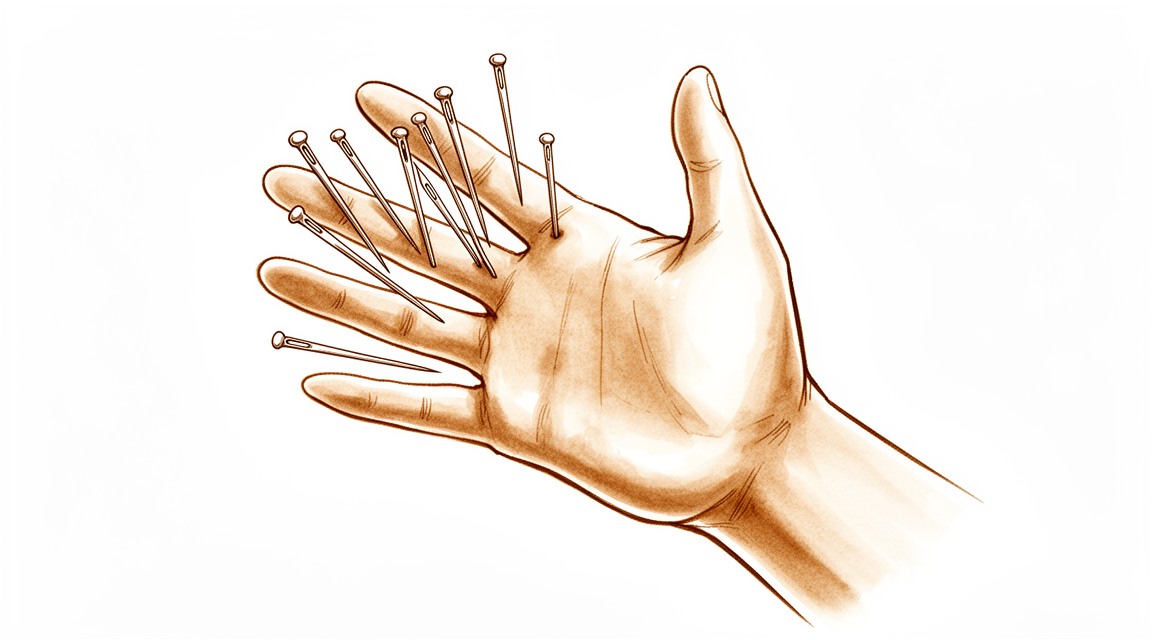
Karamihan sa mga tao ay nakapapansin ng pangingilig, tusok-tusok na pakiramdam (pins and needles) o pamamanhid sa isang bahagi ng kamay. Ang ilan ay nakararamdam ng kirot o mahapding sakit sa forearm o pulso. Ang mga sintomas ay madalas na lumalala sa gabi at maaaring gumising sa iyo mula sa pagtulog. Maaari rin itong lumala pagkatapos mong gamitin nang madalas ang iyong kamay, halimbawa pagkatapos magsulat, humawak nang mahigpit ng mga kasangkapan, o mag-type. Ang pagwagwag ng iyong kamay o pagbabago ng posisyon nito ay maaaring magpahupa sa mga sintomas nang sandali.
Ang pag-ipit ay maaaring magpahirap sa mga pang-araw-araw na gawain. Maaaring may mabitawan ka, mahirapan sa mga butones, o mahirapang buksan ang takip ng garapon. Ang mga pinong galaw ng daliri ay maaaring pakiramdam na malamya, at ang iyong grip ay maaaring pakiramdam na mas mahina kaysa karaniwan. Napapansin ng ilang tao na manhid ang mga apektadong daliri kahit na mukhang normal ang kamay.
May isang katangian ng nerve compression na mabuting malaman. Kapag naiipit ang isang nerve sa isang lugar, ang parehong nerve ay nagiging mas madaling maipit sa ibang lugar sa kahabaan nito. Nangangahulugan ito na ang mga sintomas ay minsan nagmumula sa higit sa isang lugar, at ipinapaliwanag nito kung bakit sinusuri ng iyong surgeon ang buong braso mo at hindi lamang ang lugar na masakit.
Dahil maraming nerve at maliliit na istruktura ang magkakasama sa isang makitid na espasyo, ang mga sintomas ay maaaring mahirap ilarawan at madaling maiugnay sa maling sanhi. Sa pamamagitan ng maingat na pagsusuri natutukoy ang tunay na pinagmumulan, kung minsan kasama ang mga scan o nerve conduction study, na sumusukat kung gaano kahusay dumadaloy ang mga signal sa kahabaan ng nerve.
Kung hindi humuhupa ang iyong mga sintomas, lumalala sa loob ng mga linggo, ginigising ka sa gabi, o pinipigilan kang magtrabaho o gamitin ang iyong kamay o braso, magpatingin sa iyong GP o humingi ng pagsusuri ng isang espesyalista.
Ano ang aktwal na nangyayari
Ang nerve ay medyo parang kable ng kuryente na tumatakbo mula sa iyong leeg pababa sa iyong mga daliri. Nagdadala ito ng mga mensahe sa magkabilang direksyon: mga pakiramdam mula sa iyong balat pabalik pataas sa iyong utak, at mga utos mula sa iyong utak pababa sa iyong mga kalamnan. Sa daan, kailangang dumaan ang kable sa ilang makitid na puwang sa pagitan ng mga buto, ligament at iba pang tissue.
Kapag naiipit ang isang nerve sa isa sa mga masikip na lugar na ito nang matagal, nagsisimulang magbago ang kable mismo. Kumakapal ang protective lining ng nerve, at namamaga ito sa loob ng tunnel nito, na lalong nagpapaliit sa espasyo. Naiipit din ang sariling supply ng dugo ng nerve, kaya ang ilang bahagi ng nerve ay nagkukulang sa oxygen tuwing tumataas ang pressure. Ito ang dahilan kung bakit ang mga sintomas ay madalas na pabalik-balik sa simula, lumalala kapag pinananatili mo ang iyong pulso o siko sa iisang posisyon nang ilang sandali, at humuhupa kapag nagpalit ka ng posisyon.
Kung magpapatuloy ang pag-ipit, lumalalim ang pinsala. Nagsisimulang masira ang insulation sa paligid ng bawat nerve fibre, at sa mas maraming pressure ay maaaring mapudpod ang mga fibre mismo. Hindi sabay-sabay na naaapektuhan ang bawat bahagi ng nerve. Ang mga fibre na pinakamalapit sa pinakamasikip na punto ang unang nahihirapan, kaya ang pamamanhid ay madalas na lumilitaw sa ilang daliri bago sa iba. Sa simula, nararamdaman mo ang pangingilig na pabalik-balik. Sa kalaunan, nagiging palagian ang pamamanhid dahil hindi na maayos na naipapadala ng nerve ang mga signal nito.
May isa pang bagay na mabuting malaman. Ang nerve na naipit na sa isang lugar, halimbawa sa iyong leeg, ay nahihirapang maghatid ng sustansya sa kahabaan nito. Dahil dito, hindi nito gaanong nakakayanan ang pangalawang pag-ipit sa mas ibabang bahagi, gaya ng sa pulso. Ito ang dahilan kung bakit tinitingnan ng iyong surgeon ang buong dinaraanan ng nerve, at kung bakit ang pagpapalaya sa isang lugar ay minsan hindi nagpapahupa sa lahat.
Ang magandang balita ay nangyayari ito nang paunti-unti, sa mga yugto. Kapag naalis ang pressure bago pa malubhang mapinsala ang mga nerve fibre mismo, may tunay na pagkakataon ang nerve na gumaling.
Ano ang maaari naming gawin tungkol dito
Si Dr Kieran Hirpara, isang upper-limb surgeon sa Mater Private Hospital Rockhampton, ay nagsisimula sa mga opsyon na hindi gaanong invasive na angkop sa iyong kondisyon. Ang mga pasyente ay karaniwang nirerefer sa aming klinika ng kanilang GP; kung iminungkahi ng isang physiotherapist na magpatingin sa amin, kakailanganin mo pa rin ng referral mula sa iyong GP upang maging eligible para sa Medicare rebate. Sa iyong appointment, kumukuha kami ng history, sinusuri ang iyong braso, at nag-aayos ng mga scan o nerve conduction study kung kinakailangan upang kumpirmahin kung aling nerve ang naiipit.
Para sa mga banayad na sintomas na kamakailan lamang lumitaw, madalas kaming nagsisimula sa simpleng pangangalaga sa sarili. Ang pag-aaral tungkol sa nerve, kung ano ang nagdudulot ng iyong mga sintomas at kung aling mga galaw ang dapat iwasan ay maaaring magpahupa sa mga ito nang mag-isa. Ang pagbabago sa paraan ng paggamit mo ng iyong kamay, ang pagbabawas ng pressure sa nerve at ang paggawa ng nerve gliding exercises ay pare-parehong nakatutulong. Para sa banayad na compression ng ulnar nerve sa siko, humigit-kumulang kalahati ng mga tao ang gumagaling nang walang surgery kapag nabawasan ang pressure sa nerve. Ang mga banayad na sintomas sa maagang yugto ay maaaring humupa sa impormasyong ito lamang. Karaniwan naming binibigyan ang paraang ito ng sapat na mahabang pagsubok, dahil ang mga banayad na kaso ay madalas na gumagaling nang kusa sa loob ng mga buwan.
Ang susunod na hakbang ay nakadepende sa kung gaano kalubha ang pag-ipit. Para sa katamtamang compression, hindi gumagana ang non-operative na gamutan, kaya lumilipat kami sa pag-uusap tungkol sa surgery sa halip na maghintay. Kung banayad ang iyong mga sintomas ngunit hindi humuhupa sa loob ng mga linggo, o lumalala, magpatingin sa iyong GP o humingi ng pagsusuri ng isang espesyalista.
Kapag surgery ang tamang opsyon, simple ang layunin: buksan ang masikip na espasyo upang hindi na maipit ang nerve. Tinatawag itong decompression. Ito ay isang maliit at ligtas na operasyon na nag-aalis ng pressure sa nerve kung saan ito dumadaan sa tunnel nito. Para sa karamihan ng mga problema sa nerve compression, sapat na ang release na ito lamang, at magkakaroon na ng espasyo ang nerve upang gumaling. Ang operasyon mismo, at kung ano ang kasama sa paggaling, ay may sariling pahina.
Kung mayroon kang complex regional pain syndrome, isang kondisyon ng patuloy na sakit at pagkasensitibo pagkatapos ng pinsala o surgery, iba ang pagpaplano namin sa iyong pangangalaga. Maaari kaming gumamit ng mga gamot bago at pagkatapos ng surgery upang pakalmahin ang sakit, protektahan ang kamay gamit ang maingat na dressing, at simulan nang maaga ang hand therapy. Mas matagal ang paggaling sa ganitong sitwasyon, at maaaring manatili ang kaunting paninigas.
Ano ang dapat asahan
Ang mga sintomas ng nerve compression ay madalas na pabalik-balik sa simula. Lumalala ang mga ito kapag pinananatili mo ang iyong pulso o siko sa iisang posisyon, at humuhupa kapag nagpalit ka ng posisyon. Kung magpapatuloy ang pag-ipit, nagiging palagian ang pamamanhid sa halip na pabalik-balik.
Ang kahusayan ng iyong paggaling ay lubos na nakadepende sa timing. Kapag naalis nang maaga ang pressure, bago pa malubhang mapinsala ang mga nerve fibre mismo, may tunay na pagkakataon ang nerve na gumaling. Kapag huli na ang gamutan, maaaring hindi kumpleto ang paggaling, at totoo ito anuman ang orihinal na sanhi ng compression. Ito ang dahilan kung bakit ang mga sintomas na hindi humuhupa, lumalala sa loob ng mga linggo, gumigising sa iyo sa gabi, o pumipigil sa iyong magtrabaho o gamitin ang iyong kamay o braso ay nangangailangan ng pagsusuri ng iyong GP o ng isang espesyalista, sa halip na maghintay.
Sa gamutan, ang outlook para sa maraming tao ay tuloy-tuloy na pagbuti. Napapansin ng ilang tao na tuluyang humuhupa ang kanilang mga sintomas sa loob ng tatlong buwan matapos maalis ang pressure. Ang iba ay bumubuti sa mas mahabang panahon, kung saan unti-unting gumagaan ang pamamanhid at pangingilig habang gumagaling ang nerve. Ang mga taong may diabetes ay maaaring umasa ng parehong pangmatagalang pagbuti mula sa carpal tunnel release gaya ng mga taong walang diabetes. Ang pagkawala ng pamamanhid ang madalas na pinakamalinaw na pagbabago, at maraming tao rin ang nakapapansin ng mas kaunting sakit at mas kaunting pins and needles.
Kung banayad ang compression, ang mga simpleng hakbang ay maaaring sapat na nang mag-isa sa loob ng mga buwan. Kung katamtaman o malala ito, karaniwang hindi humuhupa ang mga sintomas nang walang surgery, na tinalakay sa seksyon sa itaas.
Tapat ding sabihin na hindi lahat ng paggaling ay kumpleto. Ang ilang tao ay naiiwang may kaunting pamamanhid o panghihina, lalo na kapag huli na ang gamutan. Ang ilang tao ay patuloy na may mga sintomas pagkatapos ng isang release na maayos na ginawa. Kapag nangyari iyon, ang unang tanong ay kung may pangalawang lugar sa parehong nerve na hindi napansin, dahil ang isang nerve ay maaaring maipit sa higit sa isang lugar. Makatutulong pa rin ang karagdagang pagsusuri, at kung minsan ang karagdagang release. Ang mga nananatiling sintomas pagkatapos ng surgery ay nararapat sa panibagong pagtingin sa diagnosis sa halip na basta isantabi.
Pag-uusapan ng iyong surgeon sa iyong appointment kung nasaan ka sa saklaw na ito, batay sa iyong pagsusuri at mga scan.
Kailan dapat magpatingin
Bihirang mangailangan ng emergency care ang nerve compression, ngunit may mga eksepsiyon. Pumunta sa emergency department sa parehong araw kung ang iyong kamay o braso ay naging mainit, mapula, namamaga at masakit, lalo na kung may lagnat, o kung ang iyong mga daliri ay nagiging maputla, malamig, puti o asul. Ang biglaang pagkawala ng pakiramdam o galaw pagkatapos ng pinsala ay nangangailangan din ng emergency assessment. Ang matinding sakit na kamakailan lamang nagsimula ay maaaring mangahulugan na pagdurugo, sa halip na pamamaga, ang dumidiin sa nerve, at nangangailangan iyon ng agarang decompression upang limitahan ang pinsala sa nerve.
Magpatingin sa iyong GP o humingi ng pagsusuri ng isang espesyalista kung hindi humuhupa ang iyong mga sintomas, lumalala sa loob ng mga linggo, ginigising ka sa gabi, o pinipigilan kang magtrabaho o gamitin ang iyong kamay o braso. Ang bagong panghihina sa iyong kamay, o pamamanhid na naging palagian sa halip na pabalik-balik, ay nararapat ding ipasuri sa halip na maghintay pa.
Higit pang detalye
Advanced reading: the deeper science (optional)
Ang seksyong ito ay lumalampas sa kailangan mo para sa iyong sariling mga desisyon sa paggamot. Ang nerve compression sa braso ay karapat-dapat sa karagdagang pagbabasa dahil sa isang pigura na nagpapaliwanag sa malaking bahagi ng mga nakakadismayang resulta: isang makabuluhang minorya ng mga tao ang may higit sa isang nerve na compressed, at ang pag-release sa maling nerve ay walang binabago.
Tatlong porsyento ang nangangailangan ng pag-release ng pangalawa at ibang nerve sa loob ng isang taon
Sa isang cohort ng 7,867 na pasyenteng sumailalim sa operasyon para sa nerve compression, humigit-kumulang 3% ang sumailalim sa decompression ng ibang nerve sa parehong braso sa loob ng isang taon, at ang mga pasyenteng may parehong carpal at cubital tunnel syndrome ay maaaring makinabang mula sa simultaneous decompression, dahil ang mga outcome ay maihahambing sa single decompression [1].
Ang tatlong porsyento ay maliit sa absolute terms at malaki sa ipinapahiwatig nito. Binibilang lamang nito ang mga nagpatuloy sa pangalawang operasyon sa loob ng labindalawang buwan; hindi nito binibilang ang mga nananatiling symptomatic na hindi sumailalim, o ang mga ang pangalawang site ay nakilala bago ang unang operasyon. Ang tunay na frequency ng multi-site compression ay mas mataas kaysa sa surgical rate.
Ang clinical consequence ang mahalagang bahagi: kapag ang kamay ay nananatiling mali pagkatapos ng isang technically sound na release, ang tanong ay hindi lamang "nabigo ba ang operasyon" kundi "mayroon bang pangalawang site sa simula pa lamang".
Ang paggawa ng dalawa nang sabay ay tila walang anumang kapalit na gastos
Ang likas na pagtutol sa simultaneous decompression ay ang paniniwalang ang dalawang operasyon nang sabay sa iisang limb ay dapat magpataas ng panganib, partikular na ng complex regional pain syndrome, isang kondisyon ng persistent pain at dysfunction na hindi gaanong nauunawaan na ayon sa kasaysayan ay naiulat na may mas mataas na rate pagkatapos ng mga combined procedure.
Ang pag-aalalang iyon ay direktang sinuri. Sa pagsusuri ng 753 na pasyente, ang pagdaragdag ng carpal tunnel release sa mga nangangailangan ng fasciectomy ay nagpakita lamang ng marginal increase sa occurrence ng CRPS, na sumasalungat sa mga orihinal na ulat na nagpapakita ng lubhang mas mataas na rate, na nagpapahiwatig na walang malinaw na clinical risk na nauugnay sa simultaneous surgery [2].
Kapag binasa kasama ang natuklasan na ang combined decompression ay nagbibigay ng mga outcome na maihahambing sa single decompression [1], ang kaso para sa pagtugon sa dalawang napatunayang site sa isang anaesthetic ay makatwiran sa halip na mapusok.
Ang ideya ng double crush, at kung ano ang ipinapaliwanag at hindi nito ipinapaliwanag
Ang obserbasyon na ang kompresyon sa isang punto sa kahabaan ng nerve ay ginagawa itong mas vulnerable sa isa pang punto ay kilala bilang double crush. Ang iminumungkahing mekanismo ay ang kompresyon ay nakakasagabal sa transportasyon ng mga materyales sa kahabaan ng nerve fibre, kaya ang isang nerve na nahihirapan na sa proximal na bahagi ay hindi gaanong nakakayanan ang pangalawang insulto.
Ang konsepto ay kapaki-pakinabang at madalas na sobra kung gamitin. Nagbibigay ito ng tunay na paliwanag kung bakit ang isang tao na may pathology sa leeg ay maaaring magkaroon ng mga sintomas ng carpal tunnel sa mas mababang threshold kaysa sa inaasahan, at kung bakit ang multi-site compression ay nagkukumpol sa parehong mga indibidwal. Hindi ito lisensya upang isisi ang anumang hindi maipaliwanag na sintomas sa braso sa isang hypothetical na pangalawang lesion, at hindi nito nahuhulaan kung aling mga pasyente ang makikinabang sa aling release.
Ano ang ibig sabihin nito sa praktikal na paraan
Dalawang bagay ang kasunod nito. Bago ang operasyon, ang mga sintomas na hindi tugma sa nerve na palalayain, gaya ng pamamanhid sa ring finger at kalingkingan kapag carpal tunnel release ang nakaplano, o vice versa, ay dapat banggitin nang malinaw, dahil ang pattern ang tumutukoy sa pangalawang site.
At pagkatapos, ang mga nananatiling sintomas ay nangangailangan ng muling pagsusuri ng diagnosis sa halip na ipagpalagay na may teknikal na pagkakamali. Ang ebidensya sa itaas ay nagpapahiwatig na ang pangalawang nerve ay isang tunay at kinikilalang posibilidad, hindi isang hindi pangkaraniwang dahilan.
Mga Sanggunian
[1] Mendelaar NH, Hundepool CA, Hoogendam L, Duraku LS, Zöphel OT, Selles RW, et al. Multiple compression syndromes of the same upper extremity: prevalence, risk factors, and outcomes. J Hand Surg Am. 2023;48(5):479-88. https://doi.org/10.1016/j.jhsa.2023.01.024
[2] Buller M, Schulz S, Kasdan M, Wilhelmi BJ. The incidence of complex regional pain syndrome in simultaneous surgical treatment of carpal tunnel syndrome and Dupuytren contracture. Hand (N Y). 2017;13(4):391-4. https://doi.org/10.1177/1558944717718345
Evidence & references
This is the clinical evidence summary written for health professionals. It is technical, and it lists the research this page was built from. You do not need to read it to understand your treatment or to make a decision about it.
Overview
- Compression neuropathies of the upper extremity involve pathophysiology mechanisms including the double-crush phenomenon and systemic factors [1].
- Validated patient-reported outcome measures are utilized in the evaluation and management of upper extremity compression neuropathies [1].
- Most publications regarding uncommon compression syndromes of the radial, ulnar, and median nerves are small retrospective series or case reports [2].
- Treatment decisions for uncommon upper extremity compression neuropathies are not typically based on high levels of evidence [2].
- Proximal compression of a nerve can lessen its ability to withstand further compression more distally [3].
- Complications of compressive neuropathy management include iatrogenic injury, treatment failure, and pathologic pain syndromes [4].
- Prevention of complications in compressive neuropathy management relies on a solid understanding of normal anatomy and anatomic variations [4].
- Nerve compression is the pathologic cause of disability in quadrilateral space syndrome [5].
- Decompression of the quadrilateral space can relieve symptoms associated with the syndrome [5].
- The diagnosis and treatment of compressive neuropathies are shifting towards preoperative imaging with ultrasound and MRN [6].
- The management of failed decompressions for compressive neuropathies remains challenging [6].
- Nerve compression symptoms in the general population require careful clinical evaluation and diagnosis by the physician [7].
- Autologous vein insulator treatment for recurrent compressive neuropathy of the median or ulnar nerve resulted in reported reduction in pain and sensory disturbances in all 19 patients [8].
- Late treatment is responsible for incomplete recovery in nerve compression cases, regardless of the cause of compression [9].
- Patients with median nerve compression at the wrist due to intracarpal canal sepsis presented with significant improvement in symptoms and signs during the early follow-up period [10].
- Surgical decompression predictably affords relief of numbness in isolated compression neuropathy of the palmar cutaneous branch of the median nerve [12].
- Minimally invasive in situ decompression is technically simple, safe, and yields good results in patients with severe ulnar nerve compression at the elbow [13].
- Debulking of a collagenoma tumor along with median nerve decompression resulted in relief of neurological symptoms in a patient with carpal tunnel syndrome [15].
- Surgical decompression of the median nerve in the proximal forearm resulted in complete or partial relief of symptoms in 30 of 39 limbs [19].
- A collagen matrix wrap technique for recurrent compression neuropathies of the upper extremity has been reported with good success [22].
- Surgical decompression is associated with a greater decrease in median nerve cross-sectional area than nonsurgical treatment for carpal tunnel syndrome [27].
- Prompt surgical decompression with resection and primary anastomosis is recommended for ulnar nerve compression secondary to an ulnar artery false aneurysm at Guyon's canal to allow recovery of nerve function [33].
- Anterior interosseous nerve transfer combined with cubital and ulnar tunnel release results in sustained clinical and electrophysiological improvements in patients with severe chronic ulnar nerve compression [38].
- Anterior interosseous nerve transfer combined with cubital and ulnar tunnel release is encouraged as a standard treatment for severe chronic ulnar nerve compression [38].
- Satisfactory outcomes from endoscopic detection of compressing fascial bands support the perception that extensive decompression of the ulnar nerve beyond the cubital tunnel is not routinely needed [51].
- The literature regarding the use of internal neurolysis for ulnar nerve compression at the elbow remains controversial [53].
- Carpal tunnel release is a reasonable first step prior to proceeding with cervical spine decompression for patients with nerve compression at both the carpal tunnel and cervical spine [54].
- Surgical decompression remains the definitive treatment of cauda equina syndrome [55].
- The timing of surgery for cauda equina syndrome requires careful consideration to balance the urgency of intervention with the risks of complications [55].
Anatomy & Pathophysiology
Nerve Compression Pathophysiology
- The histopathologic changes of chronic nerve compression begin with breakdown of the blood-nerve barrier, followed by endoneurial edema and perineural thickening [72].
- Increased endoneurial pressure results in changes in microneural circulation, rendering the nerve susceptible to dynamic ischemia [72].
- With increased compression, localized demyelination occurs, followed by more diffuse demyelination and finally axonal degeneration [72].
- Neural changes do not occur uniformly across the nerve and vary depending on the distribution of compressive forces across the nerve [72].
- Fascicles susceptible to greater pressure undergo changes sooner, resulting in variable patient symptoms within a nerve’s distribution [72].
- In early carpal tunnel syndrome, superficial fascicles to the long finger and ring finger are usually affected before fascicles to the thumb and radial side of the index finger [72].
- In cubital tunnel syndrome, fascicles to the intrinsic muscles are located closer to the bony groove and are affected more than those to the flexor digitorum profundus and flexor carpi ulnaris [72].
- Patient sensory complaints parallel histopathologic neural changes, progressing from intermittent paresthesia to persistent numbness [72].
- Sensory testing varies with the degree of nerve compression, progressing from altered threshold tests (vibration and Semmes-Weinstein monofilament) to deficits in tactile discrimination testing (static and moving two-point discrimination) as compression becomes more severe [72].
- Proximal compression of a nerve could lessen its ability to withstand further compression more distally [3].
- The underlying pathophysiology of double crush syndrome involves disrupted bidirectional transport of essential nutrients along the axon, leading to morphological and functional changes [20].
- More proximal lesions closer to the cell body have a greater effect on nerve function in the context of double crush syndrome [20].
- The term "double crush" is considered misleading because it does not account for conditions where three or more sites of a given nerve are affected [20].
- The term "crush" limits the scope of the disease to purely mechanical compression, whereas other mechanical stresses such as stretch and medical or pharmacological factors may also contribute [20].
- The term "multifocal neuropathy" is proposed to expand the scope of double crush syndrome to include non-compressive mechanical stresses and non-mechanical factors [20].
- For patients with nerve compression at the carpal tunnel and cervical spine, carpal tunnel release is a reasonable first step prior to proceeding with cervical spine decompression [54].
Upper Extremity Anatomy
- The skeleton of the hand and wrist consists of 27 bones, of which 19 are long bones [41].
- The skeleton is divided into five rays, each ray making up a polyarticulated chain comprising the metacarpals and phalanges [41].
- The base of each metacarpal articulates with the distal row of the carpus [41].
- The carpus articulates with the skeleton of the forearm through its proximal row [41].
- The wrist has three axes of movement, permitting the hand to be positioned in any spatial configuration [41].
- The radial ray or first ray is the shortest, made up of only three bones: a metacarpal and two phalanges [41].
- The trapezium is angled out in front of the carpal plane so that the first metacarpal makes an angle of about 45 degrees with the second metacarpal in the sagittal plane [41].
- The thumb metacarpal is the shortest, and the index metacarpal is by far the longest [41].
- The proximal and middle phalanges of the long and ring fingers are longer than those of the index finger [41].
- The hand presents a longitudinal and transverse concavity, giving it the shape of a cup with a palmar concavity when the thumb is placed next to the index finger [41].
- The transverse axis of the palm is oblique, more distal at the metacarpophalangeal joint of the index finger and more proximal at the fifth metacarpophalangeal joint [41].
- The transverse axis forms an acute angle of approximately 75 degrees with the longitudinal axis [41].
- The epiphyseal plates are located at the proximal ends of the phalanges and the first metacarpal, whereas they are located at the distal ends of the other metacarpals [41].
- The extrinsic extensors run through six different fibroosseous retinacular compartments at the wrist level [40].
- The first extensor compartment contains the abductor pollicis longus and the extensor pollicis brevis [40].
- The second extensor compartment contains the extensor carpi radialis longus and the extensor carpi radialis brevis [40].
- The third compartment contains the extensor pollicis longus, which turns abruptly radialward about Lister tubercle [40].
- The fourth extensor compartment contains the extensor indicis proprius lying deep to the four tendons of the extensor digitorum communis [40].
- The fifth compartment contains the extensor digiti quinti [40].
- The sixth compartment contains the extensor carpi ulnaris tendon [40].
- The extensor digitorum communis tendons of the middle, ring, and little fingers are tethered together by juncturae tendinum over the dorsum of the hand proximal to the metacarpophalangeal joint [40].
- The digital extensor tendons are stabilized over the mid-line of the metacarpophalangeal joint by their attachment to sagittal band fibers [40].
- The sagittal band fibers insert onto the volar proximal phalanx and onto the lateral borders of the volar plate [40].
- With rupture or attenuation of the sagittal band fibers, the extrinsic extensor tendon can sublux to the ulnar side of the metacarpal head causing ulnar deviation of the finger [40].
- The flexor digitorum profundus inserts on the proximal volar aspect of the distal phalanx [40].
- The flexor digitorum superficialis acts as a flexor of the proximal interphalangeal and metacarpophalangeal joints [40].
- The extrinsic finger flexors are the flexor digitorum profundus and the flexor digitorum superficialis [66].
- The flexor digitorum profundus originates from the proximal ulna and the interosseous membrane [66].
- The innervation of the flexor digitorum profundus of the index and middle fingers is through the anterior interosseous branch of the median nerve [66].
- The profundus of the ring and little fingers is innervated by the ulnar nerve [66].
- The flexor digitorum superficialis has two heads: a radial head originating from the proximal shaft of the radius, and a humeral ulnar head originating from the medial humeral epicondyle and coronoid process of the ulna [66].
- The entire flexor digitorum superficialis muscle receives innervation from the median nerve [66].
- The flexor pollicis longus originates from two heads: a radial head from the proximal radius and interosseous membrane, and an accessory head from the coronoid process of the ulna and medial epicondyle of the humerus [66].
- The flexor pollicis longus is innervated by the anterior interosseous branch of the median nerve [66].
- The fibroosseous tunnel, or digital flexor sheath, consists of annular pulleys which provide mechanical stability, and cruciate pulleys which provide flexibility [66].
- The A2 and A4 pulleys are the most essential in maintaining the mechanical advantage of the flexor tendons [66].
- There are seven interosseous muscles, four dorsal and three volar [59].
- The dorsal interossei are abductors [59].
- The volar interossei are adductors [59].
- The middle finger has two dorsal interossei and no volar interossei because the central axis of the hand lies within it [59].
- Each dorsal interosseous muscle, with the exception of the third, has two muscle heads: a superficial head and a deep head [59].
- The superficial head of the dorsal interosseous muscles abducts and weakly flexes the proximal phalanx [59].
- The deep head of the dorsal interosseous muscles flexes and weakly abducts the proximal phalanx while extending the middle and distal phalanges [59].
- The volar interossei have only one muscle head and none of them insert onto the proximal phalanx [59].
- The abductor digiti quinti and flexor digiti quinti brevis are similar in structure and function to the superficial and deep heads of the dorsal interossei, respectively [59].
- The opponens digiti quinti arises from the pisohamate ligament and the hook of the hamate and inserts onto the ulnar side of the diaphysis of the fifth metacarpal [59].
- The superficial palmar fascia lies in a coronal plane beneath the palmar subcutaneous tissue [69].
- The palmaris longus tendon, when present, terminates in continuity with the fibers of the proximal corner of the superficial palmar fascia [69].
- Four central bands of fascia extend distally from the proximal corner of the superficial palmar fascia toward each of the fingers, with no central band for the thumb [69].
- At the distal palmar crease level, the central bands are bridged transversely by the superficial transverse palmar ligament [69].
- The digital neurovascular structures are surrounded by a diffuse network of thin transverse oblique fibers [69].
- Fibers dorsal to the neurovascular bundle are collectively called Cleland ligament, and those palmar to the neurovascular bundle are called Grayson ligament [69].
- A subdermal fascial layer borders the periphery of the web spaces from roughly the radial thumb sesamoid to the ulnar side of the small finger’s proximal flexion crease [69].
- The dorsal skin has a thin epidermis lined by a horny layer that is only 0.02 mm thick [68].
- The dorsal skin possesses a normal pilosebaceous system, unlike the palm [68].
- Flexion of the fingers produces a significant lengthening of the dorsal skin, with an average increase of 3 cm in the middle finger from extension to full flexion [68].
- The metacarpal arch is endowed with adaptability because of the mobility of the peripheral metacarpals [62].
- The index metacarpal is the most firmly fixed [62].
- The ring metacarpal has about 10 degrees of mobility in flexion and extension [62].
- The fifth metacarpal has a range of flexion–extension of approximately 20 degrees [62].
- The second to fifth metacarpals are bound together by various fibrous structures, the most distal of which is the deep transverse intermetacarpal ligament [62].
- The deep transverse intermetacarpal ligament is also known as the interglenoid ligament [62].
- The keystones of the longitudinal arches are the metacarpophalangeal articulations, whose thick anterior glenoid capsules, the volar plates, prevent hyperextension [62].
- The volar plates are interconnected by the transverse interglenoid ligament [62].
- The median nerve creates the “rock position” of the pronated fist [104].
- The radial nerve extends the wrist and hand forming the “paper position” [104].
- The ulnar nerve creates the “scissor position” [104].
Classification
- Ulnar tunnel syndrome is classified into two distinct types: Type 1 involves compression at the level of the pisiform with sensory and motor involvement, and Type 2 involves compression distal to the pisiform with motor involvement only [14].
- The term "double crush" is considered misleading because it does not account for conditions where three or more sites of a nerve are affected, nor does it account for mechanical stresses such as stretch [20].
- The term "double crush" is considered misleading because it limits the scope of the disease to purely mechanical compression, whereas medical and pharmacological factors also contribute [20].
- The term "multifocal neuropathy" (MFN) is proposed to expand the scope of double crush syndrome to emphasize that mechanical compression is only one part of a complex interplay of factors [20].
- Posterior interosseous nerve palsy Type 1 is characterized by drop finger and thumb, showing simultaneous compression of the recurrent branch and the descending branch at the entrance point and within the supinator [39].
- The McGowan/Goldberg (McGG) classification is used to define the clinical stage of neuropathy, with poor outcomes in cubital tunnel syndrome mainly related to severity according to this classification [89].
- A diagnostic scale for carpal tunnel syndrome was developed by collapsing 20 highest-ranked items into 8 major criteria based on commonality and redundancy [106].
- In the development of carpal tunnel syndrome diagnostic criteria, items related to the nature and distribution of sensory disturbance were combined into a single construct [106].
- In the development of carpal tunnel syndrome diagnostic criteria, items describing denervation of the thenar musculature were combined into a single construct [106].
- In the development of carpal tunnel syndrome diagnostic criteria, items related to coexisting medical conditions were combined into a single construct [106].
- In the development of carpal tunnel syndrome diagnostic criteria, items describing the response to common therapeutic interventions were combined into a single construct [106].
Clinical Presentation
- Patients often have difficulty accurately describing their symptoms and may incorrectly attribute pathology to a perceived deficit [30].
- A careful physical examination is essential to direct care and future testing if indicated [30].
- A systematic method to approaching the physical examination is essential due to the number of structures in a small space [30].
- Compression at one location on a nerve's axon would predispose that same axon to injury elsewhere [20].
- Increased susceptibility to injury in double crush syndrome resulted from disrupted bidirectional transport of essential nutrients along the axon [20].
- More proximal lesions closer to the cell body have a greater effect on nerve function [20].
- The term "double crush" is misleading because it does not take into account conditions in which three or more sites of a given nerve are affected [20].
- The term "crush" limits the scope of the disease to purely mechanical compression, whereas multiple medical and pharmacological factors are also likely to contribute [20].
- Ulnar tunnel syndrome is of two distinct types: Type 1 involves compression at the level of the pisiform with sensory and motor involvement, and Type 2 involves compression distal to the pisiform with motor involvement only [14].
- Symptoms of ulnar tunnel syndrome vary based on the anatomic location of the compression within Guyon's canal [42].
- Ganglia are the most common cause of ulnar tunnel syndrome [42].
- The most frequent described cause of compression in Guyon's canal was ganglion cyst (16%) [32].
- Ulnar nerve compression at the arch of origin of the adductor pollicis muscle is a rare condition, representing less than 1% of ulnar neuropathies at the wrist and hand [35].
- Radial tunnel syndrome is relatively uncommon but is an important cause of lateral forearm pain [45].
- Clinical examination is a crucial part of the diagnosis of radial tunnel syndrome [45].
- A short history with severe pain indicating compression secondary to haemorrhage rather than oedema should lead to urgent decompression in an attempt to minimise neural damage [16].
- Pseudogout should be considered a rare cause of acute neuropathic compression of the hand [23].
- An anomalous muscle invading the carpal canal has been reported more frequently than any other as a cause of median nerve compression [26].
- Independent recurrent motor branch compression of the median nerve appears to exist in the presence of carpal tunnel symptomatology or as an independent entity [18].
- Proximal median nerve compression can be seen in association with cubital tunnel syndrome [24].
- Concurrent carpal tunnel syndrome and pronator syndrome are rarely considered and proximal compression sites are easily overlooked [74].
- Ulnar nerve pathology may precede and increase susceptibility to median nerve compression [17].
- The vibrometer has significant potential as a clinical and research instrument in nerve compression syndromes [46].
- Nerve conduction studies should be performed in patients presenting with possible carpal tunnel syndrome to assist diagnosis [48].
- Nerve conduction studies may need to be repeated at intervals in those managed conservatively [48].
- Loss of intrinsic hand strength, ulnar sensation loss, positive elbow flexion test, positive cubital tunnel Tinel's sign, and abnormal ulnar nerve NCS result are predictors of coincident carpal and cubital tunnel syndromes [76].
- The cutoff value for the K-B score for diagnosing coincident nerve compression was 2 points, with a sensitivity of 86.6% and a specificity of 86.5% in the developmental cohort [76].
- The area under the receiver operating characteristic curve for the K-B score was 0.9217 [76].
- Cubital tunnel syndrome is the second most common upper extremity neuropathy with an incidence of 24.7 cases per 100,000 persons per year [47].
- Common sites of compression for cubital tunnel syndrome include Osborne's ligament, the 2 heads of the flexor carpi ulnaris (FCU), and the arcade of Struthers [47].
- Diagnostic ultrasound has potential utility for the preoperative evaluation of vascular anomalies that may cause ulnar nerve compression [47].
- The cause of anterior interosseous nerve palsy remains the subject of debate, with views ranging from localized compression to neuralgic amyotrophy [25].
- A distinct cause of compression of the AIN or visible changes in the AIN were seen in just three of the eight limbs that were explored [21].
Investigations
Clinical Evaluation
- Diagnostic tests such as imaging and serum laboratory studies are useful in the determination of pathology but can be expensive, time consuming, and often nonspecific [30].
- The diagnosis of each nerve compression case requires careful clinical evaluation by the physician [7].
- Ignorance of the clinical presentation of a posterior interosseous nerve palsy frequently leads to misdiagnosis [98].
Electrodiagnostic Studies
- Nerve conduction studies may need to be repeated at intervals in patients managed conservatively for carpal tunnel syndrome [48].
Imaging
- Surgical decompression was associated with a greater decrease in median nerve cross-sectional area than nonsurgical treatment [27].
- Ultrasound measurements seem to have a limited value in clinical results of patients treated for entrapment neuropathy of the ulnar nerve [97].
- High-resolution ultrasound (HRUS) is a viable method to demonstrate a punched nerve syndrome [99].
- The SCT is a useful test in clinical diagnosis and postoperative monitoring of anterior interosseous nerve compression [102].
Treatment
Non-Operative Management
- The appropriate duration of a conservative trial for compressive neuropathy is unclear [37].
- Acute compressive neurapraxias may resolve in a period of weeks [37].
- In patients with mild symptoms followed for a mean of 22 months, 90% of cases recovered spontaneously [37].
- One-half of patients suffering from mild compression of the ulnar nerve could be expected to recover without surgery if treated vigorously by minimizing pressure on the nerve [37].
- For moderate compression, non-operative treatment is completely unsuccessful [37].
- Only mild symptoms, and only then in the acute stage, would resolve solely with information [37].
- Patients with mild or moderate cubital tunnel syndrome have a good likelihood of symptom reduction or recovery without surgical treatment [37].
- Patients given an information program describing anatomy, probable causes of symptoms, and instructions on avoiding provocative movements improved as much as those treated with an elbow brace or nerve gliding exercises [37].
- Endoscopic decompression is recommended when conservative treatment fails to alleviate symptoms in anterior interosseous nerve syndrome [83].
Operative Management: General Principles and Techniques
- Minimally invasive in situ decompression is technically simple, safe, and gives good results in patients with severe nerve compression [13].
- Surgical decompression predictably affords relief of the numbness in isolated compression neuropathy of the palmar cutaneous branch of the median nerve [12].
- Surgical release was effective in relieving symptoms when the clinical picture was consistent with entrapment neuropathy, regardless of electrophysiologic studies [80].
- Debulking of a tumor along with median nerve decompression resulted in relief of neurological symptoms in a case of carpal tunnel syndrome caused by a collagenoma [15].
- Satisfactory outcomes support the perception that extensive decompression of the ulnar nerve beyond the cubital tunnel is not routinely needed [51].
- Recovery was rapid and complete following surgical relief of compression in a case of anterior interosseous nerve compression by the muscle of Gantzer, with full recovery noted three months after the intervention [50].
- Late treatment was responsible for incomplete recovery, whatever the cause of the nerve compression [9].
Operative Management: Specific Procedures and Adjuncts
- Anterior interosseous nerve transfer, along with cubital and ulnar tunnel release, results in sustained clinical and electrophysiological improvements in patients with severe chronic ulnar nerve compression [38].
- Anterior interosseous nerve transfer combined with cubital and ulnar tunnel release encourages its adoption as a standard treatment for severe chronic ulnar nerve compression [38].
- All 19 patients reported reduction in pain and sensory disturbances secondary to the compression of the median or ulnar nerve following treatment with an autologous vein insulator [8].
- The use of a collagen matrix wrap in recurrent compression neuropathies has been reported with good success [22].
- Endoscopic decompression for recurrent carpal tunnel syndrome requires rigorous patient selection, specifically for true recurrences without obvious etiology or trophic disorders [86].
- In the treatment of compression neuropathy within the context of Complex Regional Pain Syndrome (CRPS), complete release of the involved nerve is important [78].
- If there is damage to the neural bed or the neural bed is compromised during compression neuropathy surgery, modification of the neural environment is appropriate [78].
- Postoperative management for CRPS patients undergoing compression neuropathy surgery includes sympatholytic intervention with parenteral or oral agents to minimize postoperative pain and prevent a dystrophic flare-up [78].
- Patients undergoing compression neuropathy surgery for CRPS should expect prolonged rehabilitation, continued use of oral nonnarcotic agents for 3 to 6 months, and some residual disability [78].
- For a painful median nerve with CRPS, autogenous saphenous vein or allograft wrapping is selected and wrapped directly around the median nerve with an opening to allow the palmar cutaneous branch to exit [79].
- The vein graft used for median nerve wrapping should be sutured proximally and distally with a 5-0 or 6-0 nonreactive suture [79].
- Chromic suture should be avoided for median nerve wrapping because chemicals released from the suture can create a nociceptive neural focus [79].
- An injured palmar cutaneous branch of the median nerve can be resected and moved to an unscarred area or repaired by using an end-to-end interposition antebrachial cutaneous nerve [79].
- Repair of a short palmar cutaneous nerve branch is accomplished under the operating microscope with 9-0 to 10-0 nonabsorbable suture on 75- to 130-µm needles [79].
- Postoperatively, the limb is protected from pain and dystrophic flare-up by the use of continuous autonomic blockade [79].
- Motion of the affected extremity is initiated in a controlled active therapy program or by using continuous passive motion over the 3- to 5-day period of hospitalization [79].
- Surgical correction of secondary joint deformities from arthrofibrosis may be necessary after CRPS, but surgery on contracted joints should not be performed until maximal nonoperative improvement has been achieved [79].
- The waiting period for surgical correction of contracted joints after CRPS should be a minimum of 3 to 6 months after successful elimination of the active dystrophic pain [79].
- Release of the MCP or PIP joints can be performed when nonoperative improvement has plateaued and the deformity warrants intervention [79].
- Indications for surgery on contracted joints include joint pain without diffuse dystrophic symptoms and arthrofibrosis that interferes with function [79].
- If necessary, all four MCP joints and all four PIP joints can be released in a single operation [79].
- Restoration of full flexion or extension is an unreasonable goal or expectation for surgical correction of CRPS-related joint contractures [79].
- The range of motion achieved during surgery for CRPS-related joint contractures is rarely maintained after surgery as some loss is expected [79].
Post-Operative Care and Complications
- Randomized controlled studies comparing orthosis fabrication to the use of no orthosis after carpal tunnel release have failed to show any benefit to orthotics [85].
- Prevention of complications relies on a solid understanding of normal anatomy and anatomic variations [4].
- Early recognition of milder variants of reflex sympathetic dystrophy, preferably during the first or at least the second week, may prevent this serious complication [77].
- Removal or splitting of a dressing or cast to relieve pressure, elevation of an edematous hand, and intensive hand therapy are frequently helpful in preventing the development of full-blown CRPS [77].
- The surgeon should have a low threshold for performing electrodiagnostic studies and/or surgical decompression for suspected nerve entrapment in patients with CRPS [77].
- For patients who have a history of CRPS, there may be value in preemptive treatment with a long-acting sympathetic block or indwelling catheter for a regional nerve blockade for any proposed surgical procedure [77].
- Surgery on neural structures compromised by neuromas, neuroma-in-continuity, or compression is indicated if symptoms persist after nonoperative modalities, including sympatholytic medications, as long as the symptoms can be controlled medically in the perioperative period [78].
- An acute dystrophic flare-up of quiescent CRPS is possible in the postoperative period [78].
- Exacerbation of symptoms in the postoperative period can be managed effectively with oral medications or a continuous block [78].
- Surgical options for neural injury in CRPS include neurolysis, neurorrhaphy, or neural relocation [78].
- Modification of the neural bed is an option for neural injury in CRPS [78].
- Tension on the repair site should be avoided by performing nerve grafts from the sural nerve or a branch of the medial or lateral antebrachial cutaneous nerve [78].
- Adhesions between the skin and nerve should be managed by Z-plasty local flaps or distant flaps [78].
- If excessive scarring or adhesions develop, the neural bed should be modified with the use of autologous fat, rotational muscle flaps, pedicled muscle or fascial flaps, free muscle transfer, autologous or allograft venous wraps, or nerve conduits [78].
- Internal neurolysis should be minimized [78].
- Sympatholytic intervention (e.g., continuous autonomic blockade), pharmacologic palliation, physical therapy, and early active and passive range of motion should be included in postoperative care [78].
- Hemostasis should be established to prevent hematoma formation [78].
- Constrictive postoperative dressings should be avoided [78].
Complications
Iatrogenic and Surgical Complications
- Complications of compressive neuropathy management include iatrogenic injury [4].
- Prevention of iatrogenic injury relies on a solid understanding of normal anatomy and anatomic variations [4].
- Minimally invasive in situ decompression for severe ulnar nerve entrapment is technically simple and safe [13].
Treatment Failure and Recurrence
- Complications of compressive neuropathy management include treatment failure [4].
- The management of failed decompressions remains challenging [6].
- Revision decompression and collagen nerve wrap for recurrent and persistent compression neuropathies of the upper extremity has been reported with good success [22].
- All 19 patients treated with an autologous vein insulator for recurrent compressive neuropathy reported reduction in pain and sensory disturbances [8].
Pathologic Pain Syndromes
- Complications of compressive neuropathy management include pathologic pain syndromes [4].
Diagnostic and Etiologic Challenges
- A distinct cause of compression of the anterior interosseous nerve or visible changes in the nerve were seen in just three of the eight limbs that were explored [21].
Recovery
General Principles and Prognosis
- Late treatment of nerve compression is responsible for incomplete recovery [9].
- The diagnosis and treatment of compressive neuropathies continue to evolve with technology, shifting towards preoperative imaging with ultrasound and MRN, while the management of failed decompressions remains challenging [6].
- Long-term improvement in patients with diabetes remained after carpal tunnel release to the same extent as for patients without diabetes [52].
Specific Nerve Outcomes
- Recovery was rapid and complete following surgical relief of compression, with full recovery noted three months after the intervention [50].
- The patients presented here had significant improvement in median nerve symptoms and signs in the early follow-up period [10].
- All 19 patients reported reduction in pain and the sensory disturbances secondary to the compression of the median or ulnar nerve [8].
- Debulking of the tumor along with median nerve decompression was performed with relief of neurological symptoms [15].
Recurrent and Persistent Neuropathy
- The authors report on the novel technique of using a collagen matrix wrap in recurrent compression neuropathies with good success [22].
Key Evidence
- [L4] This article reviews uncommon compression syndromes of the radial, ulnar, and median nerves, noting that most publications are small retrospective series or case reports and treatment decisions are not typically based on high levels of evidence. [2] (10.1016/j.hcl.2013.04.014)
- [Paper] It is concluded that proximal compression of a nerve could lessen its ability to withstand further compression more distally. [3] (10.1016/s0363-5023(87)80207-1)
- [L5] Complications of compressive neuropathy management include iatrogenic injury, treatment failure, and pathologic pain syndromes, with prevention relying on a solid understanding of normal anatomy and anatomic variations. [4] (10.1016/j.hcl.2015.01.012)
- [L4] We believe that the nerve compression is the pathologic cause of the disability and that with decompression of this space, symptoms can be relieved. [5] (10.1016/s0363-5023(83)80056-2)
- [L5] The diagnosis and treatment of compressive neuropathies continue to evolve with technology, shifting towards preoperative imaging with ultrasound and MRN, while the management of failed decompressions remains challenging. [6] (10.1016/j.jhsg.2022.10.009)
- [L4] This tendency requires careful clinical evaluation and diagnosis of each nerve compression cases by the physician. [7] (10.1053/jhsu.2001.24972)
- [L4] All 19 patients reported reduction in pain and the sensory disturbances secondary to the compression of the median or ulnar nerve. [8] (10.1053/jhsu.2001.22528)
- [L4] Late treatment was responsible for incomplete recovery, whatever the cause of the nerve compression. [9] (10.1016/0266-7681(94)90204-6)
- [L5] The patients presented here had significant improvement in median nerve symptoms and signs in the early follow-up period. [10] (10.1016/s0363-5023(05)80445-9)
- [L5] Surgical decompression predictably affords relief of the numbness. [12] (10.1016/s0363-5023(87)80170-3)
- [L3] Minimally invasive in situ decompression is technically simple, safe and gives good results in patients with severe nerve compression. [13] (10.1177/1753193411416426)
- [L4] Ulnar tunnel syndrome is of two distinct types: Type 1 involves compression at the level of the pisiform with sensory and motor involvement, and Type 2 involves compression distal to the pisiform with motor involvement only. [14] (10.1016/0266-7681(84)90021-4)
- [Case_report] Debulking of the tumor along with median nerve decompression was performed with relief of neurological symptoms. [15] (10.1016/j.jhsa.2013.07.004)
- [L5] A short history with severe pain indicating compression secondary to haemorrhage rather than oedema should lead to urgent decompression in an attempt to minimise neural damage. [16] (10.1016/s0266-7681(84)80011-x)
- [L2] This supports the hypothesis that ulnar nerve pathology may precede and increase susceptibility to median nerve compression. [17] (10.1016/j.jhsg.2026.100970)
- [L4] Independent recurrent motor branch compression of the median nerve appears to exist in the presence of carpal tunnel symptomatology or as an independent entity. [18] (10.1016/s0363-5023(82)80155-x)
- [L4] Surgical decompression of the median nerve in the proximal forearm resulted in complete or partial relief of symptoms in 30 of 39 limbs. [19] (10.1016/0363-5023(94)90235-6)
- [L5] [20] (10.1016/j.jhsa.2016.09.009)
- [L4] A distinct cause of compression of the AIN or visible changes in the AIN were seen in just three of the eight limbs that were explored. [21] (10.1016/s0266-7681(97)80020-4)
- [L4] The authors report on the novel technique of using a collagen matrix wrap in recurrent compression neuropathies with good success. [22] (10.1097/sap.0b013e3182956475)
- [L4] Pseudogout should be considered a rare cause of acute neuropathic compression of the hand. [23] (10.1016/j.jhsg.2022.07.010)
- [L4] Proximal median nerve compression can be seen in association with cubital tunnel syndrome. [24] (10.1142/s2424835518500200)
- [L5] The cause of anterior interosseous nerve palsy remains the subject of debate, with views ranging from localized compression to neuralgic amyotrophy. [25] (10.1016/s0266-7681(98)80233-7)
- [L4] An anomalous muscle invading the carpal canal has been reported more frequently than any other as a cause of median nerve compression. [26] (10.1097/00130911-199906000-00005)
- [L3] Surgical decompression was associated with a greater decrease in median nerve cross-sectional area than nonsurgical treatment. [27] (10.1016/j.jhsa.2010.06.010)
- [L3] The most frequent described cause of compression was ganglion cyst (16%). [32] (10.1177/15589447251325827)
- [L5] Prompt surgical decompression with resection and primary anastomosis is recommended to allow recovery of nerve function. [33] (10.1016/s0363-5023(82)80077-4)
- [L4] Ulnar nerve compression at the arch of origin of the adductor pollicis muscle is a rare condition, representing less than 1% of ulnar neuropathies at the wrist and hand. [35] (10.1016/0363-5023(93)90062-8)
- [L2] [37] (10.1177/1753193408098480)
- [L4] Anterior interosseous nerve transfer, along with cubital and ulnar tunnel release, results in sustained clinical and electrophysiological improvements in patients with severe chronic ulnar nerve compression, which encourages its adoption as a standard treatment for severe chronic ulnar nerve compression. [38] (10.1177/17531934251381023)
- [L4] Type 1 (drop finger and thumb) showed a simultaneous compression of the recurrent branch and the descending branch at the entrance point and within the supinator. [39] (10.1016/s0266-7681(98)80234-9)
- [L5] The article provides a comprehensive review of the anatomy, pathophysiology, and causes of ulnar tunnel syndrome, noting that ganglia are the most common cause and that symptoms vary based on the anatomic location of the compression within Guyon's canal. [42] (10.1016/j.hcl.2007.06.006)
- [L5] Radial tunnel syndrome is relatively uncommon but is an important cause of lateral forearm pain, with clinical examination being a crucial part of the diagnosis. [45] (10.1197/j.jht.2006.02.005)
- [Paper] The vibrometer has significant potential as a clinical and research instrument in nerve compression syndromes. [46] (10.1016/s0363-5023(84)80196-3)
- [L4] [47] (10.1016/j.xrrt.2023.04.001)
- [L5] Nerve conduction studies should be performed in patients presenting with possible carpal tunnel syndrome to assist diagnosis, and may need to be repeated at intervals in those managed conservatively. [48] (10.1016/j.cnp.2018.02.005)
- [L5] Recovery was rapid and complete following surgical relief of compression, with full recovery noted three months after the intervention. [50] (10.1016/s1297-3203(01)00041-5)
- [L4] The satisfactory outcomes support the perception that extensive decompression of the ulnar nerve beyond the cubital tunnel is not routinely needed. [51] (10.1007/s11552-011-9377-x)
- [L2] Long-term improvement in patients with diabetes remained after carpal tunnel release to the same extent as for patients without diabetes. [52] (10.1016/j.jhsa.2014.01.012)
- [L5] The literature regarding the use of internal neurolysis for ulnar nerve compression at the elbow remains controversial. [53] (10.1016/s0363-5023(10)80048-6)
- [L3] For patients with nerve compression at the carpal tunnel and cervical spine, CTR is a reasonable first step prior to proceeding with cervical spine decompression. [54] (10.1177/15589447241233764)
- [L5] Surgical decompression remains the definitive treatment of CES, though the timing of surgery requires careful consideration to balance the urgency of intervention with the risks of complications. [55] (10.2106/jbjs.rvw.24.00156)
- [L4] Concurrent carpal tunnel syndrome and pronator syndrome are rarely considered and proximal compression sites are easily overlooked. [74] (10.1016/j.otsr.2016.10.009)
- [L4] [76] (10.1016/j.jhsa.2015.06.068)
- [L4] Regardless of the electrophysiologic studies, when the clinical picture was consistent with entrapment neuropathy, surgical release was effective in relieving symptoms. [80] (10.1016/s0363-5023(89)80098-x)
- [L4] The authors recommend endoscopic decompression when conservative treatment fails to alleviate symptoms. [83] (10.1016/j.jhsa.2013.07.026)
- [L1] [85] (10.1016/j.jhsa.2018.01.016)
- [L4] It requires rigorous patient selection, specifically for true recurrences without obvious etiology or trophic disorders. [86] (10.1016/j.main.2015.10.029)
- [L4] The study supports the hypothesis that poor outcomes are mainly related to the severity of the neuropathy according to the clinical stage, specifically the McGowan/Goldberg (McGG) classification. [89] (10.1016/j.hansur.2021.10.030)
- [L3] Ultrasound (US) measurements seem to have a limited value in clinical results of patients treated for entrapment neuropathy of the ulnar nerve. [97] (10.1177/1558944719857816)
- [L4] Ignorance of the clinical presentation of a posterior interosseous nerve palsy frequently leads to misdiagnosis. [98] (10.1016/j.main.2011.11.004)
- [L4] HRUS is a viable method to demonstrate a punched nerve syndrome. [99] (10.1007/s00402-015-2216-8)
- [L4] Based on our results, we believe that the SCT is a useful test in clinical diagnosis and postoperative monitoring of anterior interosseous nerve compression. [102] (10.1177/1558944716660555ku)
- [L4] The median nerve creates the “rock position” of the pronated fist, the radial nerve extends the wrist and hand forming the “paper position”, and the ulnar nerve creates the “scissor position”. [104] (10.1016/s0020-1383(02)00102-x)
- [L1] [106] (10.1016/j.jhsa.2006.03.005)
References
[1] Compression Neuropathies of the Upper Extremity. 2021.
[2] Uncommon Upper Extremity Compression Neuropathies. Hand Clinics. 2013. DOI: 10.1016/j.hcl.2013.04.014
[3] An experimental study on the “double crush” hypothesis. The Journal of Hand Surgery. 1987. DOI: 10.1016/s0363-5023(87)80207-1
[4] Complications of Compressive Neuropathy. Hand Clinics. 2015. DOI: 10.1016/j.hcl.2015.01.012
[5] Quadrilateral space syndrome. The Journal of Hand Surgery. 1983. DOI: 10.1016/s0363-5023(83)80056-2
[6] Future Considerations in the Diagnosis and Treatment of Compressive Neuropathies of the Upper Extremity. Journal of Hand Surgery Global Online. 2023. DOI: 10.1016/j.jhsg.2022.10.009
[7] The prevalence and characteristics of nerve compression symptoms in the general population. The Journal of Hand Surgery. 2001. DOI: 10.1053/jhsu.2001.24972
[8] Treatment of recurrent compressive neuropathy of peripheral nerves in the upper extremity with an autologous vein insulator. The Journal of Hand Surgery. 2001. DOI: 10.1053/jhsu.2001.22528
[9] Entrapment and Compartment Syndromes of the Upper Limb in Haemophilia. Journal of Hand Surgery. 1994. DOI: 10.1016/0266-7681(94)90204-6
[10] Median nerve compression at the wrist due to intracarpal canal sepsis. The Journal of Hand Surgery. 1995. DOI: 10.1016/s0363-5023(05)80445-9
[12] Isolated compression neuropathy of the palmar cutaneous branch of the median nerve. The Journal of Hand Surgery. 1987. DOI: 10.1016/s0363-5023(87)80170-3
[13] Severe ulnar nerve entrapment at the elbow: functional outcome after minimally invasive in situ decompression. Journal of Hand Surgery (European Volume). 2012. DOI: 10.1177/1753193411416426
[14] Ulnar Tunnel Syndrome. Journal of Hand Surgery. 1984. DOI: 10.1016/0266-7681(84)90021-4
[15] Collagenoma in a Child With Tuberous Sclerosis Complex Causing Carpal Tunnel Syndrome and Thumb Overgrowth: Case Report. The Journal of Hand Surgery. 2013. DOI: 10.1016/j.jhsa.2013.07.004
[16] Acute Carpal Tunnel Syndrome due to Spontaneous Haemorrhage. Journal of Hand Surgery. 1984. DOI: 10.1016/s0266-7681(84)80011-x
[17] Incidence of Carpal Tunnel Syndrome After the Diagnosis of Ulnar Neuropathy. Journal of Hand Surgery Global Online. 2026. DOI: 10.1016/j.jhsg.2026.100970
[18] Compression syndrome of the recurrent motor branch of the median nerve. The Journal of Hand Surgery. 1982. DOI: 10.1016/s0363-5023(82)80155-x
[19] Median nerve compression in the proximal forearm. The Journal of Hand Surgery. 1994. DOI: 10.1016/0363-5023(94)90235-6
[20] Multifocal Neuropathy: Expanding the Scope of Double Crush Syndrome. The Journal of Hand Surgery. 2016. DOI: 10.1016/j.jhsa.2016.09.009
[21] Anterior Interosseous Nerve Palsy. Journal of Hand Surgery. 1997. DOI: 10.1016/s0266-7681(97)80020-4
[22] Revision Decompression and Collagen Nerve Wrap for Recurrent and Persistent Compression Neuropathies of the Upper Extremity. Annals of Plastic Surgery. 2014. DOI: 10.1097/sap.0b013e3182956475
[23] Pseudogout: A Rare Cause of Acute Carpal Tunnel Syndrome and Acute Guyon Canal Syndrome. Journal of Hand Surgery Global Online. 2022. DOI: 10.1016/j.jhsg.2022.07.010
[24] Simultaneous Compression of the Median and Ulnar Nerve at the Elbow: A Retrospective Study. The Journal of Hand Surgery (Asian-Pacific Volume). 2018. DOI: 10.1142/s2424835518500200
[25] Recurrent Anterior Interosseous Nerve Palsies Related to Pregnancy. Journal of Hand Surgery. 1998. DOI: 10.1016/s0266-7681(98)80233-7
[26] Anatomic Anomalies and Carpal Tunnel Syndrome. Techniques in Hand & Upper Extremity Surgery. 1999. DOI: 10.1097/00130911-199906000-00005
[27] Sonographic Follow-Up of Patients With Carpal Tunnel Syndrome Undergoing Surgical or Nonsurgical Treatment: Prospective Cohort Study. The Journal of Hand Surgery. 2010. DOI: 10.1016/j.jhsa.2010.06.010
[30] Orthopaedic Knowledge Update 13 Ebook Without Multimedia. Anatomy, Evaluation, Clinical Examination, and Imaging > Evaluation and Clinical Examination: Current Concepts.
[32] Long-Term Patient-Reported Outcomes After Release of the Ulnar Nerve in Guyon’s Canal. HAND. 2025. DOI: 10.1177/15589447251325827
[33] Ulnar nerve compression secondary to ulnar artery false aneurysm at the Guyon's canal. The Journal of Hand Surgery. 1982. DOI: 10.1016/s0363-5023(82)80077-4
[35] Ulnar nerve compression at the arch of origin of the adductor pollicis muscle. The Journal of Hand Surgery. 1993. DOI: 10.1016/0363-5023(93)90062-8
[37] Conservative Treatment of the Cubital Tunnel Syndrome. Journal of Hand Surgery (European Volume). 2009. DOI: 10.1177/1753193408098480
[38] Anterior interosseous nerve transfer combined with cubital and ulnar tunnel release for severe ulnar nerve compression. Journal of Hand Surgery (European Volume). 2025. DOI: 10.1177/17531934251381023
[39] Posterior Interosseous Nerve Palsy. Journal of Hand Surgery. 1998. DOI: 10.1016/s0266-7681(98)80234-9
[40] A Lange Medical Book Current Diagnosis Treatment In Orthopedics Fifth Edition. 9Hand Surgery > Image DISORDERS OF THE MUSCULATURE OF THE HAND.
[41] Exam Of The Hand Wrist 2Ed. 1.1 SKELETON OF THE HAND > The osseous skeleton.
[42] Ulnar Tunnel Syndrome. Hand Clinics. 2007. DOI: 10.1016/j.hcl.2007.06.006
[45] Radial Tunnel Syndrome: A Surgeon's Perspective. Journal of Hand Therapy. 2006. DOI: 10.1197/j.jht.2006.02.005
[46] Vibratory sensory testing in acute peripheral nerve compression. The Journal of Hand Surgery. 1984. DOI: 10.1016/s0363-5023(84)80196-3
[47] Cubital tunnel compression neuropathy in the presence of an anomalous venous complex: a case study. JSES Reviews, Reports, and Techniques. 2023. DOI: 10.1016/j.xrrt.2023.04.001
[48] Nerve conduction studies and EMG in carpal tunnel syndrome: Do they add value?. Clinical Neurophysiology Practice. 2018. DOI: 10.1016/j.cnp.2018.02.005
[50] Compression du nerf interosseux antérieur par le muscle de Gantzer. Chirurgie de la Main. 2001. DOI: 10.1016/s1297-3203(01)00041-5
[51] Endoscopic Detection of Compressing Fascial Bands around the Ulnar Nerve within the FCU. HAND. 2011. DOI: 10.1007/s11552-011-9377-x
[52] Carpal Tunnel Release in Patients With Diabetes: A 5-Year Follow-Up With Matched Controls. The Journal of Hand Surgery. 2014. DOI: 10.1016/j.jhsa.2014.01.012
[53] Ulnar nerve neurolysis. The Journal of Hand Surgery. 1991. DOI: 10.1016/s0363-5023(10)80048-6
[54] Patients With and Without Double Crush Syndrome Achieve Similar Rates of Clinical Improvement Following Carpal Tunnel Release. HAND. 2024. DOI: 10.1177/15589447241233764
[55] Cauda Equina Syndrome: A Review of Classification, Diagnosis, Treatment, and Best Practices. JBJS Reviews. 2025. DOI: 10.2106/jbjs.rvw.24.00156
[59] Green S Operative Hand Surgery. Interosseous and Hypothenar Muscles.
[62] Exam Of The Hand Wrist 2Ed. The arches of the hand > The metacarpal arch.
[66] A Lange Medical Book Current Diagnosis Treatment In Orthopedics Fifth Edition. 9Hand Surgery > FLEXOR TENDON INJURY.
[68] Exam Of The Hand Wrist 2Ed. The dorsal skin.
[69] Green S Operative Hand Surgery. PERTINENT ANATOMY.
[72] Green S Operative Hand Surgery. Median Nerve Compression at the Elbow and Forearm > PATHOPHYSIOLOGY OF NERVE COMPRESSION.
[74] Concurrent carpal tunnel syndrome and pronator syndrome: A retrospective study of 21 cases. Orthopaedics & Traumatology: Surgery & Research. 2017. DOI: 10.1016/j.otsr.2016.10.009
[76] The Efficacy of In-Situ Cubital Tunnel Release in Management of Elbow Ulnar Compression Neuropathy in McGowen Grade 3. The Journal of Hand Surgery. 2015. DOI: 10.1016/j.jhsa.2015.06.068
[77] Green S Operative Hand Surgery. Chronic Regional Pain Syndrome.
[78] Green S Operative Hand Surgery. A Practical Guide for Complex Regional Pain Syndrome in the Acute Stage and Late Stage > Surgical Management of Neural Injury in Complex Regional Pain Syndrome.
[79] Green S Operative Hand Surgery. A Practical Guide for Complex Regional Pain Syndrome in the Acute Stage and Late Stage > CRITICAL POINTS > Management of Patients With a Painful Median Nerve and Complex Regional Pain Syndrome.
[80] Upper extremity entrapment neuropathy and tourniquet use in patients undergoing hemodialysis. The Journal of Hand Surgery. 1989. DOI: 10.1016/s0363-5023(89)80098-x
[83] Minimally Invasive Endoscopic Decompression for Anterior Interosseous Nerve Syndrome: Technical Notes. The Journal of Hand Surgery. 2013. DOI: 10.1016/j.jhsa.2013.07.026
[85] A Prospective, Randomized Trial of Splinting After Minicarpal Tunnel Release. The Journal of Hand Surgery. 2018. DOI: 10.1016/j.jhsa.2018.01.016
[86] Traitement des récidives du syndrome du canal carpien sous endoscopie. Chirurgie de la Main. 2015. DOI: 10.1016/j.main.2015.10.029
[89] Facteurs prédictifs dans le traitement chirurgical du tunnel cubital non traumatique au coude. Hand Surgery and Rehabilitation. 2021. DOI: 10.1016/j.hansur.2021.10.030
[97] Sonographic Follow-Up of Patients With Cubital Tunnel Syndrome Undergoing in Situ Open Neurolysis or Endoscopic Release: The SPECTRE Study. HAND. 2019. DOI: 10.1177/1558944719857816
[98] Les paralysies du nerf interosseux postérieur. À propos de 18 cas et revue de la littérature. Chirurgie de la Main. 2012. DOI: 10.1016/j.main.2011.11.004
[99] A rare case of a punched nerve syndrome of the deep motor branch of the ulnar nerve. Archives of Orthopaedic and Trauma Surgery. 2015. DOI: 10.1007/s00402-015-2216-8
[102] Compression of the Median Nerve in the Proximal Forearm. HAND. 2016. DOI: 10.1177/1558944716660555ku
[104] Rock–paper–scissors. Injury. 2003. DOI: 10.1016/s0020-1383(02)00102-x
[106] Development and Validation of Diagnostic Criteria for Carpal Tunnel Syndrome. The Journal of Hand Surgery. 2006. DOI: 10.1016/j.jhsa.2006.03.005