Patients › Shoulder
Fraktura ng Klavikula
Clavicle fractures — when conservative management is fine and when fixation is indicated.
Ano ang nararamdaman mo
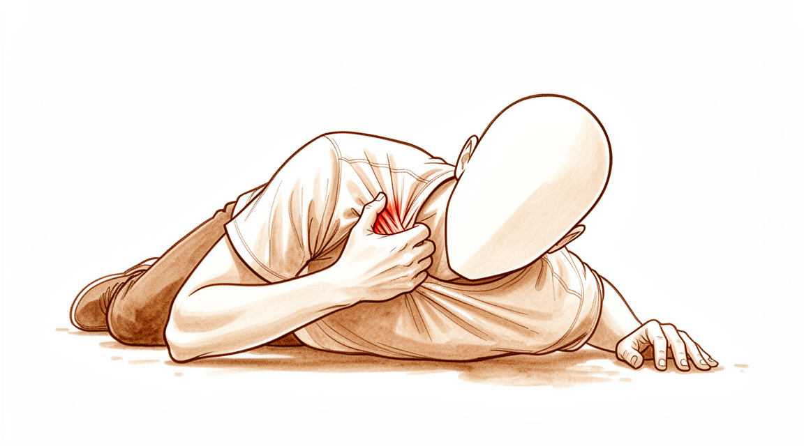
Maaaring mararamdaman mo ang matulis na sakit sa harap ng iyong balikat o sa buong collarbone. Karaniwang nangyayari ang pinsalang ito pagkatapos mahulog sa iyong balikat o matamaan nang direkta. Ito ay karaniwan sa mga lalaki, lalo na sa mga palakasan tulad ng football. Maaari mong mapansin ang pamamaga at pamumula o pagkalasong kulay sa ibabaw ng buto. Ang lugar ay magiging sensitive sa pagpikit.
Ang mga simpleng gawain sa araw-araw ay nagiging mahirap dahil ang paggalaw ng iyong braso ay humihila sa nasugatang buto. Ang pag-abot sa likod ng iyong likod upang isara ang bra o itabi ang damit ay maaaring magdulot ng sakit. Ang pag-angat ng mga bagay, kahit mga magaan, ay maaaring maramdaman na mabigat o hindi ligtas. Maaari kang makakaramdam ng hirap na panatilihin ang iyong braso na malayo sa iyong katawan. Mag-aadvice ang iyong doktor na panatilihin ang iyong braso malapit sa iyong gilid upang bawasan ang tensyon.
Ang sakit ay madalas lumala sa gabi, na nagpapatigil sa paghahanap ng komportableng posisyon sa pagtulog. Karaniwang hindi posible ang pagtulog sa nasugatang gilid. Karaniwang nagkakaroon ng katatagan sa paggising, lalo na kung gumalaw ka sa loob ng gabi. Pagkatapos ng aktibidad, maaaring lumala ang sakit. Ang pagpapahinga ay karaniwang tumutulong upang mapayapa ang sakit, ngunit kailangan mong iwasan ang anumang galaw na nagdudulot ng matulis na pagtaas ng hindi komportableng pakiramdam.
Karamihan sa mga fracture ng clavicle ay gumagaling nang maayos sa pamamagitan ng hindi pampasurgical na paggamot. Gayunpaman, kailangan mo ng malapit na pagsubaybay upang matiyak na hindi lumilipat ang buto sa maliit na posisyon. Ang pinakakaraniwang komplikasyon ay ang non-unions, kung saan nabigo ang buto na gumaling, at malunions, kung saan gumaling ito sa kaunting ibang posisyon. Maaari itong magdulot ng nananatiling sakit o isang kitang-kitang butil. Kung mayroon kang medial clavicle fracture (mas malapit sa iyong dibdib), ang buto mismo ay karaniwang gumagaling nang maayos kung makakaligtas ka sa paunang trauma. Ngunit maging alerto na ang isang mataas na proporsyon ng mga pasyente na may partikular na uri ng fracture na ito ay maaaring harapin ang seryosong panganib sa kalusugan sa loob ng tatlong taon pagkatapos ng pinsala.
Para sa mga displaced distal fractures (malapit sa joint ng balikat), ang hindi pampasurgical na paggamot ay nagdudulot ng mas mataas na rates ng non-union ngunit nag-aalok pa rin ng mahusay na function ng balikat at mababang panganib ng ibang komplikasyon. Ang mga kabataan na may ganap na displaced fractures ay madalas makakakita ng mahusay na resulta pagkatapos ng limang taon kung gagamutin nang walang pampasurgical. Ang iyong doktor ay magtatakda ng iyong paggamot base sa iyong partikular na pattern ng fracture at inaasahan.
Ano ang nangyayari talaga
Ang iyong collarbone (klawikula) ay nag-uugnay ng iyong breastbone (sternum) sa iyong shoulder blade (scapula). Ito ay nagsisilbing strut na pinapanatiling malawak at matatag ang iyong balikat. Karamihan sa mga pagkabasag ay nangyayari kapag nahulog ka nang direkta sa iyong balikat o natamaan nang mahigpit ang lugar na iyon. Karaniwang mas madalas ito sa mga lalaki at madalas na nauugnay sa mga aksidente sa kotse, pagkahulog, o mga palakasan.
Kapag nabasag ang buto, nawawala ang normal na pagkakahanay. Maaari nitong sanayin ang sakit at limitahan ang iyong paggalaw ng braso. Ang magandang balita ay ang iyong katawan ay napakahusay sa paggaling ng butong ito. Karamihan sa mga pagkabasag, lalo na sa gitnang bahagi, ay gumagaling nang maayos nang walang operasyon. Kahit pumili kang hindi magkaroon ng operasyon, susubaybayan ka ng iyong doktor nang maigi. Ito ay dahil maaaring maglisin nang bahagya ang mga piraso ng buto sa mga araw pagkatapos ng pinsala.
Para sa ilang pasyente, ang operasyon ang mas magandang pagpipilian. Madalas itong totoo kung ang pagkabasag ay ganap na nalipat o kung ang buto ay nagkaron ng pagpapapapalaki ng 2 cm o higit pa. Tumutulong ang operasyon na panatilihin ang buto sa tamang posisyon upang gumaling ito nang wasto. Ito ay isang ligtas at epektibong proseso para sa mga matatanda at mga kabataan. Sa katunayan, ang karamihan sa mga komplikasyon mula sa mga pagkabasag ng klawikula—maaring gamot nang may operasyon o walang operasyon—ay mga isyu sa paraan ng paggaling ng buto, tulad ng hindi pagkakaisa o paggaling sa kaunting maling posisyon.
Ang kabuuang rate ng komplikasyon para sa surgical care ay 8.1%. Para sa mga kabataan, ang non-surgical treatment ay madalas na nagdudulot ng mas kaunting komplikasyon at katulad na antas ng kasiyahan kumpara sa operasyon. Gayunpaman, sa mga selektibong kaso kung saan ang pagkabasag ay malala, ang operasyon ay maaaring magdulot ng mas magagandang resulta. Titingnan ng iyong doktor ang tiyak na hugis ng iyong pagkabasag at iyong mga personal na layunin upang desigunin ang pinakamainam na landas. Ang layunin ay palaging ang pagpapanumbalik ng iyong function at kaginhawaan ng balikat nang ligtas.
Mga maitutulong namin dito
Karamihan sa mga fracture ng clavicle ay dulot ng pagkabagsak sa balikat o ng direktang pagtama. Ang karamihan ay nangyayari sa mga lalaki at nauugnay sa aksidente sa sasakyan, pagkabagsak, o pinsala sa sports. Maingat na susuriin ng iyong surgeon ang anumang ibang pinsala sa rehiyon ng balikat, lalo na kung ang pinsala ay may kasamang compression. Ang karamihan sa mga pasyente na may fracture ng clavicle ay may mahusay na resulta gamit ang conservative management. Ibig sabihin, maaari kang mag-anticipate ng magandang clinical at functional results anuman ang iyong pagpili, kung gagawin mo ba ang surgery o hindi.
Maaari kang magsimula sa self-management at physiotherapy. Ang paunang nonsurgical management ay makatwiran dahil pare-pareho ang functional outcomes ng mga pasyente kahit na delayed ang surgery. Kinakailangan ang malapit na follow-up dahil sa panganib ng progressive displacement sa peri-injury period. Maaaring irekomenda ng iyong surgeon ang pagka-delay ng assessment sa 6 linggo pagkatapos ng displaced midshaft clavicle fracture. Ang timing na ito ay nagbibigay-daan sa tumpak na paghula sa mga pasyente na malamang na magkakaroon ng union sa pamamagitan ng nonoperative management. Layunin ng physiotherapy na ibalik ang galaw at lakas habang gumagaling ang buto. Kapag gumaling na ang mga fracture ng clavicle, hindi nagbibigay ng anumang makabuluhang impormasyon ang karagdagang radiographic imaging.
Ang medical management ay nakatuon sa pagpapagaan ng sakit. Maaaring magreseta ng pain medication o anti-inflammatories ang iyong surgeon upang tulungan kang pamahalaan ang discomfort sa proseso ng paggaling. Para sa mga partikular na kaso tulad ng nonunion, ang bone marrow injection ay isang pangakoang treatment na may mababang morbidity at preliminary na tagumpay. Gayunpaman, hindi sapat ang standard na plain unilateral radiographs ng clavicle upang maaasahan na matukoy ang antas ng pagpapalaki (shortening) ng mga fracture ng clavicle at ang pangangailangan para sa surgery sa mga orthopaedic surgeons na may fellowship training sa shoulder/sports medicine. Gamitin ng iyong surgeon ang clinical judgment kasama ang imaging upang gabayan ang iyong pag-aalaga.
Ang surgery ay itinuturing kapag naabot na ng conservative care ang hangganan nito. Ang high-quality na ebidensya ay nagpapakita na ang surgical treatment ng displaced clavicle fractures sa mga matatanda ay nagdudulot ng mas mataas na union rates at mas magandang early patient-reported outcomes kumpara sa nonsurgical treatment, bagama't pareho ang long-term outcomes. Ang operative treatment ng displaced midshaft clavicle fractures sa mga matatanda ay nauugnay sa mas mataas na union rates at mas magandang early patient-reported outcomes kaysa sa non-operative treatment. Ang isang selektibong grupo ng mga pasyente na may completely displaced fractures, shortening na 2 cm o higit pa, o mga partikular na indikasyon, ay nakikinabang sa surgical fixation na ipinapakita na nagdudulot ng improved outcomes kumpara sa non-operative measures. Talakayin ng iyong surgeon kung ang opsyong ito ay angkop para sa iyo batay sa iyong mga katangian ng fracture at inaasahan.
Ano ang inaasahan
Ang iyong prognosis ay nakadepende sa malaking bahagi sa iyong edad at sa lokasyon ng basag. Para sa karamihan ng mga matatanda na may basag sa gitna ng collarbone, ang operasyon ay nagdudulot ng mas mabilis na paggaling at mas magandang pag-andar ng balikat sa maagang yugto kumpara sa walang operasyong paggamot. Gayunpaman, ang mga resulta sa pangmatagalan ay katulad, anuman ang pagpili mo kung gagawa ng operasyon o hindi. Kung ikaw ay isang kabataan, madalas na mas pinipili ang walang operasyong paggamot. Ito ay may mas mababang rate ng komplikasyon at nagbibigay pa rin ng mahusay na pag-andar sa pangmatagalan, kahit na ang buto ay gumaling sa kaunting paglipat ng posisyon.
Nangangailangan ng oras ang paggaling. Sa pamamagitan ng operasyon, karaniwang mas mabilis na nag-uugnay ang buto kumpara sa kung walang operasyon. Sa ilang mga kaso ng mataas na pangangailangan, tulad ng mga propesyonal na atleta, ang paggaling ay maaaring mangyari sa loob ng humigit-kumulang 8.8 linggo. Kung walang operasyon, maaaring hintayin ng iyong doktor ang anim na linggo upang makita kung gumagaling ang buto nang sarili nito bago gumawa ng karagdagang desisyon. Karamihan ng mga basag ay dulot ng pagkabagsak o direktang pagtama. Ang mga pinakakaraniwang isyu ay ang hindi pagkakagaling (kung saan hindi gumagaling ang buto) o maling pagkakagaling (kung saan gumagaling ito sa maling posisyon). Maaaring mangyari ang mga ito sa alinman sa dalawang landas ng paggamot.
Ang mga komplikasyon ay relatibong bihira. Ang average na rate ng komplikasyon para sa operasyong paggamot ay 8.1%. Para sa mga kabataan, ang walang operasyong pamamahala ay may mas kaunting komplikasyon pa. Kung mayroon kang basag sa medial clavicle, ang iyong prognosis ay karaniwang maganda pagkatapos mong malampasan ang paunang pinsala, bagama’t mahalagang tandaan na ang isang mataas na proporsyon ng mga pasyente na may partikular na uri ng basag na ito ay maaaring harapin ang seryosong mga panganib sa kalusugan sa loob ng tatlong taon. Para sa karamihan ng ibang mga basag ng collarbone, ang panganib ng pangmatagalang sakit sa balikat o subacromial pain syndrome (sakit sa ilalim ng balikat) ay hindi pinataas ng pinsala mismo.
Kung pipili ka ng walang operasyong paggamot, mahalaga ang malapitang pagsubaybay. May panganib na ang mga piraso ng buto ay maghihiwalay pa sa mga unang linggo. Kung hindi ka gagawa ng operasyon, maaaring mas magpakita ng sintomas ang iyong balikat kumpara sa kabilang panig 10 hanggang 30 taon pagkatapos ng trauma. Kung gagawa ka ng operasyon, maaaring maranasan mo ang kaunting karagdagang maikling panahong komplikasyon, ngunit malamang na mabawi mo ang pag-andar nang mas maaga. Kapag lubos nang gumaling ang basag, karaniwang hindi nagbibigay ng bagong impormasyon ang mga karagdagang X-ray. Iiayon ng iyong doktor ang plano sa iyong partikular na pattern ng basag at mga personal na layunin.
Kailan kumonsulta sa doktor
Kumonsulta sa iyong doktor kung mayroon kang patuloy na sakit na hindi gumagaling kahit magpahinga. Humingi ng pagsusuri ng espesyalista kung napapansin mo ang kahinaan o kawalan ng katatagan sa iyong balikat. Humingi ng tulong kung ang iyong balikat ay nakakabit o biglang bumabagsak. Kontakin ang iyong surgeon kung ang mga sintomas ay nakakaapekto sa iyong pagtulog o trabaho. Humingi ng tulong para sa anumang biglaang paglala ng iyong kondisyon. Karamihan sa mga fracture ay dulot ng pagkabagsak sa balikat o direktang pagtama. Ang karamihan ay nangyayari sa mga lalaki at nauugnay sa aksidente ng sasakyan, pagkabagsak, o pinsala sa sports. Mahalaga ang malapit na pagsusuri ng mga clavicle fracture na hindi na-opera upang matiyak ang tamang paggaling.
Evidence & references
Overview
- If patients with medial clavicle fractures survive the initial trauma, they can expect good clinical and functional outcomes regardless of whether surgical or nonsurgical management is chosen [1].
- Close follow-up of nonoperatively treated clavicle fractures is warranted due to potential displacement related to patient position and progressive displacement in the peri-injury period [2].
- The most common complications following clavicle fractures, whether treated operatively or non-operatively, are non-unions and malunions [7].
- Clavicle fixation is a safe and effective procedure in the pediatric population with a lack of serious complications [12].
- Specific treatment of clavicle fractures should be individualized based on fracture characteristics and patient expectations rather than broadly applied [14].
- Nonoperative treatment of adolescent clavicle fractures demonstrated lower complication rates and similar satisfaction and functional outcomes compared to operative treatment [17].
- Most mid-shaft clavicle fractures can be treated effectively by non-operative means, but a select group of patients with completely displaced fractures, shortening of 2 cm or more, or specific indications benefit from surgical fixation which has been shown to result in improved outcomes compared with non-operative measures [28].
- Although ORIF of displaced midshaft clavicle fractures remains controversial in the adolescent population, there may be additional circumstances beyond absolute indications for surgical intervention that warrant ORIF at initial presentation [30].
- Current evidence suggests that the majority of clavicular fractures in adolescents can and should be treated nonoperatively, although operative treatment with plate and screw application has consistently good outcomes with a low complication rate in selected cases [57].
- There is an increasing trend toward stabilization and fixation of markedly displaced midshaft clavicle fractures in adolescents due to concerns about symptomatic malunion and poor functional outcomes with nonsurgical management, though definitive indications for fixation in this population remain unclear [61].
- Patient selection for surgery may influence functional outcome after midshaft clavicle fracture [64].
Anatomy & Pathophysiology
- Clavicle fractures do not increase the occurrence of later subacromial pain syndrome [3].
- Protraction of the scapula is not suggested as a major risk factor for the development of subacromial pain syndrome [3].
- Evaluation of the extent of anatomic injury and understanding its mechanical consequences regarding shoulder and arm function is key in developing treatment protocols for acromioclavicular joint injuries [34].
- Hook plate and superolateral locking plate with coracoclavicular suture fixation constructs offer superior biomechanical stability for distal third clavicle fractures with coracoclavicular ligament disruption [39].
- These constructs potentially reduce complications associated with subacromial hardware [39].
- The position of the hook portion of a clavicle hook plate implant can predispose anatomic structures to post-operative complications of subacromial impingement and bony erosion [52].
- The clinical relevance of biomechanical studies on surgical fixation of midshaft clavicle fractures is arguable because none investigate the effect of tissue adaptation over time [54].
- Clavicle hook plate fixation changes scapular kinematics and scapulohumeral rhythm [56].
- Reliable bony union and improved shoulder function can be expected with thoughtful surgical planning, appropriate implant choice, and meticulous surgical technique for clavicle nonunion and malunion [58].
- In complex scapula and ipsilateral clavicle fractures, the question of stability is preoperatively less relevant than whether dislocated fragments lead to compromised shoulder function [63].
- Biomechanical analyses of four different repair techniques for lateral clavicle fracture with coracoclavicular ligament injury did not show any significance in load to failure or displacement after cyclic loading among the study groups [66].
- Plate fixation for displaced midshaft clavicular fractures does not improve shoulder function or general symptoms, and does not decrease limitations compared with nonoperative treatment in a sling [67].
- Suture stabilization of the acromioclavicular ligament plus clavicular hook plate fixation is conducive to restoring shoulder functions and has higher economic efficiency compared to total ligament repair with loop plates for acromioclavicular joint dislocation [70].
- The biphasic plate concept is aimed at improving the biomechanics of locked plating [71].
- Biomechanical evaluation showed effective fixation across all specimens at 500 cycles for unstable lateral clavicle fractures with coracoclavicular ligament disruption (Neer type IIB) [72].
- The specific design of the Locking Compression superior anterior clavicle plate provides higher strength and stiffness when compared to seven and ten hole reconstruction plates in midshaft clavicle fracture stabilisation [73].
- Clinical outcomes for treatment of unstable distal clavicle fractures with multiple Steinmann pins were evaluated using the Constant-Murley score, the University of California at Los Angeles (UCLA) Shoulder score, and the Disabilities of the Arm, Shoulder and Hand (DASH) score [74].
- Force concentration phenomena result from morphological mismatch, such as excessive inclination and improper occupation of the subacromial space, in acromion and hook plate fixation for acromioclavicular joint dislocation [76].
- Regardless of shape, subacromial erosion did not affect clinical outcomes nor cause rotator cuff lesions after plate removal in type 5 acromioclavicular joint dislocations [77].
- Inferior plates may be better equipped to resist in vivo loads experienced by the clavicle during early rehabilitation, particularly during shoulder flexion motions associated with eating, in comminuted midshaft clavicle fractures [78].
- Three patients (18%) experienced postoperative issues including plate prominence (2) and shoulder stiffness (1) in outcomes of internal fixation of clavicle and coracoclavicular stabilization for unstable distal clavicle fractures; none required reoperation [81].
Classification
- Clavicle fractures are the most commonly occurring fracture [5].
- The middle third is the most frequent site of clavicle fractures [5].
- The incidence of clavicle fractures is 1.23% [27].
- The most common complications following clavicle fractures, whether treated operatively or non-operatively, are non-unions and malunions [7].
- Clavicle malunion is a distinct clinical entity that can be treated successfully [9].
- Complication rates following surgical clavicle fracture care averaged 8.1% [20].
- The Utrecht Score for clavicle fractures is a compact yet complete tool developed to assess functional outcome specifically in patients with a clavicle fracture, consisting of patient-reported and objective measures [24].
- The Constant score was found to be reliable for assessing patients with clavicle fractures, especially at the group level [55].
- The presented classification system for lateral clavicle fractures, along with associated treatment algorithms, showed substantial inter- and intraobserver reliability [32].
- The modified Neer classification remains the predominantly cited classification system for distal clavicle fractures [33].
- The intra- and interobserver reliability of the modified Neer classification for distal clavicle fractures has been demonstrated to be inconsistent, which can lead to incorrect treatment choices and misclassifications in research [33].
- The interrater agreement of the modified Neer classification system for lateral clavicle fractures was fair [45].
- Additional 3D CT did not improve the overall level of interrater or intrarater agreement of the modified Neer classification system or associated treatment choice for lateral clavicle fractures [45].
- A new classification system for distal clavicle fractures demonstrated moderate interobserver and substantial intraobserver reliability, as well as reliability for the associated treatment choice [35].
Clinical Presentation
- Clavicle fractures are the most commonly occurring fracture [5].
- The middle third of the clavicle is the most frequent site of fracture [5].
- Adolescent clavicle fractures occur more commonly in male patients [46].
- Adolescent clavicle fractures occur during sports activities [46].
- Adolescent clavicle fractures are secondary to a direct blow to the shoulder [46].
- Adolescent clavicle fractures occur on the nondominant side [46].
- Clinicians must carefully examine patients with isolated clavicle fractures for concomitant injuries to the ipsilateral shoulder girdle, particularly in the context of compression mechanisms [18].
- Segmental fractures of the clavicle are easily missed [26].
- Distal fractures of the clavicle in children are rare [43].
- Most distal clavicle fractures in children can be treated conservatively [43].
- The most common complications following clavicle fractures, whether treated operatively or non-operatively, are non-unions and malunions [7].
- Clavicle malunion is a distinct clinical entity that can be treated successfully [9].
Investigations
- Clavicle fractures are the most commonly occurring fracture, with the middle third being the most frequent site [5].
- Segmental fractures of the clavicle are easily missed [26].
- Delayed diagnosis of subacromial, supracoracoid dislocation of the acromioclavicular joint with ipsilateral clavicle fracture is likely if careful examination of the patient's radiographs is not performed [85].
- Clinicians must carefully examine patients with isolated clavicle fractures for concomitant injuries to the ipsilateral shoulder girdle, particularly in the context of compression mechanisms [18].
- Preoperative MRI or diagnostic arthroscopy to evaluate glenohumeral associated injuries to distal clavicle fractures should be recommended [69].
- An upright chest radiograph should be obtained to evaluate midshaft clavicle fracture displacement, as it represents the physiologic stress across the fracture when considering nonoperative management [82].
- Close follow-up of nonoperatively treated clavicle fractures is warranted [2].
- Displacement of diaphyseal clavicle fractures is related to patient position and progressive displacement in the peri-injury period [2].
- Standard plain unilateral radiographs of the clavicle are insufficient to reliably determine the degree of shortening of clavicle fractures and the need for surgery among shoulder/sports medicine fellowship–trained orthopaedic surgeons [86].
- When clavicle shortening is considered in the decision to pursue operative management, the use of plain radiograph-based measurements is not recommended [83].
- Delayed assessment at 6 weeks following displaced midshaft clavicle fracture enables an accurate prediction of patients who are likely to have union with nonoperative management [31].
- Once clavicle fractures are healed, further radiographic imaging does not provide any notable information [4].
Treatment
- If patients with medial clavicle fractures survive the initial trauma, good clinical and functional outcomes are expected regardless of whether surgical or nonsurgical management is chosen [1].
- Close follow-up of nonoperatively treated clavicle fractures is warranted due to potential for progressive displacement in the peri-injury period [2].
- Initial nonsurgical management of clavicle fractures may be reasonable because patients had similar functional outcomes even when surgery was delayed [6].
- Operative treatment of displaced medial clavicle fractures provides an excellent long-term functional outcome [8].
- Nonoperative management of adolescent mid-shaft clavicle fractures results in excellent functional outcomes at long-term follow-up [10].
- Most patients with clavicle fractures have an excellent outcome using conservative management [11].
- Clavicle fixation is a safe and effective procedure in the pediatric population with a lack of serious complications [12].
- Specific treatment of clavicle fractures should not be broadly applied but rather should be individualized based on fracture characteristics and patient expectations [14].
- A more prolonged surveillance period is recommended in children with recurrent fractures of the clavicle [15].
- Nonoperative treatment of adolescent clavicle fractures demonstrated lower complication rates and similar satisfaction and functional outcomes compared to operative treatment [17].
- Functional outcome is excellent following the treatment of both acute and non-united clavicle fractures, but recovery occurs earlier following acute treatment [19].
- Most mid-shaft clavicle fractures can be treated effectively by non-operative means, but a select group of patients with completely displaced fractures, shortening of 2 cm or more, or specific indications benefit from surgical fixation which has been shown to result in improved outcomes compared with non-operative measures [28].
- Although ORIF of displaced midshaft clavicle fractures remains controversial in the adolescent population, there may be additional circumstances beyond absolute indications for surgical intervention that warrant ORIF at initial presentation [30].
- Bone marrow injection for the treatment of clavicle nonunion is promising, with low morbidity and preliminary success justifying further trials [36].
- Plate fixation of midshaft clavicle fractures for delayed union and non-union is a cost-effective intervention but functional deficits persist at long-term follow-up [37].
- Comparably excellent outcomes of severe clavicle fractures in adolescent athletes can be achieved with non-operative treatment [38].
- High-quality evidence shows that surgical treatment of displaced clavicle fractures in adults results in higher union rates and better early patient-reported outcomes compared with nonsurgical treatment, though long-term outcomes are similar [40].
- Superiorly applied plate fixation is an effective treatment for clavicular nonunion [41].
- Operative treatment of displaced midshaft clavicle fractures in adults is associated with higher union rates and better early patient-reported outcomes than non-operative treatment, though long-term outcomes are similar [42].
- Nonsurgical and surgical management provide similar results for distal clavicle fractures [44].
- Treatment of middle-third clavicle non-union after initial failure of conservative treatment with stable fixation and bone graft is a reliable, well-suited and effective treatment [49].
- A targeted approach to the management of mid-shaft clavicle fractures is needed, with simple fractures treated nonoperatively and complex displaced fractures considered for surgery to prevent non-union [50].
- Nondisplaced clavicle fractures continue to be treated conservatively with a simple sling until the fracture is healed according to radiographs and clinical assessment [51].
- In the studied unit, there is no clearly favoured method of internal fixation of lateral clavicle fractures [59].
- Ipsilateral os acromiale may be a relative contraindication to the clavicle hook plate [62].
Complications
- Non-unions and malunions are the most common complications following clavicle fractures, regardless of whether they are treated operatively or non-operatively [7].
- Complication rates following surgical clavicle fracture care average 8.1% [20].
- Limited incision plating of midshaft clavicle fractures achieves a low complication rate comparable to standard incision techniques [23].
- Clavicle pinning results in minimal complications [48].
- Surgical treatment of clavicle fractures in the pediatric population is associated with a lack of serious complications [12].
- Nonoperative management of displaced distal clavicle fractures is associated with higher nonunion rates [29].
- Nonoperative management of displaced distal clavicle fractures carries a low risk of complications and delayed surgery [29].
- The affected shoulder side is more symptomatic than the unaffected side 10 to 30 years after conservative treatment of midshaft clavicle fractures [65].
- Clavicle fractures do not increase the occurrence of later subacromial pain syndrome [3].
- Protraction of the scapula is not suggested as a major risk factor for the development of subacromial pain syndrome [3].
- Adolescent mid-shaft clavicle fracture displacement does not predict nonunion at long-term follow-up [10].
- Children with recurrent fractures of the clavicle require a more prolonged surveillance period [15].
Recovery
- Patients with medial clavicle fractures who survive the initial trauma can expect good clinical and functional outcomes regardless of whether surgical or nonsurgical management is chosen [1].
- Medial clavicle fractures have favorable functional outcomes and pain relief at minimum 1-year follow-up among patients who survive the trauma [13].
- A high proportion of patients with medial clavicle fractures die within 3 years of the injury [13].
- Conservative management of medial clavicle fractures results in excellent functional results [16].
- Operative treatment of displaced medial clavicle fractures provides an excellent long-term functional outcome [8].
- Nonoperative management of adolescent mid-shaft clavicle fractures results in excellent functional outcomes at long-term follow-up [10].
- Adolescent mid-shaft clavicular fracture displacement does not predict nonunion or inferior functional outcome at long-term follow-up [10].
- Initial nonsurgical management of clavicle fractures may be reasonable because patients had similar functional outcomes even when surgery was delayed [6].
- Nonoperative management of displaced distal clavicle fractures results in higher nonunion rates, but shoulder function remains excellent, and the risk of complications and delayed surgery are low [29].
- Patients had very good clinical outcomes following operative management of an extra-lateral distal clavicle fracture pattern [22].
- Functional outcome is excellent following the treatment of both acute and non-united clavicle fractures, but recovery occurs earlier following acute treatment [19].
- Patients reported a good quality of life and functional outcome after plating for midshaft clavicular fractures [53].
- Surgical treatment of midshaft clavicle fractures significantly reduces the nonunion rate and shortens the time to union as compared with the nonoperative approach [47].
- Surgical treatment of midshaft clavicle fractures leads to better shoulder functional scores at short- and long-term follow-up compared with nonoperative treatment [47].
- Surgical treatment of midshaft clavicle fractures has a slightly higher incidence of complications than nonoperative treatment [47].
- A limited incision approach for plating of acute midshaft clavicle fractures achieved good functional and radiographic outcomes with a low complication rate comparable to standard incision techniques [23].
- The prognosis for obtaining bony union after infected clavicle fractures is poor, with only two of six patients achieving union [75].
- Clavicle fractures do not increase the occurrence of later subacromial pain syndrome [3].
- The results do not suggest protraction of the scapula as a major risk factor for the development of subacromial pain syndrome [3].
Key Evidence
- [L5] If patients with medial clavicle fractures can survive the initial trauma, there is every reason to expect good clinical and functional outcomes, regardless of whether surgical or nonsurgical management is chosen. [1] (10.1097/corr.0000000000001916)
- [L2] Close follow-up of nonoperatively treated clavicle fractures is warranted. [2] (10.1016/j.jse.2018.01.004)
- [L4] The results do not suggest protraction of the scapula as a major risk factor for the development of SAPS. [3] (10.1016/j.xrrt.2024.01.008)
- [L3] Once clavicle fractures are healed, further radiographic imaging does not provide any notable information. [4] (10.5435/jaaos-d-17-00598)
- [L3] Initial nonsurgical management of clavicle fractures may be reasonable because patients had similar functional outcomes even when surgery was delayed. [6] (10.5435/jaaos-d-16-00130)
- [L4] Operative treatment of displaced medial clavicle fractures provides an excellent long-term functional outcome. [8] (10.1007/s00068-018-1024-6)
- [L4] Clavicle malunion is a distinct clinical entity that can be treated successfully. [9] (10.3109/17453674.2010.480939)
- [L3] Nonoperative management of adolescent mid-shaft clavicle fractures results in excellent functional outcomes at long-term follow-up. [10] (10.1302/0301-620x.103b5.bjj-2020-1929.r1)
- [L3] Most patients with clavicle fractures have an excellent outcome using conservative management. [11] (10.1016/j.jse.2019.06.022)
- [L4] Clavicle fixation is a safe and effective procedure in the pediatric population with a lack of serious complications. [12] (10.1177/2325967119s00056)
- [L4] Medial clavicle fractures have favorable functional outcomes and pain relief at minimum 1-year follow-up among those patients who survive the trauma, but a high proportion will die within 3 years of the injury. [13] (10.1097/corr.0000000000001839)
- [L5] Specific treatment of clavicle fractures should not be broadly applied but rather should be individualized based on fracture characteristics and patient expectations. [14] (10.1016/j.jse.2011.08.053)
- [L5] The authors recommend a more prolonged surveillance period in children with recurrent fractures of the clavicle. [15] (10.1097/bpb.0000000000000231)
- [Paper] Sixty eight patients with medial clavicle fractures were identified over a 5 year period, with excellent functional results seen following conservative management. [16] (10.1016/j.injury.2016.06.011)
- [L2] Nonoperative treatment of adolescent clavicle fractures demonstrated lower complication rates and similar satisfaction and functional outcomes compared to operative treatment. [17] (10.1177/2325967119s00428)
- [L4] Clinicians must carefully examine patients with isolated clavicle fractures for concomitant injuries to the ipsilateral shoulder girdle, particularly in the context of compression mechanisms. [18] (10.1177/03635465000280062301)
- [L3] Functional outcome is excellent following the treatment of both acute and non-united clavicle fractures, but recovery occurs earlier following acute treatment. [19] (10.1016/j.otsr.2017.03.021)
- [L3] Complication rates following surgical clavicle fracture care averaged 8.1%. [20] (10.1186/s12891-022-05075-5)
- [L4] The patients had very good clinical outcomes following operative management of an extra-lateral distal clavicle fracture pattern. [22] (10.1016/j.jse.2020.10.006)
- [L5] In this large cohort with long-term follow-up, a limited incision approach for plating of acute midshaft clavicle fractures achieved good functional and radiographic outcomes with a low complication rate comparable to the reported rate for standard incision techniques. [23] (10.1016/j.jse.2025.06.002)
- [L4] The Utrecht Score for clavicle fractures is a compact yet complete tool that was developed to assess functional outcome specifically in patients with a clavicle fracture, consisting of patient-reported and objective measures. [24] (10.1007/s00068-018-0979-7)
- [Case_report] The case highlights that segmental fractures of the clavicle are easily missed. [26] (10.1177/1758573214564496)
- [L4] The incidence of clavicle fractures was 1.23%. [27] (10.1016/j.injury.2011.04.008)
- [L4] Nonoperative management of displaced distal clavicle fractures results in higher nonunion rates, but shoulder function remains excellent, and risk of complications and delayed surgery are low. [29] (10.1016/j.jse.2023.12.006)
- [Case_report] Although ORIF of displaced midshaft clavicle fractures remains controversial in the adolescent population, there may be additional circumstances beyond absolute indications for surgical intervention that warrant ORIF at initial presentation. [30] (10.1016/j.xrrt.2023.03.004)
- [L1] Delayed assessment at 6 weeks following displaced midshaft clavicle fracture enables an accurate prediction of patients who are likely to have union with nonoperative management. [31] (10.2106/jbjs.19.00955)
- [L4] The presented classification system as well as associated treatment algorithms for lateral clavicle fractures showed substantial inter- and intraobserver reliability. [32] (10.1016/j.jse.2025.04.021)
- [L5] The modified Neer classification remains the predominantly cited classification system for distal clavicle fractures, yet its intra- and interobserver reliability has been demonstrated to be inconsistent, which can lead to incorrect treatment choices and misclassifications in research. [33] (10.1097/corr.0000000000001456)
- [L5] A comprehensive clinical approach emphasizing the evaluation of the extent of the anatomic injury and understanding its mechanical consequences regarding shoulder and arm function is a key in the development of guidelines for developing operative or non-operative treatment protocols and for establishing outcomes of the treatment protocols. [34] (10.1177/17585732221122335)
- [L3] The study demonstrated moderate interobserver and substantial intraobserver reliability of the new classification system and the associated treatment choice for distal clavicle fractures. [35] (10.1016/j.otsr.2018.05.015)
- [L4] Bone marrow injection for the treatment of clavicle nonunion is promising, with low morbidity and preliminary success justifying further trials. [36] (10.1016/j.jse.2006.05.001)
- [L3] Clavicle fixation for delayed and non-union is a cost-effective intervention but outcomes are worse compared to patients that unite with non-operative management. [37] (10.1177/1758573221990367)
- [L2] Comparably excellent outcomes of severe clavicle fractures in adolescent athletes can be achieved with non-operative treatment. [38] (10.1177/2325967121s00214)
- [L5] These constructs offer superior biomechanical stability in our model and potentially reduce complications associated with subacromial hardware. [39] (10.1016/j.xrrt.2025.100645)
- [L1] High-quality evidence shows that surgical treatment of displaced clavicle fractures in adults results in higher union rates and better early patient-reported outcomes compared with nonsurgical treatment, though long-term outcomes are similar. [40] (10.5435/jaaos-d-23-00472)
- [L4] Superiorly applied plate fixation is an effective treatment for clavicular nonunion. [41] (10.1016/j.jse.2008.05.046)
- [L4] Distal fractures of the clavicle in children are rare and most can be treated conservatively. [43] (10.1016/j.rboe.2015.12.006)
- [L4] Nonsurgical and surgical management provide similar results for distal clavicle fractures. [44] (10.5435/00124635-201107000-00002)
- [L3] The interrater agreement of the modified Neer classification system for lateral clavicle fractures was fair, and additional 3D CT did not improve the overall level of interrater or intrarater agreement of the classification system or associated treatment choice. [45] (10.1177/0363546515593949)
- [L4] Adolescent clavicle fractures occurred more commonly in male patients during sports, secondary to a direct blow to the shoulder, and on the nondominant side. [46] (10.1177/2325967120921344)
- [L1] Surgical treatment of midshaft clavicle fractures significantly reduces the nonunion rate and shortens the time to union as compared with the nonoperative approach and, despite a slightly higher incidence of complications, leads to better shoulder functional scores at short- and long-term follow-up. [47] (10.1177/0363546519826961)
- [L4] The technique of clavicle pinning resulted in minimal complications, short hospital stay and excellent functional outcomes. [48] (10.4103/0973-6042.57895)
- [L4] Treatment of middle-third clavicle non-union after initial failure of conservative treatment with stable fixation and bone graft is a reliable, well-suited and effective treatment. [49] (10.1016/j.otsr.2013.09.011)
- [L5] A targeted approach to the management of mid-shaft clavicle fractures is needed, with simple fractures treated nonoperatively and complex displaced fractures considered for surgery to prevent non-union. [50] (10.1016/j.injury.2020.11.066)
- [L4] Nondisplaced clavicle fractures continue to be treated conservatively with a simple sling until the fracture is healed according to radiographs and clinical assessment. [51] (10.3810/psm.2011.09.1930)
- [L5] The observed frequency of hook contact with surrounding subacromial structures in a static shoulder confirms that the position of the hook portion of the implant can predispose anatomic structures to the post-operative complications of subacromial impingement and bony erosion. [52] (10.1016/j.injury.2009.12.012)
- [L3] Patients reported a good quality of life and functional outcome after plating for midshaft clavicular fractures. [53] (10.1016/j.injury.2017.10.032)
- [L2] The clinical relevance of the biomechanical studies may be arguable since none investigate the effect of tissue adaptation over time. [54] (10.1016/j.injury.2018.02.017)
- [L4] The Constant score was found to be reliable for assessing patients with clavicle fractures, especially at the group level. [55] (10.1016/j.jse.2016.02.022)
- [L3] Clavicle hook plate fixation changes the scapular kinematics and scapulohumeral rhythm; thus, when clavicle hook plate fixation is complete, the implant should be promptly removed. [56] (10.1007/s00264-018-4003-y)
- [L4] Current evidence suggests that the majority of clavicular fractures in adolescents can and should be treated nonoperatively, although operative treatment with plate and screw application has consistently good outcomes with a low complication rate in selected cases. [57] (10.2106/jbjs.22.01036)
- [L5] Reliable bony union and improved shoulder function can be expected with thoughtful surgical planning, appropriate implant choice, and meticulous surgical technique. [58] (10.1016/j.jse.2013.01.022)
- [L4] In our unit there is no clearly favoured method of internal fixation of lateral clavicle fractures. [59] (10.1007/s00590-021-03173-z)
- [L5] There is an increasing trend toward stabilization and fixation of markedly displaced midshaft clavicle fractures in adolescents due to concerns about symptomatic malunion and poor functional outcomes with nonsurgical management, though definitive indications for fixation in this population remain unclear. [61] (10.5435/00124635-201301000-00002)
- [L4] Ipsilateral os acromiale may be a relative contraindication to the clavicle hook plate. [62] (10.1186/s12891-021-04841-1)
- [L4] The question of stability is preoperatively less relevant than the question of whether the dislocated fragments lead to compromised shoulder function. [63] (10.1007/s00068-018-0946-3)
- [L1] This review shows that patient selection for surgery may influence functional outcome after midshaft clavicle fracture. [64] (10.1177/1758573218777996)
- [L4] The affected shoulder side was more symptomatic than the unaffected side 10 to 30 years after the trauma when midshaft clavicle fractures were treated conservatively. [65] (10.1186/s13018-023-04450-9)
- [L5] The biomechanical analyses did not show any significance in load to failure or displacement after cyclic loading among the study groups. [66] (10.1007/s00167-017-4444-7)
- [L1] In addition, the procedure does not improve shoulder function or general symptoms, and it does not decrease limitations compared with nonoperative treatment in a sling. [67] (10.2106/jbjs.15.01394)
- [L1] Preoperative MRI or diagnostic arthroscopy to evaluate glenohumeral associated injuries to distal clavicle fractures should be recommended. [69] (10.1186/s13018-022-02919-7)
- [L3] This procedure was conducive to restoring shoulder functions and had higher economic efficiency. [70] (10.1186/s13018-025-06032-3)
- [L5] The biphasic plate concept is aimed at improving the biomechanics of locked plating. [71] (10.1016/j.injury.2020.04.032)
- [L5] Biomechanical evaluation showed effective fixation across all specimens at 500 cycles. [72] (10.1016/j.jse.2022.11.008)
- [L5] The specific design of the plate provides higher strength and stiffness when compared to reconstruction plates. [73] (10.1007/s00264-012-1671-x)
- [L4] Clinical outcomes were evaluated using the Constant-Murley score, the University of California at Los Angeles (UCLA) Shoulder score, and the Disabilities of the Arm, Shoulder and Hand (DASH) score. [74] (10.1097/bot.0000000000000850)
- [L4] The prognosis for obtaining bony union after infected clavicle fractures is poor, with only two of six patients achieving union. [75] (10.1097/01.blo.0000183088.60639.05)
- [L4] The force concentration phenomenon results from cases of morphological mismatch, such as excessive inclination and improper occupation of the subacromial space. [76] (10.1007/s00167-016-3987-3)
- [L3] Regardless of shape, subacromial erosion did not affect clinical outcomes nor cause rotator cuff lesions after plate removal. [77] (10.1186/s12891-021-04987-y)
- [L5] Inferior plates may be better equipped to resist the in vivo loads experienced by the clavicle during early rehabilitation after internal fixation, particularly during the shoulder flexion motions associated with eating. [78] (10.1016/j.clinbiomech.2010.12.007)
- [L4] Three patients (18%) experienced postoperative issues: plate prominence (2) and shoulder stiffness (1); none required reoperation. [81] (10.1177/17585732261456152)
- [L4] An upright chest radiograph should be obtained to evaluate midshaft clavicle fracture displacement, as it represents the physiologic stress across the fracture when considering nonoperative management. [82] (10.1097/bot.0000000000000727)
- [L4] When clavicle shortening is considered in the decision to pursue operative management, the use of plain radiograph-based measurements is not recommended. [83] (10.4055/cios.2016.8.4.367)
- [L5] Delayed diagnosis is likely if careful examination of the patient's radiographs is not performed. [85] (10.1177/2054270414527281)
- [L3] Standard plain unilateral radiographs of the clavicle are insufficient to reliably determine the degree of shortening of clavicle fractures and the need for surgery among shoulder/sports medicine fellowship–trained orthopaedic surgeons. [86] (10.1177/0363546514523926)
References
[1] CORR Insights®: What Are the Functional Outcomes and Pain Scores after Medial Clavicle Fracture Treatment?. Clinical Orthopaedics & Related Research. 2021. DOI: 10.1097/corr.0000000000001916 [2] Displacement of diaphyseal clavicle fractures related to patient position and progressive displacement in the peri-injury period. Journal of Shoulder and Elbow Surgery. 2018. DOI: 10.1016/j.jse.2018.01.004 [3] Clavicle fractures do not increase the occurrence of later subacromial pain syndrome. A registry-based case-control study with 15-25 years of follow-up of 131.838 persons from the Danish National Patient Register. JSES Reviews, Reports, and Techniques. 2024. DOI: 10.1016/j.xrrt.2024.01.008 [4] Potential Economic Benefits of Limited Clinical and Radiographic Follow-up After Plate Fixation of Midshaft Clavicle Fractures. Journal of the American Academy of Orthopaedic Surgeons. 2019. DOI: 10.5435/jaaos-d-17-00598 [5] 7. Clavicle Fractures: Epidemiology, Clinical Evaluation, Imaging, and Classification. n.d.. [6] Factors Affecting Functional Outcomes After Clavicle Fracture. Journal of the American Academy of Orthopaedic Surgeons. 2016. DOI: 10.5435/jaaos-d-16-00130 [7] 10. Complications of Clavicle Fractures—Diagnosis and Management. n.d.. [8] Displaced medial clavicle fractures: operative treatment with locking compression plate fixation. European Journal of Trauma and Emergency Surgery. 2018. DOI: 10.1007/s00068-018-1024-6 [9] Malunion after midshaft clavicle fractures in adults. Acta Orthopaedica. 2010. DOI: 10.3109/17453674.2010.480939 [10] Adolescent mid-shaft clavicular fracture displacement does not predict nonunion or inferior functional outcome at long-term follow-up. The Bone & Joint Journal. 2021. DOI: 10.1302/0301-620x.103b5.bjj-2020-1929.r1 [11] Plate fixation of clavicle fractures: comparison between early and delayed surgery. Journal of Shoulder and Elbow Surgery. 2020. DOI: 10.1016/j.jse.2019.06.022 [12] INCREASING VALUE: CAN MIDSHAFT CLAVICLE FRACTURES BE SURGICALLY TREATED AS AN OUTPATIENT?. Orthopaedic Journal of Sports Medicine. 2019. DOI: 10.1177/2325967119s00056 [13] What Are the Functional Outcomes and Pain Scores after Medial Clavicle Fracture Treatment?. Clinical Orthopaedics & Related Research. 2021. DOI: 10.1097/corr.0000000000001839 [14] Treatment of clavicle fractures: current concepts review. Journal of Shoulder and Elbow Surgery. 2012. DOI: 10.1016/j.jse.2011.08.053 [15] Post-traumatic nonunion of a clavicle fracture in a 9-year-old child. Journal of Pediatric Orthopaedics B. 2016. DOI: 10.1097/bpb.0000000000000231 [16] Natural history of medial clavicle fractures. Injury. 2016. DOI: 10.1016/j.injury.2016.06.011 [17] Two-year Functional Outcomes Of Operative Vs. Non-operative Treatment Of Completely Displaced Midshaft Clavicle Fractures In Adolescents: Results From A Prospective, Multicenter, Level 2 Study. Orthopaedic Journal of Sports Medicine. 2019. DOI: 10.1177/2325967119s00428 [18] Ipsilateral Clavicle Fracture, Sternoclavicular Joint Subluxation, and Long Thoracic Nerve Injury: An Unusual Constellation of Injuries Sustained during Wrestling. The American Journal of Sports Medicine. 2000. DOI: 10.1177/03635465000280062301 [19] Functional recovery following early mobilization after middle third clavicle osteosynthesis for acute fractures or nonunion: A case-control study. Orthopaedics & Traumatology: Surgery & Research. 2017. DOI: 10.1016/j.otsr.2017.03.021 [20] Surgical treatment, complications, reoperations, and healthcare costs among patients with clavicle fracture in England. BMC Musculoskeletal Disorders. 2022. DOI: 10.1186/s12891-022-05075-5 [22] Operative management of an extra-lateral distal clavicle fracture pattern: a study of 48 patients and a proposed update to the modified Neer classification. Journal of Shoulder and Elbow Surgery. 2021. DOI: 10.1016/j.jse.2020.10.006 [23] Limited incision plating of midshaft clavicle fractures: a case series of 1,038 patients. Journal of Shoulder and Elbow Surgery. 2026. DOI: 10.1016/j.jse.2025.06.002 [24] Development of the Utrecht Score for clavicle fractures: a short and complete clavicle score with patient-reported and objective measures. European Journal of Trauma and Emergency Surgery. 2018. DOI: 10.1007/s00068-018-0979-7 [26] Segmental clavicle fracture and acromio-clavicular joint disruption: an unusual case report. Shoulder & Elbow. 2014. DOI: 10.1177/1758573214564496 [27] A 2-year experience, management and outcome of 200 clavicle fractures. Injury. 2012. DOI: 10.1016/j.injury.2011.04.008 [28] 9. Operative Management of Clavicle Fractures: Indications, Techniques, and Outcomes. n.d.. [29] Are displaced distal clavicle fractures associated with inferior clinical outcomes following nonoperative management? A systematic review. Journal of Shoulder and Elbow Surgery. 2024. DOI: 10.1016/j.jse.2023.12.006 [30] Consequences of delayed surgical intervention of a displaced midshaft clavicle fracture: a case report. JSES Reviews, Reports, and Techniques. 2023. DOI: 10.1016/j.xrrt.2023.03.004 [31] Displaced Midshaft Clavicle Fracture Union Can Be Accurately Predicted with a Delayed Assessment at 6 Weeks Following Injury. Journal of Bone and Joint Surgery. 2020. DOI: 10.2106/jbjs.19.00955 [32] Differentiating and treating lateral clavicle fractures: a new simple classification system. Journal of Shoulder and Elbow Surgery. 2026. DOI: 10.1016/j.jse.2025.04.021 [33] Classifications in Brief: The Modified Neer Classification for Distal-third Clavicle Fractures. Clinical Orthopaedics & Related Research. 2020. DOI: 10.1097/corr.0000000000001456 [34] Acromioclavicular joint injuries revisited: Pathoanatomy, pathomechanics, and clinical presentation. Shoulder & Elbow. 2022. DOI: 10.1177/17585732221122335 [35] Distal clavicle fractures: A new classification system. Orthopaedics & Traumatology: Surgery & Research. 2018. DOI: 10.1016/j.otsr.2018.05.015 [36] Healing of a clavicle fracture nonunion with bone marrow injection. Journal of Shoulder and Elbow Surgery. 2007. DOI: 10.1016/j.jse.2006.05.001 [37] Plate fixation of midshaft clavicle fractures for delayed union and non-union is a cost-effective intervention but functional deficits persist at long-term follow-up. Shoulder & Elbow. 2021. DOI: 10.1177/1758573221990367 [38] Operative Versus Non-Operative Treatment of Severely Shortened or Comminuted Clavicle Fractures in Older Adolescent Athletes: Results from A Prospective, Multicenter, Level 2 Cohort Study. Orthopaedic Journal of Sports Medicine. 2021. DOI: 10.1177/2325967121s00214 [39] A biomechanical comparison of hook plate vs. superolateral locking plate with coracoclavicular suture fixation for distal third clavicle fractures with coracoclavicular ligament disruption. JSES Reviews, Reports, and Techniques. 2026. DOI: 10.1016/j.xrrt.2025.100645 [40] American Academy of Orthopaedic Surgeons Clinical Practice Guideline Summary on the Treatment of Clavicle Fractures. Journal of the American Academy of Orthopaedic Surgeons. 2023. DOI: 10.5435/jaaos-d-23-00472 [41] Nonunion of the clavicle treated with plate fixation: A review of forty-seven consecutive cases. Journal of Shoulder and Elbow Surgery. 2008. DOI: 10.1016/j.jse.2008.05.046 [42] AAOS CPG: Treatment of Clavicle Fractures. 2024. [43] Distal clavicle fractures in children. Revista Brasileira de Ortopedia (English Edition). 2016. DOI: 10.1016/j.rboe.2015.12.006 [44] Management of Distal Clavicle Fractures. Journal of the American Academy of Orthopaedic Surgeons. 2011. DOI: 10.5435/00124635-201107000-00002 [45] The Interrater and Intrarater Agreement of a Modified Neer Classification System and Associated Treatment Choice for Lateral Clavicle Fractures. The American Journal of Sports Medicine. 2015. DOI: 10.1177/0363546515593949 [46] Descriptive Epidemiology of Adolescent Clavicle Fractures: Results From the FACTS (Function after Adolescent Clavicle Trauma and Surgery) Prospective, Multicenter Cohort Study. Orthopaedic Journal of Sports Medicine. 2020. DOI: 10.1177/2325967120921344 [47] Midshaft Clavicle Fractures: Surgery Provides Better Results as Compared With Nonoperative Treatment: A Meta-analysis. The American Journal of Sports Medicine. 2019. DOI: 10.1177/0363546519826961 [48] Treatment of mid-shaft clavicle fractures: A comparative study. International Journal of Shoulder Surgery. 2009. DOI: 10.4103/0973-6042.57895 [49] Outcomes from surgical treatment of middle-third clavicle fractures non-union in adults: A series of 21 cases. Orthopaedics & Traumatology: Surgery & Research. 2014. DOI: 10.1016/j.otsr.2013.09.011 [50] Midshaft clavicle fracture – Nonoperative versus operative care. Injury. 2021. DOI: 10.1016/j.injury.2020.11.066 [51] Clavicle Fractures: A Review of the Literature and Update on Treatment. The Physician and Sportsmedicine. 2011. DOI: 10.3810/psm.2011.09.1930 [52] Subacromial morphometric assessment of the clavicle hook plate. Injury. 2010. DOI: 10.1016/j.injury.2009.12.012 [53] Plating for midshaft clavicular fractures: The impact on quality of life and functional outcome. Injury. 2017. DOI: 10.1016/j.injury.2017.10.032 [54] Surgical fixation of midshaft clavicle fractures: A systematic review of biomechanical studies. Injury. 2018. DOI: 10.1016/j.injury.2018.02.017 [55] High inter-rater reliability, agreement, and convergent validity of Constant score in patients with clavicle fractures. Journal of Shoulder and Elbow Surgery. 2016. DOI: 10.1016/j.jse.2016.02.022 [56] Evaluation of three-dimensional in vivo scapular kinematics and scapulohumeral rhythm between shoulders with a clavicle hook plate and contralateral healthy shoulders. International Orthopaedics. 2018. DOI: 10.1007/s00264-018-4003-y [57] Clavicular Fractures in the Adolescent. Journal of Bone and Joint Surgery. 2023. DOI: 10.2106/jbjs.22.01036 [58] Management of clavicle nonunion and malunion. Journal of Shoulder and Elbow Surgery. 2013. DOI: 10.1016/j.jse.2013.01.022 [59] Operative management of lateral third clavicle fractures: a comparison of internal fixation methods. European Journal of Orthopaedic Surgery & Traumatology. 2021. DOI: 10.1007/s00590-021-03173-z [61] Displaced Clavicle Fractures in Adolescents: Facts, Controversies, and Current Trends. Journal of the American Academy of Orthopaedic Surgeons. 2013. DOI: 10.5435/00124635-201301000-00002 [62] Os acromiale may be a contraindication of the clavicle hook plate: case reports and literature review. BMC Musculoskeletal Disorders. 2021. DOI: 10.1186/s12891-021-04841-1 [63] Decision-making for complex scapula and ipsilateral clavicle fractures: a review. European Journal of Trauma and Emergency Surgery. 2018. DOI: 10.1007/s00068-018-0946-3 [64] Does candidate for plate fixation selection improve the functional outcome after midshaft clavicle fracture? A systematic review of 1348 patients. Shoulder & Elbow. 2018. DOI: 10.1177/1758573218777996 [65] Long-term conservative treatment outcomes for midshaft clavicle fractures: a 10-to-30-year follow-up. Journal of Orthopaedic Surgery and Research. 2023. DOI: 10.1186/s13018-023-04450-9 [66] Lateral clavicle fracture with coracoclavicular ligament injury: a biomechanical study of 4 different repair techniques. Knee Surgery, Sports Traumatology, Arthroscopy. 2017. DOI: 10.1007/s00167-017-4444-7 [67] Plate Fixation Compared with Nonoperative Treatment for Displaced Midshaft Clavicular Fractures. Journal of Bone and Joint Surgery. 2017. DOI: 10.2106/jbjs.15.01394 [69] Concomitant glenohumeral injuries in patients with distal clavicle fractures undergoing arthroscopic-assisted surgery: a systematic review. Journal of Orthopaedic Surgery and Research. 2022. DOI: 10.1186/s13018-022-02919-7 [70] Suture stabilization of the acromioclavicular ligament plus clavicular hook plate fixation vs. total ligament repair with loop plates for acromioclavicular joint dislocation. Journal of Orthopaedic Surgery and Research. 2025. DOI: 10.1186/s13018-025-06032-3 [71] Biphasic Plating – In vivo study of a novel fixation concept to enhance mechanobiological fracture healing. Injury. 2020. DOI: 10.1016/j.injury.2020.04.032 [72] Biomechanical analysis of plating techniques for unstable lateral clavicle fractures with coracoclavicular ligament disruption (Neer type IIB). Journal of Shoulder and Elbow Surgery. 2023. DOI: 10.1016/j.jse.2022.11.008 [73] Biomechanical comparison of the Locking Compression superior anterior clavicle plate with seven and ten hole reconstruction plates in midshaft clavicle fracture stabilisation. International Orthopaedics. 2012. DOI: 10.1007/s00264-012-1671-x [74] Treatment of Unstable Distal Clavicle Fractures With Multiple Steinmann Pins—A Modification of Neer's Method: A Series of 56 Consecutive Cases. Journal of Orthopaedic Trauma. 2017. DOI: 10.1097/bot.0000000000000850 [75] Infection after Clavicle Fractures. Clinical Orthopaedics and Related Research. 2005. DOI: 10.1097/01.blo.0000183088.60639.05 [76] Morphological analysis of acromion and hook plate for the fixation of acromioclavicular joint dislocation. Knee Surgery, Sports Traumatology, Arthroscopy. 2016. DOI: 10.1007/s00167-016-3987-3 [77] Effect of subacromial erosion shape on rotator cuff and clinical outcomes after hook plate fixation in type 5 acromioclavicular joint dislocations: a retrospective cohort study. BMC Musculoskeletal Disorders. 2022. DOI: 10.1186/s12891-021-04987-y [78] The comminuted midshaft clavicle fracture: A biomechanical evaluation of plating methods. Clinical Biomechanics. 2011. DOI: 10.1016/j.clinbiomech.2010.12.007 [81] Unstable distal clavicle fractures: Outcomes of internal fixation of clavicle and coracoclavicular stabilization. Shoulder & Elbow. 2026. DOI: 10.1177/17585732261456152 [82] Positional Change in Displacement of Midshaft Clavicle Fractures: An Aid to Initial Evaluation. Journal of Orthopaedic Trauma. 2017. DOI: 10.1097/bot.0000000000000727 [83] Measurement of Clavicle Fracture Shortening Using Computed Tomography and Chest Radiography. Clinics in Orthopedic Surgery. 2016. DOI: 10.4055/cios.2016.8.4.367 [85] Subacromial, supracoracoid dislocation of the acromioclavicular joint with ipsilateral clavicle fracture: a case report with review of the literature and classification. JRSM Open. 2014. DOI: 10.1177/2054270414527281 [86] Intraobserver and Interobserver Agreement in the Classification and Treatment of Midshaft Clavicle Fractures. The American Journal of Sports Medicine. 2014. DOI: 10.1177/0363546514523926