Patients › Hand
Mucous Cyst
Mucous cysts – common bumps near finger joints, often linked to arthritis, and treatment options.
Ang iyong nararamdaman
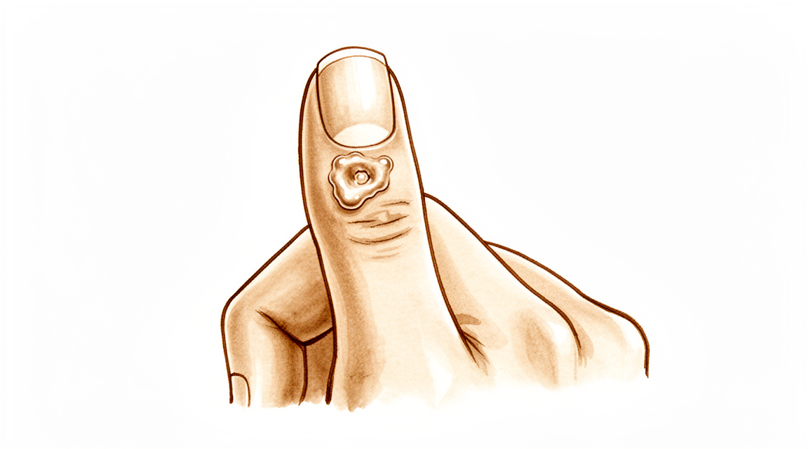
Ang mucous cyst ay isang maliit at matigas na bukol na lumilitaw sa likod ng daliri, kadalasan ay sa likod mismo ng kuko. Puno ito ng malapot at malinaw na likido. Ang bukol mismo ay madalas na hindi masakit, ngunit maaari itong dumiin sa mga katabing istruktura at magdulot ng discomfort.
Maaari mong mapansin ang mga pagbabago sa mismong kuko. Ang cyst ay nakapuwesto malapit sa nail bed, kaya maaari itong dumiin sa kuko habang lumalaki ito. Madalas itong nagdudulot ng uka o ridge na tumatakbo sa haba ng kuko. Ang balat sa ibabaw ng cyst ay maaari ring maging manipis at makintab. Sa ilang kaso, nasisira ang balat at tumatagas ang likido ng cyst sa isang maliit na daanan.
Ang bukol ay karaniwang nakapuwesto mismo kung saan bumabaluktot ang iyong daliri sa huling joint bago ang kuko. Dahil dito, nagiging asiwa ang ilang pang-araw-araw na gawain. Ang pagdukot sa masikip na bulsa, paghawak sa takip ng garapon, o pagpulot ng maliliit na barya ay maaaring hindi komportable. Ang pagta-type o pagsusulat ay maaaring dumiin sa bukol at gawin itong masakit.
Ang cyst ay may kaugnayan sa wear-and-tear arthritis sa joint ding iyon. Isang maliit na bony spur, na tinatawag na osteophyte, ang nakakairita sa joint at gumagawa ng likidong pumupuno sa cyst. Dahil dito, maaaring kumirot ang joint pagkatapos mong gamitin nang husto ang iyong kamay, at maaaring mas masakit hawakan ang bukol sa mga panahong iyon.
Kung bumuka ang balat sa ibabaw ng cyst at tumagas ang likido, panatilihing malinis ang lugar at magpatingin agad. Ang isang daanan mula sa balat papasok sa joint ng daliri ay maaaring magpapasok ng impeksyon, at ang joint na may impeksyon ay nangangailangan ng gamutan nang walang pagkaantala. Kung ang iyong daliri ay naging mainit, mapula, namamaga at masakit, lalo na kung may lagnat, pumunta sa emergency department sa parehong araw. Hindi kailangan ng referral mula sa GP.
Huwag tusukin o pisilin ang cyst nang mag-isa. Maaari itong magpapasok ng impeksyon sa joint.
Kung lumalaki ang bukol, naaabala ka sa deformity ng kuko, o hindi humuhupa ang mga sintomas sa loob ng ilang linggo, magpatingin sa iyong GP o humingi ng specialist review.
Ano ang aktwal na nangyayari
Ang huling joint ng iyong daliri ay gumagana tulad ng isang maliit na bisagra. Tulad ng anumang joint, may lining ito na isang malambot na balot na gumagawa ng kaunting likido upang mapanatiling makinis ang galaw ng mga ibabaw nito. Sa isang mucous cyst, nagkaroon ang joint na iyon ng wear-and-tear arthritis. Nairirita ang lining ng joint at nagsisimula itong gumawa ng mas maraming likido kaysa sa kailangan ng joint.
Kasabay nito, madalas na bumubuo ang arthritis ng isang maliit na bony spur sa gilid ng joint. Dumidiin ang spur na iyon sa balot ng joint at unti-unti itong binubutas mula sa loob. Pagkatapos ay tumatagas ang likido sa nabutas na bahagi at naiipon sa ilalim ng balat sa likod ng iyong daliri. Ang bukol na nakikita at nararamdaman mo ay ang nakulong na likidong iyon, na nakapuwesto sa isang panig ng joint, sa likod mismo ng kuko.
Ang cyst mismo ay isang bulsang may manipis na pader sa halip na isang tumutubong bukol. Ang pader nito ay gawa sa mga hibla ng sumusuportang tissue, at wala itong sariling tunay na lining. Dahil manipis ang pader at madalas na mas manipis pa ang balat sa ibabaw nito, ang isang maliit na pagkabagok ay maaaring magpaputok dito. Kapag nangyari iyon, tumatagas ang malinaw at parang jelly na likido sa loob.
Ipinapaliwanag nito ang mga sintomas na kababasa mo lamang. Ang bukol ay nakapuwesto malapit sa nail bed, kaya ang diin mula sa cyst ay maaaring mag-iwan ng uka sa kuko habang ito ay tumutubo. Kumikirot ang joint dahil naroon din ang arthritis, at ang cyst ay isang senyales nito. Ang spur sa gilid ng joint ang nagpapanatili sa pagdaloy ng likido, kaya hangga't naroon ito, may posibilidad na mapuno muli ang cyst kahit na matapos itong tumagas o ma-drain.
Iyan din ang dahilan kung bakit nakatuon ang gamutan sa joint sa halip na sa bukol lamang. Ang pag-alis sa spur ay pumipigil sa pagtagas sa mismong pinagmumulan nito, at humuhupa ang cyst kapag hindi na ito napupuno.
Ano ang maaari naming gawin tungkol dito
Si Dr Kieran Hirpara, isang upper-limb surgeon sa Mater Private Hospital Rockhampton, ay nagsisimula sa mga opsyon na hindi gaanong invasive na angkop sa iyong kondisyon. Ang mga pasyente ay karaniwang nirerefer sa aming klinika ng kanilang GP; kung iminungkahi ng isang physiotherapist na magpatingin sa amin, kakailanganin mo pa rin ng referral mula sa iyong GP upang maging eligible para sa Medicare rebate. Sa pagbisita sa klinika, kumukuha kami ng history, sinusuri ang iyong daliri, at nag-aayos ng imaging kung kinakailangan. Karamihan sa mga mucous cyst ay may kaugnayan sa wear-and-tear arthritis sa joint, kaya karaniwan naming sinusubukan muna ang non-operative care at isinasaalang-alang ang operasyon kapag hindi ito nagbigay ng sapat na pagbuti.
Ang unang hakbang ay madalas na ang pagbabantay at paghihintay lamang. Ang ilang cyst ay nananatiling maliit at kaunti lamang ang idinudulot na problema. Nakakatulong ang pagprotekta sa daliri: iwasan ang paulit-ulit na pagkabagok sa bukol, at mag-ingat sa mga gawaing dumidiin dito. Kung kumikirot ang joint pagkatapos ng mabigat na paggamit, ang pagbabawas sa gawaing iyon ay maaaring magpahupa nito. Makakatulong ang hand therapy sa paninigas at sa pagpapanatiling komportable ang galaw ng daliri. Bigyan ang mga simpleng hakbang na ito ng sapat na pagsubok sa loob ng ilang linggo bago magpasya ng anumang susunod na hakbang.
Simple lang ang pain relief. Ang simpleng gamot sa sakit mula sa pharmacy, na iniinom ayon sa direksyon, ay maaaring magpahupa sa kumikirot na joint. Ang anti-inflammatory na gamot ay maaaring magpagaan sa arthritis na nasa likod ng cyst. Ginagamot ng mga ito ang discomfort sa halip na ang mismong bukol.
Isang opsyon na hindi namin inaalok ay ang pag-drain ng cyst gamit ang karayom. Isa itong maginhawang procedure sa klinika, ngunit bumabalik ang cyst sa humigit-kumulang kalahati ng mga pagkakataon pagkatapos nito. May panganib din na madala ang impeksyon papasok sa joint, kaya pinapayuhan din namin na huwag tusukin o pisilin ang cyst sa bahay.
Isinasaalang-alang ang operasyon kapag patuloy na napupuno muli ang cyst, naaabala ka sa deformity ng kuko, o naging manipis o nasisira na ang balat sa ibabaw ng cyst. Ginagamot ng operasyon ang joint sa halip na ang bukol lamang. Tinatanggal ang bony spur sa gilid ng joint, na pumipigil sa pagtagas ng likido sa mismong pinagmumulan nito, at humuhupa ang cyst kapag hindi na ito napupuno. Sa ilang kaso, tinatanggal din ang cyst at isang maliit na flap ng katabing balat, na angkop para sa cyst na nagpanipis sa balat sa ibabaw nito. Ang pagtanggal sa spur kasama ang tangkay (stalk) ng cyst ang nagbibigay ng pinakamababang posibilidad na bumalik ang cyst. Pag-uusapan natin kung aling approach ang angkop sa iyong daliri, at magpapasya tayo nang magkasama.
Ano ang dapat asahan
Kung hahayaan lang, ang isang mucous cyst ay may posibilidad na patuloy na bumalik sa halip na mawala. Hangga't naroon ang bony spur sa gilid ng joint, patuloy nitong pinupuno ng likido ang cyst. Ang ilang cyst ay nananatiling maliit at kaunti lamang ang idinudulot na problema sa mahabang panahon. Ang iba naman ay dahan-dahang lumalaki, lalong nag-iiwan ng uka sa kuko, o nagpapanipis sa balat sa ibabaw ng bukol hanggang sa tumagas ito.
Kung walang gamutan, ang mga pangunahing panganib ay ang mga nabasa mo na: kukong nananatiling may ridge habang dumidiin dito ang cyst, at balat na nasisira at nagbubukas ng daanan papasok sa joint. Dapat seryosohin ang daanang iyon. Kung ang iyong daliri ay naging mainit, mapula, namamaga at masakit, lalo na kung may lagnat, pumunta sa emergency department sa parehong araw. Hindi kailangan ng referral mula sa GP.
Sa gamutang nakatuon sa joint, malinaw ang inaasahang resulta. Ang pag-alis sa spur ay pumipigil sa pagtagas ng likido sa mismong pinagmumulan nito, at humuhupa ang cyst kapag hindi na ito napupuno. Kapag tinanggal ang spur, bihirang bumalik ang cyst. Kung tinanggal din ang mismong cyst, nananatiling mababa ang pagbalik: humigit-kumulang 2 sa bawat 100 cyst na ginamot sa operasyon ang bumabalik, at humigit-kumulang 1.4 sa 100 kapag gumamit ng local skin flap upang isara ang lugar. Ang mga taong ginamot sa ganitong paraan ay karaniwang masaya sa hitsura ng peklat, at handang sumailalim muli rito.
Hindi malaking bagay ang paggaling, ngunit hindi rin ito balewala. Ang daliri ay madalas na naninigas at masakit hawakan sa loob ng ilang linggo, at ang mga pang-araw-araw na gawain tulad ng paghawak o pagkurot ay tumatagal bago maramdamang normal muli. Ang hand therapy pagkatapos ng operasyon ay kasama si Ruby Doolan sa Extend Rehabilitation. Si Ruby ay isang hand therapist: ginagabayan niya ang iyong mga ehersisyo at gumagawa ng anumang splint na kailangan mo. Karamihan sa mga tao ay nakakabalik sa normal na paggamit ng kamay sa loob ng ilang linggo hanggang ilang buwan.
Karaniwan ding bumubuti ang uka sa kuko kapag nawala na ang diin sa nail bed. Maging matiyaga sa bahaging iyon: kailangang tumubo palabas ang deformeng bahagi ng kuko, na inaabot ng ilang buwan.
Kung hindi humuhupa ang mga sintomas sa loob ng ilang linggo, o lumalala ang mga ito, magpatingin sa iyong GP o humingi ng specialist review.
Kailan dapat magpatingin
Karamihan sa mga mucous cyst ay hindi urgent, at marami ang humuhupa sa simpleng pangangalaga. May ilang senyales na nangangailangan ng aksyon.
Pumunta sa emergency department sa parehong araw kung ang iyong daliri ay naging mainit, mapula, namamaga at masakit, lalo na kung may lagnat. Tumuturo iyon sa impeksyon sa joint, na nangangailangan ng gamutan nang walang pagkaantala. Ganoon din kung bumuka ang balat sa ibabaw ng cyst at tumatagas ang likido, dahil maaaring magpapasok ng impeksyon ang butas na iyon.
Magpatingin sa iyong GP o humingi ng specialist review kung patuloy na lumalaki ang bukol, naaabala ka sa uka sa kuko, o naging manipis ang balat sa ibabaw ng cyst. Ito ang mga senyales na sulit nang pag-usapan ang gamutan. Kung hindi humuhupa ang mga sintomas sa loob ng ilang linggo, o lumalala ang mga ito, maaaring mag-ayos ang iyong GP ng referral.
Higit pang detalye
Advanced reading: the deeper science (optional)
Ang seksyong ito ay mas malalim pa kaysa sa kailangan mo para sa iyong sariling mga desisyon sa paggamot. Ang mga mucous cyst ay karapat-dapat sa karagdagang pagbabasa dahil sa isang maliit at may sapat na basehang surgical insight: ang cyst ay hindi ang problema, at ang operasyon na pinaka-epektibo ay hindi kinakailangang mag-alis nito.
Ang bone spur ang sanhi, hindi ang cyst
Ang isang mucous cyst ay nagmumula sa isang arthritic fingertip joint. Ang isang bony spur, o osteophyte, ay nairirita at binubutas ang joint capsule, at ang joint fluid ay lumalabas sa defect na iyon at naiipon sa ilalim ng balat. Ang cyst ang nakikitang dulo ng prosesong iyon, hindi ang pinagmulan nito.
Ang pag-unawang iyon ay may direktang surgical consequence, na ipinakita sa isang serye kung saan ang osteophyte excision nang walang cyst excision ay nagresulta sa kumpletong resolution sa karamihan ng mga kaso, at inilarawan bilang isang mabuting pagpipilian sa paggamot na nag-aalok ng isang less invasive na metodo [1].
Ang pagtanggal sa spur ay nagsasara sa gripo. Ang cyst, dahil hindi na napupuno, ay nawawala. Ito ang parehong lohika na namamahala sa wrist ganglion, kung saan ang stalk sa halip na ang sac ang nagtatakda ng recurrence, at ipinapaliwanag nito kung bakit ang simpleng pag-drain o pag-puncture sa isang mucous cyst ay madalas na nabibigo.
Kung saan ang cyst ay ine-excise, mabuti rin ang mga resulta
Ang alternatibong approach ay tinatanggal ang cyst kasama ang isang local skin flap upang isara ang defect. Maaasahan din ito: sa 69 na pasyente, ang surgical excision na may local advancement flap ay nagpakita ng recurrence rate na 1.4% na may mataas na kasiyahan ng pasyente tungkol sa peklat at pagkukusa na sumailalim muli sa procedure [2].
Parehong gumagana ang dalawang approach, at parehong tinutugunan ang underlying joint. Ang praktikal na pagkakaiba ay kung gaano karaming balat ang kasangkot: ang isang matagal nang cyst ay nagpapanipis sa overlying skin, kung minsan hanggang sa punto ng paglabas ng discharge, at sa sitwasyong iyon, ang nanipis na balat ay kailangang i-excise at palitan anuman ang gawin sa buto.
Bakit nagiging deforme ang kuko, at kung ito ba ay bumabalik sa dati
Ang isang uka o ridge na tumatakbo sa haba ng kuko ay isang karaniwang kasama, at mas ikinababahala ito ng mga tao kaysa sa bukol. Ang sanhi ay mechanical: ang cyst ay nakapuwesto agad sa ibabaw ng germinal matrix, ang bahagi ng nail bed na bumubuo ng kuko, at umiipit dito, kaya ang kuko ay nabubuo na may defect.
Ang kapaki-pakinabang na bahagi ay ito ay pressure sa halip na destruction. Kapag ang cyst ay na-decompress na, ang kuko ay karaniwang tumutubo nang normal, bagaman inaabot ng ilang buwan bago lumabas at mawala ang deformeng bahagi sa dulo. Samakatuwid, ang deformity ng kuko ay isang dahilan upang gamutin ang cyst sa halip na isang permanenteng bunga nito.
Ang dahilan kung bakit dapat mag-ingat sa pagtusok sa isa
Ang isang mucous cyst ay direktang nakikipag-ugnayan sa joint. Ang sadyang pagtusok dito, o ang pagkasira ng balat sa ibabaw ng isang malaking cyst, ay lumilikha ng channel mula sa labas patungo sa joint ng daliri, at ang septic arthritis ng isang maliit na joint ay isang problemang higit na mas seryoso kaysa sa cyst.
Ito ang praktikal na argumento laban sa home drainage ng isang cyst na tila isang simpleng blister ng fluid, at ang dahilan kung bakit ang isang spontaneously discharging cyst ay ginagamot nang may urgency sa halip na obserbahan lamang.
Mga Sanggunian
[1] Lee H, Kim P, Jeon I, Kyung H, Ra I, Kim T. Osteophyte excision without cyst excision for a mucous cyst of the finger. J Hand Surg Eur Vol. 2013;39(3):258-61. https://doi.org/10.1177/1753193413478549
[2] Johnson SM, Treon K, Thomas S, Cox QGN. A reliable surgical treatment for digital mucous cysts. J Hand Surg Eur Vol. 2013;39(8):856-60. https://doi.org/10.1177/1753193413508540
Evidence & references
This is the clinical evidence summary written for health professionals. It is technical, and it lists the research this page was built from. You do not need to read it to understand your treatment or to make a decision about it.
Overview
- The scientific data regarding mucous cysts consist almost entirely of retrospective studies, and much of what is done or recommended is based on expert opinion [3].
- Surgical treatment of mucous cysts is not a totally benign procedure, and complications do occur contrary to literature reporting glowing results [5].
- In-office excision is a safe and effective option for treating digital mucous cysts [6].
- Complications are rare and occur only in cysts associated with fistula, justifying their early surgical treatment [7].
- Surgery provided definitive treatment with no major long-term problems [10].
- Surgical excision was the primary treatment, with no reported recurrences [11].
- Incomplete excisions will invariably result in recurrence [12].
- Surgical excision with a local advancement skin flap is a reliable treatment for digital mucous cysts, demonstrating a low recurrence rate of 1.4% and high patient satisfaction regarding the scar and willingness to undergo the procedure again [13].
- The 2% recurrence rate (2 of 113) obtained in this series illustrates the advantage of this procedure [16].
- Osteophyte excision without cyst excision may be a good treatment choice for mucous cyst of the finger, providing a less invasive method with complete resolution in most cases [20].
- The final recurrence rate of less than 1.5% may be acceptable and comparable with other techniques [21].
- Excision with local flap cover offers benefit in terms of a reduced recurrence rate and is particularly valuable where the cyst has caused marked thinning of the skin [30].
- Excision with local flap cover would seem to offer benefit in terms of a reduced recurrence rate, and is particularly valuable where the cyst has caused marked thinning of the skin [34].
- Excision of the cyst and complete removal of the marginal osteophyte eradicates mucous cysts with extremely rare recurrence [1].
- A total dorsal capsulectomy alone was a simple treatment for mucous cysts and did not lead to any recurrence [8].
Anatomy & Pathophysiology
Clinical Presentation and Location
- Mucous cysts are small, firm, cystic masses that appear just distal to the distal interphalangeal joint [23].
- The lesion is always located to one side of the midline [23].
- Mucous cysts are rarely greater than fifteen millimeters in diameter [23].
- The skin over the mucous cyst is thinned out and occasionally may be ulcerated [23].
- Pressure on the mucous cyst does not usually result in a decrease in its size [23].
- Longitudinal grooving of the nail may be noted, occasionally occurring prior to the appearance of the cyst [23].
- Mucous cysts are usually small and located to one side as they emerge from the joint beside the extensor mechanism [28, 29].
- Occasionally, mucous cysts may spread across the whole of the dorsum of the digit between the distal interphalangeal joint and nail fold [28, 29].
- The overlying skin of a mucous cyst is frequently thin, and minimal trauma may result in rupture [28, 29].
Histology and Ultrastructure
- The histological appearance of a mucous cyst is essentially identical to that of ganglions, synovial cysts, and similar lesions appearing on other areas of the extremities [23].
- The mucous cyst is surrounded by an undemarcated fibrous capsule [23].
- The basic structure of the mucous cyst is myxomatous with interspersed fibroblasts [23].
- Areas of myxomatous degeneration tend to coalesce to form a multiloculated cyst [23].
- An epithelial lining has not been reported for mucous cysts [23].
- Digital mucous cysts and wrist ganglia show no anatomic difference at the light and scanning electron microscopic levels [19].
- The wall of a digital mucous cyst consists of multidirectional strata of collagen fibers with rare fibroblast-like cells on the inner surface [19].
- There are no signs of myxomatous degeneration in the wall of a digital mucous cyst [19].
- The surface of the digital mucous cyst wall is generally flat, with some areas showing localized elevations [19].
- Multifunctional mesenchymal cells are located beneath the lining of the ganglion and digital mucous cyst [19].
- These multifunctional mesenchymal cells contain abundant rough endoplasmic reticulum, smooth-walled vacuoles, and Golgi complexes [19].
- The ganglion fluid may be produced by these multifunctional cells and reaches the ganglion cavity by flowing through the porous collagen matrix of the ganglion wall [19].
- A digital mucous cyst has a large globular dome-shaped interconnecting cavity having a wall of collagen fibers covered by a fenestrated pseudomembrane [19].
- A porous network of collagen fibers is present in the wall of the digital mucous cyst on cross section [19].
Etiology and Pathogenesis
- Degenerative arthritis of the distal interphalangeal joint is a frequent associated finding with mucous cysts [23].
- An incidence of 78 percent for degenerative arthritis of the distal interphalangeal joint has been reported in association with mucous cysts [23].
- The etiology indicated by evidence is that the lesion arises from the joint capsule [23].
- Mucous cysts are often associated with degenerative changes at the distal interphalangeal joints [28, 29].
- Theories on the pathogenesis of mucous cysts include myxoid degeneration, repeated local trauma, vascular insufficiency, and synovial herniation [28, 29].
- Studies suggest that mucous cysts arise due to excessive hyaluronic acid production from a degenerate distal interphalangeal joint [28, 29].
Classification
- The lesion is a small, firm, cystic mass which appears just distal to the distal interphalangeal joint [23].
- The lesion is always located to one side of the mid-line [23].
- The lesion is rarely greater than fifteen millimeters in diameter [23].
- The skin over the lesion is thinned out and occasionally may be ulcerated [23].
- Pressure on the lesion does not usually result in a decrease in its size [23].
- Degenerative arthritis of the distal interphalangeal joint is a frequent associated finding [23].
- An incidence of 78 per cent has been reported for degenerative arthritis of the distal interphalangeal joint associated with mucous cysts [23].
- The histological appearance is essentially identical to that of ganglions, synovial cysts, and similar lesions appearing on other areas of the extremities [23].
- The lesion is surrounded by an undemarcated fibrous capsule [23].
- The basic structure is myxomatous with interspersed fibroblasts [23].
- An epithelial lining has not been reported [23].
- In spite of their different clinical settings, digital mucous cysts and wrist ganglia show no anatomic difference at the light and scanning electron microscopic levels [19].
- The wall of the digital mucous cyst consisted of multidirectional strata of collagen fibers with rare fibroblast-like cells on the inner surface [19].
- There were no signs of myxomatous degeneration in the wall of the digital mucous cyst [19].
- The surface of the digital mucous cyst was generally flat, with some areas showing localized elevations [19].
- Multifunctional mesenchymal cells beneath the lining of the ganglion are postulated to produce collagen fibers, elastic fibers, and the interfibrillary mucopolysaccharide matrix [19].
- The porous nature of the ganglion wall was confirmed in the ultrastructural study of digital mucous cysts [19].
- A previous ultrastructural study of digital mucous cysts demonstrated a large globular dome-shaped interconnecting cavity having a wall of collagen fibers covered by a fenestrated pseudomembrane [19].
- The current study confirmed the anatomic organization of a large globular dome-shaped interconnecting cavity having a wall of collagen fibers covered by a fenestrated pseudomembrane [19].
- A porous network of collagen fibers in the wall was revealed on cross section [19].
Clinical Presentation
- The mucous cyst presents as a small, firm, cystic mass located just distal to the distal interphalangeal joint [23].
- The lesion is easily distinguished from a Heberden's node by its placement distal to the distal interphalangeal joint [23].
- The histological appearance of the mucous cyst is essentially identical to that of ganglions, synovial cysts, and similar lesions appearing on other areas of the extremities [23].
- The basic structure of the lesion is myxomatous with interspersed fibroblasts [23].
- An epithelial lining has not been reported for the mucous cyst [23].
- The wall of the digital mucous cyst consists of a porous network of collagen fibers [19].
- The fluid within the digital mucous cyst is produced by underlying mesenchymal cells [19].
Investigations
- Ultrasound is a powerful modality for evaluation of pathologic conditions in the hand and wrist, providing a cost-effective and expedient alternative and/or adjunct to MRI [33].
- Ultrasound is best used when there is a specific clinical question regarding a well-localized abnormality [33].
- Ultra-sonography provides a new ability to study the complex anatomical structures of the hand, with the technique being quick and simple to perform [38].
- A careful physical examination is essential to direct care and future testing if indicated [35].
- Diagnostic tests such as imaging and serum laboratory studies are useful in this determination but can be expensive, time consuming, and often nonspecific [35].
Treatment
Operative Techniques
- Total dorsal capsulectomy alone is a simple treatment for mucous cysts that did not lead to any recurrence [8].
- Osteophyte-sparing treatment of mucous cysts has a final recurrence rate of less than 1.5% [21].
- Surgical excision was the primary treatment in a series with no reported recurrences [11].
- Surgery provided definitive treatment for mucous cysts with no major long-term problems [10].
- The 2% recurrence rate (2 of 113) obtained in a series of 191 excised cysts illustrates the advantage of the procedure [4, 16].
- The authors of a study on osteophyte excision without cyst excision state that their principle is not to do more damage to the friable skin because the skin is not the culprit of the cyst [15].
Outcomes and Complications
- Complications of surgical treatments include infection, recurrence, nail deformity, swelling, stiffness and pain [28].
- This report provides additional basis for prophylactic extirpation of mucous cyst of the finger [2].
- This case emphasizes the utility of a pathohistological analysis in cases where doubts arise about the initial diagnosis of a benign tumorous lesion [14].
Non-Operative and Adjunctive Considerations
- Aspiration with injection of steroid is a convenient technique that can be performed in an outpatient setting but is followed by a recurrence rate of 50% [28].
- Surgical treatment has a higher cure rate than aspiration with injection of steroid [28].
- Radical treatment with complete excision of the stalk and underlying osteophytes seems to reduce recurrence rates [28].
Complications
- Complications are rare and occur only in cysts associated with fistula [7].
- Aspiration with injection of steroid is followed by a recurrence rate of 50% [28].
- The 2% recurrence rate (2 of 113) was obtained in a series of surgically treated mucous cysts [16].
- Surgical excision with a local advancement skin flap demonstrated a low recurrence rate of 1.4% [13].
- A total dorsal capsulectomy alone did not lead to any recurrence [8].
- Eccrine porocarcinomas have a substantial risk of metastasis, high risk of local recurrence, and are potentially fatal [24].
Recovery
- Surgical excision with a local advancement skin flap demonstrated a low recurrence rate of 1.4% and high patient satisfaction regarding the scar and willingness to undergo the procedure again [13].
Key Evidence
- [L4] Excision of the cyst and complete removal of the marginal osteophyte eradicates mucous cysts with extremely rare recurrence. [1] (10.2106/00004623-197355030-00013)
- [L4] This report provides additional basis for prophylactic extirpation of mucous cyst of the finger. [2] (10.1016/s0363-5023(84)80152-5)
- [L4] The scientific data regarding mucous cysts consist almost entirely of retrospective studies, and much of what is done or recommended is based on expert opinion. [3] (10.1016/j.jhsa.2010.01.029)
- [L5] Of the 191 mucous cysts excised, 113 had at least 6 months of follow-up time. [4] (10.1016/j.jhsa.2010.07.028)
- [L4] Surgical treatment of mucous cysts is not a totally benign procedure, and complications do occur contrary to literature reporting glowing results. [5] (10.1016/s0266-7681(97)80067-8)
- [Paper] In-office excision is a safe and effective option for treating digital mucous cysts. [6] (10.1177/15589447251350168)
- [L4] Complications are rare and occur only in cysts associated with fistula, justifying their early surgical treatment. [7] (10.1016/j.main.2015.06.001)
- [L4] A total dorsal capsulectomy alone was a simple treatment for mucous cysts and did not lead to any recurrence. [8] (10.1016/j.jhsa.2014.03.004)
- [L4] Surgery provided definitive treatment with no major long-term problems. [10] (10.1053/jhsu.2003.50088)
- [L5] Incomplete excisions will invariably result in recurrence. [12] (10.1016/0363-5023(93)90302-j)
- [L4] Surgical excision with a local advancement skin flap is a reliable treatment for digital mucous cysts, demonstrating a low recurrence rate of 1.4% and high patient satisfaction regarding the scar and willingness to undergo the procedure again. [13] (10.1177/1753193413508540)
- [L5] This case emphasizes the utility of a pathohistological analysis in cases where doubts arise about the initial diagnosis of a benign tumorous lesion. [14] (10.1007/s00402-008-0794-4)
- [L5] The authors state that their principle is not to do more damage to the friable skin because the skin is not the culprit of the cyst. [15] (10.1177/1753193414546990)
- [L4] The 2% recurrence rate (2 of 113) obtained in this series illustrates the advantage of this procedure. [16] (10.1016/0363-5023(94)90071-x)
- [L5] [19] (10.1016/s0363-5023(88)80143-6)
- [L4] Osteophyte excision without cyst excision may be a good treatment choice for mucous cyst of the finger, providing a less invasive method with complete resolution in most cases. [20] (10.1177/1753193413478549)
- [L4] The final recurrence rate of less than 1.5% may be acceptable and comparable with other techniques. [21] (10.5435/jaaosglobal-d-21-00164)
- [L4] [23] (10.2106/00004623-197254070-00008)
- [L4] Prompt recognition and appropriate treatment are critical because eccrine porocarcinomas have a substantial risk of metastasis, high risk of local recurrence, and are potentially fatal. [24] (10.1016/j.jhsa.2016.07.112)
- [L4] [28] (10.1177/1753193408103498)
- [L4] [29] (10.1177/1753193409103498)
- [L4] Excision with local flap cover offers benefit in terms of a reduced recurrence rate and is particularly valuable where the cyst has caused marked thinning of the skin. [30] (10.1016/0266-7681(90)90064-b)
- [L5] Ultrasound is a powerful modality for evaluation of pathologic conditions in the hand and wrist, providing a cost-effective and expedient alternative and/or adjunct to MRI, best used when there is a specific clinical question regarding a well-localized abnormality. [33] (10.1016/j.jhsa.2009.02.010)
- [L4] Excision with local flap cover would seem to offer benefit in terms of a reduced recurrence rate, and is particularly valuable where the cyst has caused marked thinning of the skin. [34] (10.1016/0266-7681_90_90064-b)
- [L4] Ultra-sonography provides a new ability to study the complex anatomical structures of the hand, with the technique being quick and simple to perform. [38] (10.1016/0266-7681(90)90133-o)
References
[1] Marginal Osteophyte Excision in Treatment of Mucous Cysts. The Journal of Bone & Joint Surgery. 1973. DOI: 10.2106/00004623-197355030-00013
[2] Infected mucous cyst of the finger. The Journal of Hand Surgery. 1984. DOI: 10.1016/s0363-5023(84)80152-5
[3] Mucous Cysts. The Journal of Hand Surgery. 2010. DOI: 10.1016/j.jhsa.2010.01.029
[4] Mucous Cysts. The Journal of Hand Surgery. 2010. DOI: 10.1016/j.jhsa.2010.07.028
[5] Complications Following Mucous Cyst Excision. Journal of Hand Surgery. 1997. DOI: 10.1016/s0266-7681(97)80067-8
[6] Safety and Cost-Effectiveness of In-Office Digital Mucous Cyst Excisions. HAND. 2025. DOI: 10.1177/15589447251350168
[7] Surgical treatment of mucous cysts by subcutaneous excision and osteophyte resection: Results in 68 cases at a mean 6.63 years’ follow-up. Chirurgie de la Main. 2015. DOI: 10.1016/j.main.2015.06.001
[8] Total Dorsal Capsulectomy for the Treatment of Mucous Cysts. The Journal of Hand Surgery. 2014. DOI: 10.1016/j.jhsa.2014.03.004
[10] Treatment of mucous cysts of the fingers: Review of 134 cases with minimum 2-year follow-up evaluation. The Journal of Hand Surgery. 2003. DOI: 10.1053/jhsu.2003.50088
[11] 10.1177-15589447261433068. n.d..
[12] Juvenile hyaline fibromatosis of the hand in an adult. The Journal of Hand Surgery. 1993. DOI: 10.1016/0363-5023(93)90302-j
[13] A reliable surgical treatment for digital mucous cysts. Journal of Hand Surgery (European Volume). 2013. DOI: 10.1177/1753193413508540
[14] Malignant Natural-Killer cell neoplasm presenting as a mucous cyst on the distal interphalangeal joint of the finger. Archives of Orthopaedic and Trauma Surgery. 2008. DOI: 10.1007/s00402-008-0794-4
[15] Re: Lee, H.-J., Kim, P.-T., Jeon, I.-H., Kyung, H.-S., Ra, I.-H. and Kim, T.-K. Osteophyte excision without cyst excision for a mucous cyst of the finger. J Hand Surg Eur. 2014, 39: 258–61. Journal of Hand Surgery (European Volume). 2014. DOI: 10.1177/1753193414546990
[16] Outcome of surgically treated mucous cysts of the hand. The Journal of Hand Surgery. 1994. DOI: 10.1016/0363-5023(94)90071-x
[19] A surface ultrastructure study of ganglia and digital mucous cysts. The Journal of Hand Surgery. 1988. DOI: 10.1016/s0363-5023(88)80143-6
[20] Osteophyte excision without cyst excision for a mucous cyst of the finger. Journal of Hand Surgery (European Volume). 2013. DOI: 10.1177/1753193413478549
[21] Osteophyte-Sparing Treatment of Mucous Cysts: Case Analysis and Surgical Technique. JAAOS: Global Research and Reviews. 2021. DOI: 10.5435/jaaosglobal-d-21-00164
[23] Etiology and Treatment of the So-Called Mucous Cyst of the Finger. The Journal of Bone & Joint Surgery. 1972. DOI: 10.2106/00004623-197254070-00008
[24] Eccrine Porocarcinoma Presenting as a Hand Cyst. The Journal of Hand Surgery. 2016. DOI: 10.1016/j.jhsa.2016.07.112
[28] Use of Wolfe Graft for the Treatment of Mucous Cysts. Journal of Hand Surgery (European Volume). 2009. DOI: 10.1177/1753193408103498
[29] Use of Wolfe Graft for the Treatment of Mucous Cysts. Journal of Hand Surgery (European Volume). 2009. DOI: 10.1177/1753193409103498
[30] Mucous cyst of the distal interphalangeal joint: Treatment by simple excision or excision and rotation flap. The Journal of Hand Surgery: Journal of the British Society for Surgery of the Hand. 1990. DOI: 10.1016/0266-7681(90)90064-b
[33] Ultrasound of the Hand and Wrist. The Journal of Hand Surgery. 2009. DOI: 10.1016/j.jhsa.2009.02.010
[34] Mucous Cyst of the Distal Interphalangeal Joint: Treatment by Simple Excision or Excision and Rotation Flap. Journal of Hand Surgery. 1990. DOI: 10.1016/0266-7681_90_90064-b
[35] Orthopaedic Knowledge Update 13 Ebook Without Multimedia. Anatomy, Evaluation, Clinical Examination, and Imaging > Evaluation and Clinical Examination: Current Concepts.
[38] Diagnostic medical ultrasound in the management of hand injuries. The Journal of Hand Surgery: Journal of the British Society for Surgery of the Hand. 1990. DOI: 10.1016/0266-7681(90)90133-o