Patients › Shoulder
Arthroscopy ng Balikat
What shoulder arthroscopy is, what it's used for, and how to prepare and recover.
Bakit iminungkahi ang operasyong ito
Si Dr Kieran Hirpara, isang upper-limb surgeon sa Mater Private Hospital Rockhampton, ay nagsisimula sa mga opsyon na hindi gaanong invasive na angkop sa iyong kondisyon. Ang mga pasyente ay karaniwang nirerefer sa aming klinika ng kanilang GP; kung iminungkahi ng isang physiotherapist na magpatingin sa amin, kakailanganin mo pa rin ng referral mula sa iyong GP upang maging eligible para sa Medicare rebate. Sa iyong appointment, kumukuha kami ng history, sinusuri ang iyong balikat, at nag-aayos ng imaging kung saan ito kinakailangan. Ang pagsusuring iyon ang nagsasabi sa amin kung ano ang problema sa loob ng joint.
Para sa mga matagal nang problema, karaniwan naming sinusubukan muna ang non-operative care. Maaaring mangahulugan ito ng pagbabago sa mga aktibidad, physiotherapy, o mga injection. Pumapasok sa usapan ang surgery kapag hindi nagbigay sa iyo ng sapat na pagbuti ang mga hakbang na iyon. Para sa ilang problema sa istruktura, tulad ng napunit na tendon o balikat na hindi matatag (unstable), maaari naming irekomenda agad ang surgery.
Ang shoulder arthroscopy ay isang keyhole operation. Isang maliit na camera ang inilalagay sa loob ng joint, at ipinapasok ang mga instrumento sa pamamagitan ng maliliit na hiwa sa paligid ng balikat. Pinapayagan kami nitong makita at magamot ang mga problema tulad ng labral tears, rotator cuff tears, at pinsala sa biceps tendon sa iisang operasyon. Ang layunin ay mas kaunting sakit, mas mabuting galaw, at mas matatag na balikat.
Bago ang operasyon
Sa mga araw bago ang operasyon, kukumpirmahin namin kung aling mga scan ang kailangan upang maplano ang iyong operasyon. Maaaring ito ay X-ray, MRI, o ultrasound. Bibigyan ka ng malinaw na mga tagubilin tungkol sa iyong mga gamot. Maaaring kailanganing itigil ang ilan sa mga ito sa maikling panahon. Kung umiinom ka ng blood thinner o may diabetes ka, sabihin sa amin nang maaga upang maplano namin ang mga ito. Sa araw ng operasyon, magdala ng listahan ng lahat ng iyong iniinom o ginagamit, kabilang ang mga tableta, injection at supplement. Magsuot ng maluwag at komportableng damit na madaling isuot sa ibabaw ng balikat na may bendahe. Mag-ayos ng taong maghahatid sa iyo pauwi pagkatapos, dahil hindi ka makapagmamaneho sa araw na iyon. Kailangan mong itigil ang pagkain at pag-inom pitong oras bago ang iyong operasyon. Humihingi kami ng pitong oras upang maiuna ang iyong operasyon kung mas maaga ang takbo ng listahan sa theatre. Kung mayroon kang iba pang kondisyong medikal, maaaring kailanganin mo ng mga blood test o pagsusuri kasama ang anaesthetist bago ang araw ng operasyon.
Sa araw ng operasyon
Darating kayo sa surgical admissions unit ng ospital, kung saan kayo ay i-che-check in at ihahanda para sa theatre. Pagkatapos ay makikipagkita kayo sa anaesthetist, ang doktor na nagbibigay ng anaesthetic at nag-aalaga sa inyo habang isinasagawa ang operasyon. Ang operasyong ito ay ginagawa sa ilalim ng general anaesthetic na pinagsama sa isang regional nerve block. Kayo ay tulog nang tuluyan para sa operasyon, at ang block (isang injection na nagpamanhid sa mga nerve na nagsusuplay sa braso bago kayo magising) ay nagbibigay ng relief sa sakit para sa unang 12 hanggang 24 oras pagkatapos ng surgery. Makikipagkita sa inyo ang anaesthetist bago ang operasyon at ipapaliwanag sa inyo ang dalawang bahagi nito.
Pagkatapos nito, dadalhin kayo sa operating theatre, kung saan isasagawa ang operasyon. Kapag tapos na ito, magigising kayo sa recovery area. Mananatili sa inyo ang mga nurse at babantayan kayo habang nawawala ang bisa ng anaesthetic. Kapag stable na kayo, ililipat kayo sa ward o uuwi na. Kung alin dito ang mangyayari ay depende sa procedure na ginawa sa inyo at sa takbo ng inyong paggaling.
Ano ang kinapapalooban ng operasyon
Ang shoulder arthroscopy ay isang keyhole operation. Ang iyong surgeon ay gagawa ng ilang maliliit na hiwa sa paligid ng iyong balikat, kabilang ang isa sa likuran, at magpapasok ng isang maliit na camera sa loob ng joint. Ipinapadala ng camera sa isang screen ang larawan ng loob ng iyong balikat, upang direktang makita ng iyong surgeon ang mga ibabaw ng joint, ang labrum (isang rim ng cartilage sa paligid ng socket), at ang mga rotator cuff tendon. Nagbobomba ng likido papasok upang panatilihing nakabukas ang joint at malinaw ang tanaw.
Ang susunod na mangyayari ay depende sa kung ano ang matagpuan. Ang napunit na labrum ay maaaring ikabit muli sa buto. Ang napunit na rotator cuff tendon ay maaaring tahiin pabalik sa kinakapitan nito sa buto ng braso gamit ang maliliit na anchor na nakabaon sa buto. Ang maluwag o napinsalang tissue ay maaaring tabasin, at ang namamagang tissue o mga bone spur na dumidiin sa tendon ay maaaring tanggalin. Dahil maaaring igalaw ang camera upang tumingin mula sa iba't ibang anggulo, masusuri ng iyong surgeon ang buong joint at magagamot ang higit sa isang problema sa iisang operasyon.
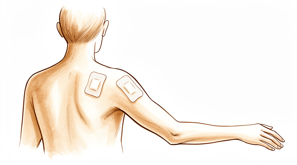
Sa dulo, inilalabas ang mga instrumento at isinasara ang maliliit na hiwa gamit ang mga tahi. Tinatakpan ng dressing ang iyong balikat, na mananatili nang mga 10 araw gaya ng inilarawan sa seksyon tungkol sa paggaling.
Pagkatapos ng operasyon
Karamihan sa mga pasyente ay nananatili ng isang gabi sa ospital pagkatapos ng operasyong ito, bagaman ang ilan ay nakakauwi sa mismong araw. Magigising ka sa recovery area, pagkatapos ay ililipat sa ward para sa gabi. Ang iyong braso ay nakalagay sa isang simpleng sling para sa ginhawa, na tinatanggal para sa mga ehersisyo at paghuhugas. Ang unang 24 oras ang pinakamahalaga para sa pagkontrol ng sakit, kaya babantayan namin ito nang mabuti gamit ang iyong mga gamot sa sakit. Inaasahan ang pamamanhid at panghihina sa braso habang nawawala ang bisa ng nerve block, karaniwan sa loob ng mga 24 oras. Kung hindi mo maigalaw ang braso, kamay o mga daliri, o may pamamanhid ka pa kapag nawala na ang bisa ng block, tumawag sa klinika. Dapat may taong sumama sa iyo sa unang 24 oras. Hinahayaan naming nakalagay ang dressing sa loob ng mga 10 araw; pakiusap huwag itong tanggalin bago ang panahong iyon maliban kung sinabi namin sa iyo. Papalitan o tatanggalin namin ito kapag nakita ka namin.
Paggaling
Ang unang isa o dalawang araw pagkatapos ng operasyon ang panahong pinakamatindi ang sakit, kaya inumin nang tuloy-tuloy ang iyong mga gamot sa sakit mula sa simula. Mawawala ang bisa ng nerve block mula sa theatre, at habang nangyayari ito ay maaari kang makaramdam ng mapurol na kirot sa balikat. Ang ilang pamamaga sa paligid ng balikat at itaas na bahagi ng braso ay normal at humuhupa habang lumilipas ang mga araw. Ang pahinga, yelo at ang pain relief na inireseta sa iyo ay pawang nakatutulong na pagaanin ang maagang discomfort na ito.
Ang iyong braso ay nakalagay sa isang simpleng sling para sa ginhawa, na tinatanggal para sa iyong mga ehersisyo at para sa paghuhugas. Gagabayan ka ng iyong physiotherapist sa mga banayad na galaw, pagkatapos ay unti-unting lilipat sa mga ehersisyong pampalakas ayon sa kaya ng iyong balikat. Maaari mong gamitin ang iyong kamay at pulso para sa mga magaan na gawain tulad ng pagkain at pagsusulat kapag kaya mo na. Iwasang magbuhat gamit ang inoperahang braso hanggang sabihin ng iyong surgeon o physiotherapist na ligtas na ito. Ang pagtulog nang nakaupo o nakasandal sa mga unan ay madalas na mas komportable sa mga unang araw, at maraming tao ang mas madaling nakakatulog sa recliner o may dagdag na mga unan sa likod.
Ang paggaling ay nag-iiba-iba sa bawat tao, kaya maaaring magkaiba ang iyong timeline sa iba. Gagabayan ka ng iyong surgeon at physiotherapist sa bawat review. Kapag humupa na ang pamamaga at bumalik ang iyong galaw, nagiging mas madali ang mga pang-araw-araw na gawain. Hindi ka magmamaneho hanggang sa payagan ka ng iyong surgeon, karaniwan ay sa review sa ikaanim na linggo; tingnan ang Driving after upper-limb surgery para sa kumpletong mga panuntunan. Ang pagbabalik sa trabaho, sport at gym ay nangyayari nang paunti-unti, ayon sa pakiramdam ng iyong balikat at sa napagkasunduan ng iyong surgeon at physiotherapist na handa ka na.
Ano ang maaaring maging problema
Karamihan sa mga pasyente ay gumagaling nang maayos, ngunit paminsan-minsan ay maaaring magkaroon ng mga problema. Binabantayan kayo nang maigi ng inyong surgeon at ng team upang maagang matukoy ang anumang isyu.
Hindi karaniwan ang impeksyon pagkatapos ng keyhole surgery sa balikat. Maaari itong lumabas bilang lagnat, pamumulang kumakalat mula sa sugat, o likido o nana na tumutulo mula rito. Tumawag sa klinika sa mismong araw na iyon kung mapansin ninyo ang alinman sa mga ito. Ang sakit na patuloy na lumalala sa kabila ng inyong mga gamot sa sakit ay dahilan din upang tumawag sa klinika sa mismong araw na iyon.
Bihira ngunit seryoso ang blood clot. Pumunta sa emergency department kung may pamamaga o sakit kayo sa inyong calf (binti), o kahirapan sa paghinga o pananakit ng dibdib. Ang mga ito ay maaaring palatandaan ng blood clot.
Maaaring magkaroon ng paninigas (stiffness) pagkatapos ng ilang operasyon sa balikat, at kung minsan ay maaaring magkaroon ng frozen shoulder. Maaari ninyong mapansin na humihigpit ang balikat at nawawalan ito ng galaw, lalo na sa pag-abot sa inyong likod o palabas sa gilid. Banggitin ito sa inyong susunod na review, dahil nakatutulong ang maagang paggamot.
Dumadaan ang mga nerve malapit sa balikat, at paminsan-minsan ay maaaring mairita o mapinsala ang mga ito. Maaari kayong makaramdam ng pamamanhid, tingling, o panghihina sa braso, kamay o mga daliri. Inaasahan ang pamamanhid at panghihina sa unang 24 oras pagkatapos ng nerve block. Tumawag sa klinika kung may pamamanhid kayo, o hindi ninyo maigalaw ang braso, kamay o mga daliri, kapag nawala na ang bisa ng block (mga 24 oras). Kung hindi ninyo makontak ang klinika, pumunta sa pinakamalapit na emergency department.
Kung inoperahan kayo upang patatagin ang balikat na nadi-dislocate, kung minsan ay maaaring dumulas o ma-dislocate muli ang balikat. Maaari ninyong maramdaman na lumilipat, bumibigay, o dumudulas palabas sa puwesto ang balikat. Sabihin ito sa inyong surgeon sa review, o tumawag sa klinika kung mangyari ito nang mas maaga.
Ang complications table sa pahinang ito ay naglilista ng mga tipikal na rate kung nais ninyo ang mga detalye.
Kailan dapat tumawag sa amin
Karamihan sa mga paggaling ay maayos, ngunit may ilang palatandaan na nangangailangan ng mabilis na aksyon. Pumunta sa emergency department kung may pamamaga o sakit kayo sa inyong calf (binti), o kahirapan sa paghinga o pananakit ng dibdib. Ang mga ito ay maaaring palatandaan ng blood clot. Tumawag sa klinika sa mismong araw na iyon kung may lagnat kayo, kumakalat na pamumula sa paligid ng sugat, o likido o nana na tumutulo mula rito. Tumawag din sa klinika sa mismong araw na iyon kung patuloy na lumalala ang inyong sakit sa kabila ng inyong mga gamot sa sakit. Tumawag sa klinika kung may pamamanhid kayo, o hindi ninyo maigalaw ang braso, kamay o mga daliri, kapag nawala na ang bisa ng nerve block (mga 24 oras). Kung hindi ninyo makontak ang klinika, pumunta sa pinakamalapit na emergency department.
Saan maaaring magbasa nang higit pa tungkol sa kondisyon
Ang pahinang ito ay tungkol sa operasyon mismo. Ang kondisyong ginagamot nito, kabilang ang kung ano ang ipinapakita ng ebidensya tungkol sa kung kailan nakatutulong ang operasyon at kung kailan hindi, ay tinalakay nang mas detalyado sa pahinang Rotator Cuff Disorders.
Evidence & references
This is the clinical evidence summary written for health professionals. It is technical, and it lists the research this page was built from. You do not need to read it to understand your treatment or to make a decision about it.
Overview
- Diagnostic arthroscopy in the beach-chair position can effectively identify and characterize intra-articular shoulder pathologies when properly executed [1].
- Refined techniques are crucial for addressing complex shoulder pathologies effectively as shoulder arthroscopy continues to evolve [2].
- Performing diagnostic shoulder arthroscopy with the patient under local anesthesia is a demanding procedure for the patient and may not always be possible [3].
- Supine-position shoulder arthroscopy using the anterior portal as the initial approach serves as a safer, more cost-effective, and more accessible complementary approach for shoulder arthroscopy positioning and is worthy of routine clinical application [4].
- Resident involvement in shoulder arthroscopy procedures is not associated with increased risk for medical or surgical 30-day postoperative complications [5].
- Current guidelines for thromboprophylaxis in shoulder arthroscopy lack consensus and need patient-specific considerations [6].
- Arthroscopic Instruments represented the most frequent category among the top 100 most-cited shoulder arthroscopy patents [7].
- Complications in arthroscopic shoulder instability surgery are frequently technique-specific and can be prevented by familiarity with the common pitfalls inherent in each arthroscopic repair procedure [9].
- The arthroscopic suspensionplasty technique is relatively simple, with low morbidity for a surgeon with experience in shoulder arthroscopy and rotator cuff surgery using common devices [10].
- Earlier Orthopaedic Surgeon evaluation of workers' compensation patients with shoulder injuries was associated with a higher return to full duty after shoulder arthroscopic surgery [11].
- The high-low positioned bag technique for fluid management in shoulder arthroscopy is a simple, effective, and cost-efficient approach [12].
- Dynamic anterior glenohumeral capsular ligament tensioning is considered a reliable surgical technique for traumatic anterior instability of the dominant shoulder in athletes who wish to return to overhead-throwing sports [13].
- The use of tranexamic acid in shoulder arthroscopy has shown to have significantly improved visual clarity in comparison to saline irrigation alone [14].
- Strong consideration is recommended for performing arthroscopy prior to open Latarjet if a preoperative MRI is not obtained or if a preoperative MRI identifies additional intra-articular pathology [15].
- A reproducible and teachable method exists for safely positioning a patient in the beach-chair position for shoulder arthroscopy, with associated advantages and disadvantages described [16].
- Lateral decubitus positioning is safe and effective for arthroscopic treatment of various shoulder pathologies with appropriate technique [20].
Anatomy & Pathophysiology
Bony Anatomy
- The proximal humerus comprises four main parts: the humeral head, greater tuberosity, lesser tuberosity, and humeral shaft [53].
- The articular head of the humerus is spherical with a diameter of 37 to 57 mm [53].
- The most superior portion of the articular surface of the humeral head averages 8 mm above the greater tuberosity [53].
- Humeral version averages 29.8 degrees, with a range of 10 to 55 degrees [53].
- The humeral head is inclined approximately 130 degrees with respect to the humeral shaft [53].
- The bicipital groove lies between the greater and lesser tuberosities and serves as a pathway for the long head of the biceps [53].
- The distal aspect of the bicipital groove is internally rotated with respect to the proximal portion [53].
- The anatomic neck of the proximal humerus is located at the junction of the articular surface and the tuberosities [53].
- The surgical neck represents an indistinct region below the tuberosities but above the humeral shaft [53].
- The greater tuberosity serves as the attachment site for the supraspinatus, infraspinatus, and teres minor tendons [53].
- The lesser tuberosity serves as the attachment site for the subscapularis tendon [53].
- The glenoid is a convex structure of shallow depth shaped like an inverted pear [53].
- The acromion, coracoacromial ligament, and coracoid process form the coracoacromial arch [53].
- The rotator cuff, subacromial bursa, and subdeltoid bursa pass underneath the coracoacromial arch [53].
- The humeral head averages 19° of retroversion and 41° of inclination [55].
- The glenoid averages 5° of retroversion in relation to the axis of the scapular body [55].
- The humeral head is retroverted 30 degrees relative to the transepicondylar axis of the humerus [64].
- The head height is approximately 5.6 cm above the superior border of the pectoralis major tendon [64].
- The scapula is anteverted on the chest wall approximately 30 degrees relative to the body [64].
- The glenoid is retroverted approximately 5 degrees relative to the scapular body [64].
- The articular surface of the humeral head is essentially spherical with an arc of approximately 160 degrees covered by articular cartilage [62].
- The radius of curvature of the humeral head is approximately 25 mm [62].
- The glenoid articular surface radius of curvature is 2 to 3 mm larger than that of the humeral head [62].
- The average neck-shaft angle is 45 degrees with a range of 30 to 50 degrees [62].
- The superior margin of the humeral head articular surface is normally superior to the top of the greater tuberosity by 8 to 10 mm [62].
- The distance from the lateral base of the coracoid process to the lateral margin of the greater tuberosity is called the lateral humeral offset [62].
- The glenoid cavity is a shallow socket, approximately one third the size of the humeral head [54].
- The neck-shaft angle measures an average of 135 degrees [54].
- The humeral head is retroverted an average of 30 degrees [54].
- The clavicle is the first bone to ossify at the fifth week of gestation [55].
- The medial (sternal) epiphysis of the clavicle is the last ossification center to fuse, at age 20 to 25 years [55].
- The scapula has only one true diarthrodial articulation, the acromioclavicular joint [55].
- Normal shoulder motion is approximately two-thirds glenohumeral and one third scapulothoracic [55].
- The acromion has three ossification centers: the metacromion, mesoacromion, and preacromion [55].
- Failure of fusion of the acromial ossification centers results in os acromiale [55].
- The coracobrachialis muscle and the short head of the biceps tendon originate from the coracoid process [55].
- The pectoralis minor muscle inserts onto the medial coracoid process [55].
- The subchondral bone of the glenoid is relatively flat, and the articular concavity is augmented by cartilage and a circumferential labrum [55].
- The proximal humerus has three centers of ossification: the humeral head, greater tuberosity, and lesser tuberosity [55].
- The humeral head ossification center is usually present at birth [59].
- The greater tuberosity ossification center appears by 1 to 3 years of age [59].
- The lesser tuberosity ossification center appears by 5 years of age [59].
- The proximal humeral physis closes by 14 to 17 years of age in girls and by 16 to 18 years in boys [59].
- Humeral retroversion averages 65 degrees in infants and young children and gradually decreases to adult values by 11 years of age [59].
- Eighty percent of subsequent humeral growth comes from the proximal humeral physis [59].
- The proximal humeral physis is irregularly shaped, with its apex located on the posteromedial portion of the proximal humerus [59].
- The periosteum is thicker and stronger in the posteromedial portion of the proximal humerus than in the anterolateral portion [59].
- The glenoid diameter ranges from 18-30 mm superior anteroposterior, 21-35 mm inferior anteroposterior, and 30-48 mm superoinferior [62].
- The glenoid inclination averages 4.2 degrees with a range of –7 to 20 degrees [62].
- The humeral head inclination ranges from 30-55 degrees [62].
- The glenoid version is 1.5 degrees retroversion with a range of 10.5-9.5 degrees anteversion [62].
- The humeral head version is 0-55 degrees retroversion [62].
- The glenoid surface area is 4-6 mm and the humeral head surface area is 11-19 mm [62].
- The glenoid cartilage thickness is 2.16 mm and the humeral head cartilage thickness is 1.44 mm [62].
- The glenoid radius of curvature is 22-28 mm and the humeral head radius of curvature is 23-28 mm [62].
- The medial (coronal) humeral offset is 4-14 mm and the posterior (transverse) humeral offset is –2 to 10 mm [62].
Vascular Supply
- The proximal humerus receives its blood supply from the anterior and posterior humeral circumflex branches from the third division of the axillary artery [53].
- The posterior humeral circumflex artery travels with the axillary nerve, enters the quadrilateral space posteriorly, and anastomoses with a branch of the anterior circumflex to supply the posterior cuff [53].
- The anterior humeral circumflex artery arises from the axillary artery at the inferior border of the subscapularis [53].
- The anterior humeral circumflex artery provides vascular inflow to the humeral head by way of its terminal anterolateral branch known as the artery of Laing or arcuate artery [53].
- The ascending branch of the anterior humeral circumflex artery courses parallel to the lateral aspect of the long head biceps tendon and enters the humeral head at the interface of the bicipital groove and greater tuberosity [53].
- Injury to the arcuate artery may result in osteonecrosis of the humeral head [53].
- Additional extraosseous collateral branches can permit humeral head perfusion despite complete ligation of the arcuate artery [53].
- The anterolateral ascending branch of the anterior humeral circumflex artery provides the primary blood supply to the humeral head [55].
- The terminal intraosseous portion of the anterior humeral circumflex artery enters at the proximal aspect of the intertubercular groove as the arcuate artery [55].
- The major blood supply to the humeral head is through the ascending branch of the anterior humeral circumflex artery, which penetrates the head at the bicipital groove and becomes the arcuate artery [54].
- Recent quantitative assessment has shown that 64% of the humeral head blood supply arises from the posterior humeral circumflex artery [59].
- The brachial plexus and axillary artery are anterior to the coracoid process of the scapula and humeral head [54].
Nerve Anatomy
- Nerves innervating muscles around the shoulder include the axillary, suprascapular, subscapular, and musculocutaneous nerves [54].
- The axillary nerve is a terminal branch coming off the posterior cord of the brachial plexus just proximal to the coracoid process [58].
- The axillary nerve passes beneath the conjoined tendon anterior to the subscapularis 3 to 5 mm medial to the musculotendinous junction [58].
- The axillary nerve is adjacent to the inferior capsule before entering the quadrilateral space posteriorly [58].
- The axillary nerve splits into the anterior and posterior branches within the quadrangular space [58].
- The anterior and middle deltoid muscle receives sole innervation from the anterior branch of the axillary nerve [58].
- Posterior deltoid muscle innervation varies, with supply only from the anterior branch in 2.3% of cases, from the posterior branch in 8.5%, and from both branches in 89.1% [58].
- The posterior branch of the axillary nerve branches to supply the teres minor muscle and then terminates as the superior lateral brachial cutaneous nerve [58].
- In the anterolateral deltoid splitting approach, the axillary nerve crosses approximately 5 cm inferior to the anterolateral acromial corner [58].
- In the posterior deltoid splitting approach, the axillary nerve is approximately 7 cm from the posterior acromial corner [58].
- The axillary nerve circles the humeral neck just inferior to the glenohumeral joint as it courses posteriorly [59].
- The suprascapular artery runs superior to the superior transverse scapular ligament, and the nerve runs deep to the ligament [55].
- Entrapment of the suprascapular nerve at the superior transverse scapular ligament causes denervation of both the supraspinatus and the infraspinatus [55].
- The spinoglenoid ligament overlies the suprascapular nerve at the spinoglenoid notch [55].
- Entrapment, traction, or compression of the suprascapular nerve at the spinoglenoid notch causes denervation of the infraspinatus [55].
- The acromial branch of the thoracoacromial artery runs on the medial aspect of the coracoacromial ligament [64].
- The axillary nerve lies 2.2 to 2.6 cm above the midpoint on the vertical plane of the deltoid [35].
- In over a third of specimens, the arthroscopic portal in the suprapectoral shoulder region either directly pierced or came within 5 mm of the axillary nerve [22].
Joints and Ligaments
- The shoulder joint is composed of four articulations: the sternoclavicular, acromioclavicular, glenohumeral, and scapulothoracic [63].
- The sternoclavicular joint is the only true diarthrodial articulation between the upper appendicular and axial skeletons [55].
- The posterior SC joint capsule and ligaments are the primary stabilizers to anterior and posterior translation of the medial clavicle [55].
- The AC joint is a small diarthrodial joint with an interposed fibrocartilaginous disk [55].
- The superior and posterior AC ligaments are the primary stabilizers to anterior and posterior translation of the clavicle [55].
- The coracoclavicular ligaments are the primary stabilizers to superior translation of the distal clavicle [55].
- The conoid ligament is medial and the trapezoid ligament is lateral [55].
- The superior shoulder suspensory complex provides a stable connection between the scapula and the axial skeleton [55].
- The superior shoulder suspensory complex is composed of the glenoid, coracoid process, coracoclavicular ligaments, distal clavicle, AC joint, and acromion [55].
- The superior strut of the superior shoulder suspensory complex comprises the middle clavicle [55].
- The inferior strut of the superior shoulder suspensory complex comprises the lateral scapular border and spine of the scapula [55].
- The glenohumeral joint static stabilizers include articular congruity, the glenoid labrum, concavity-compression, negative intra-articular pressure, and the glenohumeral capsule and ligaments [55].
- The glenoid labrum provides concavity and up to 50% of marginal glenoid socket depth [55].
- The rotator interval is defined medially by the base of the coracoid, superiorly by the supraspinatus tendon, and inferiorly by the subscapularis tendon [55].
- The rotator interval contains the coracohumeral ligament, the superior glenohumeral ligament, and the intra-articular portion of the long head of the biceps tendon [55].
- Laxity of the rotator interval results in inferior laxity, known as the sulcus sign [55].
- Contracture of the rotator interval is seen with adhesive capsulitis [55].
- The coracohumeral ligament restricts external rotation in adduction [55].
- The coracohumeral ligament is a static restraint to inferior and posterior translation in adduction and external rotation [55].
- The superior glenohumeral ligament is a primary static restraint against anterior translation with the arm at the side [55].
- With the coracohumeral ligament, the superior glenohumeral ligament forms a pulley that provides restraint against medial subluxation of the long head of the biceps tendon [55].
- The middle glenohumeral ligament is a primary static restraint against anterior translation with the arm in external rotation and 45° of abduction [55].
- The anterior band of the inferior glenohumeral ligament is a primary static restraint against anterior-inferior dislocation of the glenohumeral joint in 90° of abduction and external rotation [55].
- The posterior band of the inferior glenohumeral ligament is a primary static restraint against posterior-inferior translation in internal rotation and adduction [55].
- The coracohumeral ligament restrains inferior translation and external rotation of the adducted arm [64].
- The superior glenohumeral ligament restrains external rotation and inferior translation of the adducted or slightly abducted arm [64].
- The middle glenohumeral ligament is absent in up to 30% of shoulders [64].
- The middle glenohumeral ligament restrains anterior translation with the arm abducted to 45 degrees [64].
- The inferior glenohumeral ligament anterior band restrains anterior and inferior translation with the arm externally rotated and abducted to 90 degrees [64].
- The inferior glenohumeral ligament posterior band restrains posterior and inferior translation with the arm internally rotated and abducted to 90 degrees [64].
- The superior glenohumeral ligament is the primary restraint to inferior humeral subluxation in 0 degrees of abduction [63].
- The superior glenohumeral ligament is the primary stabilizer to anterior and posterior stress in 0 degrees of abduction [63].
- Tightening of the rotator interval decreases posterior and inferior translation [63].
- The middle glenohumeral ligament limits external rotation when the arm is in the lower and middle ranges of abduction [63].
- The middle glenohumeral ligament has little effect when the arm is in 90 degrees of abduction [63].
- The inferior glenohumeral ligament is composed of an anterior band, a posterior band, and a thinner intervening axillary pouch [63].
- With external rotation, the inferior glenohumeral ligament hammock slides anteriorly and superiorly, the anterior band tightens, and the posterior band fans out [63].
- With internal rotation, the inferior glenohumeral ligament hammock slides posteriorly and inferiorly, the posterior band tightens, and the anterior band fans out [63].
- The anteroinferior glenohumeral ligament complex is the main stabilizer to anterior and posterior stresses when the shoulder is abducted 45 degrees or more [63].
- The coracoacromial ligament contributes to anterosuperior stability in rotator cuff deficiency [64].
Classification
- The Wright and Cofield classification divides periprosthetic humeral fractures associated with shoulder arthroplasty into three categories [101].
- Type A fractures in the Wright and Cofield classification propagate proximally from the distal stem [101].
- Type B fractures in the Wright and Cofield classification are centered over the distal stem [101].
- Type C fractures in the Wright and Cofield classification are located distal to the tip of the stem [101].
Clinical Presentation
History and Physical Examination Principles
- The clinical evaluation of the shoulder is the beginning of the doctor-patient relationship, with the goal of carrying out an evaluation that leads to a reasonable management plan rather than just a diagnosis [41].
- The four factors determining treatment outcome are the patient, the shoulder problem experienced, the procedure to treat the patient and problem, and the physician rendering the treatment [41].
- A "no touch" approach to physical examination involves asking patients to demonstrate difficult actions and describe what they feel is happening before the examiner touches the patient [41].
- If a shoulder problem is not apparent on history, physical examination, and plain radiographs, or if the patient does not appear to be an excellent surgical candidate, nonoperative management is likely recommended [41].
- Nonoperative management is recommended even if MRIs show findings such as acromioclavicular arthrosis, labral fraying, humeral avulsion of the glenohumeral ligament (HAGL) lesions, or supraspinatus tendinosis, if the problem is not apparent on clinical evaluation [41].
- Imaging studies must be interpreted in light of a thorough history and physical examination and must not be used as a stand-alone method to direct patient care [77].
- Most physical examination tests for shoulder conditions are sensitive for a wide range of conditions but very few are specific to the presence of a single disorder [77].
- The use of any single physical examination test to establish a pathognomonic diagnosis for shoulder problems could not be recommended [77].
- Combinations of shoulder physical examination tests provided better diagnostic accuracy than single tests, but only marginally so [77].
- The history and physical examination are paramount in the diagnosis of a stiff shoulder, with ancillary studies being helpful in certain circumstances [30].
Specific Clinical Findings and Tests
- The anterior apprehension test, relocation test, and surprise test have a specificity exceeding 95% for anterior shoulder instability [77].
- For anterior shoulder instability, glenohumeral translation is used as the sine qua non for diagnosis rather than pain [77].
- In patients older than 60 years of age, the combination of weakness in external rotation, a positive drop-arm sign, and a painful arc of motion indicates a 91% chance of a full-thickness rotator cuff tear [77].
- The apprehension-relocation test (Fowler test) is the most sensitive test for shoulder instability, where the arm is placed into abduction and external rotation to elicit a sense of instability that is relieved by a posterior force [82].
- The load-and-shift test classifies degrees of instability based on the distance of humeral head translation: 1+ for 0 to 1 cm translation to before the glenoid rim, 2+ for 1 to 2 cm translation to the glenoid rim, and 3+ for greater than 2 cm translation or over the glenoid rim [82].
- The hallmark of a frozen shoulder is the corresponding loss of both passive and active range of motion [79].
- Testing range of motion in the initial evaluation is paramount to avoid misdirecting subsequent testing and treatment in patients with frozen shoulder [79].
- Glenohumeral motion should be differentiated from humeroscapular motion during examination, as patients with glenohumeral stiffness can compensate with scapulothoracic motion [79].
- The classic features of a posterior shoulder dislocation include limited external rotation (often to <0 degrees), limited elevation (often to <90 degrees), posterior prominence and rounding of the shoulder, flattening of the anterior aspect of the shoulder, and prominence of the coracoid process [76].
- Asymmetry of shoulder contours in posterior dislocation can often best be visualized by viewing the shoulders from above while standing behind the patient [76].
- An acutely dislocated anterior shoulder is usually very painful with muscle spasm, a palpable humeral head anteriorly, a hollow beneath the acromion on the posterior and lateral aspect, and the arm held in slight abduction [76].
- Assessment of the neurovascular status of the upper extremity is an essential part of the physical examination of an anteriorly dislocated shoulder before reduction [76].
- The history for shoulder instability should define the mechanism of injury, including the position of the arm, amount of force applied, and point of force application [76].
- Injury with the arm in extension, abduction, and external rotation favors anterior dislocation, while electoshock, seizures, or a fall on the flexed and adducted arm are commonly associated with posterior dislocation [76].
- The history for recurrent instability should define the initial injury, the position or action resulting in instability, how long the shoulder stays out, and what means were necessary to reduce the shoulder [76].
- The history should solicit evidence of neurologic or rotator cuff problems after previous episodes of shoulder instability [76].
- The history for biceps pathology should ascertain patient age, speed of onset, duration of symptoms, and the possibility of a precipitating injury [85].
- A young athlete with pain only on throwing or heavy use raises the suspicion of a SLAP tear [85].
- An elderly individual with a degenerative biceps tendon related to concomitant cuff tears contrasts with the young athlete presentation [85].
- Inspection may reveal the loss of biceps muscle contour following long head of biceps tendon (LHBT) rupture [85].
- Palpation for biceps pain is typically felt more anteriorly in the shoulder over the bicipital groove with the arm in internal 10-degree rotation [85].
- O’Brien’s active compression test and O’Driscoll’s dynamic labral shear test are considered useful for SLAP pathology [85].
- Biceps-specific tests have poor specificity and are not conclusive for biceps pathology when coexisting pathology involves the subacromial space, acromioclavicular joint, and rotator cuff [85].
- The evaluation of the overhead athlete requires the close integration of history and physical examination findings while utilizing a systematic approach [86].
- Many traditional examination tests of the shoulder have not been validated or critically evaluated to a significant extent and should be used only as an adjunct to a wider global assessment [86].
- In throwers, pain during cocking is often a result of instability or internal impingement with a type II SLAP lesion [86].
- In throwers, pain during follow-through arises from rotator cuff or posterior capsular problems [86].
- In swimmers, pain often occurs during the catch or recovery when the shoulder is in the provocative impingement position [86].
- Symptoms elicited with the arm in adduction and internal rotation may suggest posterior instability in athletes [86].
- Symptoms reproduced by holding objects with the arms at the sides often indicate inferior instability in athletes [86].
- The costoclavicular maneuver is used to cause compression of the subclavian vessels in the costoclavicular space by performing a backward and downward bracing of the shoulders [77].
- Obliteration of the radial pulse is considered a positive test for the costoclavicular maneuver [77].
Diagnostic Arthroscopy and Imaging in Clinical Context
- Lateral decubitus positioning presents technical challenges including increased theoretical risk to the musculocutaneous and axillary nerves, risk of traction injury to the brachial plexus, and limitations in range of motion that make procedures like rotator cuff repair more challenging [27].
- Diagnostic arthroscopy is critical for finalizing the surgical plan for shoulder instability and includes evaluation of the glenoid labrum, capsular redundancy, tissue quality, size of the humeral Hill-Sachs defect, anterior-inferior bony defects of the glenoid, osteochondral loose bodies, and glenohumeral ligament detachment [72].
- Arthroscopic inspection of the intra-articular and bursal surfaces of the rotator cuff should be performed, particularly in older patients who tend to have a high prevalence of concomitant rotator cuff pathology [72].
- Approximately 20% to 25% of patients with instability undergoing arthroscopy have associated loose bodies, rotator cuff tears, biceps tendon pathology, or SLAP lesions [72].
- MRI has proven useful in identifying capsulolabral avulsions (HAGL and reverse HAGL lesions) and rotator cuff pathology in patients with glenohumeral dislocation [72].
- Computed tomography (CT) allows for a more precise quantification of bone loss on the glenoid and humerus compared to plain radiography [72].
- The best CT views to evaluate the glenoid are sagittal cuts and three-dimensional (3D) reconstructed en face glenoid views with the humerus subtracted [72].
- A 1.5-mm osseous lesion corresponds to 5% glenoid bone loss [72].
- Glenoid bone loss greater than 18% to 25% of the glenoid surface area increases the risk of failure of nonoperative and operative management that does not address the bone loss [72].
- Recent analysis suggests 18% bone loss as the threshold for concern in combined glenoid and humeral head bone loss (bipolar lesions) [72].
- In a series of high-demand military personnel, anteroinferior glenoid bone loss greater than 13.5% was associated with unacceptably low Western Ontario Shoulder Instability (WOSI) scores following arthroscopic Bankart repair [72].
- An examination under anesthesia is critical to the success of arthroscopic stabilization and is more sensitive for determining both the degree and direction of instability [72].
- The axial load test or load-and-shift test conducted under anesthesia grades instability as 1+ for translation to the edge of the glenoid, 2+ if the humeral head can be subluxated over the glenoid rim but reduces spontaneously, and 3+ if a frank dislocation does not reduce spontaneously [72].
- Ultrasound is useful to assess for rotator cuff tendinopathy, partial tears, and subluxation but is user-dependent and does not visualize the intra-articular portion or the superior labrum [85].
- MRI provides distinct advantages over ultrasound for evaluating the proximal biceps in younger, more active patients where more subtle lesions may be present [85].
- MRI enhanced with a gadolinium arthrogram is the investigation of choice if there is any clinical concern about SLAP pathology [85].
- The diagnostic performance of MRI and ultrasonography may be similar for detection of any rotator cuff tears [87].
- The sensitivity of ultrasonography may be much lower than that of MRI for detecting partial thickness rotator cuff tears [87].
- CT is the first-line imaging modality for the evaluation of glenoid bone loss and Hill-Sachs lesions [87].
- MRI can be used to identify Hill-Sachs lesions, glenoid bone loss, and soft tissue injuries [87].
- Diagnostic arthroscopy is useful to look at the subtleties of internal lesions such as deep surface cuff or bicipital lesions [87].
- Asymptomatic individuals greater than 60 years old had a 28% incidence of full-thickness rotator cuff tears and 26% partial-thickness tears [87].
- The incidence of full-thickness rotator cuff tears increases to 80% in the eighth decade of life [87].
- Dual-echo T2-weighted oblique coronal MRIs evaluated in 100 asymptomatic patients aged 19 to 88 years showed that 75% were diagnosed with acromioclavicular joint arthrosis [77].
- In a study of 50 asymptomatic shoulders, acromial joint arthrosis was diagnosed using MRI in 41 (82%) of the shoulders [77].
- In the over-30 age group, 93% of asymptomatic shoulders exhibited arthritic changes on MRI [77].
- Eight (40%) of 20 asymptomatic elite overhead athletes had findings of partial or full-thickness tears of the rotator cuff on MRI [77].
- A study presented at the 2015 annual meeting of the American Academy of Orthopaedic Surgeons noted a high rate (72%) of superior glenoid labral tears on MRI in a cohort of 45- to 60-year-old asymptomatic patients [77].
- The authors recommend strong consideration of performing arthroscopy prior to open Latarjet if a preoperative MRI is not obtained or if a preoperative MRI identifies additional intra-articular pathology [15].
Investigations
Imaging Modalities
- MRI is the modality of choice for evaluating the rotator cuff, biceps, and subacromial/subdeltoid bursa [68].
- T1-weighted MRI can reveal Hill-Sachs lesions and is often used with magnetic resonance arthrograms to provide a more detailed picture of the joint surfaces [68].
- T2-weighted MRI provides better visualization of full thickness rotator cuff tears [68].
- MR arthrography is considered the benchmark for evaluation for labral tears and rarely is indicated for evaluation of rotator cuff pathology [68].
- CT arthrography is indicated when MRI or MR arthrography is contraindicated, such as in patients with pacemakers or vascular clips [68].
- CT with three-dimensional reconstructions is the advanced imaging study of choice for determining the extent of glenoid bone loss in the setting of shoulder instability [69].
- Ultrasonography is a low-cost alternative to MRI and arthrography for evaluating both skeletal and soft-tissue structures of the shoulder [68].
- Ultrasonography can provide immediate, real-time visualization of the rotator cuff, biceps tendon, and calcific deposits [68].
- Ultrasonography can be used to measure the subacromial space and detect atrophy of rotator cuff muscles [68].
- Ultrasonography is highly operator dependent and is not as useful for evaluating labral tears or rotator cuff tears that are very small or larger than 3 cm [68].
- The sensitivity of ultrasonography for the detection of full-thickness rotator cuff tears is 98% [69].
- The specificity of ultrasonography for the detection of full-thickness rotator cuff tears is 80% [69].
- The positive predictive value of ultrasonography for the detection of full-thickness rotator cuff tears is 90% [69].
- The negative predictive value of ultrasonography for the detection of full-thickness rotator cuff tears is 95% [69].
- The accuracy of ultrasonography for the detection of full-thickness rotator cuff tears is 94% [69].
- The sensitivity of MRI for the detection of full-thickness rotator cuff tears is 100% [69].
- The specificity of MRI for the detection of full-thickness rotator cuff tears is 68% [69].
- The positive predictive value of MRI for the detection of full-thickness rotator cuff tears is 85% [69].
- The negative predictive value of MRI for the detection of full-thickness rotator cuff tears is 100% [69].
- The accuracy of MRI for the detection of full-thickness rotator cuff tears is 89% [69].
Radiographic Views
- The standard shoulder series should include orthogonal views of the shoulder, including a true AP view in the scapular plane, an AP view, an axillary view, and a scapular Y view [69].
- The true AP view in the scapular plane visualizes the anterior greater tuberosity in profile [69].
- The AP view is taken with the arm held in internal rotation and visualizes the posterior aspect of the greater tuberosity and the lesser tuberosity in profile [69].
- The axillary view is a necessary view in evaluation of glenohumeral joint instability and enables determination of the humeral head position in the glenoid fossa [69].
- The axillary view may detect occult, locked posterior shoulder dislocation in a patient who exhibits a lack of passive external rotation [69].
- The scapular Y view provides visualization of the coracoacromial arch and can reveal coracoacromial spurs [69].
- The scapular Y view is a reliable alternative for evaluation of glenohumeral subluxation and dislocation [69].
- The acromiohumeral distance is normally 7 to 14 mm [69].
- The width of the glenohumeral joint space should be symmetric superiorly and inferiorly [69].
- The coracoclavicular distance is normally 1.1 to 1.3 cm [69].
- Neer classified acromial morphology as type I (flat), type II (curved), and type III (hooked) [69].
- Type III acromial morphology has been shown to have a correlation with the presence of rotator cuff disease, although no direct causal relationship has been demonstrated [69].
- The Neer classification of acromial morphology has shown relatively poor interobserver reliability [69].
- The West Point view is indicated for evaluating anterior glenoid bone loss [69].
- The Zanca view is indicated for evaluating the acromioclavicular joint [69].
- The Stryker notch view is indicated to evaluate Hill-Sachs lesions after dislocation [69].
- The apical oblique view is indicated to evaluate for glenoid rim fracture in instability [69].
- Standardized plain films are almost always sufficient to garner the information needed for shoulder evaluation [39].
- CT scans may offer increased precision in the measurement of glenoid version, but this precision does not necessarily improve the quality of the surgery or the clinical outcome [39].
- The axillary view taken with the arm in the functional position of elevation is referred to as the "truth view" because it demonstrates the glenohumeral relationships in that position [39].
- CT scans have the disadvantage of being taken with the arm in the adducted position, unlike the axillary truth view [39].
- The axillary truth view can show posterior subluxation or "functional decentering" that is not evident in images taken with the arm at the side [39].
Clinical Evaluation
- The diagnosis of a stiff shoulder depends on awareness of the problem, with history and physical examination being paramount [30].
- If the problem is not apparent on history, physical examination, and plain radiographs, or if the patient does not appear to be an excellent surgical candidate, nonoperative management is likely to be recommended [41].
- This recommendation for nonoperative management remains the case even if MRIs show acromioclavicular arthrosis, labral fraying, a humeral avulsion of the glenohumeral ligament lesion, or supraspinatus tendinosis [41].
- The purpose of imaging of the shoulder is to help establish the diagnosis, determine the severity of the pathoanatomy, assist in surgical planning, and enable the surgeon to illustrate the condition of the shoulder to the patient [39].
- Unless a specific research protocol is in place, the temptation to "overimage" should be resisted, obtaining only the scans or reconstructions that are necessary for the care of the patient [39].
Arthroscopic Diagnostic Findings
- When properly executed, diagnostic arthroscopy in the beach-chair position can effectively identify and characterize intra-articular shoulder pathologies [1].
Treatment
Positioning and Setup
- The supine position using the anterior portal as the initial approach serves as a safer, more cost-effective, and more accessible complementary approach for shoulder arthroscopy positioning [4].
- The beach-chair position presents technical challenges including increased theoretical risk to the musculocutaneous and axillary nerves, risk of traction injury to the brachial plexus, and limitations in range of motion that make procedures like rotator cuff repair more challenging [27].
- A beach-chair traction technique combines the operative benefits of traction and lateral distraction during shoulder arthroscopy in the conventional beach chair position with minimal impact on cost and setup time [36].
- A reproducible and teachable method exists for safely positioning a patient in the beach-chair position for shoulder arthroscopy [16].
Anesthesia and Fluid Management
- The use of tranexamic acid (TXA) in shoulder arthroscopy has significantly improved visual clarity in comparison to saline irrigation alone [14].
Surgical Techniques and Procedures
- Complications in arthroscopic shoulder instability surgery are frequently technique-specific and can be prevented by familiarity with common pitfalls inherent in each repair procedure [9].
- The arthroscopic suspensionplasty technique for hemiplegic shoulder painful inferior subluxation is relatively simple, with low morbidity for a surgeon with experience in shoulder arthroscopy and rotator cuff surgery [10].
- Arthroscopy is recommended for consideration prior to open Latarjet if a preoperative MRI is not obtained or if a preoperative MRI identifies additional intra-articular pathology [15].
- Arthroscopic and open repair techniques for the treatment of recurrent traumatic shoulder instability yield comparable results if the procedure is selected on the basis of the pathologic findings at the time of surgery [31].
- Recent randomized trials and systematic reviews have not shown the superiority of modern arthroscopic techniques compared with open repairs for shoulder instability [31].
- Open repair resulted in a significantly lower risk of recurrence compared to arthroscopic repair in terms of patient quality of life, suggesting open surgical repair may be recommended to reduce the risk of recurrent instability in younger male patients with a Hill-Sachs lesion [31].
- Arthroscopic approaches are not as effective as open approaches in preventing recurrent instability or enabling patients to return to work [31].
- Misplaced suture anchors during arthroscopic instability repairs can give rise to secondary degenerative joint disease or "anchor arthropathy" [31].
- Intra-articular infusion of local antibiotics via a pain pump after arthroscopic instability repairs results in a risk of glenohumeral chondrolysis [31].
- The healing time for a labral reattachment in arthroscopic stabilization is likely to be the same as the time to heal a subscapularis tenotomy, so the time to return to activity should not be different with the two approaches [31].
- Arthroscopic surgery is frequently used for shoulders with apparent instability, but it is important to note that recent randomized trials and systematic reviews have not shown the superiority of modern arthroscopic techniques compared with open repairs [31].
- Operative procedures for traumatic recurrent anterior instability can be done open or arthroscopically with comparable results [48].
- Arthroscopic Bankart or capsular plication procedures are preferred surgical procedures for anterior instability as indicated [48].
- Moderately sized (20% to 30%) humeral head defects are treated with an arthroscopic remplissage procedure and Bankart repair [48].
- Larger humeral head defects (35% to 45%) are treated indirectly by increasing the glenoid arc using a Latarjet procedure or by allograft repair of the defect [48].
- In a contact or collision athlete, any significant Hill-Sachs lesion is treated [48].
- Arthroscopy can serve as both a diagnostic and therapeutic tool for patients who have pain after shoulder arthroplasty [32].
- Failure of standard investigations to identify the cause of pain after shoulder arthroplasty is an indication for diagnostic arthroscopy [32].
- Indications for the use of arthroscopy as a therapeutic tool in the postarthroplasty shoulder range from removal of a loose glenoid component to treatment of shoulder instability [32].
- When performing arthroscopy after arthroplasty, care should be taken to avoid damage to the metal or polyethylene surfaces [32].
- A 30-degree arthroscope should be used and turned away from the humeral head or glenosphere to avoid the "mirror effect" that can distort anatomy and confuse the surgeon [32].
- Arthroscopic examination for more than 2 mm of motion at the interface correctly diagnosed all five of the loose glenoid components in a series of nine cases [32].
- Arthroscopic removal of an all-polyethylene glenoid component to convert a failed total shoulder arthroplasty to a hemiarthroplasty provided some pain relief and increased function in all five patients, with three having complete relief of symptoms [32].
- Arthroscopic subacromial decompression (SAD) yielded excellent or good results in five of six patients with impingement syndrome after total shoulder arthroplasty [32].
- Arthroscopy in 12 painful shoulder arthroplasties included procedures such as SAD, distal clavicle excision, capsular release, mini-open rotator cuff repairs, biceps tenodesis or debridement, and loose body or suture granuloma removal, with all patients showing significant improvement in Hospital for Special Surgery shoulder scores [32].
- Arthroscopic rotator cuff repair offers advantages over traditional open repair techniques including more thorough visualization, diagnosis, and treatment of lesions within the joint [96].
- Arthroscopy allows a more comprehensive assessment of intra-articular pathology and rotator cuff tear configuration by viewing from multiple angles [96].
- Tendon mobilization in arthroscopic rotator cuff repair is facilitated by precise releases of adhesions that limit tendon excursion, leading to an improved ability to anatomically reduce the edge and create a tension-free repair [96].
- Injury to the deltoid muscle is minimized in arthroscopic rotator cuff repair because the acromial deltoid origin is preserved, eliminating the risk of deltoid dehiscence [96].
- A key theoretical benefit of arthroscopic rotator cuff repair is decreased postoperative pain secondary to less soft tissue trauma, which aids in postoperative rehabilitation and earlier resumption of range of motion [96].
- Anatomic footprint restoration is now possible in arthroscopic rotator cuff repair with fixation at both the suture-tendon interface and the anchor-bone interface that approximates traditional open transosseous repairs [96].
- The double-row technique in arthroscopic rotator cuff repair has been advocated as a better biomechanical construct and a more anatomic repair strategy [96].
- While biomechanical studies show double-row repair outperforms single-row repair in failure strength, superior clinical results with double-row fixation over single-row fixation is still controversial [96].
- For the posterosuperior rotator cuff, adequate mobilization of the tear is key to a tensionless repair without damaging the overlying suprascapular nerve [98].
- An arthroscopic elevator can be placed in the interval between the tendon and glenoid neck/scapula to break up adhesions for retracted posterosuperior rotator cuff tears [98].
- For the superior aspect of the cuff, an arthroscopic cautery device is preferred to free adhesions, as any ablation in proximity of the nerve will cause the muscle to contract [98].
- For the subscapularis, a thorough release of tissue using arthroscopic cautery from the inferolateral coracoid is essential for mobilization [98].
- A coracoidplasty can be performed to prevent further impingement on the subscapularis tendon and provide easier access to the retracted tendon [98].
- For severely retracted subscapularis tears beyond the glenoid margin, open repair should be considered [98].
- The anterior interval slide frees the contracted leading edge of the supraspinatus from the rotator interval and the coracohumeral ligament [98].
- The posterior interval slide involves incising the interval between the supraspinatus and infraspinatus to free up the tendon [98].
- The scapular spine should be visualized during a posterior interval slide to prevent migration anteriorly that could lead to inadvertent injury to the suprascapular nerve [98].
- The posterior interval slide technique should be used sparingly as the intact cuff is being incised to release the tendon [98].
- The arthroscopic shoulder kite technique affords more control with soft-tissue grafts in an arthroscopic setting and allows for passage of sutures into the graft from outside the joint to avoid iatrogenic injury [51].
- Arthroscopic labral repair using a knotless all-suture anchor with suture tape is a described technique [21].
Complications and Safety
- Overall, complications of shoulder arthroscopy are low [33].
- Most of the literature describing complications of shoulder arthroscopy is limited to single-institution case series with conflicting data in several areas [33].
- Surgeons must have a thorough understanding of the potential arthroscopic complications in shoulder surgery to prevent, recognize, and manage them when they occur [33].
- Dermal burns associated with shoulder arthroscopy are more common than originally thought [25].
- The first and most common sensory impairment after arthroscopic shoulder surgery involved the skin over the deltoid muscle in the distribution of the axillary nerve [35].
- Accepted mechanisms of neurovascular damage accompanying shoulder arthroscopy include lesions at the portal sites, excessive traction, manipulation with the patient under general anesthesia, and extravasation of fluid with joint distension [35].
- Positioning the patient for surgery may change the position of nerves relative to arthroscopic portals [35].
Outcomes and Patient Factors
- Higher resilience scores (BRS) were associated with improved patient-reported outcomes (PROs) following arthroscopic shoulder stabilization surgery [19].
- A patient with minocycline-induced black bone disease in shoulder arthroscopy achieved 180 degrees of forward flexion and 160 degrees of abduction of the right shoulder without pain [17].
Training and Simulation
- The Mentice shoulder arthroscopy simulator demonstrates construct validity by differentiating users with different levels of surgical skill [28].
Operational Efficiency
- Intervention strategies including parallel anesthetic induction, a dedicated porter system, and starting the first case within 10 minutes of scheduled start time reduced turnover time in arthroscopic shoulder surgery by 9 minutes (18%) [46].
- These intervention strategies resulted in a minimum net financial benefit of $430 per day to the day surgery facility [46].
Complications
General Complication Profile
- Most literature describing complications of shoulder arthroscopy is limited to single-institution case series with conflicting data in several areas [33].
- Complications are frequently technique-specific and can be prevented by familiarity with the common pitfalls inherent in each arthroscopic repair procedure [9].
Neurovascular Injuries
- There is a risk of neurovascular damage accompanying shoulder arthroscopy as there is with arthroscopic surgery of any joint [35].
- Accepted mechanisms of neurovascular complications in shoulder arthroscopy include lesions at the portal sites, excessive traction, manipulation with the patient under general anesthesia, and extravasation of fluid with joint distension [35].
- The first and most common sensory impairment reported in a study of cutaneous nerve lesions involved the skin over the deltoid muscle in the distribution of the axillary nerve [35].
- The posterior portal is generally placed no lower than 3 cm below the posterior angle of the acromion [35].
- Unusual complications following shoulder arthroscopy include injuries to the medial pectoral and anterior interosseous nerves [37].
- The incidence of neurovascular complications in open stabilization surgery has been reported as 1% to 8% [104].
- Neurovascular complications and subscapularis rupture are rare but devastating complications of open stabilization surgery [104].
- Subscapularis rupture and neurologic injury are exceedingly rare in arthroscopic stabilization [104].
Thermal and Chemical Injuries
- Unusual complications following shoulder arthroscopy include thermal burns [37].
- Dermal burns are a complication of shoulder arthroscopy that is more common than originally thought [25].
- Use of intra-articular infusion of local antibiotics via a pain pump after arthroscopic instability repairs results in a risk of glenohumeral chondrolysis [31].
- Most cases of early osteoarthritis after arthroscopic stabilization have been associated with anchor complications or chondrolysis from thermal capsulorrhaphy [104].
Anchor and Hardware Complications
- Misplaced suture anchors can give rise to secondary degenerative joint disease or "anchor arthropathy" [31].
- Rhee et al. treated five cases of early glenohumeral osteoarthritis an average of 12 months after arthroscopic stabilization and found intra-articular metal anchors in all cases [104].
- Absorbable suture anchors can displace, break, and cause chondral injury or synovitis [104].
- Survivorship curves for two nonresorbable anchors (Pushloc and Labrafix) showed over 90% stability out to 3 years and 4 years, respectively [38].
Vascular Complications
- Venous pseudoaneurysm is a complication of shoulder arthroscopy with no previous report identified in the literature at the time of publication [26].
- Operative duration is an independent risk factor for lower-extremity deep vein thrombosis following shoulder arthroscopy [24].
Other Complications
- Unusual complications following shoulder arthroscopy include heterotopic ossification [37].
Recovery
- Diagnostic shoulder arthroscopy performed under local anesthesia is a demanding procedure for the patient and may not always be possible [3].
- Resident involvement in shoulder arthroscopy procedures is not associated with an increased risk for medical or surgical 30-day postoperative complications [5].
- Current guidelines for thromboprophylaxis in shoulder arthroscopy lack consensus and require patient-specific considerations [6].
- In a case report of minocycline-induced black bone disease, the patient achieved 180 degrees of forward flexion and 160 degrees of abduction of the right shoulder without pain [17].
- Higher resilience scores were associated with improved patient-reported outcomes following arthroscopic shoulder stabilization surgery [19].
- In a study of 809 patients undergoing knee and shoulder arthroscopy, 218 patients underwent shoulder arthroscopy [47].
- Among the 218 shoulder arthroscopy patients in the resilience study, 100 identified as male (45.9%) and 118 identified as female (54.1%) [47].
- The mean preoperative resiliency score for all 809 patients in the study was 3.96 ± 0.62 [47].
- In the resilience study, 35 patients (4%) belonged to the low-resilience category (1.00 to 2.99) [47].
- In the resilience study, 536 patients (66%) were classified as having normal resilience (3.00 to 4.30) [47].
- In the resilience study, 238 patients (29%) scored within the range considered to be high resilience (4.31 to 5.00) [47].
Key Evidence
- [L5] When properly executed, diagnostic arthroscopy in the beach-chair position can effectively identify and characterize intra-articular shoulder pathologies. [1] (10.1016/j.eats.2024.103083)
- [L5] As shoulder arthroscopy continues to evolve, adopting such refined techniques will be crucial for addressing complex shoulder pathologies effectively. [2] (10.1016/j.eats.2025.103901)
- [L4] However, performing diagnostic shoulder arthroscopy with the patient under local anesthesia is a demanding procedure for the patient and may not always be possible. [3] (10.1016/1058-2746(93)90008-5)
- [Paper] It serves as a safer, more cost-effective, and more accessible complementary approach for shoulder arthroscopy positioning and is worthy of routine clinical application. [4] (10.1002/atn2.70109)
- [L3] Resident involvement in shoulder arthroscopy procedures is not associated with increased risk for medical or surgical 30-day postoperative complications. [5] (10.5435/jaaosglobal-d-20-00138)
- [L4] Current guidelines for thromboprophylaxis in shoulder arthroscopy lack consensus and need patient-specific considerations. [6] (10.2106/jbjs.rvw.23.00228)
- [Paper] Arthroscopic Instruments represented the most frequent category among the top 100 most-cited shoulder arthroscopy patents. [7] (10.1016/j.xrrt.2026.100828)
- [L5] Complications are frequently technique-specific and can be prevented by familiarity with the common pitfalls inherent in each arthroscopic repair procedure. [9] (10.1016/s0278-5919(05)70183-6)
- [L5] The arthroscopic suspensionplasty technique is relatively simple, with low morbidity for a surgeon with experience in shoulder arthroscopy and rotator cuff surgery using common devices. [10] (10.1016/j.eats.2023.02.037)
- [L4] Earlier Orthopaedic Surgeon evaluation of WC patients with shoulder injuries was associated with a higher return to full duty after shoulder arthroscopic surgery. [11] (10.5435/jaaosglobal-d-24-00269)
- [Paper] The high-low positioned bag technique for fluid management in shoulder arthroscopy is a simple, effective, and cost-efficient approach. [12] (10.1016/j.eats.2025.103852)
- [L5] Therefore, we consider this surgical technique to be reliable for traumatic anterior instability of the dominant shoulder in athletes who wish to return to overhead-throwing sports. [13] (10.1016/j.eats.2024.103069)
- [L2] The use of TXA in shoulder arthroscopy has shown to have significantly improved visual clarity in comparison to saline irrigation alone. [14] (10.1016/j.otsr.2024.103844)
- [L4] The authors recommend strong consideration of performing arthroscopy prior to open Latarjet if a preoperative MRI is not obtained or if a preoperative MRI identifies additional intra-articular pathology. [15] (10.1177/23259671261415839)
- [L5] The purpose of this Technical Note and video is to present and demonstrate a reproducible and teachable method for safely positioning a patient in the beach-chair position for shoulder arthroscopy, as well as to describe the associated advantages and disadvantages. [16] (10.1016/j.eats.2024.103082)
- [L5] He achieved 180 degrees of forward flexion and 160 degrees of abduction of the right shoulder without pain. [17] (10.1016/j.xrrt.2026.100854)
- [L4] Higher BRS scores were associated with improved PROs following shoulder stabilization. [19] (10.1177/2325967126s00528)
- [L5] With appropriate technique, lateral decubitus positioning is safe and effective for arthroscopic treatment of various shoulder pathologies. [20] (10.1016/j.eats.2024.103080)
- [L5] [21] (10.1016/j.eats.2025.103598)
- [L5] In over a third of our specimens, the arthroscopic portal in the suprapectoral shoulder region either directly pierced or came within 5 mm of the axillary nerve. [22] (10.1016/j.xrrt.2026.100842)
- [L2] These findings highlight the need to reconsider VTE risk assessment in shoulder arthroscopy and support further research into risk-stratified prevention strategies. [24] (10.1177/23259671261451735)
- [L4] [25] (10.1016/j.arthro.2011.06.005)
- [L5] This study reports the development of a venous pseudoaneurysm as a complication of shoulder arthroscopy, a condition with no previous report identified in the literature. [26] (10.1016/s1058-2746(96)80073-5)
- [L5] However, it presents technical challenges including increased theoretical risk to the musculocutaneous and axillary nerves, risk of traction injury to the brachial plexus, and limitations in range of motion that make procedures like rotator cuff repair more challenging. [27] (10.1016/j.eats.2024.103081)
- [L4] Our results support the construct validity of the Mentice shoulder arthroscopy simulator by demonstrating its ability to differentiate users with different levels of surgical skill. [28] (10.1016/j.jse.2003.12.009)
- [L4] [35] (10.1016/s1058-2746(05)80017-5)
- [L4] This BCT technique combines the operative benefits of traction and lateral distraction during shoulder arthroscopy in the conventional and anatomically familiar beach chair position, with only minimal impact on cost and setup time. [36] (10.1097/bte.0b013e31826db6cf)
- [L4] The authors report 9 cases of unusual complications following shoulder arthroscopy, including injuries to the medial pectoral and anterior interosseous nerves, thermal burns, and heterotopic ossification. [37] (10.1067/mse.2000.106319)
- [L3] Survivorship curves for the 2 nonresorbable anchors were more encouraging with over 90% stable out to 3 years (Pushloc) and 4 years (Labrafix). [38] (10.1097/bte.0000000000000106)
- [Paper] [46] (10.1097/bte.0b013e31823920e8)
- [L3] [47] (10.5435/jaaosglobal-d-23-00207)
- [L5] The technique affords more control with soft-tissue grafts in an arthroscopic setting and allows for passage of sutures into the graft from outside the joint to avoid iatrogenic injury. [51] (10.1016/j.eats.2024.103134)
References
[1] Basics of Shoulder Arthroscopy Part II: Diagnostic Arthroscopy in the Beach‐Chair Position. Arthroscopy Techniques. 2024. DOI: 10.1016/j.eats.2024.103083
[2] Subacromial Access in Shoulder Arthroscopy Using a Posterior‐to‐Anterior Cannula‐Guided Technique. Arthroscopy Techniques. 2025. DOI: 10.1016/j.eats.2025.103901
[3] Shoulder arthroscopy with the use of local anesthesia. Journal of Shoulder and Elbow Surgery. 1993. DOI: 10.1016/1058-2746(93)90008-5
[4] Supine‐Position Shoulder Arthroscopy Using the Anterior Portal as the Initial Approach. Arthroscopy Techniques. 2026. DOI: 10.1002/atn2.70109
[5] The Impact of Resident Involvement on Postoperative Complications After Shoulder Arthroscopy: A Propensity-Matched Analysis. JAAOS: Global Research and Reviews. 2020. DOI: 10.5435/jaaosglobal-d-20-00138
[6] Venous Thromboembolism Chemical Prophylaxis in Patients Undergoing Shoulder Arthroscopy. JBJS Reviews. 2024. DOI: 10.2106/jbjs.rvw.23.00228
[7] A bibliometric analysis of the top 100 most-cited shoulder arthroscopy patents. JSES Reviews, Reports, and Techniques. 2026. DOI: 10.1016/j.xrrt.2026.100828
[9] ARTHROSCOPIC SHOULDER INSTABILITY SURGERY. Clinics in Sports Medicine. 1999. DOI: 10.1016/s0278-5919(05)70183-6
[10] Arthroscopic Shoulder Biceps Suspensionplasty for Hemiplegic Shoulder Painful Inferior Subluxation. Arthroscopy Techniques. 2023. DOI: 10.1016/j.eats.2023.02.037
[11] Earlier Orthopaedic Surgeon Evaluation of Workers' Compensation Associated With Higher Return to Full Duty After Shoulder Arthroscopy. JAAOS: Global Research and Reviews. 2025. DOI: 10.5435/jaaosglobal-d-24-00269
[12] An Improved Fluid Management Technique Using High‐Low Positioned Fluid Bags in Shoulder Arthroscopy. Arthroscopy Techniques. 2025. DOI: 10.1016/j.eats.2025.103852
[13] Dynamic Anterior Glenohumeral Capsular Ligament Tensioning During Arthroscopic Shoulder Stabilization in Overhead‐Throwing Athletes. Arthroscopy Techniques. 2024. DOI: 10.1016/j.eats.2024.103069
[14] The effect of tranexamic acid and epinephrine on visual clarity during arthroscopic shoulder surgery: A meta-analysis of RCTs. Orthopaedics & Traumatology: Surgery & Research. 2024. DOI: 10.1016/j.otsr.2024.103844
[15] The Utility of Shoulder Arthroscopy at the Time of Open Latarjet. Orthopaedic Journal of Sports Medicine. 2026. DOI: 10.1177/23259671261415839
[16] Basics of Shoulder Arthroscopy Part I: Beach‐Chair Patient Positioning and Operating Room Setup. Arthroscopy Techniques. 2024. DOI: 10.1016/j.eats.2024.103082
[17] Minocycline-Induced Black Bone Disease in Shoulder Arthroscopy: A Case Report. JSES Reviews, Reports, and Techniques. 2026. DOI: 10.1016/j.xrrt.2026.100854
[19] Poster 233. Resilience Reduces the Impact of Recurrent Shoulder Instability on Patient-Reported Outcomes After Arthroscopic Shoulder Stabilization Surgery. Orthopaedic Journal of Sports Medicine. 2026. DOI: 10.1177/2325967126s00528
[20] Basics of Shoulder Arthroscopy Part III: Lateral Decubitus Patient Positioning and Operating Room Setup. Arthroscopy Techniques. 2024. DOI: 10.1016/j.eats.2024.103080
[21] Arthroscopic Shoulder Labral Repair Using Knotless All‐Suture Anchor With Suture Tape. Arthroscopy Techniques. 2025. DOI: 10.1016/j.eats.2025.103598
[22] The course of the anterior branch of the axillary nerve and its implications on arthroscopic shoulder surgery. JSES Reviews, Reports, and Techniques. 2026. DOI: 10.1016/j.xrrt.2026.100842
[24] Operative Duration as an Independent Risk Factor for Lower-Extremity Deep Vein Thrombosis Following Shoulder Arthroscopy: A Prospective Cohort Study With Systematic Ultrasonographic Screening. Orthopaedic Journal of Sports Medicine. 2026. DOI: 10.1177/23259671261451735
[25] Dermal Burns Associated With Shoulder Arthroscopy. Arthroscopy. 2011. DOI: 10.1016/j.arthro.2011.06.005
[26] Venous pseudoaneurysm as a complication of shoulder arthroscopy. Journal of Shoulder and Elbow Surgery. 1996. DOI: 10.1016/s1058-2746(96)80073-5
[27] Basics of Shoulder Arthroscopy Part IV: Diagnostic Arthroscopy in the Lateral Decubitus Position. Arthroscopy Techniques. 2024. DOI: 10.1016/j.eats.2024.103081
[28] Initial evaluation of a shoulder arthroscopy simulator: establishing construct validity. Journal of Shoulder and Elbow Surgery. 2004. DOI: 10.1016/j.jse.2003.12.009
[30] Rockwood And Matsen S The Shoulder. Arthroscopic Management of Prearthritic and Arthritic Conditions of the Shoulder and the Postarthroplasty Shoulder > SUMMARY.
[31] Rockwood And Matsen S The Shoulder. Arthroscopic Management of Prearthritic and Arthritic Conditions of the Shoulder and the Postarthroplasty Shoulder > Instability.
[32] Rockwood And Matsen S The Shoulder. Arthroscopic Management of Prearthritic and Arthritic Conditions of the Shoulder and the Postarthroplasty Shoulder > OSTEONECROSIS > Arthroscopy After Shoulder Arthroplasty.
[33] Rockwood And Matsen S The Shoulder. Arthroscopic Management of Prearthritic and Arthritic Conditions of the Shoulder and the Postarthroplasty Shoulder > INTRODUCTION.
[35] Cutaneous nerve lesions of the shoulder and arm after arthroscopic shoulder surgery. Journal of Shoulder and Elbow Surgery. 1995. DOI: 10.1016/s1058-2746(05)80017-5
[36] Economical and Efficacious Beach Chair Traction Positioning for Shoulder Arthroscopy. Techniques in Shoulder & Elbow Surgery. 2012. DOI: 10.1097/bte.0b013e31826db6cf
[37] Unusual complications of shoulder arthroscopy. Journal of Shoulder and Elbow Surgery. 2000. DOI: 10.1067/mse.2000.106319
[38] A Comparison of Permanent Anchors Versus Biodegradable Anchors and Tacks for Arthroscopic Shoulder Stabilization. Techniques in Shoulder & Elbow Surgery. 2018. DOI: 10.1097/bte.0000000000000106
[39] Rockwood And Matsen S The Shoulder. Arthroscopic Management of Prearthritic and Arthritic Conditions of the Shoulder and the Postarthroplasty Shoulder > Radiographic Evaluation.
[41] Rockwood And Matsen S The Shoulder. Developmental Anatomy of the Shoulder and Anatomy of the Glenohumeral Joint > SENIOR EDITOR COMMENTARY.
[46] Turnover Time in Arthroscopic Shoulder Surgery. Techniques in Shoulder & Elbow Surgery. 2011. DOI: 10.1097/bte.0b013e31823920e8
[47] Resilience of Patients Undergoing Knee and Shoulder Arthroscopy Procedures. JAAOS: Global Research and Reviews. 2023. DOI: 10.5435/jaaosglobal-d-23-00207
[48] Campbell S Operative Orthopaedics 4 Volume Set. POSTERIOR SURGICAL APPROACH FOR QUADRILATERAL SPACE SYNDROME > ANTERIOR INSTABILITY OF THE SHOULDER.
[51] Dermal Allograft Augmentation of Rotator Cuff Repair via the Arthroscopic Shoulder Kite Technique. Arthroscopy Techniques. 2024. DOI: 10.1016/j.eats.2024.103134
[53] Rockwood And Matsen S The Shoulder. Shoulder and Elbow Specialty Clinic Workers’ Survey > ANATOMY.
[54] A Lange Medical Book Current Diagnosis Treatment In Orthopedics Fifth Edition. 2Musculoskeletal Trauma Surgery > SHOULDER AND ARM INJURIES.
[55] Aaos Comprehensive Orthopaedic Review 3. Anatomy of the Shoulder, Arm, and Elbow > I. Shoulder.
[58] Rockwood And Matsen S The Shoulder. Developmental Anatomy of the Shoulder and Anatomy of the Glenohumeral Joint > EDITOR COMMENTARY.
[59] Rockwood And Matsen S The Shoulder. Fractures, Dislocations, and Acquired Problems of the Shoulder in Children > FRACTURES OF THE PROXIMAL HUMERUS.
[62] Campbell S Operative Orthopaedics 4 Volume Set. RECONSTRUCTIVE PROCEDURES OF THE SHOULDER AND ELBOW IN ADULTS > ANATOMY AND BIOMECHANICS.
[63] Campbell S Operative Orthopaedics 4 Volume Set. ANTERIOR CRUCIATE LIGAMENT RECONSTRUCTION WITH BONE-PATELLAR TENDON-BONE GRAFT > SHOULDER INJURIES > ANATOMY AND BIOMECHANICS.
[64] Miller S Review Of Orthopaedics. Genetics of musculoskeletal conditions and abnormalities are summarized in Table 1.27 > UPPER EXTREMITY > SHOULDER.
[68] Orthopaedic Knowledge Update 13 Ebook Without Multimedia. Shoulder Anatomy and Biomechanics, Clinical Evaluation, Imaging > Clinical Evaluation > Imaging.
[69] Aaos Comprehensive Orthopaedic Review 3. Imaging of the Shoulder and Elbow > I. Shoulder.
[72] Rockwood And Matsen S The Shoulder. Fractures, Dislocations, and Acquired Problems of the Shoulder in Children > DIAGNOSIS AND DIAGNOSTIC ARTHROSCOPY IN SHOULDER INSTABILITY.
[76] Rockwood And Matsen S The Shoulder. Fractures, Dislocations, and Acquired Problems of the Shoulder in Children > Clinical Findings.
[77] Rockwood And Matsen S The Shoulder. Developmental Anatomy of the Shoulder and Anatomy of the Glenohumeral Joint > Costoclavicular Maneuver.
[79] Rockwood And Matsen S The Shoulder. Arthroscopic Management of Prearthritic and Arthritic Conditions of the Shoulder and the Postarthroplasty Shoulder > Physical Examination.
[82] Miller S Review Of Orthopaedics. SHOULDER INSTABILITY > History and physical examination.
[85] Rockwood And Matsen S The Shoulder. Arthroscopic Management of Prearthritic and Arthritic Conditions of the Shoulder and the Postarthroplasty Shoulder > Acknowledgements > Patient Evaluation.
[86] Rockwood And Matsen S The Shoulder. Arthroscopic Management of Prearthritic and Arthritic Conditions of the Shoulder and the Postarthroplasty Shoulder > EVALUATION OF THE OVERHEAD ATHLETE.
[87] Extreme Sports Medicine. 34. Rehabilitation of Extreme Sports Injuries > 34.3 Shoulder Dislocation > 34.3.1 Diagnosis and Management.
[96] Rockwood And Matsen S The Shoulder. Fractures, Dislocations, and Acquired Problems of the Shoulder in Children > ARTHROSCOPIC ROTATOR CUFF REPAIR.
[98] Rockwood And Matsen S The Shoulder. Fractures, Dislocations, and Acquired Problems of the Shoulder in Children > ADVANCED ARTHROSCOPIC REPAIRS.
[101] Orthopaedic Knowledge Update Trauma. Periprosthetic Fractures > Upper Extremity Periprosthetic Fractures > Periprosthetic Fractures Associated With Shoulder Arthroplasty.
[104] Rockwood And Matsen S The Shoulder. Fractures, Dislocations, and Acquired Problems of the Shoulder in Children > COMPLICATIONS OF STABILIZATION SURGERY.