Patients › Wrist
Sakit ni Kienböck
Kienböck’s disease — progressive wrist pain from lunate avascular necrosis; diagnosis and treatment options.
Ano ang nararamdaman mo
Ang Kienböck disease ay isang kondisyon na nakakaapekto sa mga maliit na buto sa iyong pulso. Ito ay hindi karaniwan. Ang problema ay karaniwang lumalala sa paglipas ng panahon kung hindi ito gagamutin. Maaaring mapansin mo ang pamamaga o pagkahigpit sa iyong pulso. Ang sakit ay madalas na nagsisimula nang dahan-dahan at umuunlad.
Maaaring mararamdaman mo ang pagkakaalipin kapag pinindot ang itaas ng iyong pulso. Dito nakatira ang lunate bone. Ang sakit ay maaaring magpalihim ng mga simpleng gawain. Maaari kang mahirap itulak ang iyong sarili upang tumayo mula sa upuan. Ang pag-angat ng mga bagay ay maaaring maging masakit at magdulot ng kahinaan. Kahit ang pag-ikot ng hawakan ng pinto o pagbubukas ng bote ay maaaring maging masakit.
Ang iyong pulso ay maaaring maging higpit sa umaga. Ang pagkahigpit na ito ay madalas na bumababa habang gumagalaw ka sa loob ng araw. Gayunpaman, maaaring bumalik ang sakit pagkatapos mong gamitin ang iyong mga kamay nang ilang oras. Ang mga gawaing naglalagay ng bigat sa iyong pulso, tulad ng paggawa ng push-ups o pagdadala ng mga bilihin, ay maaaring mag-trigger ng mga flare-up. Maaari mo ring mapansin na mas mahina ang iyong grip strength kaysa dati.
Sa ilang kaso, maaaring matalas o malabo ang sakit. Maaari itong mag-radiate pataas sa iyong forearm. Maaari ring magkaroon ng sakit sa gabi, lalo na kung matutulog ka sa gilid na iyon. Maaari nitong mapinsala ang iyong pahinga at mag-iwan sa iyo na pagod. Maaari kang mahirap umabot sa likod ng iyong likod upang isara ang bra o itabi ang isang kamiseta. Ang mga galaw na ito ay humihigpit sa pulso at maaaring magpalala sa inflamed na buto.
Ang kabiguan ng iyong mga sintomas ay nakadepende sa kung paano ang hugis ng mga buto sa iyong pulso. Ang hugis na ito ay nakakaapekto sa kung gaano karaming pressure ang inilalagay sa lunate. Ang sanhi ng sakit ay malamang na dulot ng maraming salik na nagtutulungan. Hindi laging malinaw kung bakit ang isang tao ang magkakaroon nito at ang isa ay hindi.
Kung balewalain mo ang mga sintomas, ang kondisyon ay maaaring umunlad sa mas advanced na yugto. Maaari nitong magdulot ng mga pagbabago sa hugis ng mga buto sa iyong pulso. Titingnan ng iyong surgeon ang mga X-ray upang makita kung gaano karami ang mga buto ang nalipat. Gayunpaman, ang mga karaniwang X-ray ay hindi laging nagpapakita ng buong larawan ng bone collapse. Ito ang dahilan kung bakit maaaring mag-order ang iyong surgeon ng mas detalyadong mga scan upang makakuha ng malinaw na pananaw kung ano ang nangyayari sa loob ng iyong pulso.
Ano ang nangyayari talaga
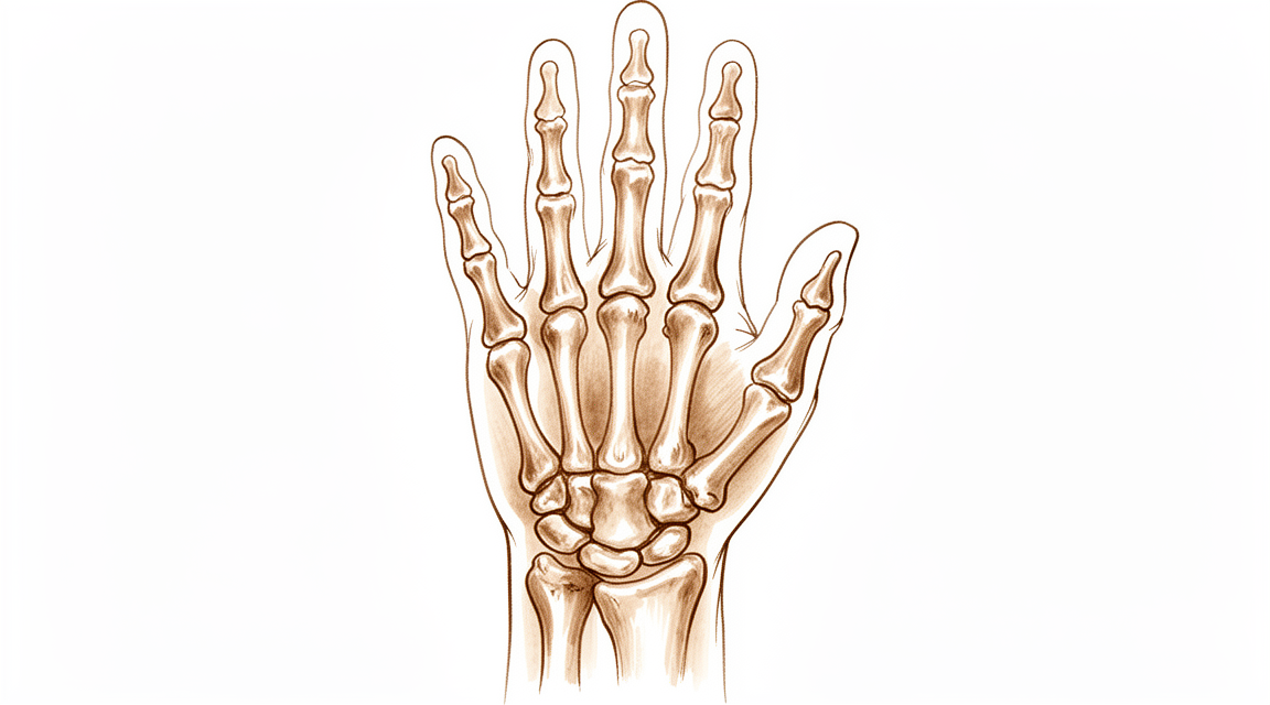
Ang Kienböck’s disease ay isang kondisyon kung saan nababawasan o nawawala ang suplay ng dugo sa isang maliit na buto sa iyong pulso, na tinatawag na lunate. Dahil sa kakulangan ng dugo, unti-unting humihina ang butong ito at maaari itong mag-collapse sa huli. Isipin ang lunate bilang isang sentral na shock absorber sa iyong pulso. Kapag nawala ang structural integrity nito, hindi na ito makakapag-cushion ng impact sa pagitan ng mga buto ng iyong forearm at ng natitirang bahagi ng iyong kamay.
Ang pag-collapse na ito ay nagbabago sa paraan ng paggalaw ng iyong pulso. Karaniwan, ang mga buto sa iyong pulso ay madulas at maayos na dumudulas at dumudurog sa isa’t isa. Kapag sira ang lunate, nagiging irregular ang galaw na ito. Maaaring lumipat ang mga buto mula sa kanilang normal na alignment, isang problema na kilala bilang rotational malalignment. Nagdudulot ang maling alignment na ito ng karagdagang stress sa ibang bahagi ng wrist joint, lalo na kung saan nagtatagpo ang radius bone at scaphoid bone. Sa paglipas ng panahon, maaaring magdulot ang hindi pantay na pagsuot na ito ng arthritis sa mga partikular na lugar na ito.
Tinitingnan ng iyong surgeon ang mga pagbabagong ito upang desyuminin ang pinakamainam na landas. Sa mga mas batang pasyente, madalas na layunin ang pagligtas sa buto. Ang mga prosedura tulad ng radial osteotomies, kung saan ina-adjust ang forearm bone, ay maaaring mapabuti ang mga resulta at radiographic findings sa mga teenager. Para sa iba, maaaring irekomenda ang capitate shortening o vascularized bone grafting upang ibalik ang balanse at suplay ng dugo. Layunin ng mga paggamot na ito na panatilihin ang paggalaw ng iyong pulso na kasingkalikasan ng normal.
Sa mga mas advanced na kaso kung saan malaki ang pag-collapse ng buto, ang pokus ay lumilipat sa pag-stabilize ng joint. Ang mga operasyon tulad ng scaphocapitate arthrodesis, na nagf-fuse ng dalawang buto, ay maaaring magbigay ng long-term relief mula sa sakit at mapabuti ang lakas ng hawak. Habang limitado ng mga prosedurang ito ang ilang galaw, nililikha nito ang isang matatag na platform na nagbibigay-daan upang maging epektibo ang paggamit ng iyong kamay. Pipiliin ng iyong surgeon ang opsyon na pinaka-angkop sa stage ng iyong sakit at sa iyong personal na pangangailangan.
Ano ang maaari naming gawin dito
Simulan namin ang hindi operasyong paggamot upang pamahalaan ang iyong mga sintomas at protektahan ang iyong pulso. Ang paraang ito ay nakatuon sa pagbawas ng stress sa mga buto sa iyong kamay. Maaaring irekomenda ng iyong surgeon ang pahinga o mga partikular na gawain na iwasan. Tumutulong ang pisyoterapiya upang mapanatili ang galaw at lakas ng iyong pulso at kamay. Layunin ng mga pamamaraang ito na panatilihing komportable ka habang sinusubaybayan namin ang kondisyon.
Ipakikita ng mga pananaliksik na ang mga hindi operasyong paggamot ay maaaring makamit ang magagandang at mahusay na resulta sa mga mas batang pasyente na patuloy pa ring lumalaki ang kanilang mga buto. Para sa marami, sapat ang konservatibong daang ito upang pamahalaan ang sakit at pagganap. Gayunpaman, karaniwang isang progressive na kondisyon ang Kienböck’s disease. Ibig sabihin, maaari itong maglala sa paglipas ng panahon, na maaaring magdulot ng mga advanced na pagbabago sa joint ng pulso kung hindi ito babantayan. Mahigpit namin pinagmamasdan ang mga senyales na ito sa iyong mga regular na check-up.
Kung hindi magbigay ng sapat na ginhawa ang mga hindi operasyong hakbang, tatalakayin namin ang mga opsyon sa medikal na pamamahala. Ang mga gamot pang-sakit at anti-inflammatories ay tumutulong upang kontrolin ang hindi komportableng pakiramdam at pamamaga. Sa ilang kaso, maaari naming isaalang-alang ang mga injection upang bawasan ang pamamaga nang direkta sa loob ng joint. Nag-aalok ang mga paggamot na ito ng pansamantalang ginhawa at tumutulong sa iyo na manatiling aktibo. Hindi nila inuulit ang mga pagbabago sa ilalim na buto ngunit maaari nilang mapabuti ang iyong araw-araw na kalidad ng buhay habang nagpaplano kami para sa susunod na mga hakbang.
Isinasalang-alang ang operasyon kapag naabot na ng konservatibong paggamot ang hangganan nito o kung ang sakit ay patuloy na lumalala kahit mayroong paggamot. Layunin ng operasyon na i-unload ang apektadong buto, ibalik ang daloy ng dugo, o istabilisa ang joint. Kasama sa mga opsyon ang mga pamamaraan upang baguhin ang hugis ng butong radius, mag- graft ng bagong buto kasama ang sarili nitong suplay ng dugo, o i-fuse ang mga partikular na buto nang sama-sama upang bawasan ang sakit. Ang pagpili ay nakadepende sa yugto ng iyong sakit at sa iyong mga indibidwal na pangangailangan.
Para sa mga pasyenteng kabataan, epektibo ang mga radial osteotomies (pagbabago ng hugis ng buto ng forearms) sa pagpapabuti ng parehong mga sintomas at mga resulta sa X-ray. Maaaring magbigay ang mga pamamaraang ito ng pagpapabuti na tumatagal ng dekada sa 75% ng mga pasyente. Sa mga advanced na kaso kung saan na-collapse na ang pulso, nag-aalok ang mga pamamaraang pag-fuse tulad ng scaphocapitate arthrodesis ng malaking pagbawas ng sakit at pagpapabuti ng pagganap. Ipapaliwanag ng iyong surgeon kung aling opsyon ang angkop sa iyong partikular na sitwasyon.
Ginagamit din namin ang mga imaging tulad ng X-ray at MRI upang subaybayan ang mga pagbabago. Habang minsan ay maaaring malampasan ng plain X-ray ang mga maagang senyales ng collapse, tumutulong ang advanced na imaging upang makita namin ang tunay na estado ng iyong butong lunate. Tinitiyak nito na pumili kami ng angkop na paggamot sa tamang oras. Maging kailangan mo ng simpleng pahinga o isang operasyon, ang aming layunin ay mapanatili ang pagganap ng iyong pulso at bawasan ang sakit.
Ano ang inaasahan
Ang Kienböck’s disease ay isang kondisyon kung saan nababawasan ang suplay ng dugo sa isang maliit na buto sa pulso, na nagdudulot ng pagpapahina at pagkabagsak nito. Ang prosesong ito ay karaniwang progressive, ibig sabihin ay may tendency na lumala ito sa loob ng panahon imbes na mag-settle nang sarili. Kung walang paggamot, ang kondisyon ay maaaring umabot sa Stage IV changes, na may kasamang malaking wear-and-tear arthritis sa wrist joint. Ang eksaktong landas ng sakit ay hindi ganap na kilala, ngunit madalas itong nagdudulot ng persistent na sakit at stiffness kung iiwan na lang.
Sa may kasamang paggamot, ang iyong outlook ay malaki ang pag-unlad. Maraming surgical options ang dinisenyo upang itigil ang pag-unlad na ito at bawasan ang sakit. Halimbawa, ang radial shortening osteotomy—isang procedure na inaayos ang haba ng iyong forearm bone—ay nagbibigay ng decade-long improvement sa 75% ng mga pasyente na may symptomatic disease. Ang paggamot na ito ay partikular na epektibo para sa long-lasting na pagpapagaan ng sakit sa mga pasyente na may negative ulnar variance, isang tiyak na alignment ng buto sa pulso. Ang ibang mga procedure, tulad ng scaphocapitate arthrodesis (pagkakaisa ng dalawang buto) o proximal row carpectomy (pag-alis ng isang row ng maliit na buto sa pulso), ay nag-aalok ng matibay, long-term na benepisyo. Ang mga operasyong ito ay karaniwang nagpapagaan ng sakit, nagpapanatili ng functional mobility, at nagpapanatili ng sapat na grip strength sa loob ng maraming taon.
Kahit may paggamot, hindi laging mabilis ang pag-unlad ng sakit. Ang radiographic progression ng Kienböck disease sa loob ng 1 taon o higit pa ay bahagya lamang sa average, anuman ang paggamot na pinili. Ibig sabihin, habang maaaring manatili ang underlying condition, ang mga visible na pagbabago sa X-ray ay madalas ay nagse-stabilize. Inaasahan mong i-tailor ng iyong surgeon ang plano batay sa iyong tiyak na stage at sintomas. Maging ikaw ay isang teenager o adult, ang layunin ay pamahalaan ang sakit at panatilihin ang functional na pulso. Habang ang mga outcomes ay karaniwang positive, mahalagang magkaroon ng realistic na expectations. Ang sakit ay multifactorial, at iba-iba ang individual na resulta, ngunit ang mga modernong paggamot ay nag-aalok ng reliable na landas para sa pagpapagaan ng sakit at pag-unlad ng hand function.
Kailan kumonsulta sa doktor
Ang Kienböck’s disease ay isang bihirang kondisyon na nakakaapekto sa pulso. Karaniwang itinuturing itong isang unti-unting lumalalang isyu na maaaring magdulot ng malubhang pagbabago sa kasukasuan. Hindi ganap na kilala ang eksaktong landas nito. Dapat mong kumonsulta sa iyong doktor kung mayroon kang patuloy na sakit na hindi gumagaling sa pamamagitan ng pahinga. Humingi ng pagsusuri ng espesyalista kung napapansin mo ang kahinaan o kawalan ng katatagan sa iyong pulso. Humingi ng tulong kung ang iyong kamay ay nakakabit o biglang nababagsak. Kontakin ang iyong surgeon kung ang mga sintomas ay nakakaapekto sa iyong pagtulog o trabaho. Biglaang paglala ng sakit ay dahilan din upang humingi ng medikal na tulong. Ang maagang pagsusuri ay tumutulong upang ma-manahang nang epektibo ang komplikadong kondisyong ito.
Evidence & references
Overview
- Radial osteotomies are effective in improving short-term clinical outcomes and radiographic findings in teenage patients with Kienböck disease [1].
- Scaphocapitate arthrodesis demonstrates long-term clinical benefits for the treatment of collapsed Kienböck disease [2].
- Vascularized bone grafting for stage III Kienböck disease demonstrates favorable long-term results and is recommended as a surgical treatment [3].
- Good and excellent clinical and radiological outcomes can be achieved with both nonsurgical and surgical treatments in skeletally immature patients with Kienböck disease [5].
- Temporary scaphotrapezoidal joint fixation is recommended for the surgical treatment of adolescent Kienböck's disease [7].
- Radial shortening osteotomy provides decade-long improvement in 75% of patients and is a reasonable treatment for symptomatic Kienböck’s disease [8].
- Scaphocapitate arthrodesis is an effective procedure for the treatment of Kienböck disease, associated with satisfactory functional outcomes and significant improvement in pain scores and grip strength [13].
- Capitate shortening is a safe and effective approach for the treatment of early stages of Kienböck's disease and can be associated with a satisfying outcome [14].
- Advanced Kienböck's disease with carpal collapse is not a contraindication for carpal-sparing surgery radial shortening osteotomy [17].
- Lunate excision, capitate osteotomy, and intercarpal arthrodesis should be used with caution for advanced Kienböck's disease because it does not have good long-term results and is no longer widely used in Europe [25].
- The acceptance rate for negative outcomes studies regarding Kienböck's disease is higher than for other surgical disorders, indicating a relative decrease in positive outcome bias among published Kienböck's disease studies compared with other surgical disorders [41].
Anatomy & Pathophysiology
- Tendon ball arthroplasty and proximal carpal stabilization with tendon graft restore the integrity of the proximal carpal row in advanced Kienböck’s disease [28].
- Surgical treatments for scapholunate advanced collapse result in decreased wrist kinematic motion and functional performance compared with individuals with normal wrists [30].
- Lunate morphology affects the 3-dimensional kinematics of the carpus during wrist flexion and extension [31].
- Three- and 4-corner fusions produce motion that is smoother and more closely replicates the normal axis and functional motion of the wrist [32].
- Computed fiber elongations of the dorsal carpal ligaments vary linearly with wrist position [33].
- Four-dimensional computed tomography (4DCT) is a non-invasive and affordable method to assess and quantify wrist kinematics by incorporating the temporal dimension [34].
- During simple unresisted wrist motions, force in the scapholunate interosseous ligament does not exceed 20 N [35].
- Kinematic changes in scapholunate instability may predict the development of radioscaphoid arthritis and help identify a kinematically abnormal wrist [36].
- Rotational malalignment of the wrist has significant effects on carpal, distal radial, and distal radioulnar joint measurements [37].
- Scaphoid nonunions have a dramatic impact on carpal kinematics by partially uncoupling the proximal and distal carpal rows [42].
- Dynamic imaging may enable the derivation of a standardized protocol for mapping carpal motion that is clinically applicable and reproducible [44].
- Computer-aided CT analysis provides guidelines for measuring and quantifying carpal alignment three-dimensionally and establishes a database for normal values [45].
- More than half the motion of the carpus when the wrist was loaded in extension occurred at the midcarpal joint [47].
- Radioscapholunate fusion shows the most biomechanically similar behavior to the healthy wrist among three fusion types [51].
- Contact areas between the scaphoid and distal radius are maximized during full extension of the wrist, which helps stabilize the radiocarpal joint [52].
- A dorsally applied PLA plate restores carpal kinematics for 1,000 cycles of motion in unstable wrists where fixation is not compromised by carpal size or osteoporosis [53].
- The modification of the wrist center of rotation during flexion and extension indicates that stability is considered more important than mobility in clinical conditions [54].
- Correction of scapholunate dissociation may correlate with improved carpal dynamics and improved clinical outcomes [56].
- Postarthroscopic lunate excision alters normal carpal kinematics but maintains joint congruity in the short-term [57].
Classification
- Kienböck disease is generally considered a progressive condition that can end in Stage IV changes [6].
- Lunate morphology may affect the severity of Kienböck disease at the time of initial presentation [9].
- The patterns of carpal collapse differ between stage IIIb Kienböck disease and scapholunate dislocation in terms of radioscaphoid joint congruity [50].
- Traditional radiographic indices measured on plain radiographs have poor diagnostic performance in the detection of carpal collapse in Kienböck's disease [15].
Clinical Presentation
- Kienböck disease is a relatively infrequent carpal pathology [4].
- Kienböck disease is generally considered a progressive condition that can end in Stage IV changes [6].
- The natural history of Kienböck disease is not fully known [6].
- Lunate morphology may affect the severity of Kienböck disease at the time of initial presentation [9].
- The development of Kienböck disease is probably multifactorial [10].
- Traditional radiographic indices measured on plain radiographs have poor diagnostic performance in the detection of carpal collapse in Kienböck's disease [15].
Investigations
- Radial osteotomies are effective in improving short-term clinical outcomes and radiographic findings in teenage patients with Kienböck disease [1].
- Scaphocapitate arthrodesis demonstrates long-term clinical benefits for the treatment of collapsed Kienböck disease [2].
- Vascularized bone grafting for stage III Kienböck disease demonstrates favorable long-term results and is recommended as a surgical treatment [3].
- Good and excellent clinical and radiological outcomes can be achieved with both nonsurgical and surgical treatments in skeletally immature patients with Kienböck disease [5].
- Temporary scaphotrapezoidal joint fixation is recommended for the surgical treatment of adolescent Kienböck's disease [7].
- Radial shortening osteotomy provides decade-long improvement in 75% of patients and is a reasonable treatment for symptomatic Kienböck’s disease [8].
- Lunate morphology may affect the severity of Kienböck disease at the time of initial presentation [9].
- Traditional radiographic indices measured on plain radiographs have poor diagnostic performance in the detection of carpal collapse in Kienböck's disease [15].
- The Camembert osteotomy improved function in patients with early stage Kienböck disease, with MRI aspects improving in most cases and no patients experiencing lunate collapse [21].
- Radiographic progression of Kienböck over 1 year or more seems slight on average regardless of treatment [26].
- Computed tomography of the lunate in Kienböck disease is an important investigative tool [43].
- Lunates with advanced Kienböck's disease exhibit significantly denser, thicker, and more plate-like trabecular microstructure compared to normal lunates [58].
- Following medial femoral trochlea reconstruction of the proximal lunate for advanced Kienböck disease, cessation of radiocarpal collapse was observed [59].
Treatment
Non-Operative and General Considerations
- Good and excellent clinical and radiological outcomes can be achieved with both nonsurgical and surgical treatments in skeletally immature patients with Kienböck disease [5].
- The natural history of Kienböck's disease is generally considered a progressive condition that can end in Stage IV changes [6].
- Treatment strategies for Kienböck's disease focus on biomechanical unloading, vascularized bone grafts, or salvage procedures depending on the stage [6].
- There is limited, low-quality evidence that surgical treatment slows progression of Kienböck's disease [18].
- Many uncontrolled case series document slight improvement in motion and grip after surgical treatment without clear evidence that this is better than placebo or no intervention [18].
Radial Osteotomies and Shortening
- Radial osteotomies are effective in improving not only short-term clinical outcomes, but also radiographic findings in teenage patients with Kienböck disease [1].
- Radial shortening osteotomy provides decade-long improvement in 75% of patients and seems to be a reasonable treatment for symptomatic Kienböck’s disease [8].
- The medium- and long-term results of radial shortening osteotomy for Kienböck's disease in patients with negative ulnar variance are comparable to short-term results, providing long-lasting pain relief [16].
- Advanced Kienbock's disease with carpal collapse is not a contraindication for carpal-sparing surgery radial shortening osteotomy [17].
- Radius core decompression demonstrated favorable long-term results and could be considered as a surgical alternative for stage IIIA of Kienböck disease [61].
Vascularized Bone Grafts
- Vascularized bone grafting for stage III Kienböck disease demonstrated favorable long-term results and is recommended as a surgical treatment [3].
- Vascularized bone grafts are indicated for Kienböck's disease, while being contraindicated in advanced carpal collapse with degenerative changes [40].
Arthrodesis and Salvage Procedures
- The long-term clinical benefits of scaphocapitate arthrodesis for treatment of collapsed Kienböck disease are demonstrated [2].
- Scaphocapitate arthrodesis is an effective procedure for treatment of Kienböck disease associated with satisfactory functional outcomes and significant improvement in pain scores and grip strength [13].
- Scaphocapitate arthrodesis should be considered as a treatment option for wrist salvage in the patient with advanced Kienbock's disease given the significant postoperative reduction in associated pain symptoms at the time of follow-up [24].
- Scaphotrapeziotrapezoid arthrodesis with lunate excision for advanced Kienböck disease provided favorable clinical results in terms of pain relief and functional improvement [22].
- The procedure of lunate excision, capitate osteotomy, and intercarpal arthrodesis should be used with caution for advanced Kienböck's disease because it does not have good long-term results and is no longer widely used in Europe [25].
- Improved outcomes following proximal row carpectomy were associated with Kienbock's disease [55].
Capitate Shortening
- Capitate shortening is a safe and effective approach for treatment of the early stages of Kienböck's disease and can be associated with a satisfying outcome [14].
Arthroscopic Procedures
- Arthroscopic lunate core decompression appears to be an effective and safe surgery for treating Kienböck disease on the basis of mid-term follow-up [20].
Adolescent-Specific Interventions
- Temporary scaphotrapezoidal joint fixation is recommended for the surgical treatment of adolescent Kienböck's disease [7].
Complications
- Kienböck's disease is generally considered a progressive condition that can end in Stage IV changes [6].
- The natural history of Kienböck's disease is not fully known [6].
- Lunate morphology may affect the severity of Kienböck disease at the time of initial presentation [9].
- Negative ulnar variance has a longitudinal relationship with progressive Kienböck disease, though additional long-term study is needed to confirm this [19].
- The development of Kienböck disease is probably multifactorial [10].
- The observation of Kienböck disease and carpal coalition in one wrist is fortuitous [10].
Recovery
- Radial osteotomies improve short-term clinical outcomes in teenage patients with Kienböck disease [1].
- Radial osteotomies improve radiographic findings in teenage patients with Kienböck disease [1].
- Scaphocapitate arthrodesis provides long-term clinical benefits for collapsed Kienböck disease [2].
- Vascularized bone grafting demonstrates favorable long-term results for stage III Kienböck disease [3].
- Radial shortening osteotomy provides decade-long improvement in 75% of patients with symptomatic Kienböck’s disease [8].
- Radial shortening osteotomy provides long-lasting pain relief for patients with negative ulnar variance [16].
- Titanium lunate arthroplasty (TLA) shows promising longer-term results for stage III Kienböck disease [23].
- Scaphocapitate arthrodesis with lunate excision significantly alleviates pain in advanced Kienböck disease at a mean follow-up of 10.7 years [38].
- Scaphocapitate arthrodesis with lunate excision preserves functional mobility in advanced Kienböck disease at a mean follow-up of 10.7 years [38].
- Scaphocapitate arthrodesis with lunate excision maintains satisfactory grip strength in advanced Kienböck disease at a mean follow-up of 10.7 years [38].
- Proximal row carpectomy (PRC) is a durable long-term treatment option for radiocarpal degenerative arthritis and Kienböck's disease [46].
- Proximal row carpectomy allows for maintenance of wrist range of motion [46].
- Proximal row carpectomy improves grip strength [46].
- Proximal row carpectomy is a reliable and durable procedure for Lichtman stage IIIA or IIIB Kienböck's disease at an average follow-up of 10 years [49].
- Scaphocapitate arthrodesis (SCA) results in improved grip strength in patients with advanced stages of Kienböck disease in medium-term follow-up [48].
- Scaphocapitate arthrodesis (SCA) corrects carpal alignment in patients with advanced stages of Kienböck disease in medium-term follow-up [48].
- Radiographic progression of Kienböck disease over 1 year or more is slight on average regardless of treatment [26].
Key Evidence
- [L4] The current results indicate that radial osteotomies are effective in improving not only short-term clinical outcomes, but also radiographic findings in teenage patients with Kienböck disease. [1] (10.1097/01.blo.0000173254.46899.72)
- [L4] The long-term clinical benefits of scaphocapitate arthrodesis for treatment of collapsed Kienböck disease are demonstrated. [2] (10.1177/1753193413496177)
- [L3] Vascularized bone grafting for stage III Kienböck disease demonstrated favorable long-term results and is recommended as a surgical treatment. [3] (10.1016/j.jhsa.2013.02.010)
- [L5] This review article discusses the history, etiology, and course of Kienböck disease and reviews the literature on both the diagnosis and management of this relatively infrequent carpal pathology. [4] (10.5435/jaaos-d-20-00020)
- [L4] Good and excellent clinical and radiological outcomes can be achieved with both nonsurgical and surgical treatments in skeletally immature patients with Kienböck disease. [5] (10.1016/j.jhsa.2018.02.029)
- [L5] The natural history of Kienbock's disease is not fully known, though it is generally considered a progressive condition that can end in Stage IV changes; treatment strategies focus on biomechanical unloading, vascularized bone grafts, or salvage procedures depending on the stage. [6] (10.1016/j.hcl.2006.07.003)
- [L4] We therefore recommend this procedure for the surgical treatment of adolescent Kienböck's disease. [7] (10.1016/j.jhsa.2008.09.019)
- [L4] Radial shortening osteotomy provides decade-long improvement in 75% of patients and seems to be a reasonable treatment for symptomatic Kienböck’s disease. [8] (10.1177/1753193413512222)
- [L3] Lunate morphology may affect the severity of Kienböck disease at the time of initial presentation. [9] (10.1016/j.jhsa.2014.12.024)
- [Letter] The observation of Kienböck disease and carpal coalition in one wrist is fortuitous, and the development of Kienböck disease is probably multifactorial. [10] (10.1016/j.jhsa.2016.11.010)
- [L4] Scaphocapitate arthrodesis is an effective procedure for treatment of Kienböck disease associated with satisfactory functional outcomes and significant improvement in pain scores and grip strength. [13] (10.1016/j.jhsg.2023.03.014)
- [L2] Capitate shortening is a safe and effective approach for treatment of the early stages of Kienböck's disease and can be associated with a satisfying outcome. [14] (10.1177/15589447221081564)
- [L3] Traditional radiographic indices measured on plain radiographs have poor diagnostic performance in the detection of carpal collapse in Kienböck's disease. [15] (10.1177/17531934231153966)
- [L3] The medium- and long-term results of radial shortening osteotomy for Kienböck's disease in patients with negative ulnar variance are comparable to short-term results, providing long-lasting pain relief. [16] (10.1097/blo.0b013e318041d309)
- [L5] There is limited, low-quality evidence that surgical treatment slows progression of Kienböck's disease, and many uncontrolled case series document slight improvement in motion and grip after surgical treatment without clear evidence that this is better than placebo or no intervention. [18] (10.1016/j.jhsa.2009.10.013)
- [L2] Additional long-term study is needed to confirm the longitudinal relationship of negative ulnar variance with progressive Kienböck disease. [19] (10.1016/j.jhsa.2017.06.107)
- [L4] Arthroscopic lunate core decompression appears to be an effective and safe surgery for treating Kienböck disease on the basis of mid-term follow-up. [20] (10.1016/j.jhsa.2023.02.011)
- [L4] Scaphotrapeziotrapezoid arthrodesis with lunate excision for advanced Kienböck disease provided favorable clinical results in terms of pain relief and functional improvement. [22] (10.1016/j.jhsa.2012.08.031)
- [L4] The longer-term results of TLA for stage III Kienböck disease are promising. [23] (10.1016/j.jhsa.2018.02.009)
- [L4] Given the significant postoperative reduction in associated pain symptoms at the time of follow-up, scaphocapitate arthrodesis should be considered as a treatment option for wrist salvage in the patient with advanced Kienbock's disease. [24] (10.1007/s11552-014-9705-z)
- [L5] The procedure of lunate excision, capitate osteotomy, and intercarpal arthrodesis should be used with caution for advanced Kienböck's disease because it does not have good long-term results and is no longer widely used in Europe. [25] (10.1177/1753193418807360)
- [L4] Radiographic progression of Kienböck over 1 year or more seems slight on average regardless of treatment. [26] (10.1016/j.jhsa.2016.02.016)
- [L4] The technique demonstrated reduced wrist pain and improved wrist motion and grip strength while restoring the integrity of the proximal carpal row. [28] (10.1177/17531934241238939)
- [L2] Both surgical groups demonstrated decreased wrist kinematic motion and functional performance compared with individuals with normal wrists. [30] (10.1016/j.jhsa.2015.04.035)
- [L5] This study describes the effect of lunate morphology on 3-dimensional carpal kinematics during wrist flexion and extension. [31] (10.1016/j.jhsa.2014.09.019)
- [L3] Motion was smoother and more closely replicated the normal axis and functional motion of the wrist. [32] (10.1016/j.jhsa.2015.02.027)
- [L5] Despite complex carpal bone anatomy and kinematics, computed fiber elongations were found to vary linearly with wrist position. [33] (10.1016/j.jhsa.2012.04.025)
- [L5] Four-dimensional computed tomography (4DCT) is a promising, non-invasive, and affordable method to assess and quantify wrist kinematics, extending conventional CT by incorporating the temporal dimension. [34] (10.1177/17531934251326028)
- [L5] However, during simple unresisted wrist motions, the force did not exceed 20 N. [35] (10.1016/j.jhsa.2015.04.007)
- [L3] These kinematic changes may predict the development of radioscaphoid arthritis and help identify a kinematically abnormal wrist. [36] (10.1177/17531934241242676)
- [L4] Rotational malalignment of the wrist has significant effects on carpal, distal radial and distal radioulnar joint measurements. [37] (10.1177/1753193408090393)
- [L4] Scaphocapitate arthrodesis with lunate excision performed in an advanced stage of Kienböck disease significantly alleviates pain, while preserving functional mobility and satisfactory grip strength in the long term. [38] (10.1177/1753193417739247)
- [L4] This manuscript offers a current review of the techniques and outcomes of VBGs to the carpal bones, noting that VBGs are indicated for scaphoid nonunion with proximal pole AVN, Kienböck's disease, Preiser's disease, and capitate osteonecrosis, while being contraindicated in advanced carpal collapse with degenerative changes. [40] (10.1007/s11552-012-9479-0)
- [L2] The acceptance rate for negative outcomes studies regarding Kienböck's disease is higher than for other surgical disorders, indicating a relative decrease in positive outcome bias among published Kienböck's disease studies compared with other surgical disorders. [41] (10.1016/j.jhsa.2009.12.003)
- [L4] Scaphoid nonunions have a dramatic impact on carpal kinematics, partially uncoupling the proximal and distal carpal rows. [42] (10.1016/j.jhsa.2008.03.008)
- [L4] Computed tomography of the lunate in Kienböck disease is an important investigative tool. [43] (10.1016/j.jhsa.2018.05.008)
- [L4] With the increased focus on dynamic imaging for wrist motion, it may be possible to derive a standardized protocol for mapping the carpal motion that is clinically applicable and reproducible. [44] (10.1016/j.jhsg.2022.10.001)
- [L4] This study provides guidelines on how to measure and quantify carpal alignment three-dimensionally and establishes a database for normal values, which may be useful when analysing various wrist pathologies and kinematics. [45] (10.1177/17531934231160100)
- [L4] PRC is a durable long-term treatment option for radiocarpal degenerative arthritis and Kienböck's disease allowing for maintenance of wrist range of motion and improvement in grip strength. [46] (10.1016/s0363-5023(10)60087-1)
- [L4] More than half the motion of the carpus when the wrist was loaded in extension occurred at the midcarpal joint. [47] (10.1016/j.jhsa.2012.10.035)
- [L4] SCA resulted in improved grip strength with correction of carpal alignment in patients with advanced stages of Kienböck disease in medium-term follow-up. [48] (10.1016/j.jhsa.2014.12.013)
- [L4] At an average follow-up of 10 years, proximal row carpectomy is a reliable and durable procedure for patients with Lichtman stage IIIA or IIIB Kienböck's disease. [49] (10.1016/j.jhsa.2008.02.031)
- [L2] The patterns of carpal collapse differed between stage IIIb Kienböck disease and scapholunate dislocation in terms of radioscaphoid joint congruity. [50] (10.1016/j.jhsa.2014.10.035)
- [L5] The contact areas between the scaphoid and distal radius are maximized during full extension of the wrist, which helps stabilize the radiocarpal joint and potentially reduces the risk of injury to the carpus and the distal radius. [52] (10.1177/1753193413507810)
- [L5] The study shows that in the unstable wrist, following ligament sectioning, where fixation is not compromised by carpal size or osteoporosis, a dorsally applied PLA plate does restore carpal kinematics for 1,000 cycles of motion. [53] (10.1016/j.jhsa.2008.01.016)
- [L4] The study also characterized the modification of the wrist CoR during flexion and extension, noting that stability is considered more important than mobility in clinical conditions. [54] (10.1016/s0749-0712(03)00008-8)
- [L3] Improved outcomes were associated with age over 40, Kienbock's disease, concomitant neurectomy, non-labourer status, and surgery after 1990, while radiocapitate arthrosis did not correlate with clinical outcomes. [55] (10.1177/1753193415597096)
- [L5] This correction might correlate with improved carpal dynamics and improved clinical outcomes. [56] (10.1016/j.jhsa.2010.06.029)
- [L4] Although postarthroscopic lunate excision alters normal carpal kinematics, the joint congruity is maintained in the short-term. [57] (10.1016/j.jhsa.2025.01.024)
- [L4] Lunates with advanced Kienböck's disease exhibit significantly denser, thicker, and more plate-like trabecular microstructure compared to normal lunates. [58] (10.1177/1753193411422337)
- [L4] Following medial femoral trochlea reconstruction of the proximal lunate for advanced Kienböck disease, we observed a cessation of radiocarpal collapse. [59] (10.1016/j.jhsa.2019.12.008)
- [L4] In this limited series, the radius core decompression demonstrated favorable long-term results and could be considered as a surgical alternative for stage IIIA of Kienböck disease. [61] (10.1016/j.jhsa.2017.05.017)
References
[1] Radial Osteotomies for Teenage Patients with Kienb??ck Disease. Clinical Orthopaedics and Related Research. 2005. DOI: 10.1097/01.blo.0000173254.46899.72 [2] Scaphocapitate arthrodesis for treatment of late stage Kienböck disease. Journal of Hand Surgery (European Volume). 2013. DOI: 10.1177/1753193413496177 [3] Long-Term Results of Vascularized Bone Graft for Stage III Kienböck Disease. The Journal of Hand Surgery. 2013. DOI: 10.1016/j.jhsa.2013.02.010 [4] Osteonecrosis of the Lunate: Kienböck Disease. Journal of the American Academy of Orthopaedic Surgeons. 2020. DOI: 10.5435/jaaos-d-20-00020 [5] Kienböck Disease in the Skeletally Immature Patient. The Journal of Hand Surgery. 2018. DOI: 10.1016/j.jhsa.2018.02.029 [6] Kienböck's Disease: An Approach to Treatment. Hand Clinics. 2006. DOI: 10.1016/j.hcl.2006.07.003 [7] Temporary Scaphotrapezoidal Joint Fixation for Adolescent Kienböck's Disease. The Journal of Hand Surgery. 2009. DOI: 10.1016/j.jhsa.2008.09.019 [8] Long-term outcome (20 to 33 years) of radial shortening osteotomy for Kienböck’s lunatomalacia. Journal of Hand Surgery (European Volume). 2013. DOI: 10.1177/1753193413512222 [9] The Effect of Lunate Morphology in Kienböck Disease. The Journal of Hand Surgery. 2015. DOI: 10.1016/j.jhsa.2014.12.024 [10] Letter Regarding “Kienböck Disease and Carpal Coalitions: A Potential Correlation”. The Journal of Hand Surgery. 2017. DOI: 10.1016/j.jhsa.2016.11.010 [13] Clinical and Radiological Outcomes of Scaphocapitate Fusion in Kienböck Disease: A Systematic Review and Meta-Analysis. Journal of Hand Surgery Global Online. 2023. DOI: 10.1016/j.jhsg.2023.03.014 [14] Comparing the Radiologic and Functional Outcome of Radial Shortening Versus Capitate Shortening in Management of Kienböck’s Disease. HAND. 2022. DOI: 10.1177/15589447221081564 [15] Diagnostic performance of traditional radiographic indices in detection of carpal collapse in Kienböck’s disease. Journal of Hand Surgery (European Volume). 2023. DOI: 10.1177/17531934231153966 [16] Outcome of Kienböck's Disease 22 Years after Distal Radius Shortening Osteotomy. Clinical Orthopaedics & Related Research. 2007. DOI: 10.1097/blo.0b013e318041d309 [17] 10.1055-s-0039-1688947. n.d.. [18] Kienböck's Disease. The Journal of Hand Surgery. 2009. DOI: 10.1016/j.jhsa.2009.10.013 [19] Risk Factors of Lunate Collapse in Kienböck Disease. The Journal of Hand Surgery. 2017. DOI: 10.1016/j.jhsa.2017.06.107 [20] Arthroscopic Treatment of Kienböck Disease: Mid-Term Outcome of Arthroscopic Lunate Core Decompression. The Journal of Hand Surgery. 2024. DOI: 10.1016/j.jhsa.2023.02.011 [21] 10.1055-s-0039-1683931. n.d.. [22] Scaphotrapeziotrapezoid Arthrodesis and Lunate Excision for Advanced Kienböck Disease. The Journal of Hand Surgery. 2012. DOI: 10.1016/j.jhsa.2012.08.031 [23] Long-Term Clinical Outcome After Titanium Lunate Arthroplasty for Kienböck Disease. The Journal of Hand Surgery. 2018. DOI: 10.1016/j.jhsa.2018.02.009 [24] Limited Intercarpal Fusion as a Salvage Procedure for Advanced Kienbock Disease. HAND. 2014. DOI: 10.1007/s11552-014-9705-z [25] Lunate excision, capitate osteotomy, and intercarpal arthrodesis should be used with caution for advanced Kienböck’s disease. Journal of Hand Surgery (European Volume). 2018. DOI: 10.1177/1753193418807360 [26] Radiographic Progression of Kienböck Disease: Radial Shortening Versus No Surgery. The Journal of Hand Surgery. 2016. DOI: 10.1016/j.jhsa.2016.02.016 [28] Tendon ball arthroplasty and proximal carpal stabilization with tendon graft for advanced Kienböck’s disease. Journal of Hand Surgery (European Volume). 2024. DOI: 10.1177/17531934241238939 [30] Surgical Treatments for Scapholunate Advanced Collapse Wrist: Kinematics and Functional Performance. The Journal of Hand Surgery. 2015. DOI: 10.1016/j.jhsa.2015.04.035 [31] The Effect of Lunate Morphology on the 3-Dimensional Kinematics of the Carpus. The Journal of Hand Surgery. 2015. DOI: 10.1016/j.jhsa.2014.09.019 [32] Comparison of the Clinical and Functional Outcomes Following 3- and 4-Corner Fusions. The Journal of Hand Surgery. 2015. DOI: 10.1016/j.jhsa.2015.02.027 [33] Elongation of the Dorsal Carpal Ligaments: A Computational Study of In Vivo Carpal Kinematics. The Journal of Hand Surgery. 2012. DOI: 10.1016/j.jhsa.2012.04.025 [34] Dynamic wrist imaging: How it works and how to assess kinematic changes in wrists with scapholunate instability. Journal of Hand Surgery (European Volume). 2025. DOI: 10.1177/17531934251326028 [35] Force in the Scapholunate Interosseous Ligament During Active Wrist Motion. The Journal of Hand Surgery. 2015. DOI: 10.1016/j.jhsa.2015.04.007 [36] Radiocarpal and midcarpal kinematics in scapholunate instability: a four-dimensional CT study in vivo. Journal of Hand Surgery (European Volume). 2024. DOI: 10.1177/17531934241242676 [37] The Effect of Rotational Malalignment on X-rays of the Wrist. Journal of Hand Surgery (European Volume). 2009. DOI: 10.1177/1753193408090393 [38] Results of scaphocapitate arthrodesis with lunate excision in advanced Kienböck disease at 10.7-year mean follow-up. Journal of Hand Surgery (European Volume). 2017. DOI: 10.1177/1753193417739247 [40] Vascularized Bone Grafts for the Treatment of Carpal Bone Pathology. HAND. 2013. DOI: 10.1007/s11552-012-9479-0 [41] Publication Bias in Kienböck's Disease: Systematic Review. The Journal of Hand Surgery. 2010. DOI: 10.1016/j.jhsa.2009.12.003 [42] Interfragmentary Motion in Patients With Scaphoid Nonunion. The Journal of Hand Surgery. 2008. DOI: 10.1016/j.jhsa.2008.03.008 [43] Fixation of the Fractured Lunate in Kienböck Disease. The Journal of Hand Surgery. 2019. DOI: 10.1016/j.jhsa.2018.05.008 [44] Radiographic Evaluation of Carpal Mechanics and the Scapholunate Angle in a Clenched Fist with Dynamic Computed Tomography Imaging. Journal of Hand Surgery Global Online. 2023. DOI: 10.1016/j.jhsg.2022.10.001 [45] Three-dimensional carpal alignment: computer-aided CT analysis of carpal axes and normal ranges. Journal of Hand Surgery (European Volume). 2023. DOI: 10.1177/17531934231160100 [46] Minimum Twenty-year Follow-up of Proximal Row Carpectomy. The Journal of Hand Surgery. 2010. DOI: 10.1016/s0363-5023(10)60087-1 [47] In Vivo Kinematics of the Scaphoid, Lunate, Capitate, and Third Metacarpal in Extreme Wrist Flexion and Extension. The Journal of Hand Surgery. 2013. DOI: 10.1016/j.jhsa.2012.10.035 [48] Scaphocapitate Arthrodesis for Kienböck Disease. The Journal of Hand Surgery. 2015. DOI: 10.1016/j.jhsa.2014.12.013 [49] Proximal Row Carpectomy for Advanced Kienböck's Disease: Average 10-Year Follow-Up. The Journal of Hand Surgery. 2008. DOI: 10.1016/j.jhsa.2008.02.031 [50] In Vivo 3-Dimensional Analysis of Stage III Kienböck Disease: Pattern of Carpal Deformity and Radioscaphoid Joint Congruity. The Journal of Hand Surgery. 2015. DOI: 10.1016/j.jhsa.2014.10.035 [51] Load_transfer_through_the_radiocarpal_joint_and_the_effects_of_partial_wrist_art_1753193412441761. 1934. [52] Contact areas of the scaphoid and lunate with the distal radius in neutral and extension: correlation of falling strategies and distal radial anatomy. Journal of Hand Surgery (European Volume). 2013. DOI: 10.1177/1753193413507810 [53] Treatment of Scapholunate Dissociation With a Bioresorbable Polymer Plate: A Biomechanical Study. The Journal of Hand Surgery. 2008. DOI: 10.1016/j.jhsa.2008.01.016 [54] Electrogoniometric and radiologic evaluation of scapho-trapezo-trapezoid arthrodesis. Hand Clinics. 2003. DOI: 10.1016/s0749-0712(03)00008-8 [55] Factors associated with improved outcomes following proximal row carpectomy: a long-term outcome study of 144 patients. Journal of Hand Surgery (European Volume). 2015. DOI: 10.1177/1753193415597096 [56] Radiographic Evaluation of the Modified Brunelli Technique Versus the Blatt Capsulodesis for Scapholunate Dissociation in a Cadaver Model. The Journal of Hand Surgery. 2010. DOI: 10.1016/j.jhsa.2010.06.029 [57] Three-Dimensional In Vivo Kinematic Analysis of Kienböck Disease Treated with Arthroscopic Lunate Excision. The Journal of Hand Surgery. 2025. DOI: 10.1016/j.jhsa.2025.01.024 [58] Trabecular microstructure of the human lunate in Kienböck’s disease. Journal of Hand Surgery (European Volume). 2011. DOI: 10.1177/1753193411422337 [59] Preliminary Clinical, Radiographic, and Patient-Reported Outcomes of the Medial Femoral Trochlea Osteochondral Free Flap for Lunate Reconstruction in Advanced Kienböck Disease. The Journal of Hand Surgery. 2020. DOI: 10.1016/j.jhsa.2019.12.008 [61] Radius Core Decompression for Kienböck Disease Stage IIIA: Outcomes at 13 Years Follow-Up. The Journal of Hand Surgery. 2017. DOI: 10.1016/j.jhsa.2017.05.017