Patients › Shoulder
Pag-aalis ng Distal Clavicle (Prosedura ni Mumford)
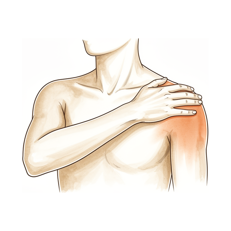
Ang prosedura ng Mumford (pag-aalis ng distal na bahagi ng klavikula) ay nag-aalis ng maliit na nabasag na panlabas na dulo ng klavikula upang bawasan ang sakit sa balikat sa AC joint. Ano ang kasalukuyang ginagawa sa pamamagitan ng keyhole operation at paano nakikita ang paggaling.
Bakit inirerekomenda ang operasyong ito
Inirekomenda ng iyong doktor ang distal clavicle excision, kilala rin bilang Mumford procedure. Ang operasyong ito ay nag-aalis ng labas na dulo ng collarbone upang pigilan itong magkuskos sa shoulder blade. Posibleng kailangan mo ito dahil sa patuloy na sakit o arthritis na dulot ng pagkasira na hindi nakababagay sa mga non-surgical na paggamot.
Karaniwang inaalok ang operasyong ito sa mga pasyenteng may lumang dislokasyon o kronikong sakit na gumagawa ng mabigat na trabaho o madalas na itinataas ang kanilang mga braso. Ang pangunahing layunin ay bawasan ang iyong sakit at mapabuti ang pag-andar ng iyong balikat. Bagama't parehong epektibo ang bukas na paraan at keyhole (arthroscopic) na paraan, ginagamit ng iyong doktor ang arthroscopic na pamamaraan. Ito ay nangangailangan ng maliliit na hiwa at isang kamera upang makapagbalik ka sa iyong mga gawain nang mas mabilis na may katulad na matagalang resulta.
Bago ang operasyon
Kailangan mong mag-fasting bago ang iyong operasyon at itigil ang pag-inom ng ilang gamot ayon sa payo ng iyong surgeon. Mangyaring mag-arrange ng taong magpapadala sa iyo pauwi at magsuot ng komportableng damit. Maaaring kailanganin ng X-ray, MRI, blood tests, o pagsusuri ng anestesia upang suriin ang iyong kalusugan at magplano ng prosedura. Isasagawa ng iyong surgeon ang operasyong ito gamit ang arthroscopic (keyhole) na paraan na may dalawa o tatlong maliit na hiwa at isang maliit na camera sa loob ng kasukasuan. Ang paraang ito ay tumutulong sa mabilis na pagbabalik sa mga aktibidad habang naiiwasan ang malalaking peklat. Dalhin ang listahan ng lahat ng iyong kasalukuyang gamot sa iyong appointment.
Sa araw ng operasyon
Dadating ka sa ospital at tatagpo ang iyong surgeon at anesthesiologist. Ang operasyong ito ay isasagawa sa ilalim ng general anesthesia na pinagsama ng regional nerve block. Ikaw ay lubos na matutulog habang ginagawa ang operasyon, at ang nerve block (isang injeksyon na nagpapabango sa mga nerbiyong nagpapadala ng sensasyon sa braso bago ka gumising) ay nagbibigay ng pagpapagaan ng sakit sa unang 12 hanggang 24 oras pagkatapos ng operasyon. Tatagpuin ka ng anesthesiologist bago ang operasyon at ipaliwanag ang parehong bahagi ng proseso.
Pagkatapos, pupunta ka sa operating theatre kung saan isasagawa ng iyong surgeon ang pamamaraan gamit ang keyhole approach. Ito ay nangangailangan ng dalawa o tatlong maliit na hiwa at isang maliit na camera sa loob ng kasu-kasuan upang gabayan ang paggawa. Pagkatapos ng operasyon, magigising ka sa recovery area kung saan hahawakan ng team ang iyong kaginhawaan habang unti-unti nang nawawala ang epekto ng pagkabango.
Mga Detalye ng Operasyon
Isasagawa ng iyong doktor ang operasyong ito gamit ang mga teknik na keyhole (laparoscopic). Gagawa sila ng dalawa o tatlong maliit na hiwa, bawat isa ay humigit-kumulang 1 cm ang haba, sa malapit sa iyong balikat. Sa pamamagitan ng mga bukasang ito, ipapasok ang isang maliit na kamera at espesyal na mga kasangkapan upang makita ang loob ng kasukasuan. Ang paraang ito ay nagbibigay-daan sa iyong doktor na maabot ang panlabas na dulo ng iyong collarbone nang hindi gumagawa ng malaking hiwa.
Ang pangunahing layunin ay alisin ang isang maliit na piraso ng buto mula sa panlabas na dulo ng iyong collarbone. Maingat na aalisin ng iyong doktor ang butong ito upang hindi na ito magkuskos sa scapula (shoulder blade). Ang mga ebidensya ay nagpapakita na ang pag-aalis ng humigit-kumulang 5 mm ng buto ay nagtitiyak na hindi na magkikita ang mga buto, habang ang pag-aalis ng 2.5 mm ay naging matagumpay sa maraming kaso. Ilalagay ng iyong doktor ang kamera at mga kasangkapan nang tumpak upang maiwasan ang pinsala sa mga kalapit na estruktura.
Pagkatapos alisin ang buto, isasara ang mga maliit na hiwa. Maaaring gumamit ang doktor ng natutunaw na tahi o pandikit upang isara ang balat. Inaasahan mong mabilisang babalik sa mga gawain sa pamamagitan ng paraang keyhole na ito kumpara sa mas malaking bukas na hiwa, habang nakukuha ang katulad na pangmatagalang resulta. Ang prosedura ay nakatuon sa pagpapagaan ng iyong sakit sa pamamagitan ng pag-aalis ng pinagmumulan ng kuskos sa kasukasuan.
Pagkatapos ng operasyon
Gising ka sa isang recovery area kung saan ang iyong team ang mag-aalaga sa iyong sakit. Gumagamit ang iyong surgeon ng keyhole technique na may dalawa o tatlong maliit na hiwa at isang maliit na camera sa loob ng joint. Magdudulot ka ng sling at may mga dressing sa mga maliit na hiwa. Maaari kang simulan ang paggalaw ng iyong mga daliri at pulso nang dahan-dahan agad-agad. Karamihan sa mga pasyente ay nananatili ng isang gabi sa ospital pagkatapos ng operasyong ito, bagaman may mga makakapagpunta sa bahay sa parehong araw. Kailangan mong magkaroon ng kasama na manatili sa iyo sa loob ng unang 24 na oras. Inaasahan mong makabalik sa pagmamaneho sa loob ng 4 linggo at sa trabaho sa loob ng 6 linggo.
Pagbawi
Maaaring maranasan mo ang ilang sakit at pamamaga sa iyong balikat sa unang ilang araw. Ito ay normal habang ang iyong katawan ay nagpapagaling mula sa maliliit na incisions na keyhole. Maaaring imungkahi ng iyong doktor ang mga ice pack at gamot para sa sakit upang matulungan kang maging komportable. Karamihan sa mga tao ay nakakakita na ang hindi komportableng pakiramdam ay unti-unting bumababa habang bumababa ang pamamaga.
Magdudulot ka ng sling upang suportahan ang iyong braso habang nagpapahinga. Ang iyong physiotherapist ay gabayin ka sa mga banayad na ehersisyo upang panatilihin ang paggalaw ng iyong balikat nang hindi ito pinapabigatan. Maaari kang gumawa ng magaan na mga gawain sa bahay kapag handa ka na, ngunit iwasan ang pag-angat ng mabibigat na bagay o pag-abot sa itaas. Ang pagtulog ay maaaring mahirap sa simula; ang pagtayo sa iyong sarili gamit ang mga unan ay madalas tumutulong upang makahanap ng komportableng posisyon.
Habang bumabalik ang iyong paggalaw at bumababa ang pamamaga, mas magkakaroon ka ng kumpiyansa sa iyong balikat. Ang iyong doktor at physiotherapist ay sasabihin sa iyo kung kailan mo maaaring muling magmaneho, bumalik sa trabaho, o maglaro ng sports. Maaaring magkaiba ang iyong personal na timeline mula sa iba, kaya sundin ang kanilang tiyak na payo para sa iyong pagbawi.
Maaaring mangyari
Karamihan sa mga pasyente ay gumagaling nang maayos, ngunit minsan ay maaaring magkaroon ng mga problema. Ang iyong manggagamot at ang koponan ay masusing sinusubaybayan ka upang maagang makita ang anumang isyu.
Kung patuloy kang nararamdaman ang sakit o parang may pagkagiling sa iyong balikat pagkatapos ng operasyon, maaaring hindi sapat ang pagtanggal ng buto. Minsan, maaaring lumaki muli ang buto sa parehong lokasyon. Ito ay maaaring magdulot ng malalim na sakit na hindi nawawala kahit gamitan ng simpleng gamot pang-alis ng sakit. Ipabatid sa iyong manggagamot kung nararamdaman mo ito upang masuri ang iyong paggaling.
Kung mapansin mo ang biglaang matulis na sakit o pakiramdam ng kawalan ng katatagan kung saan nagtatagpo ang iyong collarbone at balikat, maaaring sobra ang tinanggal na buto. Ito ay maaaring gawing maluwag o hindi matatag ang kasukasuan. Iulat agad ang anumang bagong pakiramdam ng pag-click o pagkagiling.
Sa bihirang kaso, maaaring magkaroon ng fracture sa collarbone o sa buto sa ilalim nito. Maaaring maranasan ang biglaang pag-ikot o matinding sakit na nagpapahirap sa paggalaw ng iyong braso. Ito ay seryosong isyu na nangangailangan ng agarang atensyon. Pumunta sa emergency department kung ikaw ay nag-aakala na may fracture.
Ginagamit ng iyong manggagamot ang keyhole approach na may dalawa o tatlong maliit na putol at isang maliit na camera sa loob ng kasukasuan. Kahit sa maingat na pamamaraang ito, maaari pa ring mangyari ang mga komplikasyon. Ang table ng mga komplikasyon sa pahinang ito ay naglalaman ng karaniwang rates kung gusto mo ng mga detalye.
Kailan tawagan ang aming klinika
Tawagan ang iyong doktor kung ikaw ay may lagnat, lumalalang pamumula, o paglabas ng likido mula sa iyong maliliit na incision. Pumunta sa emergency care kung ikaw ay biglaang nararamdaman ang matinding sakit, napansin na ang iyong binti ay namamaga at masakit, o may hirap sa paghinga. Makipag-ugnayan agad sa amin kung mawalan ng pakiramdam ang iyong braso o hindi mo maigalaw ang iyong balikat. Ang mga senyales na ito ay nangangailangan ng agad na pagsusuri upang mapanatili ang iyong kaligtasan.
Evidence & references
Overview
- A well-performed distal clavicle excision will likely perform better than a poorly performed one, regardless of whether an open or arthroscopic approach is chosen [1].
- Patients undergoing arthroscopic distal clavicle excision through the direct approach can expect a faster return to activities compared with the open procedure while obtaining similar long-term outcomes [2].
- Arthroscopic distal clavicle resection has provided more 'good or excellent' results than has the open procedure, though this finding is comprised of low-level evidence [3].
- Simple excision of the outer end of the clavicle has yielded satisfactory results with no residual upward displacement disturbing patients [4].
- Patients with displacement greater than 100% of the thickness of the distal clavicle had poorer postoperative clinical outcomes [5].
- Incomplete excision and regrowth of the distal clavicle are the most common causes of revision surgery [6].
- Portal placement remains paramount in facilitating surgery and avoiding injury to adjacent extra-articular structures regardless of the technique chosen for distal clavicle resection [7].
- In appropriately selected patients, open or arthroscopic distal clavicle resection is necessary to relieve symptoms [8].
- Distal clavicle excision with 2.5 mm of bone was successful in many specimens, but a 5 mm resection guaranteed no bone-to-bone abutment [9].
- Arthroscopic and open distal clavicle excisions both provide significant pain reduction at 1 year with no significant difference in outcome measures between groups, except for VAS pain score improvement [10].
- Excision of the outer end of the clavicle is preferred for old dislocations, while open reduction and internal fixation are not recommended due to complications and poor functional results [15].
- Both the direct superior approach and the indirect subacromial approach to the arthroscopic distal clavicle resection result in successful clinical outcome with clinically insignificant difference at final follow-up [16].
Anatomy & Pathophysiology
- A precise, easy to use and low-cost non-invasive method able to draw and analyze the kinematics of the shoulder complex has not been developed yet [25].
- Normative kinematic values of scapulothoracic movements in the shoulder girdle have been provided [26].
- No reconstruction strategy completely restores the shoulder girdle to its preinjured state, although each technique restores different elements of joint kinematics [27].
- The trapezoid and conoid ligaments have unique functions in normal shoulder kinematics because of their anatomic attachments [28].
- Kinematic changes could be a potential source of pain and dysfunction in the shoulder with AC joint dislocation [29].
- Scapular and clavicular kinematics were affected in AC separation models [30].
- A comprehensive clinical approach emphasizing the evaluation of the extent of the anatomic injury and understanding its mechanical consequences regarding shoulder and arm function is key in the development of guidelines for developing operative or non-operative treatment protocols and for establishing outcomes of the treatment protocols [31].
- The inconsistency of AC joint testing parameters and the lack of thorough translation studies indicate a necessity for increased attention in the overall assessment of shoulder stability to close the gap in the foundational biomechanical research [32].
- Anatomically, the pectoralis minor tendon provides sufficient tissue length, excursion, and width [33].
- Biomechanically, the pectoralis minor tendon is as strong as the coracoacromial ligament [33].
- No significant biomechanical differences in displacement or stiffness were seen between the anatomical landmark technique and the coracoid-based landmarks technique for coracoclavicular stabilization [34].
- New surgical techniques continue to evolve as more biomechanical data emerge and kinematic understanding improves [35].
- Emerging concepts and strategies regarding horizontal and rotational instability and scapular biomechanics aim to lay the foundation for future studies aimed at improving treatment outcomes and patient management [36].
- Preliminary findings revealed no detectable differences between surgically reconstructed and uninjured sides in ACJ biomechanics, range of motion, and isometric strength [37].
- Nonoperatively treated shoulders showed increased internal rotation, upward rotation, and posterior tilting [37].
- Type I and II acromioclavicular joint disruptions impair long-term shoulder function in about half of patients 10 years after injury [40].
- At 150 to 200 N of loading, coracoacromial ligament excision and acromioplasty increase the rotator cuff force required to maintain normal glenohumeral biomechanics by 25% to 30% [41].
- Centre of pressure measurement detected sensorimotor functional deficits following surgical treatment of the shoulder joint in patients with confirmed successful clinical and functional outcomes [42].
Classification
- A well-performed distal clavicle excision will likely perform better than a poorly performed one, regardless of whether an open or arthroscopic approach is chosen [1].
- Patients undergoing an arthroscopic procedure specifically through the direct approach can expect a faster return to activities while obtaining similar long-term outcomes compared with the open procedure [2].
- Arthroscopic distal clavicle resection has provided more 'good or excellent' results than has the open procedure, but this finding is comprised of low-level evidence [3].
- Simple excision of the outer end of the clavicle has yielded satisfactory results with no residual upward displacement disturbing the patients [4].
- Patients with displacement greater than 100% of the thickness of the distal clavicle had poorer postoperative clinical outcomes [5].
- Incomplete excision and regrowth of the distal clavicle are the most common causes of revision [6].
- Portal placement remains paramount in both facilitating surgery and avoiding injury to adjacent extra-articular structures regardless of the technique chosen for distal clavicle resection [7].
- In appropriately selected patients, open or arthroscopic distal clavicle resection is necessary to relieve symptoms [8].
- Distal clavicle excision with 2.5 mm of bone was successful in many specimens, but a 5 mm resection guaranteed no bone-to-bone abutment [9].
- Arthroscopic and open distal clavicle excisions both provide significant pain reduction at 1 year with no significant difference in outcome measures between groups, except for VAS pain score improvement [10].
- Horizontal instability of the clavicle is evident with distal clavicle resection of greater than 10 mm [11].
- The new operative procedure combines resection arthroplasty with fixation of the clavicle in an anatomical position [12].
- A records review found that 10 of 894 (1.1%) rotator cuff repairs underwent subsequent distal clavicle resection [23].
- The cross-sectional A-frame morphology of the superior cortex of the distal clavicle provides a reproducible landmark that is eliminated approximately 1.0 cm medial to the distal, lateral end of the clavicle, which can be used intraoperatively to determine when adequate resection has been completed [24].
- Severe chronic symptomatic AC joint separations (Rockwood types III through V) can be repaired entirely by arthroscopy safely and effectively by transferring the coracoacromial ligament with a bone block in the distal clavicle [47].
Clinical Presentation
- A well-performed distal clavicle excision will likely perform better than a poorly performed one, regardless of whether an open or arthroscopic approach is chosen [1].
- Patients having an arthroscopic procedure, specifically through the direct approach, can expect a faster return to activities while obtaining similar long-term outcomes compared with the open procedure [2].
- Arthroscopic distal clavicle resection has provided more 'good or excellent' results than has the open procedure, but is comprised of low-level evidence [3].
- Simple excision of the outer end of the clavicle has yielded satisfactory results in patients with complete dislocation and subluxation of the acromioclavicular joint, with no residual upward displacement disturbing the patients [4].
- Patients with displacement greater than 100% of the thickness of the distal clavicle had poorer postoperative clinical outcomes [5].
- Incomplete excision and regrowth of the distal clavicle are the most common causes of revision surgery [6].
- Portal placement remains paramount in both facilitating surgery and avoiding injury to adjacent extra-articular structures regardless of the technique chosen for distal clavicle resection [7].
- In appropriately selected patients, open or arthroscopic distal clavicle resection is necessary to relieve symptoms [8].
- Although distal clavicle excision with 2.5 mm of bone was successful in many specimens, a 5 mm resection guaranteed no bone-to-bone abutment [9].
- Arthroscopic and open distal clavicle excisions both provide significant pain reduction at 1 year with no significant difference in outcome measures between groups, except for VAS pain score improvement [10].
- Horizontal instability of the clavicle is evident with distal clavicle resection of greater than 10 mm [11].
- Late loss of reduction was common, and clavicular resection reliably produced significant improvement in patients with persistent pain or posttraumatic arthritis [13].
- In carefully selected patients with isolated ACJ pathology, arthroscopic distal clavicle excision results in statistically and clinically significant improvements in range of motion and patient-reported outcome measures [14].
- Excision of the outer end of the clavicle is preferred for old dislocations, while open reduction and internal fixation are not recommended due to complications and poor functional results [15].
- Methods to diagnose both superior and posterior translation of the clavicle need further debate [17].
- Clinical examination and surgical treatment should address anatomic restoration of individual structures to optimize the mechanical capability of the claviscapular segment [18].
- For chronic symptomatic injuries, partial claviculectomy is believed to be the best procedure, offering negligible morbidity and rapid return to function [19].
- Operation should be considered only in thin patients with a prominent clavicle, those doing heavy work, or those whose work requires frequent shoulder abduction and flexion [20].
- Older patients and females were more likely to experience postoperative complications requiring reoperations, including revision ACJR, distal clavicle excision, and irrigation and debridement [21].
- Excellent clinical results were achieved with acromioclavicular joint reconstruction with coracoacromial ligament transfer using the docking technique, decreasing the risk of recurrent distal clavicle instability [46].
Investigations
- A well-performed distal clavicle excision will likely perform better than a poorly performed one, regardless of whether an open or arthroscopic approach is chosen [1].
- Patients having an arthroscopic procedure, specifically through the direct approach, can expect a faster return to activities while obtaining similar long-term outcomes compared with the open procedure [2].
- Arthroscopic distal clavicle resection has provided more 'good or excellent' results than has the open procedure, but is comprised of low-level evidence [3].
- Simple excision of the outer end of the clavicle has yielded satisfactory results in this group of patients, with no residual upward displacement disturbing the patients [4].
- Patients with displacement greater than 100% of the thickness of the distal clavicle had poorer postoperative clinical outcomes [5].
- Incomplete excision and regrowth of the distal clavicle are the most common causes of revision [6].
- Portal placement remains paramount in both facilitating surgery and avoiding injury to adjacent extra-articular structures regardless of the technique chosen for distal clavicle resection [7].
- In appropriately selected patients, open or arthroscopic distal clavicle resection is necessary to relieve symptoms [8].
- Distal clavicle excision with 2.5 mm of bone was successful in many specimens, but a 5 mm resection guaranteed no bone-to-bone abutment [9].
- Horizontal instability of the clavicle is evident with distal clavicle resection of greater than 10 mm [11].
- The new operative procedure combines resection arthroplasty with fixation of the clavicle in an anatomical position [12].
- In carefully selected patients with isolated ACJ pathology, arthroscopic distal clavicle excision results in statistically and clinically significant improvements in range of motion and patient-reported outcome measures [14].
- Methods to diagnose both superior and posterior translation of the clavicle need further debate [17].
- Clinical examination and surgical treatment should address anatomic restoration of individual structures to optimize the mechanical capability of the claviscapular segment [18].
- A 5-mm distal clavicle resection guaranteed no abutment but decreased joint stiffness [22].
- The cross-sectional A-frame morphology of the superior cortex of the distal clavicle provides a reproducible landmark that is eliminated approximately 1.0 cm medial to the distal, lateral end of the clavicle, which can be used intraoperatively to determine when adequate resection has been completed [24].
- Weighted stress radiographs significantly increased the measured elevation of the clavicle and the coracoclavicular distance compared to non-weighted views [54].
- There was no significant difference between open or arthroscopic distal clavicle excision (DCE) [55].
- Although radiological assessment showed a statistically significant immediate superior clavicular displacement after hardware removal following ACJ stabilization, with an increased incidence in the first year following stabilization, this may not negatively influence the results of ACJ stabilization in a clinically relevant way [56].
- Fifteen years postoperatively, good clinical results persisted and anatomic reduction was overall maintained, often with asymptomatic ossification of the coracoclavicular ligaments [57].
Treatment
- A well-performed distal clavicle excision will likely perform better than a poorly performed one, regardless of whether an open or arthroscopic approach is chosen [1].
- Patients undergoing arthroscopic distal clavicle excision via the direct approach can expect a faster return to activities compared with the open procedure while obtaining similar long-term outcomes [2].
- Arthroscopic distal clavicle resection has provided more 'good or excellent' results than the open procedure, though this is based on low-level evidence [3].
- Simple excision of the outer end of the clavicle has yielded satisfactory results with no residual upward displacement disturbing patients [4].
- Patients with displacement greater than 100% of the thickness of the distal clavicle had poorer postoperative clinical outcomes [5].
- Incomplete excision and regrowth of the distal clavicle are the most common causes of revision surgery [6].
- Portal placement remains paramount in facilitating surgery and avoiding injury to adjacent extra-articular structures regardless of the technique chosen for distal clavicle resection [7].
- In appropriately selected patients, open or arthroscopic distal clavicle resection is necessary to relieve symptoms [8].
- Distal clavicle excision with 2.5 mm of bone was successful in many specimens, but a 5 mm resection guaranteed no bone-to-bone abutment [9].
- Arthroscopic and open distal clavicle excisions both provide significant pain reduction at 1 year with no significant difference in outcome measures between groups, except for VAS pain score improvement [10].
- Horizontal instability of the clavicle is evident with distal clavicle resection of greater than 10 mm [11].
- Late loss of reduction was common, and clavicular resection reliably produced significant improvement in patients with persistent pain or posttraumatic arthritis [13].
- Excision of the outer end of the clavicle is preferred for old dislocations, while open reduction and internal fixation are not recommended due to complications and poor functional results [15].
- Both the direct superior approach and the indirect subacromial approach to arthroscopic distal clavicle resection result in successful clinical outcomes with clinically insignificant difference at final follow-up [16].
- A 5-mm distal clavicle resection guaranteed no abutment but decreased joint stiffness [22].
- Surgical treatment may offer early benefits in pain relief and coracoclavicular distance improvement but does not enhance long-term functional outcomes and is associated with higher specific complication rates [49].
- The slight increase in the in situ graft force only in the posterosuperior and posterior direction after distal clavicle excision suggests only a marginal protective role of the acromioclavicular articulation [50].
- A bone anchor system for distal fixation in the base of the coracoid process and a medialized hole in the clavicle restored anatomy best [52].
Complications
- A well-performed distal clavicle excision performs better than a poorly performed one, regardless of whether an open or arthroscopic approach is chosen [1].
- Incomplete excision and regrowth of the distal clavicle are the most common causes of revision surgery [6].
- Portal placement is paramount in facilitating surgery and avoiding injury to adjacent extra-articular structures [7].
- Distal clavicle excision with 2.5 mm of bone was successful in many specimens, but a 5 mm resection guaranteed no bone-to-bone abutment [9].
- Horizontal instability of the clavicle is evident with distal clavicle resection of greater than 10 mm [11].
- Patients with displacement greater than 100% of the thickness of the distal clavicle had poorer postoperative clinical outcomes [5].
- Older patients and females were more likely to experience postoperative complications requiring reoperations, including revision ACJR, distal clavicle excision, and irrigation and debridement [21].
- The incidence of complications in operative acromioclavicular joint separations in an active population was 1.35 per 100 person-years [59].
- Clavicle and coracoid fractures occurred in 1.9 out of 100 cases of operative acromioclavicular joint separations [59].
- Fracture of the distal clavicle or coracoid process after CC ligament repair or reconstruction is a rare but serious complication that can occur independent of bone tunnels created during the index procedure [62].
- Coracoclavicular ligament reconstruction is an effective surgical approach for decreasing the incidence of subacromial osteolysis [60].
- Excellent results can be obtained with coracoacromial ligament transfer using the docking technique, decreasing the risk of recurrent distal clavicle instability [61].
Recovery
- A well-performed distal clavicle excision will likely perform better than a poorly performed one, regardless of whether an open or arthroscopic approach is chosen [1].
- Patients undergoing an arthroscopic procedure, specifically through the direct approach, can expect a faster return to activities compared with the open procedure while obtaining similar long-term outcomes [2].
- Arthroscopic distal clavicle resection has provided more 'good or excellent' results than has the open procedure, though this is comprised of low-level evidence [3].
- Simple excision of the outer end of the clavicle has yielded satisfactory results with no residual upward displacement disturbing the patients [4].
- Patients with displacement greater than 100% of the thickness of the distal clavicle had poorer postoperative clinical outcomes [5].
- Incomplete excision and regrowth of the distal clavicle are the most common causes of revision [6].
- Arthroscopic and open distal clavicle excisions both provide significant pain reduction at 1 year with no significant difference in outcome measures between groups, except for VAS pain score improvement [10].
- Clavicular resection reliably produced significant improvement in patients with persistent pain or posttraumatic arthritis, although late loss of reduction was common [13].
- For chronic symptomatic injuries, partial claviculectomy is believed to be the best procedure, offering negligible morbidity and rapid return to function [19].
- Operation should be considered only in thin patients with a prominent clavicle, those doing heavy work, or those whose work requires frequent shoulder abduction and flexion [20].
- More than 90% of patients manage to return to driving within 4 weeks and to work within 6 weeks following arthroscopic subacromial decompression and acromio-clavicular joint excision [38].
- Late reconstruction of the ligaments in young patients with complete acromioclavicular separations can yield better results than excision of the lateral clavicle, allowing patients to return to strenuous sports or heavy labor [43].
- The described single-tunnel technique for coracoclavicular and acromioclavicular ligament reconstruction results in satisfactory objective and patient-reported outcomes and return to sports while avoiding coracoid and clavicle fractures [44].
- The anatomic reconstruction complex could withstand early rehabilitation, but the decrease in the structural properties and stiffness of the clavicle should be considered in optimizing the anatomic reconstruction technique [45].
- Satisfactory outcome depends upon restoring the stability of the clavicle as well as the acromioclavicular joint [53].
- The arthroscopic partial distal clavicle beveling procedure for nonincarcerated type IV AC separations resulted in a significant reduction in pain, improved daily function, and early return to sport [58].
Key Evidence
- [L5] A well-performed distal clavicle excision will likely perform better than a poorly performed one, regardless of whether an open or arthroscopic approach is chosen. [1] (10.1016/j.arthro.2018.03.004)
- [L3] Among patients undergoing distal clavicle excision for acromioclavicular joint pathology, those having an arthroscopic procedure, specifically through the direct approach, can expect a faster return to activities while obtaining similar long-term outcomes compared with the open procedure. [2] (10.1016/j.arthro.2009.12.007)
- [L3] Arthroscopic distal clavicle resection has provided more 'good or excellent' results than has the open procedure, but is comprised of low-level evidence. [3] (10.1097/blo.0b013e31802f5450)
- [L3] Patients with displacement greater than 100% of the thickness of the distal clavicle had poorer postoperative clinical outcomes. [5] (10.1186/s12891-025-09190-x)
- [L4] Incomplete excision and regrowth of the distal clavicle are the most common causes of revision. [6] (10.1016/j.arthro.2009.06.010)
- [Case_report] Regardless of the technique chosen for distal clavicle resection, portal placement remains paramount in both facilitating surgery and avoiding injury to adjacent extra-articular structures. [7] (10.1016/j.jse.2010.08.032)
- [L5] In appropriately selected patients, open or arthroscopic distal clavicle resection is necessary to relieve symptoms. [8] (10.5435/00124635-199905000-00004)
- [Abstract] Although distal clavicle excision with 2.5 mm of bone was successful in many specimens, a 5 mm resection guaranteed no bone-to-bone abutment. [9] (10.1016/j.jse.2007.02.105)
- [L1] Arthroscopic and open distal clavicle excisions both provide significant pain reduction at 1 year with no significant difference in outcome measures between groups, except for VAS pain score improvement. [10] (10.1016/j.jse.2006.10.006)
- [L4] Horizontal instability of the clavicle is evident with distal clavicle resection of greater than 10 mm. [11] (10.1016/j.xrrt.2021.05.003)
- [L4] The new operative procedure combines resection arthroplasty with fixation of the clavicle in an anatomical position. [12] (10.2106/00004623-197254060-00005)
- [L3] Late loss of reduction was common, and clavicular resection reliably produced significant improvement in patients with persistent pain or posttraumatic arthritis. [13] (10.2106/00004623-198769070-00013)
- [L4] In carefully selected patients with isolated ACJ pathology, arthroscopic distal clavicle excision results in statistically and clinically significant improvements in range of motion and patient-reported outcome measures. [14] (10.1016/j.jseint.2023.07.014)
- [L4] Excision of the outer end of the clavicle is preferred for old dislocations, while open reduction and internal fixation are not recommended due to complications and poor functional results. [15] (10.2106/00004623-196345080-00024)
- [L2] Both the direct superior approach and the indirect subacromial approach to the arthroscopic distal clavicle resection result in successful clinical outcome with clinically insignificant difference at final follow-up. [16] (10.1177/0363546506294855)
- [L4] Methods to diagnose both superior and posterior translation of the clavicle need further debate. [17] (10.1016/j.jseint.2019.11.006)
- [L5] Clinical examination and surgical treatment should address anatomic restoration of individual structures to optimize the mechanical capability of the claviscapular segment. [18] (10.5435/jaaos-d-24-00360)
- [L1] Operation should be considered only in thin patients with a prominent clavicle, those doing heavy work, or those whose work requires frequent shoulder abduction and flexion. [20] (10.2106/00004623-198668040-00011)
- [L4] Older patients and females were more likely to experience postoperative complications requiring reoperations, including revision ACJR, distal clavicle excision, and irrigation and debridement. [21] (10.1007/s00167-016-4206-y)
- [L5] A 5-mm distal clavicle resection guaranteed no abutment but decreased joint stiffness. [22] (10.1016/j.arthro.2007.07.004)
- [L3] This records review found that 10 of 894 (1.1%) rotator cuff repairs underwent subsequent distal clavicle resection. [23] (10.1177/2325967119844295)
- [L5] The cross-sectional A-frame morphology of the superior cortex of the distal clavicle provides a reproducible landmark that is eliminated approximately 1.0 cm medial to the distal, lateral end of the clavicle, which can be used intraoperatively to determine when adequate resection has been completed. [24] (10.1016/j.jse.2021.10.013)
- [L5] Despite technology innovations, a precise, easy to use and low-cost non-invasive method able to draw and analyze the kinematics of the shoulder complex has not been developed yet. [25] (10.1177/17585732221090226)
- [L5] This study provided normative kinematic values of scapulothoracic movements in the shoulder girdle. [26] (10.1016/j.jseint.2022.09.014)
- [L5] Although each technique was able to restore different elements of the joint kinematics, none of the strategies completely restored the shoulder girdle to its preinjured state. [27] (10.1177/03635465221095231)
- [L5] The trapezoid and conoid ligaments have unique functions in normal shoulder kinematics because of their anatomic attachments. [28] (10.1016/j.arthro.2009.12.031)
- [L5] The kinematic changes could be a potential source of pain and dysfunction in the shoulder with AC joint dislocation. [29] (10.1177/0363546512458571)
- [L5] Scapular and clavicular kinematics were affected in AC separation models. [30] (10.1016/j.jse.2013.01.004)
- [L5] A comprehensive clinical approach emphasizing the evaluation of the extent of the anatomic injury and understanding its mechanical consequences regarding shoulder and arm function is a key in the development of guidelines for developing operative or non-operative treatment protocols and for establishing outcomes of the treatment protocols. [31] (10.1177/17585732221122335)
- [L4] The inconsistency of AC joint testing parameters and the lack of thorough translation studies indicate a necessity for increased attention in the overall assessment of shoulder stability to close the gap in the foundational biomechanical research. [32] (10.1016/j.xrrt.2024.06.009)
- [L5] Anatomically, it provides sufficient tissue length, excursion, and width, and biomechanically, it is as strong as the coracoacromial ligament. [33] (10.1016/j.jse.2006.09.007)
- [L5] No significant biomechanical differences in displacement or stiffness were seen between the anatomical landmark technique and the coracoid-based landmarks technique. [34] (10.1177/23259671221132541)
- [L5] New surgical techniques continue to evolve as more biomechanical data emerge and kinematic understanding improves. [35] (10.5435/jaaos-d-16-00776)
- [L5] By exploring emerging concepts and strategies regarding horizontal and rotational instability and scapular biomechanics, the article aims to lay the foundation for future studies aimed at improving treatment outcomes and patient management. [36] (10.1016/j.jseint.2023.11.018)
- [L4] Preliminary findings revealed no detectable differences between surgically reconstructed and uninjured sides in ACJ biomechanics, range of motion, and isometric strength, while nonoperatively treated shoulders showed increased internal rotation, upward rotation, and posterior tilting. [37] (10.1177/23259671241274707)
- [L3] The results obtained in the present study suggest that more than 90% of the patients manage to return to driving within 4 weeks and to work within 6 weeks following arthroscopic subacromial decompression and acromio-clavicular joint excision. [38] (10.1111/j.1758-5740.2010.00048.x)
- [L4] Type I and II acromioclavicular joint disruptions impair long-term shoulder function in about half of patients 10 years after injury. [40] (10.1177/0363546508319047)
- [L5] At 150 to 200 N of loading, CAL excision and acromioplasty increase the rotator cuff force required to maintain normal glenohumeral biomechanics by 25% to 30%. [41] (10.1016/j.jse.2015.10.022)
- [L3] Centre of pressure measurement detected sensorimotor functional deficits following surgical treatment of the shoulder joint in patients with confirmed successful clinical and functional outcomes. [42] (10.1007/s00167-021-06751-0)
- [L4] Late reconstruction of the ligaments in young patients with complete acromioclavicular separations can yield better results than excision of the lateral clavicle, allowing patients to return to strenuous sports or heavy labor. [43] (10.2106/00004623-197658060-00008)
- [L4] The described technique results in satisfactory objective and patient-reported outcomes and return to sports while avoiding coracoid and clavicle fractures. [44] (10.1016/j.jse.2017.11.032)
- [L5] The low level of permanent elongation after cyclic loading suggests that the anatomic reconstruction complex could withstand early rehabilitation; however, the decrease in the structural properties and stiffness of the clavicle should be considered in optimizing the anatomic reconstruction technique. [45] (10.1177/0363546504264637)
- [L4] Excellent clinical results were achieved, decreasing the risk of recurrent distal clavicle instability. [46] (10.1186/1471-2474-10-6)
- [L4] Severe chronic symptomatic AC joint separations (Rockwood types III through V) can be repaired entirely by arthroscopy safely and effectively by transferring the coracoacromial ligament with a bone block in the distal clavicle. [47] (10.1016/j.arthro.2009.08.008)
- [L1] Surgical treatment may offer early benefits in pain relief and coracoclavicular distance improvement but does not enhance long-term functional outcomes and is associated with higher specific complication rates. [49] (10.1186/s12891-024-08100-x)
- [L5] The slight increase in the in situ graft force only in the posterosuperior and posterior direction after distal clavicle excision suggests only a marginal protective role of the acromioclavicular articulation. [50] (10.1177/0363546510374447)
- [L5] A bone anchor system for distal fixation in the base of the coracoid process and a medialized hole in the clavicle restored anatomy best. [52] (10.1007/s001670050182)
- [L4] Satisfactory outcome depends upon restoring the stability of the clavicle as well as the acromioclavicular joint. [53] (10.1111/j.1758-5740.2010.00102.x)
- [L4] Weighted stress radiographs significantly increased the measured elevation of the clavicle and the coracoclavicular distance compared to non-weighted views. [54] (10.1016/j.jseint.2023.06.011)
- [L4] There was no significant difference between open or arthroscopic distal clavicle excision (DCE). [55] (10.1177/17585732231157090)
- [L4] Although radiological assessment showed a statistically significant immediate superior clavicular displacement after this rarely required procedure, with an increased incidence in the first year following stabilization, this may not negatively influence the results of ACJ stabilization in a clinically relevant way. [56] (10.1007/s00167-022-06978-5)
- [L3] Fifteen years postoperatively, good clinical results persisted and anatomic reduction was overall maintained, often with asymptomatic ossification of the coracoclavicular ligaments. [57] (10.1177/03635465251355958)
- [L4] The arthroscopic partial distal clavicle beveling procedure for nonincarcerated type IV AC separations resulted in a significant reduction in pain, improved daily function, and early return to sport. [58] (10.1016/j.arthro.2016.06.013)
- [L3] This review demonstrated an incidence of 1.35 complications per 100 person-years, with clavicle and coracoid fractures occurring in 1.9 out of 100 cases. [59] (10.1177/2325967121s00330)
- [L1] The current analysis suggests coracoclavicular ligament reconstruction as an effective surgical approach for decreasing the incidence of subacromial osteolysis. [60] (10.1016/j.jse.2024.03.018)
- [Abstract] Excellent results can be obtained with this technique, decreasing the risk of recurrent distal clavicle instability. [61] (10.1016/j.jse.2007.02.104)
- [L4] Fracture of the distal clavicle or coracoid process after CC ligament repair or reconstruction is a rare but serious complication that can occur independent of bone tunnels created during the index procedure. [62] (10.1177/03635465211036713)
References
[1] Editorial Commentary: The “Mumford” & Sons: For Distal Clavicle Excisions, What Are Our Young Surgeons Doing, and How Well Are They Doing It?. Arthroscopy. 2018. DOI: 10.1016/j.arthro.2018.03.004 [2] Open Versus Arthroscopic Distal Clavicle Resection. Arthroscopy. 2010. DOI: 10.1016/j.arthro.2009.12.007 [3] Surgical Treatment of Symptomatic Acromioclavicular Joint Problems. Clinical Orthopaedics and Related Research. 2007. DOI: 10.1097/blo.0b013e31802f5450 [4] Complete Dislocation and Subluxation of the Acromioclavicular Joint: End Result in Seventy-three Cases.. The Journal of Bone and Joint Surgery. American Volume. 1961. [5] Predicting reduction loss risk after acromioclavicular joint dislocation treated with the endobutton device. BMC Musculoskeletal Disorders. 2025. DOI: 10.1186/s12891-025-09190-x [6] Open Versus Arthroscopic Acromioclavicular Joint Resection: A Retrospective Comparison Study. Arthroscopy. 2009. DOI: 10.1016/j.arthro.2009.06.010 [7] Acromioclavicular dislocation after arthroscopic distal clavicle resection: a case report. Journal of Shoulder and Elbow Surgery. 2011. DOI: 10.1016/j.jse.2010.08.032 [8] Painful Conditions of the Acromioclavicular Joint. Journal of the American Academy of Orthopaedic Surgeons. 1999. DOI: 10.5435/00124635-199905000-00004 [9] Arthroscopic Distal Clavicle Resection: A Biomechanical Analysis In A Cadaver Model. Journal of Shoulder and Elbow Surgery. 2007. DOI: 10.1016/j.jse.2007.02.105 [10] Arthroscopic versus open distal clavicle excision: Comparative results at six months and one year from a randomized, prospective clinical trial. Journal of Shoulder and Elbow Surgery. 2007. DOI: 10.1016/j.jse.2006.10.006 [11] The reverse coracoacromial ligament transfer for “horizontal” acromioclavicular joint instability. JSES Reviews, Reports, and Techniques. 2021. DOI: 10.1016/j.xrrt.2021.05.003 [12] Treatment of Acromioclavicular Injuries, Especially Complete Acromioclavicular Separation. The Journal of Bone & Joint Surgery. 1972. DOI: 10.2106/00004623-197254060-00005 [13] Dislocation of the acromioclavicular joint. An end-result study.. The Journal of Bone & Joint Surgery. 1987. DOI: 10.2106/00004623-198769070-00013 [14] Patient outcomes following arthroscopic distal clavicle excision: a prospective case series. JSES International. 2023. DOI: 10.1016/j.jseint.2023.07.014 [15] COMPLETE DISLOCATION OF THE ACROMIOCLAVICULAR JOINT. The Journal of Bone & Joint Surgery. 1963. DOI: 10.2106/00004623-196345080-00024 [16] Arthroscopic Distal Clavicle Resection in Athletes. The American Journal of Sports Medicine. 2007. DOI: 10.1177/0363546506294855 [17] Methods used to assess the severity of acromioclavicular joint separations in Japan: a survey. JSES International. 2020. DOI: 10.1016/j.jseint.2019.11.006 [18] Effect of Acromioclavicular Joint Injuries on the Acromioclavicular Joint Complex and Scapulohumeral Rhythm: A Functional and Mechanical Perspective. Journal of the American Academy of Orthopaedic Surgeons. 2025. DOI: 10.5435/jaaos-d-24-00360 [19] Acromioclavicular-Joint Injury: AN END-RESULT STUDY.. The Journal of Bone and Joint Surgery. American Volume. 1966. [20] Conservative or surgical treatment of acromioclavicular dislocation. A prospective, controlled, randomized study.. The Journal of Bone & Joint Surgery. 1986. DOI: 10.2106/00004623-198668040-00011 [21] Early complications of acromioclavicular joint reconstruction requiring reoperation. Knee Surgery, Sports Traumatology, Arthroscopy. 2016. DOI: 10.1007/s00167-016-4206-y [22] Arthroscopic Distal Clavicle Resection: A Biomechanical Analysis of Resection Length and Joint Compliance in a Cadaveric Model. Arthroscopy. 2007. DOI: 10.1016/j.arthro.2007.07.004 [23] Preoperative Factors Associated With Subsequent Distal Clavicle Resection After Rotator Cuff Repair. Orthopaedic Journal of Sports Medicine. 2019. DOI: 10.1177/2325967119844295 [24] Distal clavicle “A-frame” morphology: a reliable intraoperative guide for arthroscopic distal clavicle excision. Journal of Shoulder and Elbow Surgery. 2022. DOI: 10.1016/j.jse.2021.10.013 [25] Evaluation of the range of motion of scapulothoracic, acromioclavicular and sternoclavicular joints: State of the art. Shoulder & Elbow. 2022. DOI: 10.1177/17585732221090226 [26] Kinematic analysis of scapulothoracic movements in the shoulder girdle: a whole cadaver study. JSES International. 2023. DOI: 10.1016/j.jseint.2022.09.014 [27] Differences between Coracoclavicular, Acromioclavicular, or Combined Reconstruction Techniques on the Kinematics of the Shoulder Girdle. The American Journal of Sports Medicine. 2022. DOI: 10.1177/03635465221095231 [28] A Biomechanical Analysis of the Native Coracoclavicular Ligaments and Their Influence on a New Reconstruction Using a Coracoid Tunnel and Free Tendon Graft. Arthroscopy. 2010. DOI: 10.1016/j.arthro.2009.12.031 [29] The Function of the Acromioclavicular and Coracoclavicular Ligaments in Shoulder Motion. The American Journal of Sports Medicine. 2012. DOI: 10.1177/0363546512458571 [30] Acromioclavicular joint ligamentous system contributing to clavicular strut function: a cadaveric study. Journal of Shoulder and Elbow Surgery. 2013. DOI: 10.1016/j.jse.2013.01.004 [31] Acromioclavicular joint injuries revisited: Pathoanatomy, pathomechanics, and clinical presentation. Shoulder & Elbow. 2022. DOI: 10.1177/17585732221122335 [32] Acromioclavicular joint biomechanics: a systematic review. JSES Reviews, Reports, and Techniques. 2024. DOI: 10.1016/j.xrrt.2024.06.009 [33] Anatomy of the pectoralis minor tendon and its use in acromioclavicular joint reconstruction. Journal of Shoulder and Elbow Surgery. 2007. DOI: 10.1016/j.jse.2006.09.007 [34] Comparing the Anatomical Landmarks Versus the Coracoid-Based Landmarks Techniques for Coracoclavicular Stabilization After High-Grade Acromioclavicular Injury: A Biomechanical Study. Orthopaedic Journal of Sports Medicine. 2022. DOI: 10.1177/23259671221132541 [35] Challenges in Treating Acromioclavicular Separations: Current Concepts. Journal of the American Academy of Orthopaedic Surgeons. 2018. DOI: 10.5435/jaaos-d-16-00776 [36] Current trends in surgical treatment of the acromioclavicular joint injuries in 2023: a review of the literature. JSES International. 2024. DOI: 10.1016/j.jseint.2023.11.018 [37] Using Dynamic Stereo X-ray Imaging for In Vivo Acromioclavicular Joint Kinematics Assessment: A Preliminary Investigation. Orthopaedic Journal of Sports Medicine. 2024. DOI: 10.1177/23259671241274707 [38] Return to Work and Driving following Arthroscopic Subacromial Decompression and Acromioclavicular Joint Excision. Shoulder & Elbow. 2010. DOI: 10.1111/j.1758-5740.2010.00048.x [40] Long-Term Shoulder Function after Type I and II Acromioclavicular Joint Disruption. The American Journal of Sports Medicine. 2008. DOI: 10.1177/0363546508319047 [41] The effect of coracoacromial ligament excision and acromioplasty on the amount of rotator cuff force production necessary to restore intact glenohumeral biomechanics. Journal of Shoulder and Elbow Surgery. 2016. DOI: 10.1016/j.jse.2015.10.022 [42] Center of pressure (COP) measurement in patients with confirmed successful outcomes following shoulder surgery show significant sensorimotor deficits. Knee Surgery, Sports Traumatology, Arthroscopy. 2021. DOI: 10.1007/s00167-021-06751-0 [43] Late reconstruction of the ligaments following acromioclavicular separation. The Journal of Bone & Joint Surgery. 1976. DOI: 10.2106/00004623-197658060-00008 [44] Clinical outcomes of a single-tunnel technique for coracoclavicular and acromioclavicular ligament reconstruction. Journal of Shoulder and Elbow Surgery. 2018. DOI: 10.1016/j.jse.2017.11.032 [45] Biomechanical Rationale for Development of Anatomical Reconstructions of Coracoclavicular Ligaments after Complete Acromioclavicular Joint Dislocations. The American Journal of Sports Medicine. 2004. DOI: 10.1177/0363546504264637 [46] Acromioclavicular joint reconstruction with coracoacromial ligament transfer using the docking technique. BMC Musculoskeletal Disorders. 2009. DOI: 10.1186/1471-2474-10-6 [47] All‐Arthroscopic Weaver‐Dunn‐Chuinard Procedure With Double‐Button Fixation for Chronic Acromioclavicular Joint Dislocation. Arthroscopy. 2009. DOI: 10.1016/j.arthro.2009.08.008 [49] Comparative efficacy of operative versus conservative treatment for Rockwood type III acromioclavicular joint dislocation: a systematic review and meta-analysis of randomized controlled trials. BMC Musculoskeletal Disorders. 2024. DOI: 10.1186/s12891-024-08100-x [50] The Effect of Distal Clavicle Excision on in Situ Graft Forces in Coracoclavicular Ligament Reconstruction. The American Journal of Sports Medicine. 2010. DOI: 10.1177/0363546510374447 [52] Which stabilization technique corrects anatomy best in patients with AC‐separation?. Knee Surgery, Sports Traumatology, Arthroscopy. 1999. DOI: 10.1007/s001670050182 [53] Fracture Clavicle with Acromioclavicular Dislocation: A Complex Injury. Shoulder & Elbow. 2011. DOI: 10.1111/j.1758-5740.2010.00102.x [54] Position of scapula and clavicle in acute acromioclavicular joint dislocations: depressed scapula or elevated distal clavicle?. JSES International. 2023. DOI: 10.1016/j.jseint.2023.06.011 [55] A systematic review of the treatment of primary acromioclavicular joint osteoarthritis. Shoulder & Elbow. 2023. DOI: 10.1177/17585732231157090 [56] Low rate of substantial loss of reduction immediately after hardware removal following acromioclavicular joint stabilization using a suspensory fixation system. Knee Surgery, Sports Traumatology, Arthroscopy. 2022. DOI: 10.1007/s00167-022-06978-5 [57] Long-term Follow-up After Arthroscopically Assisted 2-Bundle Anatomic Reduction of Acute Acromioclavicular Joint Separations. The American Journal of Sports Medicine. 2025. DOI: 10.1177/03635465251355958 [58] Posterior Distal Clavicle Beveling for Chronic Nonincarcerated Type IV Acromioclavicular Separations: Surgical Technique and Early Clinical Outcomes. Arthroscopy. 2016. DOI: 10.1016/j.arthro.2016.06.013 [59] Characteristics and Complications of Operative Acromioclavicular Joint Separations in an Active Population (222). Orthopaedic Journal of Sports Medicine. 2021. DOI: 10.1177/2325967121s00330 [60] Subacromial osteolysis following hook plate fixation for acromioclavicular dislocation: a systematic review and meta-analysis. Journal of Shoulder and Elbow Surgery. 2024. DOI: 10.1016/j.jse.2024.03.018 [61] Ac Joint Reconstruction With Ca Ligament Transfer Using The Docking Technique. Journal of Shoulder and Elbow Surgery. 2007. DOI: 10.1016/j.jse.2007.02.104 [62] Coracoid or Clavicle Fractures Associated With Coracoclavicular Ligament Reconstruction. The American Journal of Sports Medicine. 2021. DOI: 10.1177/03635465211036713