Patients › Elbow
Pagsira ng Distal Biceps
Distal biceps rupture causes sudden elbow pain, bruising, and weakness—often needing surgical repair.
Ano ang nararamdaman mo¶
Maaaring mapansin mo ang biglaang matulis na sakit sa harap ng iyong itaas na braso malapit sa siko. Karaniwang nangyayari ito kapag nagbuhat ka ng mabigat o kung ikaw ay nag-strain ng iyong braso. Maraming tao ang naglalarawan nito bilang pakiramdam na parang pinukpok o pinatukan sa bahaging iyon. Maaari mo ring makita o maramdaman ang pamamaga sa iyong itaas na braso, minsan ay tinatawag na "Popeye muscle." Nangyayari ito dahil ang tendon ay humiwalay sa buto.
Maaaring mahina ang iyong braso kapag sinusubukan mong yumuko ng iyong siko o ikutlit ang palad pataas. Maaaring maging mahirap ang mga simpleng gawain. Maaaring mahirapan kang magbuhat ng bag ng bilihin, magbukas ng lata, o gumamit ng screwdriver. Maaaring maging awkward o masakit ang pag-abot sa likod ng iyong likod upang i-fasten ang bra o ang pagtupi ng isip na damit. May mga tao na mahirap matulog sa apektadong gilid dahil ang presyon ay nagpapalala sa masakit na lugar.
Karaniwang lumala ang sakit pagkatapos ng aktibidad. Maaaring mas malala ito kapag gumising ka sa umaga kung natulog ka na may yumuyuko na braso. Ang pahinga ay karaniwang tumutulong upang mapahinahon ang sakit, ngunit maaaring tumagal ang kahinaan. Kung mayroon kang bahagyang sugat, maaaring mas mababa ang sintomas kumpara sa kumpletong pagputol, ngunit apektado pa rin nito ang iyong pang-araw-araw na gawain. Mas malaki ang posibilidad na magkaroon ng bahagyang sugat ang mga kababaihan kaysa sa kumpletong sugat.
Kung mayroon kang iisang sugat ng maikling ulo ng biceps, maaaring maramdaman mo ang puwang o sulcus sa kalamnan. Ito ay bihira, ngunit nangangailangan ng maagang atensyon kung mayroon kang malaking kahinaan. Susuriin ng iyong doktor ang mga senyales na ito upang desisyunin ang pinakamainam na landas. Karamihan sa mga taong may distal na sugat ng biceps ay nakakaranas ng katulad na mga pattern ng sakit at kahinaan, anuman kung ang sugat ay acute o chronic. Ang magandang balita ay ang surgical repair ay nagbibigay ng consistently magandang resulta para sa patient-scored outcomes. Maaari kang mag-expect ng mahusay na functional outcomes sa higit sa 1 taon pagkatapos ng repair, anuman kung gumagamit ng bioabsorbable o nonabsorbable screws.
Ano ang nangyayari talaga¶
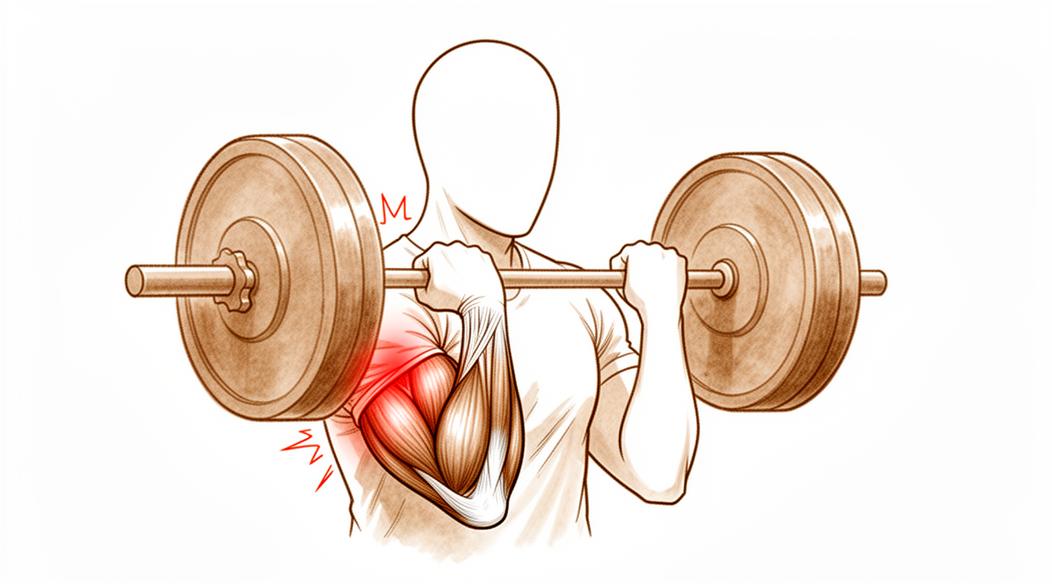
Ang kalamnan ng iyong biceps ay nagtatapos sa iyong siko bilang isang makapal at matibay na tendon. Isipin ang tendon na ito bilang isang lubid na may mataas na kapasidad na nag-uugnay sa kalamnan ng iyong itaas na braso sa buto ng iyong babaeng braso. Ang pangunahing tungkulin nito ay tumulong sa pagbaluktot ng iyong siko at pag-ikot ng palad pataas. Kapag naputol o naputol ang tendon na ito, nababasag ang ugnayang iyon. Hindi na maaaring mahigpit na hilahin ng kalamnan ang buto, kaya nararamdaman mo ang biglaang kahinaan at sakit.
Karaniwang nangyayari ang pinsalang ito kapag sinusubukan mong mag-angat ng mabigat na bagay o kumuhin ng nahuhulog na bagay habang tuwid ang iyong braso. Ang puwersa ay nagdudulot ng sobrang stress sa tendon, na nagdudulot ng pagputol nito. Maaaring marinig mo ang isang "pop" o mararamdaman ang matulis na pagputol. Pagkatapos ng pinsala, maaaring magpukpok ang kalamnan sa iyong itaas na braso, na lumilikha ng kitang-kitang bulge. Nangyayari ito dahil hindi na hawak ng tendon ang kalamnan sa kanyang normal na posisyon malapit sa siko.
Ang siko ay isang kumplikadong hinge joint. Umaasa ito sa mga buto, ligamento, at tendon na magtrabaho nang sama-sama para sa katatagan. Kapag nasira ang tendon ng biceps, maaari itong makaapekto sa paraan ng paggalaw ng iyong siko at pagdala ng bigat. Titingnan ng iyong surgeon ang putol upang malaman kung ito ay kumpleto o bahagya. Ang bahagyang putol ay nangangahulugang may ilang hibla pa rin ang nakakabit, habang ang kumpletong putol ay nangangahulugang ganap na nawala ang pagkakabit ng tendon.
Ang pag-aayos ng tendon ay muling nagdadala ng mahalagang ugnayan sa pagitan ng kalamnan at buto. Ang mga makabagong teknik sa operasyon ay nagbibigay-daan sa iyong surgeon na muling ikabit ang tendon nang ligtas sa buto ng babaeng braso. Anuman ang paggamit ng mga absorbable o non-absorbable anchors, ang layunin ay ibalik ang tendon sa kanyang orihinal na lokasyon. Pinapayagan nito ang tissue na gumaling at muling makuha ang lakas.
Ang oras ay mahalaga para sa iyong paggaling. Karamihan sa mga problema pagkatapos ng operasyon ay nagmumula sa paghintong masyado bago mag-operasyon o paggamit ng malaking incision na nakakaabala sa mga nakapaligid na tissue. Kung hihintayin mo ang higit sa 21 araw, maaaring magkaroon ng scar ang tendon sa isang maikli na posisyon, na nagpapatibay sa pag-aayos. Gayunpaman, maaari pa ring magbigay ng mahusay na functional outcomes ang mga delayed repairs. Pipiliin ng iyong surgeon ang pinakamainam na paraan upang bawasan ang mga komplikasyon at tulungan kang bumalik sa mga normal na gawain nang ligtas.
Ano ang maaari naming gawin para dito¶
Maaari kang magsimula sa pagpapahinga ng iyong braso at pag-iwas sa mabibigat na pag-angat o mga galaw ng pag-ikot na nagdudulot ng tensyon sa harap ng iyong siko. Maaaring irekomenda ng iyong surgeon ang isang panahon ng pisikal na terapiya upang matulungan ang pagbawas ng pamamaga at mabagal na pagbalik ng galaw. Ang layunin ay panatilihing flexible ang kasukasuan habang nagpapahinga ang sugatang tendon. Dapat mong bigyan ng patas na pagkakataon ang konservatibong pamamaraan na ito bago isaalang-alang ang mas invasive na mga hakbang.
Kung mararamdaman mo ang sakit, maaaring imungkahi ng iyong surgeon ang mga over-the-counter na gamot pang-alis ng sakit o anti-inflammatory na gamot upang matulungan kang pamahalaan ang hindi komportableng pakiramdam. Ang mga gamot na ito ay hindi nagpapagaling sa tendon, ngunit maaari nilang gawing mas matibay ang mga araw-araw na gawain habang nagre-recover ka. Sa ilang kaso, maaaring gamitin ang mga injeksyon tulad ng cortisone upang paitiisin ang pamamaga. Gayunpaman, ang mga treatment na ito ay hindi nag-aayos ng sarili ng napunit na tendon. Ang kanilang epekto ay pansamantala, at hindi sila solusyon sa pangmatagalan para sa isang kumpletong pagputol.
Ang operasyon ay itinuturing kapag ang non-surgical na pag-aalaga ay hindi nakakapagpahinga sa iyong mga sintomas o kapag kailangan mong muling makuha ang buong lakas para sa trabaho o sports. Usapin ng iyong surgeon ang pinakamainam na oras para sa pag-aayos, na binabanggit na ang mga resulta ay napakahusay kahit na maantala ang pag-aayos ng higit sa 21 araw. Ang operasyon ay kinabibilangan ng pag-attach muli ng napunit na tendon sa buto upang muling ibalik ang lakas ng iyong braso. Ang pamamaraang ito ay nagbibigay ng consistently magagandang resulta sa mga resulta na sinusukat ng pasyente at lakas. Para sa mga partial na pagputol na hindi nakakabuti sa pagpapahinga, ang operasyon ay isang viable na opsyon din. Sa bihirang mga kaso kung saan hindi maiaattach muli nang direkta ang tendon, maaaring gumamit ang iyong surgeon ng graft upang mabuo muli ang lugar. Ang kabuuang rate ng seryosong komplikasyon mula sa operasyon ay mababa, anuman ang ginamit na teknika.
Ano ang inaasahan¶
Maaari kang mag-expect ng mahusay na pangmatagalang resulta pagkatapos ng operasyon upang ayusin ang puting tendon ng biceps. Anuman kung ang iyong surgeon ay gumamit ng mga absorbable na turnilyo na natutunaw o nonabsorbable na turnilyo na nananatili sa lugar, ang iyong mga functional outcome sa higit sa 1 taon ay mahusay. Karamihan sa mga pasyente ay nagsasabi ng patuloy na magagandang resulta batay sa kanilang pakiramdam at pagganap sa pang-araw-araw na buhay. Kahit na mayroon kang partial na tear na hindi gumaling sa pamamagitan ng pahinga, ang surgical repair ay nananatiling isang viable na option pagkatapos na mabigo ang nonsurgical treatment.
Ang timing ay may malaking papel sa iyong karanasan sa paggaling. Kung maghihintay ka ng higit sa 21 araw bago makuha ang repair, maaari pa ring mag-expect ng mga functional outcome na katulad ng mga naiproseso agad. Gayunpaman, ang delayed repair ay may kaakibat na mataas na rate ng mga unang komplikasyon. Karamihan sa mga isyu mula sa operasyon ay pangunahing naa-attribute sa delay na ito sa timing. Ito ay sekundaryong nauugnay din sa pangangailangan para sa malawak na exposure ng harap ng braso sa panahon ng proseso. Kung ang iyong tear ay chronic o recurrent, ang reconstruction na gumagamit ng donor tissue (allograft) ay ligtas at epektibo. Ang pamamaraang ito ay nagdudulot ng mahusay na patient-reported outcomes at range of motion na walang malalaking komplikasyon sa mga pinag-aralan na grupo.
Ang mga seryosong komplikasyon ay bihira. Ang major complication rate pagkatapos ng distal biceps tendon repair ay 4.6%. Sa kabuuan, ang isang sa 20 na pasyente ay magkakaroon ng major complication, at ang isang sa limang pasyente ay magkakaroon ng minor complication. Ang nerve injury ang pinakakaraniwang isyung maaari mong harapin. Sa kabila ng mga risk na ito, ang kabuuang rate ng seryosong komplikasyon ay nananatiling mababa anuman ang surgical approach. Ang antas ng pagsasanay ng iyong surgeon ay hindi malaki ang pagbabago sa mga profile ng risk na ito kumpara sa malalaking pag-aaral.
Kung pipili kang hindi magkaroon ng operasyon, o kung ang tear ay pamamahalaan nang conservative, ang mga outcome ay karaniwang mas mahina kaysa sa surgical repair. Ang operasyon ang nagbibigay ng pinakamagandang pagkakataon para sa pagpapanumbalik ng lakas at function. Bagama't ang mga chronic repair ay maaaring may bahagyang mas mataas na agad na rate ng komplikasyon kaysa sa mga acute, ang mga long-term functional result ay katulad. Dapat mong inaasahan ang isang panahon ng pag-aadjust. Ang mga unang linggo ay maaaring magdulot ng discomfort at limitadong galaw, ngunit ang layunin ay ang pagbabalik sa normal na paggamit. Karamihan sa morbidity ay nagmumula sa mga delay o malawak na surgical exposure, kaya ang pagsunod sa payo ng iyong surgeon tungkol sa timing at aftercare ay mahalaga para sa pinakamagandang posibleng outcome.
Kailan kumonsulta sa doktor¶
Kumonsulta sa iyong doktor kung mayroon kang patuloy na sakit na hindi gumagaling kahit magpahinga. Humingi ng pagsusuri ng espesyalista kung napapansin mo ang kahinaan o kawalan ng katatagan sa iyong braso. Humingi ng tulong kung ang iyong siko ay nakakabit o biglang bumabagsak. Kontakin ang iyong doktor kung ang mga sintomas ay nakakaapekto sa iyong tulog o trabaho. Humingi ng tulong para sa anumang biglaang paglala ng sakit. Maaaring magpahiwatig ang mga senyales na ito ng pagputol ng tendon ng distal biceps. Inirerekomenda ang maagang interbensyon kung nararamdaman mo ang puwang sa kalamnan o mayroon kang malaking kahinaan. Ang pagkaantala sa pagkukumpuni ay maaaring magpalaki ng panganib ng komplikasyon. Susuriin ka ng iyong surgeon upang matukoy ang pinakamainam na hakbang.
Evidence & references
Overview¶
- Clinical and functional outcomes at more than 1 year after distal biceps tendon repair are excellent for patients treated with bioabsorbable screws [1].
- Clinical and functional outcomes at more than 1 year after distal biceps tendon repair are excellent for patients treated with nonabsorbable screws [1].
- Selective disruption of the short head of the biceps distal tendon may be effectively treated with anatomic repair when diagnosed appropriately [2].
- Patients have excellent overall outcomes after distal biceps tendon rupture repair [3].
- Functional outcomes for chronic distal biceps ruptures are comparable to those seen in acute distal biceps ruptures, despite a potentially slightly higher immediate complication rate [4].
- Patients treated with delayed distal biceps tendon repair (>21 days) can expect similar functional outcomes to those treated acutely, despite a high rate of initial complications [5].
- Most morbidity from repair of the distal biceps tendon is attributed primarily to a delay in the timing of the repair [6].
- Most morbidity from repair of the distal biceps tendon is attributed secondarily to an extensive anterior exposure [6].
- Bioabsorbable interference screw fixation of distal biceps ruptures through a single anterior incision is a safe and successful technique [7].
- Surgical repair of distal biceps ruptures provides consistently good results in terms of patient-scored outcomes [8].
- The surgical repair of distal biceps tendon ruptures has an overall low rate of serious complications, regardless of approach or technique [9].
- Surgical treatment of partial distal biceps tendon tears is a viable option after failed nonsurgical treatment [11].
- Utilizing a double-incision technique with a combination of cortical button and interference screw fixation helps determine the best method of fixation and approach to minimize postoperative complications in patients with distal biceps ruptures [14].
- Two-incision distal biceps repairs demonstrate a high degree of patient satisfaction [21].
- Two-incision distal biceps repairs demonstrate a low complication rate [21].
Anatomy & Pathophysiology¶
- The elbow's stability and complex kinematics are accounted for by a combination of bony articulation and soft-tissue stabilizers [29].
- Musculoskeletal ultrasonography provides a dynamic, functional assessment of elbow structures, allowing visualization of pathology under stress and motion [30].
- The medial elbow joint space increases under a valgus load [31].
- The medial elbow joint space decreases when a maximal grip contraction is performed [31].
- A low carrying angle is measured in elite tennis players just before ball impact during the forehand, suggesting a dynamic varus instant accommodation moving towards full extension [32].
- The decrease in the carrying angle is a consequence of an increase in elbow flexion position dictated by the transition from a closed to open, semi-open stances [32].
- Determining which structures need to be repaired is important to avoid complications that could lead to elbow instability [33].
- Ulnar collateral ligament reconstruction using a suspension button fixation technique reliably restored elbow kinematics to the intact state [34].
- Limiting the number of innings pitched is likely the best way to reduce elbow injury in youth pitchers because biomechanical variables correlated with peak valgus torque are not easily modifiable [35].
- The medial elbow joint space was significantly reduced under 60-N valgus stress plus 50% MVC compared to 60-N valgus stress alone [37].
- No significant relationships between adaptations in shoulder strength or ROM were related to chronic structural adaptations of the elbow in professional baseball pitchers [38].
- The spin move is a simple maneuver that can improve exposure of the coronoid process regardless of the degree of elbow instability [39].
- Reattachment of the flexor and extensor tendons at the epicondyle should be considered in the development of improved repair techniques for stabilizers of the elbow [40].
- An internal joint stabilizer with a standardized treatment protocol could maintain concentric reduction while allowing early functional motion and improve clinical outcomes for patients with complex persistent elbow instability [41].
- Activation of lateral forearm muscles decreased elbow varus angulation throughout flexion [42].
- In patients treated with surgery for snapping triceps syndrome, it is crucial to make sure full resolution of the snapping by examining all dislocating structures during passive elbow motion and/or myoelectrical stimulation to achieve excellent results [43].
- Pre-operative evaluations in elbow stiffness should identify involved articular and periarticular tissues and determine whether articular surfaces and osteoarticular congruence are preserved [44].
- Elbow vascularized composite allotransplantation may be technically feasible on the basis of its consistent vascular anatomy and a proposed surgical technique [45].
- Elbow valgus stress might correlate with ball velocity at 75% partial valgus stress pitch [46].
- Physical examination of the elbow is a critical component in formulating an accurate diagnosis [47].
Classification¶
- Distal biceps tendon tears in women present differently than in men [10].
- The incidence of women sustaining a distal biceps tendon tear is 3.2% [12].
- Partial tears are statistically more common than complete ruptures in women [12].
- Partial ruptures of the distal biceps brachii tendon represent a spectrum of patterns with varying involvement of the long head (LH) and short head (SH) tendons [16].
- Selective disruption of the short head of the biceps distal tendon may be effectively treated with anatomic repair when diagnosed appropriately [2].
Clinical Presentation¶
- Distal biceps tendon tears in women present differently than in men [10].
- The incidence of women sustaining a distal biceps tendon tear is 3.2% [12].
- Partial tears are statistically more common than complete ruptures in women [12].
- Isolated rupture of the short head of the biceps brachii is a rare injury [15].
- Early intervention for isolated short head rupture is recommended if a sulcus or gap in the musculotendinous unit is palpable or considerable weakness is present [15].
- Partial ruptures of the distal biceps brachii tendon represent a spectrum of patterns with varying involvement of the long head (LH) and short head (SH) tendons [16].
- The Hook test has imperfect validity for diagnosing distal biceps tendon ruptures and should not be used alone [17].
- The prevalence of distal biceps tendon signal changes on MRI in asymptomatic patients is very low [18].
- Patients with a smaller radioulnar window have a higher risk of rupturing the distal biceps tendon [19].
Investigations¶
- The Hook test has imperfect validity and should not be used alone to diagnose distal biceps tendon ruptures [17].
- The prevalence of distal biceps tendon signal changes on MRI in asymptomatic patients is very low [18].
- Patients with a smaller radioulnar window have a higher risk of rupturing the distal biceps tendon [19].
- Distal biceps tendon tears in women present differently than in men [10].
- Isolated rupture of the short head of the biceps brachii is a rare injury [15].
- Early intervention is recommended for isolated rupture of the short head of the biceps brachii if a sulcus or gap in the musculotendinous unit is palpable or considerable weakness is present [15].
Treatment¶
Operative Repair¶
- Clinical and functional outcomes at more than 1 year after distal biceps tendon repair are excellent for both bioabsorbable and nonabsorbable screw fixation groups [1].
- Selective disruption of the short head of the biceps distal tendon may be effectively treated with anatomic repair when diagnosed appropriately [2].
- Patients have excellent overall outcomes after distal biceps tendon rupture repair [3].
- Functional outcomes in chronic distal biceps rupture repairs are comparable to those in acute distal biceps rupture populations, despite a potentially slightly higher immediate complication rate [4].
- Patients treated with distal biceps tendon repair after a delay of more than 21 days can expect similar functional outcomes to those treated acutely, despite a high rate of initial complications [5].
- Most morbidity from repair of the distal biceps tendon is attributed primarily to a delay in the timing of the repair and secondarily to an extensive anterior exposure [6].
- Bioabsorbable interference screw fixation of distal biceps ruptures through a single anterior incision is a safe and successful technique [7].
- Surgical repair of distal biceps ruptures provides consistently good results in terms of patient-scored outcomes [8].
- The surgical repair of distal biceps tendon ruptures has an overall low rate of serious complications, regardless of approach or technique [9].
- Surgical treatment of partial distal biceps tendon tears is a viable option after failed nonsurgical treatment [11].
- Reinsertion of the distal biceps tendon on the brachialis tendon is a safe and effective procedure with a low complication rate, providing satisfactory subjective and objective results at long-term follow-up [13].
- Utilizing a double-incision technique with a combination of cortical button and interference screw fixation helps determine the best method of fixation and approach with the goal of minimizing postoperative complications [14].
- Distal biceps reconstruction using an Achilles tendon allograft, transosseous EndoButton, and Pulvertaft weave with tendon wrap technique is recommended for patients with retracted, irreparable distal biceps ruptures [26].
- A two-incision repair with suture anchor attachment is a safe and effective method for treatment of distal biceps tendon ruptures [36].
- Repair of distal biceps ruptures using an Endobutton fixation results in a nearly normal return of strength and function, which is significantly better than in those managed nonoperatively [50].
Non-Operative Management¶
- Incomplete distal triceps tears with active elbow extension against resistance are managed nonsurgically [52].
Complications¶
- Distal biceps tendon repair has an overall low rate of serious complications, regardless of approach or technique [9].
- The major complication rate after distal biceps tendon repair is 4.6% [23].
- One in five patients will have a minor complication after surgery on the distal biceps tendon [24].
- One in twenty patients will have a major complication after surgery on the distal biceps tendon [24].
- Nerve injury is the most common complication after distal biceps tendon repair [22].
- Most morbidity from repair of the distal biceps tendon is attributed primarily to a delay in the timing of the repair [6].
- Most morbidity from repair of the distal biceps tendon is attributed secondarily to an extensive anterior exposure [6].
- Chronic distal biceps rupture repair may have a slightly higher immediate complication rate compared to acute repair [4].
- Delayed repair of distal biceps tendon ruptures (>21 days) is associated with a high rate of initial complications [5].
- Allograft reconstruction of distal biceps injuries for chronic and recurrent tears results in no significant complications among the studied cohort [20].
- Distal biceps repairs using the two-incision technique demonstrate a low complication rate [21].
- Reinsertion of the distal biceps tendon on the brachialis tendon is associated with a low complication rate [13].
- Complication rates after distal biceps tendon repair performed by newly trained surgeons are similar to those previously reported in large cohort studies [22].
Recovery¶
- Clinical and functional outcomes at more than 1 year after distal biceps tendon repair are excellent for patients treated with bioabsorbable screws [1].
- Clinical and functional outcomes at more than 1 year after distal biceps tendon repair are excellent for patients treated with nonabsorbable screws [1].
- Selective disruption of the short head of the biceps distal tendon may be effectively treated with anatomic repair when diagnosed appropriately [2].
- Patients have excellent overall outcomes after distal biceps tendon rupture repair [3].
- Functional outcomes for chronic distal biceps ruptures are comparable to those seen in acute distal biceps ruptures, despite a potentially slightly higher immediate complication rate [4].
- Patients treated with distal biceps tendon repair after a delay of more than 21 days can expect similar functional outcomes to those treated acutely, despite a high rate of initial complications [5].
- Most morbidity from repair of the distal biceps tendon is attributed primarily to a delay in the timing of the repair [6].
- Most morbidity from repair of the distal biceps tendon is attributed secondarily to an extensive anterior exposure [6].
- Surgical repair of distal biceps ruptures provides consistently good results in terms of patient-scored outcomes [8].
- Surgical treatment of partial distal biceps tendon tears is a viable option after failed nonsurgical treatment [11].
- Reinsertion of the distal biceps tendon on the brachialis tendon is a safe and effective procedure with a low complication rate, providing satisfactory subjective and objective results at long-term follow-up [13].
- Allograft reconstruction of distal biceps injuries for chronic and recurrent tears results in excellent patient-reported outcomes and range of motion [20].
- Allograft reconstruction of distal biceps injuries for chronic and recurrent tears results in no significant complications among the studied cohort [20].
- Complication rates after distal biceps tendon repair performed by newly trained surgeons are similar to those previously reported in large cohort studies [22].
- Nerve injury is the most common complication after distal biceps tendon repair performed by newly trained surgeons [22].
- One in 5 patients will have a minor complication after surgery on the distal biceps tendon [24].
- One in 20 patients will have a major complication after surgery on the distal biceps tendon [24].
- Patients with biceps tendon repair etiology had a trend toward greater motion improvement than that of patients with a traumatic etiology regarding anconeus interposition for proximal radioulnar synostosis [25].
- Nearly all complete distal biceps ruptures can be effectively reapproximated to the radial tuberosity within the first 6 weeks of injury using single-incision repair with suture anchors [28].
- Primary repair of chronic distal biceps tendon tears greater than 6 weeks from injury demonstrated excellent patient-reported outcome measures (PROMs) and elbow range of motion (ROM) [48].
- Delayed reconstruction of irreparable distal biceps tendon ruptures with semitendinosus autograft produces similar strength, range of motion, and complication rates compared with delayed primary repair [55].
- Delayed reconstruction of irreparable distal biceps tendon ruptures with semitendinosus autograft produces slightly worse functional outcome scores compared with delayed primary repair [55].
Key Evidence¶
- [L3] Clinical and functional outcome at more than 1 year after distal biceps tendon repair was excellent in both groups. [1] (10.1016/j.jse.2015.12.007)
- [L4] When diagnosed appropriately, selective disruption of the short head of the biceps distal tendon may be effectively treated with anatomic repair. [2] (10.1016/j.jse.2016.09.050)
- [L3] Overall, patients have excellent outcome after distal biceps tendon rupture repair. [3] (10.1007/s00402-018-3018-6)
- [L4] Although there may be a slightly higher immediate complication rate, the functional outcomes remain comparable with those seen in the patient population with acute distal biceps. [4] (10.1177/23259671211065772)
- [L3] Despite a high rate of initial complications, patients treated with distal biceps tendon repair after a delay (>21 days) can expect similar functional outcomes to those treated acutely. [5] (10.1016/j.jse.2017.02.025)
- [L4] Most morbidity from repair of the distal biceps tendon can be attributed primarily to a delay in the timing of the repair and secondarily to an extensive anterior exposure. [6] (10.2106/00004623-200011000-00010)
- [Paper] This is a safe and successful technique for the management of distal biceps tendon ruptures. [7] (10.1007/s00402-009-0974-x)
- [L4] Surgical repair of distal biceps ruptures provides consistently good results in terms of patient-scored outcomes. [8] (10.1016/j.injury.2012.10.029)
- [L3] The surgical repair of distal biceps tendon ruptures has an overall low rate of serious complications, regardless of approach or technique. [9] (10.1177/0363546517720200)
- [L4] Distal biceps tendon tears in women present differently than in men. [10] (10.1016/j.jse.2010.01.015)
- [L4] Surgical treatment of partial distal biceps tendon tears is a viable option after failed nonsurgical treatment. [11] (10.1016/j.jhsa.2010.04.024)
- [L4] The incidence of women sustaining a distal biceps tendon tear is 3.2%, with partial tears being statistically more common than complete ruptures. [12] (10.1016/j.jse.2014.02.006)
- [L4] Reinsertion of the distal biceps tendon on the brachialis tendon can be considered a safe and effective procedure with low complication rate, providing satisfactory subjective and objective results at long-term follow-up. [13] (10.1007/s00167-008-0705-9)
- [L4] This will help surgeons determine the best method of fixation and approach with the goal of minimizing postoperative complications in patients with distal biceps ruptures. [14] (10.1177/17585732241312212)
- [L4] Isolated rupture of the short head of the biceps brachii is a rare injury and early intervention is recommended if a sulcus or gap in musculotendinous unit is palpable or considerable weakness is present. [15] (10.1007/s00167-010-1108-2)
- [L4] Partial ruptures of the distal biceps brachii tendon represent a spectrum of patterns with varying involvement of the LH and SH tendons. [16] (10.1016/j.jse.2020.04.021)
- [L2] The authors caution against using the Hook test alone to diagnose distal biceps tendon ruptures due to its imperfect validity. [17] (10.1016/j.jhsa.2023.07.004)
- [L3] The prevalence of distal biceps tendon signal changes on MRI in asymptomatic patients is very low. [18] (10.1016/j.jhsa.2022.01.020)
- [L3] Therefore, patients with a smaller radioulnar window have a higher risk of rupturing the distal biceps tendon. [19] (10.1016/j.jse.2023.09.020)
- [L5] Allograft reconstruction of distal biceps injuries for chronic and recurrent tears results in excellent patient-reported outcomes and range of motion, with no significant complications among our cohort. [20] (10.1016/j.xrrt.2025.100661)
- [L3] In this large cohort of 2-incision distal biceps repairs, we found a high degree of patient satisfaction and a low complication rate. [21] (10.1016/j.jse.2014.12.032)
- [L4] Complication rates after distal biceps tendon repair performed by newly trained surgeons were similar to those previously reported in large cohort studies, with nerve injury as the most common complication. [22] (10.1016/j.jse.2022.09.014)
- [L2] This is the largest analysis of complications after distal biceps repair, indicating a major complication rate of 4.6%. [23] (10.1177/0363546519899933)
- [L4] Patients should be counseled that 1 in 5 patients will have a minor complication and 1 in 20 patients will have a major complication after surgery on the distal biceps tendon. [24] (10.1016/j.jse.2016.02.032)
- [L3] Patients with biceps tendon repair etiology had a trend toward greater motion improvement than that of patients with a traumatic etiology. [25] (10.1016/j.jse.2014.07.011)
- [L4] This technique is recommended for patients who have a retracted, irreparable distal biceps rupture. [26] (10.1016/j.jse.2016.01.014)
- [L4] The study demonstrates that nearly all complete distal biceps ruptures can be effectively reapproximated to the radial tuberosity within the first 6 weeks of injury. [28] (10.1016/j.jse.2006.03.002)
- [L5] This article discusses the basic anatomy of the elbow and the biomechanics of this joint, noting that a combination of bony articulation and soft-tissue stabilizers accounts for the elbow's stability and complex kinematics. [29] (10.1016/j.csm.2004.06.008)
- [L5] Musculoskeletal ultrasonography provides a dynamic, functional assessment of elbow structures, allowing visualization of pathology under stress and motion. [30] (10.5435/jaaos-d-20-00935)
- [L4] Medial elbow joint space increases under a valgus load and then decreases when a maximal grip contraction is performed. [31] (10.1177/0363546518755149)
- [L4] The observed decrease in the carrying angle is a consequence of an increase in elbow flexion position dictated by the transition from a closed to open, semi‐open stances. [32] (10.1002/ksa.12016)
- [L3] It is important to determine which structures need to be repaired to avoid complications that could lead to elbow instability. [33] (10.1016/j.jse.2019.07.006)
- [L5] Ulnar collateral ligament reconstruction using a suspension button fixation technique reliably restored elbow kinematics to the intact state. [34] (10.1177/0363546509350109)
- [L5] Given that the biomechanical variables correlated with peak valgus torque are not easily modifiable, limiting the number of innings pitched is likely the best way to reduce elbow injury in youth pitchers. [35] (10.1016/j.jse.2004.01.013)
- [L4] Based on our data, we believe that a two-incision repair with suture anchor attachment is a safe and effective method for treatment of distal biceps tendon ruptures. [36] (10.1007/s001670050134)
- [L5] The medial elbow joint space was significantly reduced under 60-N valgus stress plus 50% MVC compared to 60-N valgus stress alone. [37] (10.1016/j.jse.2022.03.027)
- [L3] However, no significant relationships between adaptations in shoulder strength or ROM were related to chronic structural adaptations of the elbow. [38] (10.1177/03635465251317509)
- [L5] The spin move is a simple maneuver that can improve exposure of the coronoid process regardless of the degree of elbow instability. [39] (10.1016/j.jse.2022.11.020)
- [L5] Thus, it should be considered in the development of improved repair techniques for stabilizers of the elbow. [40] (10.1186/s12891-018-2341-y)
- [L4] An internal joint stabilizer with a standardized treatment protocol could maintain concentric reduction while allowing early functional motion and improve clinical outcomes for patients with complex persistent elbow instability. [41] (10.1097/corr.0000000000002159)
- [L5] In addition, activation of lateral forearm muscles decreased elbow varus angulation throughout flexion. [42] (10.1016/j.jhsg.2024.11.006)
- [L4] In patients treated with surgery, it is crucial to make sure full resolution of the snapping by examining all dislocating structures during passive elbow motion and/or myoelectrical stimulation to achieve excellent results. [43] (10.1016/j.xrrt.2025.08.017)
- [L5] Pre-operative evaluations in elbow stiffness should identify involved articular and periarticular tissues and determine whether articular surfaces and osteoarticular congruence are preserved. [44] (10.1016/j.jisako.2023.10.009)
- [L5] Elbow VCA may be technically feasible on the basis of its consistent vascular anatomy and our proposed surgical technique. [45] (10.1016/j.jse.2017.04.014)
- [L5] Elbow valgus stress might correlate with ball velocity at 75% partial valgus stress pitch. [46] (10.1016/j.jse.2022.08.009)
- [L5] Physical examination of the elbow is a critical component in formulating an accurate diagnosis. [47] (10.5435/jaaos-d-16-00622)
- [L4] Primary repair of chronic distal biceps tendon tears greater than 6 weeks from injury demonstrated excellent PROMs and elbow ROM. [48] (10.1177/15589447221107691)
- [L3] Repair of distal biceps ruptures using an Endobutton fixation results in nearly normal return of strength and function, which is significantly better than in those managed nonoperatively. [50] (10.1016/j.jse.2015.10.008)
- [L5] Incomplete tears with active elbow extension against resistance are managed nonsurgically. [52] (10.5435/00124635-201001000-00005)
- [L3] Delayed reconstruction of irreparable distal biceps tendon ruptures with semitendinosus autograft produces similar strength, range of motion, and complication rates but slightly worse functional outcome scores compared with delayed primary repair. [55] (10.1016/j.jse.2019.01.006)
References¶
[1] Distal biceps tendon repair: comparison of clinical and radiological outcome between bioabsorbable and nonabsorbable screws. Journal of Shoulder and Elbow Surgery. 2016. DOI: 10.1016/j.jse.2015.12.007 [2] Rupture of the short head component of a bifurcated distal biceps tendon. Journal of Shoulder and Elbow Surgery. 2017. DOI: 10.1016/j.jse.2016.09.050 [3] Functional outcome in patients who underwent distal biceps tendon repair. Archives of Orthopaedic and Trauma Surgery. 2018. DOI: 10.1007/s00402-018-3018-6 [4] Surgical Results of Chronic Distal Biceps Ruptures: A Systematic Review. Orthopaedic Journal of Sports Medicine. 2022. DOI: 10.1177/23259671211065772 [5] Delayed repair of distal biceps tendon ruptures is successful: a case-control study. Journal of Shoulder and Elbow Surgery. 2017. DOI: 10.1016/j.jse.2017.02.025 [6] Complications of Repair of the Distal Biceps Tendon with the Modified Two-Incision Technique†. The Journal of Bone and Joint Surgery-American Volume. 2000. DOI: 10.2106/00004623-200011000-00010 [7] Bioabsorbable interference screw fixation of distal biceps ruptures through a single anterior incision: a single-surgeon case series and review of the literature. Archives of Orthopaedic and Trauma Surgery. 2009. DOI: 10.1007/s00402-009-0974-x [8] Distal biceps tendon rupture: Current concepts. Injury. 2013. DOI: 10.1016/j.injury.2012.10.029 [9] Surgical Treatment of Distal Biceps Tendon Ruptures: An Analysis of Complications in 784 Surgical Repairs. The American Journal of Sports Medicine. 2017. DOI: 10.1177/0363546517720200 [10] Distal biceps tendon tears in women. Journal of Shoulder and Elbow Surgery. 2010. DOI: 10.1016/j.jse.2010.01.015 [11] Surgical Treatment of Partial Distal Biceps Tendon Ruptures. The Journal of Hand Surgery. 2010. DOI: 10.1016/j.jhsa.2010.04.024 [12] Re-rupture rate of primarily repaired distal biceps tendon injuries. Journal of Shoulder and Elbow Surgery. 2014. DOI: 10.1016/j.jse.2014.02.006 [13] Surgical repair of the distal biceps brachii tendon: clinical and isokinetic long‐term follow‐up. Knee Surgery, Sports Traumatology, Arthroscopy. 2009. DOI: 10.1007/s00167-008-0705-9 [14] Outcomes of utilizing double-incision technique with combination of cortical button and interference screw fixation for distal biceps rupture: A case series. Shoulder & Elbow. 2025. DOI: 10.1177/17585732241312212 [15] Traumatic isolated closed rupture of the short head of the biceps brachii in military paratrooper. Knee Surgery, Sports Traumatology, Arthroscopy. 2010. DOI: 10.1007/s00167-010-1108-2 [16] Partial rupture of the distal biceps brachii tendon: a magnetic resonance imaging analysis. Journal of Shoulder and Elbow Surgery. 2020. DOI: 10.1016/j.jse.2020.04.021 [17] Reliability and Validity of the Hook Test for Diagnosis of Distal Biceps Tendon Ruptures. The Journal of Hand Surgery. 2023. DOI: 10.1016/j.jhsa.2023.07.004 [18] Evaluation of MRI Signal Changes of the Distal Biceps Tendon in Asymptomatic Patients. The Journal of Hand Surgery. 2022. DOI: 10.1016/j.jhsa.2022.01.020 [19] Smaller radioulnar window is associated with a distal biceps tendon rupture in patients with limited forearm rotation: a 3-dimensional computed tomography comparison study of proximal impingement caused by radial tuberosity hypertrophy—a single-center case series. Journal of Shoulder and Elbow Surgery. 2024. DOI: 10.1016/j.jse.2023.09.020 [20] Allograft reconstruction of distal biceps ruptures: surgical technique and analysis of outcomes in chronic tears and failed repairs. JSES Reviews, Reports, and Techniques. 2026. DOI: 10.1016/j.xrrt.2025.100661 [21] Endobutton versus transosseous suture repair of distal biceps rupture using the two-incision technique: a comparison series. Journal of Shoulder and Elbow Surgery. 2015. DOI: 10.1016/j.jse.2014.12.032 [22] Trends and complications of distal biceps tendon repair among American Board of Orthopaedic Surgery part II oral examination candidates. Journal of Shoulder and Elbow Surgery. 2023. DOI: 10.1016/j.jse.2022.09.014 [23] Complications After Distal Biceps Tendon Repair: A Systematic Review. The American Journal of Sports Medicine. 2020. DOI: 10.1177/0363546519899933 [24] Factors associated with adverse events after distal biceps tendon repair or reconstruction. Journal of Shoulder and Elbow Surgery. 2016. DOI: 10.1016/j.jse.2016.02.032 [25] Outcomes of anconeus interposition for proximal radioulnar synostosis. Journal of Shoulder and Elbow Surgery. 2014. DOI: 10.1016/j.jse.2014.07.011 [26] Distal biceps reconstruction using an Achilles tendon allograft, transosseous EndoButton, and Pulvertaft weave with tendon wrap technique for retracted, irreparable distal biceps ruptures. Journal of Shoulder and Elbow Surgery. 2016. DOI: 10.1016/j.jse.2016.01.014 [28] Single-incision repair of acute distal biceps ruptures by use of suture anchors. Journal of Shoulder and Elbow Surgery. 2007. DOI: 10.1016/j.jse.2006.03.002 [29] Elbow anatomy and structural biomechanics. Clinics in Sports Medicine. 2004. DOI: 10.1016/j.csm.2004.06.008 [30] Ultrasound Examination Techniques for Elbow Injuries in Overhead Athletes. Journal of the American Academy of Orthopaedic Surgeons. 2020. DOI: 10.5435/jaaos-d-20-00935 [31] Medial Elbow Joint Space Increases With Valgus Stress and Decreases When Cued to Perform A Maximal Grip Contraction. The American Journal of Sports Medicine. 2018. DOI: 10.1177/0363546518755149 [32] A low carrying angle is measured in elite tennis players just before ball impact during the forehand, suggesting a dynamic varus instant accommodation moving towards full extension. Knee Surgery, Sports Traumatology, Arthroscopy. 2024. DOI: 10.1002/ksa.12016 [33] Isolated displaced type II partial articular radial head fracture: correlation of preoperative imaging with intraoperative findings of lateral ulnar collateral ligament tear. Journal of Shoulder and Elbow Surgery. 2020. DOI: 10.1016/j.jse.2019.07.006 [34] Revision Ulnar Collateral Ligament Reconstruction Using a Suspension Button Fixation Technique. The American Journal of Sports Medicine. 2009. DOI: 10.1177/0363546509350109 [35] Valgus torque in youth baseball pitchers: a biomechanical study. Journal of Shoulder and Elbow Surgery. 2004. DOI: 10.1016/j.jse.2004.01.013 [36] Repair of distal biceps tendon rupture with suture anchors. Knee Surgery, Sports Traumatology, Arthroscopy. 1999. DOI: 10.1007/s001670050134 [37] Changes in medial elbow joint space with differences in contraction strength of flexor-pronator muscle under elbow valgus stress. Journal of Shoulder and Elbow Surgery. 2022. DOI: 10.1016/j.jse.2022.03.027 [38] Chronic Structural Adaptations of the Shoulder and Elbow Are Correlated in Professional Baseball Pitchers. The American Journal of Sports Medicine. 2025. DOI: 10.1177/03635465251317509 [39] The spin move to facilitate antegrade coronoid fixation in terrible triad injuries. Journal of Shoulder and Elbow Surgery. 2023. DOI: 10.1016/j.jse.2022.11.020 [40] Reattachment of the flexor and extensor tendons at the epicondyle in elbow instability: a biomechanical comparison of techniques. BMC Musculoskeletal Disorders. 2018. DOI: 10.1186/s12891-018-2341-y [41] Does an Internal Joint Stabilizer and Standardized Protocol Prevent Recurrent Instability in Complex Persistent Elbow Instability?. Clinical Orthopaedics & Related Research. 2022. DOI: 10.1097/corr.0000000000002159 [42] The Effect of Arm Abduction and Forearm Muscle Activation on Kinematics During Elbow Flexion. Journal of Hand Surgery Global Online. 2025. DOI: 10.1016/j.jhsg.2024.11.006 [43] Snapping triceps syndrome: a review of the literature and proposed operative treatment algorithm. JSES Reviews, Reports, and Techniques. 2026. DOI: 10.1016/j.xrrt.2025.08.017 [44] Elbow stiffness: Arthritis and heterotopic ossification. Journal of ISAKOS. 2024. DOI: 10.1016/j.jisako.2023.10.009 [45] Elbow vascularized composite allotransplantation—surgical anatomy and technique. Journal of Shoulder and Elbow Surgery. 2017. DOI: 10.1016/j.jse.2017.04.014 [46] Effect of quantitative partial valgus stress during baseball pitching on ball velocity and subjective pitch-effort. Journal of Shoulder and Elbow Surgery. 2023. DOI: 10.1016/j.jse.2022.08.009 [47] Comprehensive Review of the Elbow Physical Examination. Journal of the American Academy of Orthopaedic Surgeons. 2018. DOI: 10.5435/jaaos-d-16-00622 [48] Primary Repair of Chronic Distal Biceps Tendon Tears. HAND. 2022. DOI: 10.1177/15589447221107691 [50] A comparison of nonoperative vs. Endobutton repair of distal biceps ruptures. Journal of Shoulder and Elbow Surgery. 2016. DOI: 10.1016/j.jse.2015.10.008 [52] Distal Triceps Rupture. American Academy of Orthopaedic Surgeon. 2010. DOI: 10.5435/00124635-201001000-00005 [55] Management of chronic distal biceps tendon ruptures: primary repair vs. semitendinosus autograft reconstruction. Journal of Shoulder and Elbow Surgery*. 2019. DOI: 10.1016/j.jse.2019.01.006