Patients › Shoulder
Calcific Tendinitis
Calcific tendinitis causes shoulder pain from calcium deposits; treatment ranges from observation to washing out the calcium.
Ang iyong nararamdaman
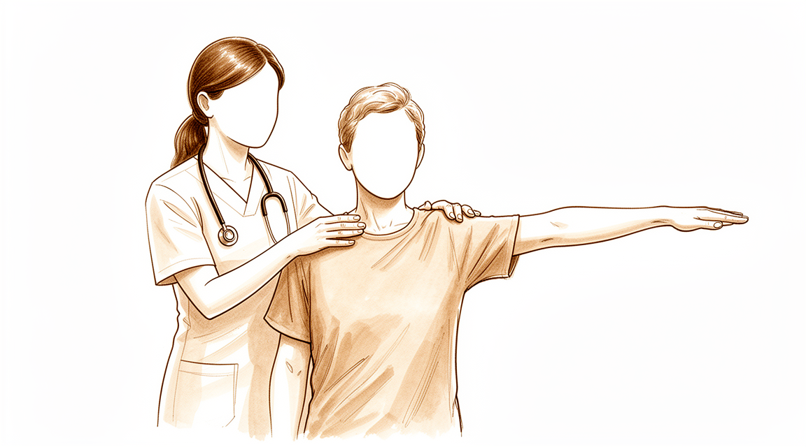
Ang calcific tendinitis ay nangangahulugang may maliit na naipong calcium sa loob ng isa sa mga tendon ng iyong rotator cuff, ang grupo ng mga tendon na humahawak at nagpapagalaw sa iyong balikat. Ang sakit ay karaniwang nasa gilid o ibabaw ng iyong balikat, sa bahagi ng supraspinatus tendon, na siyang tendon na kadalasang apektado. Karaniwang lumalala ito kapag iniaangat mo ang iyong braso palabas sa gilid o lampas sa ulo, at ang pag-abot sa mataas na istante o pagsasampay ng labada ay maaaring maging mahirap.
Madalas lumala ang sakit sa gabi, lalo na kung nakahiga ka sa balikat na iyon, at maaari ka nitong gisingin. Maaari rin itong maramdaman pagkatapos ng aktibidad o sa unang paggising sa umaga. Napapansin ng ilang tao na dumarating ang sakit nang pana-panahon, na may mas tahimik na mga panahon sa pagitan, at maaari itong tumagal nang ilang buwan. Ang ganoong katagal na hindi komportableng pakiramdam ay maaaring mangahulugan ng pagliban sa trabaho at maaaring magpababa sa kalidad ng iyong buhay.
Pinakakaraniwan ang kondisyong ito sa mga kababaihang may edad sa pagitan ng 30 at 60, bagaman nakaaapekto rin ito sa iba. Ang deposito mismo ay karaniwang nasa pagitan ng 0.5 at 1.5 cm ang haba, at karamihan ay siksik sa halip na parang bulak ang itsura. Sa humigit-kumulang 9% ng mga tao, higit sa isang tendon ang apektado, na maaaring magpakalat ng sakit sa paligid ng balikat.
Isang bagay na mabuting malaman: ang deposito ng calcium na nakita sa scan ay hindi nangangahulugang walang punit sa parehong tendon. Kung hindi humuhupa ang iyong sakit, lumalala ito sa loob ng ilang linggo, ginigising ka nito sa gabi, o pinipigilan ka nitong magtrabaho o gamitin ang iyong braso, magpatingin sa iyong GP o humingi ng pagsusuri ng isang espesyalista.
Ano ang aktwal na nangyayari
Ang iyong rotator cuff ay isang grupo ng apat na tendon na humahawak at nagpapagalaw sa iyong balikat. Dumudulas ang mga ito sa isang makitid na espasyo sa ilalim ng isang maliit na arko ng buto sa itaas ng balikat, na may manipis na unan na may lamang likido sa pagitan ng mga ito. Isipin ang tendon bilang isang lubid na dumadaan sa isang masikip na pulley.
Sa calcific tendinitis, nagbabago ang isang bahagi ng lubid na iyon. Dahan-dahang nagiging mga cell na kahawig ng cartilage ang mga cell sa loob ng tendon, at nagsisimulang maipon ang calcium sa nabagong tissue. Nangyayari ito nang payugto-yugto: una ay nagbabago ang tendon, pagkatapos ay nabubuo ang calcium at nananatili roon, at sa huli ay sinisira ng katawan ang deposito at sinisipsip ito. Isa itong aktibong proseso, hindi simpleng pagkaluma dahil sa gamit (wear and tear), at iba ito sa pag-ipon ng calcium na maaaring makita sa isang mas matanda at pudpod nang tendon sa ibang bahagi ng katawan.
Mahalaga ang yugto dahil iba-iba ang pakiramdam ng bawat isa. Habang tahimik na nananatili ang calcium sa tendon, maaaring kaunti o walang sakit kang nararamdaman. Kapag sinimulan na itong sipsipin ng katawan, madalas na nagiging matindi at biglaan ang sakit, at maaari itong pinakamalala sa gabi. Maaari ring magdulot ng problema ang deposito dahil sa laki nito: habang namamaga ito sa loob ng masikip na espasyo, maaari itong sumiksik laban sa arko ng buto sa itaas, na humaharang sa maayos na paggalaw ng braso. Kaya naman ang pag-angat ng iyong braso palabas sa gilid ay maaaring maramdamang matigas o parang may humaharang.
Ang deposito ay kadalasang nasa supraspinatus tendon, mga 1.5 hanggang 2 cm mula sa kung saan ito kumakapit sa ibabaw ng buto ng braso. Ipinapaliwanag ng lokasyong iyon kung bakit doon nararamdaman ang sakit, at kung bakit ang pag-abot lampas sa ulo ang pinakamahirap na galaw.
Isa pang bagay na mabuting malaman: maraming tao ang may ganitong mga deposito nang walang anumang sakit, at humigit-kumulang isang katlo lamang ng mga deposito ang nagdudulot ng mga sintomas. Kaya ang calcium sa iyong scan ay isang tunay na natuklasan, ngunit kung ito nga ang sanhi ng iyong sakit ay nakadepende sa yugto nito at sa kung paano tumutugon ang iyong katawan dito.
Ano ang maaari naming gawin tungkol dito
Si Dr Kieran Hirpara, isang upper-limb surgeon sa Mater Private Hospital Rockhampton, ay nagsisimula sa mga opsyon na hindi gaanong invasive na angkop sa iyong kondisyon. Ang mga pasyente ay karaniwang nirerefer sa aming klinika ng kanilang GP; kung iminungkahi ng isang physiotherapist na magpatingin sa amin, kakailanganin mo pa rin ng referral mula sa iyong GP upang maging eligible para sa Medicare rebate. Sa iyong unang pagbisita, kumukuha kami ng history, sinusuri ang iyong balikat, at nagsasaayos ng imaging kung saan ito kinakailangan. Para sa matagal nang problemang tulad nito, karaniwan muna naming sinusubukan ang non-operative na pangangalaga at isinasaalang-alang ang operasyon kapag hindi ito nagbigay ng sapat na pagbuti.
Simple ang unang hakbang: baguhin ang mga aktibidad na nagpapalala ng sakit, at ipahinga ang iyong balikat mula sa mga gawaing lampas sa ulo. Layunin ng physiotherapy na pahupain ang sakit at ibalik ang maayos at kontroladong paggalaw ng braso. Bigyan ito ng sapat na pagkakataon bago ito husgahan, dahil ang kondisyong ito ay maaaring tumagal nang ilang buwan. Kung hindi humuhupa ang iyong mga sintomas, lumalala ang mga ito sa loob ng ilang linggo, ginigising ka ng mga ito sa gabi, o pinipigilan ka nitong magtrabaho o gamitin ang iyong braso, magpatingin sa iyong GP o humingi ng pagsusuri ng isang espesyalista.
Ang mga anti-inflammatory na gamot ay karaniwang unang gamutan para sa sakit. Kung hindi sapat ang mga ito, maaari kaming mag-alok ng mga procedure na direktang tumutugon sa deposito. Ang isang opsyon ay shock wave therapy, kung saan itinututok ang mga sound wave sa calcium mula sa labas ng katawan upang madurog ang deposito. Ito ay noninvasive, ibig sabihin ay walang tumutusok sa balat, at ginagawa ito sa nakatakdang lakas at bilang ng pulse upang matanggal ang calcium. Ang isa pang opsyon ay ultrasound-guided needling, kung saan inilalagay ang isang karayom sa deposito sa tulong ng ultrasound, isang scan na nagpapakita ng live na larawan ng tendon, upang durugin at higupin palabas ang calcium. Ang parehong pamamaraan ay maaaring magpaginhawa ng sakit, magbalik ng function at mag-alis ng calcium. Maaaring ulitin ang needling kung nagpapatuloy ang mga sintomas, na may hindi bababa sa anim na linggo sa pagitan ng mga procedure.
Isinasaalang-alang ang operasyon kapag hindi gumana ang mga gamutang ito. Kinapapalooban ito ng pagtanggal ng deposito ng calcium mula sa tendon, at pagkumpuni rin sa tendon kung ito ay labis na apektado. Kung hindi humupa ang iyong sakit pagkatapos ng tunay na pagsubok sa mga opsyon sa itaas, pag-uusapan natin kung makatuwiran ang operasyon para sa iyo at magdedesisyon tayo nang magkasama.
Ano ang dapat asahan
Sa karamihan ng kaso, maganda ang pananaw para sa calcific tendinitis. Maraming tao ang may ganitong mga deposito nang hindi nila nalalaman kailanman, at humigit-kumulang isang katlo lamang ng mga ito ang nagdudulot ng sakit. Kapag nagdudulot nga ng mga sintomas ang deposito, malaki ang posibilidad na tuluyan itong mawala sa pangmatagalan. Kayang sipsipin ng iyong katawan ang calcium nang mag-isa, at nagsisimula na ang prosesong iyon mula pa sa sandaling mabuo ang deposito.
Gayunpaman, hindi palaging mabilis ang daan patungo roon. Maaaring tumagal nang ilang buwan ang mga sintomas, at ang ganoong katagal na hindi komportableng pakiramdam ay maaaring mangahulugan ng pagliban sa trabaho at tunay na pagbaba ng kalidad ng iyong buhay. Madalas dumating ang sakit nang pana-panahon, na may mas tahimik na mga panahon sa pagitan, at maaari itong maging matindi at biglaan kapag sinimulan ng iyong katawan na sirain ang deposito. Ang ilang deposito ay humuhupa sa loob ng ilang buwan, ang iba ay mas matagal, at walang maaasahang paraan upang mahulaan nang eksakto kung paano kikilos ang sa iyo.
Karamihan sa mga tao ay gumagaling nang maayos nang walang operasyon. Ang conservative care, ibig sabihin ay gamutang hindi kinapapalooban ng operasyon, ang karaniwang unang pagpipilian, at nagdudulot ito ng makabuluhang pagbuti sa karamihan ng tao: 72% ng mga taong ginamot sa ganitong paraan ang nag-uulat ng mabuti o napakahusay na resulta, anuman ang laki o posisyon ng deposito. Ang shock wave therapy at ultrasound-guided needling ay parehong nagpapaginhawa ng sakit at nag-aalis ng calcium sa maraming tao, at pareho silang may matibay na rekord ng kaligtasan. Mas mahusay na gumagana ang needling kapag mas maikli pa ang panahon ng mga sintomas at mas maliit ang deposito. Kung hindi angkop ang isa sa mga procedure na ito sa iyong partikular na deposito, pag-uusapan namin kasama mo ang iba pang opsyon.
Ang operasyon ay inilalaan para sa mga taong hindi humupa ang sakit pagkatapos ng tunay na pagsubok sa mga gamutang ito. Tinatanggal nito ang deposito ng calcium mula sa tendon, at nagbibigay ito ng mabuting resulta sa 88.5% ng mga taong ang sakit ay hindi tumugon sa ibang gamutan. Unti-unti ang paggaling pagkatapos ng operasyon: dahan-dahang bumubuti ang function ng balikat sa unang anim na buwan, at karamihan sa pagbuti sa ginhawa at paggalaw ay nabubuo sa panahong iyon sa halip na dumating nang sabay-sabay.
Kung hindi humuhupa ang iyong mga sintomas, lumalala ang mga ito sa loob ng ilang linggo, ginigising ka ng mga ito sa gabi, o pinipigilan ka nitong magtrabaho o gamitin ang iyong braso, magpatingin sa iyong GP o humingi ng pagsusuri ng isang espesyalista.
Kailan dapat magpatingin
Karamihan sa mga deposito ng calcium ay kusang humuhupa, at marami ang hindi nagdudulot ng anumang problema. Ngunit ang kondisyong ito ay maaaring tumagal nang ilang buwan, at maaari kang mawalan ng oras sa trabaho at bumaba ang kalidad ng iyong buhay. Magpatingin sa iyong GP o humingi ng pagsusuri ng isang espesyalista kung hindi humuhupa ang pananakit ng iyong balikat, lumalala ito sa loob ng ilang linggo, ginigising ka nito sa gabi, o pinipigilan ka nitong magtrabaho o gamitin ang iyong braso. Banggitin din ito kung parang may humaharang o sumasabit kapag iniaangat mo ang iyong braso palabas sa gilid, dahil ang namamagang deposito ay maaaring pisikal na pumigil sa paggalaw ng tendon sa ilalim ng arko ng buto sa itaas nito. Isa pang bagay na mabuting banggitin: ang deposito ng calcium sa iyong scan ay hindi nangangahulugang walang punit sa parehong tendon, kaya hindi sapat ang natuklasan sa scan lamang upang matiyak ang diagnosis. Kung hindi mo makontak ang klinika sa labas ng oras ng opisina o sa weekend, pumunta sa pinakamalapit na emergency department.
Higit pang detalye
Advanced reading: the deeper science (optional)
Ang seksyong ito ay higit pa sa kailangan mo para sa iyong sariling mga desisyon sa paggamot. Ang calcific tendinitis ay karapat-dapat sa karagdagang pagbabasa dahil kumikilos ito nang naiiba sa karamihan ng mga kondisyon sa balikat: ang deposito ay karaniwan sa mga taong walang nararamdamang sakit, karaniwang tinutunaw ito ng katawan nang walang tulong, at ang paggamot na nag-aalis ng calcium ay hindi ang paggamot na pinakamabilis na nagpapaginhawa sa sakit.
Ang deposit ay isang karaniwang natuklasan, hindi awtomatikong isang diagnosis
Ang isang calcium deposit na naiulat sa iyong scan ay madaling basahin bilang sanhi ng problema, dahil ito ay nakikita at ang sakit ay totoo. Pinakomplikado ito ng mga prevalence data. Sa mga 1,219 na matatanda, ang mga deposit ay naroon sa 7.8% ng mga taong walang sintomas at 42.5% ng mga may subacromial pain syndrome [1]. Sa isang hiwalay na serye ng 302 na mga balikat, ang mga deposit ay madalas sa pangkalahatang populasyon at mga isang katlo lamang ang masakit [2].
Kaya ang deposit ay nagpapataas ng probabilidad na ito ang pinagmumulan, nang malaki, ngunit humigit-kumulang isa sa labintatlong walang sakit na mga balikat ay mayroon din nito. Ang tila naglilipat sa isang deposit mula sa pagiging silent patungo sa symptomatic ay bahagyang ang laki at posisyon: ang pinakamataas na posibilidad ng tunay na symptomatic disease ay nasa mga kababaihang may edad 30 hanggang 60 na may subacromial pain at may deposit na mas mahaba sa 1.5 cm [1], at ang sakit ay may kinalaman sa lokasyon sa supraspinatus at sa pagkakasangkot ng higit sa isang tendon [2].
Karaniwan itong natutunaw, at iyon ang humuhubog sa lahat
Ang natural na takbo ay patungo sa resorption, kung kaya't maraming paggamot ang tila gumagana. Ang pinakamalinaw na halimbawa ay nagmula sa isang randomised trial kung saan ang bawat pasyente ay sumailalim sa needling at lavage at pagkatapos ay na-randomise sa isang steroid o saline injection: sa loob ng labindalawang buwan, ang calcification ay na-resorb sa 83% ng saline group at 74% ng steroid group [3].
Basahin iyan nang mabuti, dahil naglalaman ito ng counter-intuitive na resulta. Ang steroid ay nagpahusay sa sakit sa loob ng anim na linggo at sa function sa loob ng tatlong buwan, at walang makabuluhang epekto kung nawala ba ang calcium [3]. Ang pagkawala ng sakit at ang paglinis ng calcium ay magkahiwalay na proseso. Ang isang paggamot ay maaaring magbigay ng isa nang wala ang isa, at ang nakikitang bagay sa scan ay hindi ang bagay na nagpapatakbo ng mga sintomas linggo-linggo.
Kaya naman ang mga non-operative na opsyon ay may magkakatulad na resulta
Kung ang deposit ay malaki nang nawawala anuman ang mangyari, ang mga gamutan ay naglalayong gawing tolerable ang interval sa halip na gamutin ito nang lubos. Iyan ang ipinapakita ng mga paghahambing. Sa 257 na pasyente, ang physical therapy, corticosteroid injection at ultrasound-guided barbotage ay nagresulta sa magkakatulad na rate ng pag-iwas sa operasyon [4]. Sa 239 na pasyente, ang parehong tatlong approach ay naging matagumpay sa malaking bahagi, kung saan ang physiotherapy lamang ang may pinakamataas na failure rate [5].
Sa mga procedural na opsyon, ang high-energy shockwave therapy ang pinaka-masusing pinag-aralang minimally invasive treatment at napatunayang ligtas at epektibo sa short to medium term, habang ang ultrasound-guided needling ay hindi napatunayang superior sa isang ultrasound-guided subacromial injection [6]. Sa pagsasama-sama ng 1,258 na pasyente, ang shockwave, needling at arthroscopy ay lahat nagbigay ng mabuting clinical outcomes [7], at ang barbotage sa 908 na pasyente ay ligtas na may mataas na success rate ngunit hindi pa kailanman naihambing nang head-to-head laban sa iba pang pangunahing opsyon [8].
Ano ang idinaragdag ng operasyon, na nakasaad nang tumpak
Ang operasyon ay hindi walang bentahe, at ang laki nito ay mas mahalagang banggitin kaysa ilarawan. Sa pagsasama-sama ng 2,352 na pasyente mula sa mga randomised trial, ang surgical treatment ay nagresulta sa mas malaking pagbuti sa functional scores at katulad na pagbawas ng sakit kumpara sa non-operative treatment, partikular na ang ultrasound-guided needling, kung saan ang parehong pamamaraan ay nakamit ang clinically significant improvement [9]. Sa pagitan ng mga surgical technique mismo, walang makabuluhang pagkakaiba, at ang pagtanggal lamang ng deposit ay nagpakita ng katulad na resulta sa pagtanggal nito kasama ang subacromial decompression [10].
Ang pagkakaiba ng function-versus-pain ang kapaki-pakinabang na detalye. Kung sakit ang pangunahing reklamo, ang ebidensya ay hindi malinaw na pumapabor sa operasyon. Kung paninigas at pagkawala ng function ang nangingibabaw at nagpatuloy, mas pumapabor ito rito.
Ang asosasyon na madalas nakakaligtaan
Ang calcific tendinitis ay hindi pantay ang distribusyon. Sa 102 na pasyente, ang mga may kaugnay na endocrine disorder, pangunahin na ang sakit sa thyroid at diabetes, ay nagkaroon ng mga sintomas sa mas batang edad, nagkaroon ng signipikanteng mas matagal na kurso, at mas madalas na nangailangan ng operasyon [11].
Karapat-dapat itong banggitin sa iyong GP kung ang iyong kurso ay naging hindi pangkaraniwang haba o nagsimula nang maaga, hindi dahil ang paggamot sa endocrine condition ay nakakalutas sa balikat, kundi dahil binabago nito kung ano ang itsura ng isang makatotohanang timeline. Ang pagsasabing ang isang kondisyon ay self-limiting ay mahirap itugma sa dalawang taon ng pananakit; ang endocrine association ay isang paliwanag kung bakit ang karaniwang pagpapanatag ng loob ay hindi angkop sa bawat kaso.
Mga Sanggunian
[1] Louwerens JK, Sierevelt IN, van Hove RP, van den Bekerom MP, van Noort A. Prevalence of calcific deposits within the rotator cuff tendons in adults with and without subacromial pain syndrome: clinical and radiologic analysis of 1219 patients. J Shoulder Elbow Surg. 2015;24(10):1588-93. https://doi.org/10.1016/j.jse.2015.02.024
[2] Sansone V, Consonni O, Maiorano E, Meroni R, Goddi A. Calcific tendinopathy of the rotator cuff: the correlation between pain and imaging features in symptomatic and asymptomatic female shoulders. Skeletal Radiol. 2015;45(1):49-55. https://doi.org/10.1007/s00256-015-2240-3
[3] Darrieutort-Laffite C, Varin S, Coiffier G, Albert J, Planche L, Maugars Y, et al. Are corticosteroid injections needed after needling and lavage of calcific tendinitis? Randomised, double-blind, non-inferiority trial. Ann Rheum Dis. 2019;78(6):837-43. https://doi.org/10.1136/annrheumdis-2018-214971
[4] Gilbert R, Dadoo S, Lin R, Bhardwaj N, McMahon S, Steuer F, et al. Comparison of physical therapy, corticosteroid injections, and ultrasound-guided barbotage for nonoperative management of calcific tendinitis. Orthop J Sports Med. 2026;14(4). https://doi.org/10.1177/23259671261434919
[5] Drummond M, Ayinon C, Lin A, Dunn R. Relative efficacy of three nonsurgical treatments for calcific tendinitis: physical therapy vs steroid injection vs barbotage. Orthop J Sports Med. 2021;9(7_suppl4). https://doi.org/10.1177/2325967121S00210
[6] Louwerens JK, Sierevelt IN, van Noort A, van den Bekerom MP. Evidence for minimally invasive therapies in the management of chronic calcific tendinopathy of the rotator cuff: a systematic review and meta-analysis. J Shoulder Elbow Surg. 2014;23(8):1240-9. https://doi.org/10.1016/j.jse.2014.02.002
[7] Louwerens JK, Veltman ES, van Noort A, van den Bekerom MP. The effectiveness of high-energy extracorporeal shockwave therapy versus ultrasound-guided needling versus arthroscopic surgery in the management of chronic calcific rotator cuff tendinopathy: a systematic review. Arthroscopy. 2015;32(1):165-75. https://doi.org/10.1016/j.arthro.2015.06.049
[8] Gatt DL, Charalambous CP. Ultrasound-guided barbotage for calcific tendonitis of the shoulder: a systematic review including 908 patients. Arthroscopy. 2014;30(9):1166-72. https://doi.org/10.1016/j.arthro.2014.03.013
[9] Angileri HS, Gohal C, Comeau-Gauthier M, Owen MM, Shanmugaraj A, Terry MA, et al. Chronic calcific tendonitis of the rotator cuff: a systematic review and meta-analysis of randomized controlled trials comparing operative and nonoperative interventions. J Shoulder Elbow Surg. 2023;32(8):1746-60. https://doi.org/10.1016/j.jse.2023.03.017
[10] Anam E, Zahran S, Roy A, Daneshvar P, Bicknell RT, Janssen I. Surgical approaches of shoulder calcific tendonitis: a systematic review and meta-analysis. JSES Rev Rep Tech. 2024;4(3):353-8. https://doi.org/10.1016/j.xrrt.2024.03.013
[11] Harvie P, Pollard TC, Carr AJ. Calcific tendinitis: natural history and association with endocrine disorders. J Shoulder Elbow Surg. 2007;16(2):169-73. https://doi.org/10.1016/j.jse.2006.06.007
Evidence & references
This is the clinical evidence summary written for health professionals. It is technical, and it lists the research this page was built from. You do not need to read it to understand your treatment or to make a decision about it.
Overview
Epidemiology and Natural History
- Calcific tendon deposits of the shoulder are a frequent occurrence in the general population [2].
- Only one third of calcific tendon deposits in the shoulder are painful [2].
- Symptomatic calcific tendinitis of the shoulder has a good likelihood to completely resolve in the long-term [3].
Non-Operative Treatment
- The primary choice of treatment for calcific tendinitis is conservative, especially in patients with acute calcific tendinitis [26].
- Ultrasound treatment helps resolve calcifications and is associated with short-term clinical improvement in patients with symptomatic calcific tendinitis of the shoulder [1].
- Conservative treatment for calcific tendinitis of the shoulder showed clinically significant improvement, with 72% of excellent or good results regardless of the location, radiologic type and size, and initial symptoms of calcific deposits [5].
- Extracorporeal shock wave therapy (ESWT) has produced a high rate of success in pain relief and functional restoration with negligible associated complications [11].
- Shock wave therapy is a safe and effective noninvasive treatment for patients with calcific tendinitis of the shoulder, producing a high rate of success in pain relief and functional restoration with negligible complications [19].
- ESWT can be considered as an alternative treatment for chronic calcific tendinitis of the shoulder due to its good tolerance, safety, and clinical radiological response [25].
- Both ultrasound-guided needling and extracorporeal shock wave therapy improved clinical outcomes and eliminated calcium deposits in the treatment of calcific tendinitis [6].
- Utilization of barbotage as a treatment for calcific tendonitis of the shoulder appears to produce notable pain reduction in the short term (specifically at the 2-month follow-up), but begins to lose some efficacy over long-term evaluation [27].
- Patients with calcific tendinitis of the shoulder who have the factors identified for a poor outcome after ESWT should undergo a different procedure [42].
Operative Treatment
- Surgical treatment of calcific tendinitis gives good results in 88.5% of patients resistant to medical treatment [12].
- The aim of arthroscopic treatment in calcific tendinitis is to remove the calcific deposit [15].
- Arthroscopic removal of calcification leads to improved clinical outcomes in patients with chronic calcific tendinitis, but at least 6 months of follow-up is needed for these improvements to become statistically significant [13].
- Patients undergoing arthroscopic treatment of a calcific deposit in the shoulder had satisfactory clinical and radiological outcomes at the final follow-up, with functional scores improving slowly and reaching more than 75 percent at six months after surgery [7].
- Arthroscopic treatment of calcifying tendinitis provides good to excellent clinical results [20].
- Arthroscopic treatment of chronically painful calcific tendinitis of the rotator cuff promises to be successful in more than 90% of the patients if conservative treatment fails [37].
- Arthroscopic removal of calcific deposits of the shoulder shows good clinical results for pain reduction, shoulder function and patient satisfaction [44].
- Endoscopic and open surgery are equally effective in the treatment of chronic calcifying tendinopathy, showing similar clinical and sonographic results [34].
- Preoperative ultrasound-guided marking of calcific deposits is a procedure that statistically significantly improves the clinical results of arthroscopic surgery as seen at 6 weeks and 2 years [33].
- The short-term functional outcome of patients with calcific tendonitis after arthroscopic bursectomy and debridement of the calcific deposit is not influenced if performed in combination with or without a subacromial decompression [40].
- Routine diagnostic glenohumeral exploration does not appear beneficial in arthroscopic treatment of calcific tendinitis due to the low prevalence of intraarticular pathologies which most frequently do not require surgical treatment [23].
Prognostic Factors
- Patients with calcific lesions >1 cm had a 2.8 increased likelihood to undergo operative treatment in the setting of calcific tendinitis of the shoulder [4].
Anatomy & Pathophysiology
Epidemiology & Demographics
- Calcific tendon deposits of the shoulder are a frequent occurrence in the general population, although only one third are painful [2].
- In approximately 80% of patients, calcifying tendinitis occurs in the supraspinatus tendon [115].
- Most patients with calcifying tendinitis are 30-50 years old [115].
- Women are approximately 1.5 times more often affected by calcifying tendinitis than men [115].
- The incidence of calcifying tendinitis in the general population is 2.5-20% [115].
- In about 10% of individuals, calcific deposits are found bilaterally [17].
- In 20% of cases, subjects with rotator cuff calcific tendinopathy are asymptomatic [17].
- Subjects with rotator cuff calcific tendinopathy were often female aged between 30 and 60 [17].
- Demographic, radiographic, and clinical features of calcific tendinitis of the shoulder in the Korean population were not different from those of Western populations [14].
Anatomical Location & Structures
- Calcific deposits are most commonly located at the level of the supraspinatus tendon [17].
- In calcifying tendinitis, deposits are typically located 1.5-2 cm from the tendinous insertion at the greater tuberosity [115].
- The greater tuberosity serves as the attachment site for the supraspinatus, infraspinatus, and teres minor tendons of the rotator cuff [70].
- The rotator cuff consists of four muscles: the subscapularis, supraspinatus, infraspinatus, and teres minor [71].
- The acromion, the coracoacromial ligament, and the coracoid process form the coracoacromial arch [70].
- The rotator cuff, subacromial bursa, and subdeltoid bursa pass underneath the coracoacromial arch [70].
- The subacromial bursa separates the rotator cuff tendons from the coracoacromial arch and allows them to glide [78].
- The infraspinatus and teres minor fuse near their musculotendinous junctions [83].
- The tendons of the infraspinatus and supraspinatus muscles join approximately 15 mm proximal to their insertion [83].
Pathogenesis & Staging
- The pathogenesis of calcific tendinitis remains unclear, and different theories have been proposed [30].
- Calcific tendinitis of the rotator cuff tendons is thought to have a different pathophysiologic mechanism than calcific tendinitis at other sites, such as the Achilles or patellar tendons [30].
- Uhthoff and Loehr described an active, cell-mediated process that is now widely accepted for the pathogenesis of calcific tendinitis [30].
- Uhthoff and Loehr described cartilage metaplasia as a reactionary process in an active biologic environment [30].
- The reactive calcification theory proposed by Uthoff in 1997 consists of a three-stages process: pre-calcific, calcific, and post-calcific stage [17].
- The precalcific stage consists of predominantly fibrocartilaginous metaplasia presumably within less vascular areas of the tendon [30].
- During the pre-calcific stage, tenocytes undergo a metaplastic transformation into chondrocytes within the site where the calcium deposits will subsequently form [17].
- The calcific stage is subdivided into three separate phases: formative, resting, and resorptive [30].
- In the formative phase of the calcific stage, matrix vesicles unite to become calcific deposits that are separated by fibrocartilage [30].
- The resting phase is characterized by the presence of calcium deposits in the rotator cuff [17].
- The resorptive phase is associated with acute, sudden onset of extremely severe pain [49].
- Calcific deposits consist of two different forms of hydroxyapatite: A type and B type [30].
- The composition of an individual calcific deposit can have a mixture of the two types of hydroxyapatite [30].
- Each individual phase of calcific tendinitis has a different composition of hydroxyapatite [30].
- Calcium is deposited in the fibrocartilaginous matrix of the tendon as calcium carbonate apatite [49].
- Endocrine and metabolic disorders were often described as comorbidity in subjects with rotator cuff calcific tendinopathy [17].
- Endocrine disorders probably play an important role in the development of rotator cuff calcific tendinopathy, but the effect of this process is still unknown [17].
- Shorter length of the infraspinatus tendon and specific subacromial bursa anatomy may explain the intramuscular migration of calcium deposits leading to the lesion [10].
Clinical Presentation & Pain Mechanisms
- Subjects with rotator cuff calcific tendinopathy often reported nightly, acute, unilateral and severe pain with spontaneous onset [17].
- Subjects with rotator cuff calcific tendinopathy often reported deficit in active and passive range of motion, mainly in abduction and flexion [17].
- Calcific tendinitis of the supraspinatus does not typically cause loss of external rotation but is frequently associated with mild isolated restriction of abduction [22].
- Patients with calcific tendinitis had less passive abduction, forward flexion, internal rotation, and external rotation than the control group [65].
- The main mechanism of abduction contracture lies in the mechanical blocking between the bulged calcified deposits and the coracoacromial arch [45].
- Abduction contracture is presumably caused by mechanical blocking by the calcified deposits and the coracoacromial arch and by reflex muscle spasm caused by pain [125].
- Patients with calcific tendinitis had significantly greater nerve counts compared with the rotator cuff repair group and the control group [65].
- A greater nerve count positively correlated with a greater degree of neovascularization and also positively correlated with greater frequency of extreme pain [65].
- Neovascularization positively correlated with more frequent pain during sleep and more frequent extreme pain [65].
- The pathophysiology of calcific tendinitis remains largely unknown, with proposed theories including degenerative calcification, repetitive trauma, necrosis of tenocytes and intracellular calcium accumulation, reactive calcification, endochondral ossification, and chondral metaplasia [65].
- Calcific tendinitis of the rotator cuff with tuberosity osteolysis is a distinctive form of calcific tendinitis that should be considered in clinical and surgical practice [28].
- A rotator cuff tear was the only factor affecting the complete recovery of shoulder function in patients treated for calcific tendinitis [113].
Classification
- The supraspinatus is the most frequently affected tendon in rotator cuff calcific tendinopathy, followed by the infraspinatus and subscapularis [38].
- In 9% of patients with rotator cuff calcific deposits, two or more tendons are affected [38].
- The median length of calcific deposits in the rotator cuff is 1.16 cm [38].
- 55.8% of calcific deposits in the rotator cuff have a length between 0.5 and 1.5 cm [38].
- 28.6% of calcific deposits in the rotator cuff have a length greater than 1.5 cm [38].
- Gartner type II morphology is the most common radiographic classification for calcific deposits, present in 47.0% of cases [38].
- Gartner type I morphology is present in 38.4% of calcific deposits [38].
- Gartner type III morphology is present in 14.6% of calcific deposits [38].
- Approximately 76% of calcifications in the shoulder are characterized as dense (type A or B) using the Mole et al. classification scheme [39].
- Approximately 24% of calcifications in the shoulder are characterized as fluffy (type C) using the Mole et al. classification scheme [39].
- Type A calcific deposits are dense, rounded, and sharply delineated [67].
- Type B calcific deposits are multilobular in appearance, radiodense, and sharply outlined [67].
- Type C calcific deposits are more radiolucent and heterogeneous with irregular outlines [67].
- Type D calcific deposits are dystrophic calcific lesions of the tendon insertion [67].
- The pathogenesis of calcific tendinitis involves a three-stage process: precalcific, calcific, and postcalcific [30].
- The calcific stage is subdivided into three phases: formative, resting, and resorptive [30].
- The precalcific stage consists of predominantly fibrocartilaginous metaplasia within less vascular areas of the tendon [30].
- In the formative phase of the calcific stage, matrix vesicles unite to become calcific deposits separated by fibrocartilage [30].
- The composition of an individual calcific deposit can have a mixture of A type and B type hydroxyapatite [30].
- Disease evolution in calcific tendinitis can be classified into four stages: precalcific, calcific, resorptive, and postcalcific [107].
- The resorptive phase of calcific tendinitis is the most painful stage owing to vascular invasion, increased phagocytic cells, and edema from intratendinous pressure [107].
Clinical Presentation
- Subjects complaining of rotator cuff calcific tendinopathy were often female aged between 30 and 60 [17].
- Calcific tendinitis is a poorly understood condition in which symptoms can be protracted, resulting in time off work and impaired quality of life [8].
- The calcium deposits are most commonly located at the level of the supraspinatus tendon [17].
- The supraspinatus was the most frequently affected tendon in 82.7% of patients with calcific deposits, followed by the subscapularis tendon in 8.9% and the infraspinatus in 8.4% [38].
- Two or more tendons were affected in 9% of patients with calcific deposits [38].
- The median length of the calcific deposit was 1.16 cm, with 55.8% ranging from 0.5 to 1.5 cm and 28.6% greater than 1.5 cm [38].
- Approximately 76% of calcifications were characterized as dense (type A or B) and 24% as fluffy (type C) [39].
- Recognition of atypical presentations of calcific tendinitis with bone erosion may prevent unnecessary biopsy and overtreatment [16].
- The presence of a calcium deposit in the rotator cuff of a patient with painful shoulder does not rule out the possibility of a coexisting rotator cuff tear [60].
- Calcific tendinitis in the elbow of a child has not been reported previously [24].
- Atypical calcific tendinitis involving teres minor can affect overhead movement [9].
- Calcific tendinitis of the trapezius insertion is a recognized presentation [18].
- Of twelve histologically verified cases of calcific periarthritis adjacent to joints other than the shoulder, in only one patient was the preoperative clinical diagnosis accurate [59].
Investigations
Plain Radiography
- Standardized plain films are almost always sufficient to garner the information needed for shoulder imaging [47].
- The purpose of imaging the shoulder is to help establish the diagnosis, determine the severity of the pathoanatomy, assist in surgical planning, and enable the surgeon to illustrate the condition of the shoulder to the patient [47].
- At least two X-ray views should be obtained for shoulder imaging: an anteroposterior in the plane of the glenoid and an axillary projection with the arm in abduction [87].
- The axillary view is taken with the arm in the functional position of elevation in the plane of the scapula and oriented so that both the spinoglenoid notch and the scapular neck are visible [47].
- The axillary view demonstrates glenohumeral relationships in the functional position of elevation, which is referred to as the "truth view" [47].
- CT scans may offer a few degrees of increased precision in the measurement of glenoid version, but this precision does not improve the quality of the surgery or the clinical outcome [47].
- The temptation to "overimage" should be resisted, obtaining only the scans or reconstructions that are necessary for the care of the patient [47].
- Calcific tendinitis can be observed on plain radiograph [94].
- Calcific deposits in the fingers may not be seen on standard views and oblique views may be necessary [111].
Ultrasonography
- Ultrasound is a simple and accurate test for identifying rotator cuff tears and calcific tendinitis [87].
- Ultrasound can be useful in guiding injections or barbotage (aspirating calcific deposits in the rotator cuff) [87].
- Ultrasonography is a low-cost alternative to MRI and arthrography for evaluating both skeletal and soft-tissue structures of the shoulder [94].
- Ultrasonography can provide immediate, real-time visualization of the rotator cuff, biceps tendon, and calcific deposits [94].
- Ultrasonography can be used to measure the subacromial space and detect atrophy of rotator cuff muscles [94].
- Ultrasonography can evaluate impingement in various positions and motions due to real-time imaging [94].
- Ultrasonography is highly operator dependent and is not as useful for evaluating labral tears or rotator cuff tears that are very small or larger than 3 cm [94].
- The accuracy of rotator cuff ultrasonography depends on the skill of the scanner operator and an awareness of pitfalls that are encountered [85].
- US elastography is recommended for all patients with calcific tendinosis, except for those with the arc morphological type, before making management decisions [31].
- The surgeon performing ultrasound of the shoulder can diagnose intraosseous calcifying tendinitis [56].
- Calcific tendinitis of the supraspinatus is frequently associated with mild isolated restriction of abduction but does not typically cause loss of external rotation [22].
Magnetic Resonance Imaging
- MRI is the modality of choice for evaluating the rotator cuff, biceps, and subacromial/subdeltoid bursa [94].
- T2-weighted MRI provides better visualization of full thickness rotator cuff tears [94].
- MRI can identify labral tears and rotator cuff tears, although the accuracy for these is enhanced by combining the scan with arthrography [87].
- Shoulder surgeons should be aware of the accuracy limitations of sonographic or MRI evaluation regarding rotator cuff tears as a comorbidity in calcific tendinitis [32].
- The incidence of rotator cuff tears in cases of calcific tendonitis is higher than previously reported in cohorts undergoing MRI [41].
Computed Tomography
- CT imaging is frequently used to evaluate fractures of the shoulder, to assess for bony lesions in recurrent instability cases, or for preoperative templating for shoulder arthritis [94].
- CT is helpful for planning fracture surgery and shoulder joint replacement [87].
Diagnostic Findings and Pathology
- The paper illustrates the dynamic pathological process of calcific tendinopathy, including migration patterns of calcium deposits [29].
Treatment
Non-Operative Management
- Nonoperative treatment consists of physical therapy, therapeutic modalities, and injections [49].
- Anti-inflammatory medications and steroid use are a common first line treatment option [36].
- Extracorporeal shock wave therapy (ESWT) is a safe and effective noninvasive treatment for patients with calcific tendinitis of the shoulder, producing a high rate of success in pain relief and functional restoration with negligible complications [19].
- Treatment of calcific tendinitis of the shoulder with shock waves has produced a high rate of success in pain relief and functional restoration with negligible associated complications [11].
- The effectiveness of ESWT in calcific tendinitis of the shoulder is dose-dependent [43].
- Disintegration of calcific deposits via ESWT requires an energy of at least 0.42 mJ/mm² and 2000 impulses [58].
- Shock wave therapy is a heterogeneous but effective treatment for calcifying tendinitis of the shoulder [53].
- ESWT in calcific tendinitis of the shoulder is very effective [55].
- Both ultrasound-guided needling and extracorporeal shock wave therapy improved clinical outcomes and eliminated calcium deposits in patients with calcific tendinitis [6].
- Ultrasound-guided needling showed a higher decrease in calcium deposits compared to radial shockwave therapy at 6 weeks [104].
- Ultrasound-guided needling showed a significantly higher improvement in the Oxford Score pre- and post-treatment compared to radial shockwave therapy [104].
- After one year, there was no significant difference in NRS and Oxford Score between ultrasound-guided needling and radial shockwave therapy groups [104].
- Patients with calcific tendinitis of the shoulder who have factors identified for a poor outcome after ESWT should undergo a different procedure [42].
- Needle aspiration of calcific deposits (NACD) is safe and effective for calcific tendinitis [35].
- In case of persisting symptoms, patients may be scheduled for another NACD procedure with a minimal term of six weeks between procedures [35].
- Percutaneous needle barbotage of calcium deposits has been demonstrated to provide pain relief and improved symptoms in patients suffering from calcific tendonitis [36].
- Symptoms of calcific tendinitis of the trapezius insertion responded to needle aspiration and injection, with the patient reporting normal shoulder function approximately 1 year after the procedure [18].
Operative Management
- Arthroscopic or open removal of the deposit is occasionally necessary for calcific tendinitis [49].
- In athletes with calcifying tendinitis of the supraspinatus tendon with failed nonoperative treatment, complete arthroscopic removal of calcific deposits and tendon repair without acromioplasty results in significant pain relief and improvement in functional outcomes [102].
- Arthroscopic debridement of calcific tendinitis with intraosseous involvement is a safe and effective treatment method similar to that of pure tendinous involvement [100].
- Complete removal of calcium was achieved in 44 patients (88%) after surgery, with faint specks apparent in 6 (12%) [39].
- Routine exploration of the gleno-humeral joint during arthroscopic removal of rotator cuff calcifications is not advisable as it significantly increases the risk of adhesive capsulitis without identifying a sufficient number of lesions requiring a therapeutic procedure [110].
- The rotator cuff should be repaired if it is significantly involved during treatment for calcific tendinitis [49].
Complications
- Shoulder surgeons should be cautious about rotator cuff tears as a comorbidity in calcific tendinitis and aware of the accuracy limitations of sonographic or MRI evaluation [32].
- The incidence of rotator cuff tears in cases of calcific tendonitis is higher than previously reported in a cohort of patients who underwent MRI [41].
- The incidence and type of intraarticular lesions in calcifying tendinitis are comparable to age-matched shoulders with partial- rather than full-thickness rotator cuff tears [61].
- It is speculated that the main mechanism of abduction contracture lies in the mechanical blocking between the bulged calcified deposits and the coracoacromial arch [45].
- Periarticular calcific deposits are self-limiting and respond to conservative treatment, whereas recurrence and persistent symptoms are not rare in patients with peritendinous calcific deposits in the hand and wrist [21].
Recovery
Natural History and Prognosis
- Symptoms of calcific tendinitis can be protracted, resulting in time off work and impaired quality of life [8].
- The onset of calcium resorption in acute calcific tendinitis occurs most frequently in the summer in Japan [130].
Conservative and Minimally Invasive Management
- Extracorporeal shock wave therapy (ESWT) can be considered as an alternative treatment for chronic calcific tendinitis of the shoulder due to its good tolerance, safety, and clinical radiological response [25].
- Calcific deposits disappeared in the same percentage of patients in both groups of a randomized clinical trial comparing two different energy levels of extracorporeal shock-wave therapy [69].
- Percutaneous needle aspiration and lavage is effective in the short term and in the long term in calcific tendinitis of the shoulder, with results similar to or better than those published for other techniques, and it is only slightly invasive and painful [62].
- There was no significant difference in short- and long-term clinical outcomes between single- and double-needle US-guided percutaneous irrigation of calcific tendinopathy [63].
- A symptom duration of ≤10 months or calcification size of ≤10.82 mm represented the clinical scenarios most likely to show resorption after ESWT [126].
- In SWT studies, Type III calcifications and shorter symptom duration were associated with better outcomes [129].
Operative Management
- Improved Constant scores at 24 months after arthroscopic treatment were inversely related to the number and size of residual calcifications in all patients [67].
Prognostic Factors and Complications
Key Evidence
- [L1] In patients with symptomatic calcific tendinitis of the shoulder, ultrasound treatment helps resolve calcifications and is associated with short-term clinical improvement. [1] (10.1056/nejm199905203402002)
- [L3] Calcific tendon deposits of the shoulder are a frequent occurrence in the general population, although only one third are painful. [2] (10.1007/s00256-015-2240-3)
- [L1] Symptomatic calcific tendinitis of the shoulder has a good likelihood to completely resolve in the long-term. [3] (10.1097/phm.0000000000000939)
- [L3] Patients with calcific lesions >1 cm had a 2.8 increased likelihood to undergo operative treatment in the setting of calcific tendinitis of the shoulder. [4] (10.1016/j.jseint.2021.01.013)
- [L2] Conservative treatment for calcific tendinitis of the shoulder showed clinically significant improvement, with 72% of excellent or good results regardless of the location, radiologic type and size, and initial symptoms of calcific deposits. [5] (10.1016/j.jse.2009.07.008)
- [L2] Both treatment modalities for calcific tendinitis improved clinical outcomes and eliminated calcium deposits. [6] (10.1016/j.jse.2014.06.036)
- [Paper] Patients undergoing arthroscopic treatment of a calcific deposit in the shoulder had satisfactory clinical and radiological outcomes at the final follow-up, with functional scores improving slowly and reaching more than 75 percent at six months after surgery. [7] (10.1016/j.otsr.2020.03.005)
- [L3] Calcific tendinitis is a poorly understood condition in which symptoms can be protracted, resulting in time off work and impaired quality of life. [8] (10.1016/j.jse.2006.06.007)
- [Case_report] [9] (10.1016/j.jisako.2025.101055)
- [L4] Shorter length of the infraspinatus tendon and specific subacromial bursa anatomy may explain the intramuscular migration of calcium deposits leading to the lesion. [10] (10.1016/j.jse.2022.01.092)
- [L2] Treatment of calcific tendinitis of the shoulder with shock waves has produced a high rate of success in pain relief and functional restoration with negligible associated complications. [11] (10.1016/j.jse.2007.03.023)
- [L4] Surgical treatment of calcific tendinitis gives good results in 88.5% of patients resistant to medical treatment. [12] (10.1007/s100670050108)
- [L4] Arthroscopic removal of calcification leads to improved clinical outcomes in patients with chronic calcific tendinitis, but at least 6 months of follow-up is needed for these improvements to become statistically significant. [13] (10.5397/cise.2018.21.2.75)
- [L4] This study reported demographic, radiographic, and clinical features of calcific tendinitis of the shoulder in the Korean population, which were not different from those of Western populations. [14] (10.5397/cise.2020.00010)
- [L4] The aim of arthroscopic treatment in calcific tendinitis is to remove the calcific deposit. [15] (10.1016/s1058-2746(98)90180-x)
- [L5] Recognition of atypical presentations of calcific tendinitis with bone erosion may prevent unnecessary biopsy and overtreatment. [16] (10.1016/j.jse.2009.02.009)
- [L3] [17] (10.1177/17585732241244515)
- [L5] Symptoms responded to needle aspiration and injection, with the patient reporting normal shoulder function approximately 1 year after the procedure. [18] (10.1016/s1058-2746(99)90011-3)
- [L3] Shock wave therapy is a safe and effective noninvasive treatment for patients with calcific tendinitis of the shoulder, producing a high rate of success in pain relief and functional restoration with negligible complications. [19] (10.1177/03635465030310031701)
- [L3] Arthroscopic treatment of calcifying tendinitis provides good to excellent clinical results. [20] (10.1177/03635465211037690)
- [L3] Periarticular calcific deposits are self-limiting and respond to conservative treatment, whereas recurrence and persistent symptoms are not rare in patients with peritendinous calcific deposits in the hand and wrist. [21] (10.1177/1753193413478393)
- [L3] Calcific tendinitis of the supraspinatus does not typically cause loss of external rotation but is frequently associated with mild isolated restriction of abduction. [22] (10.1177/2325967117752907)
- [L3] Routine diagnostic glenohumeral exploration does not appear beneficial in arthroscopic treatment of calcific tendinitis due to the low prevalence of intraarticular pathologies which most frequently do not require surgical treatment. [23] (10.1186/s12891-017-1839-z)
- [L5] Calcific tendinitis in the elbow of a child has not been reported previously. [24] (10.1067/mse.2002.119854)
- [L2] Because of its good tolerance, safety, and clinical radiological response, ESWT can be considered as an alternative treatment for chronic calcific tendinitis of the shoulder. [25] (10.1136/ard.62.3.248)
- [L5] The primary choice of treatment for calcific tendinitis is conservative, especially in patients with acute calcific tendinitis. [26] (10.5397/cise.2020.00318)
- [L4] Utilization of barbotage as a treatment for calcific tendonitis of the shoulder appears to produce notable pain reduction in the short term (specifically at the 2-month follow-up), but begins to lose some efficacy over long-term evaluation. [27] (10.1016/j.jseint.2024.06.005)
- [L2] Imaging and functional data indicate that calcific tendinitis of the rotator cuff with tuberosity osteolysis is a distinctive form of calcific tendinitis that should be considered in clinical and surgical practice. [28] (10.1016/j.jse.2008.09.016)
- [L5] The paper illustrates the dynamic pathological process of calcific tendinopathy, including migration patterns of calcium deposits, and discusses clinical and sonographic assessment to optimize diagnosis, rehabilitation, and interventional management for functional recovery. [29] (10.3390/diagnostics12123097)
- [L5] [30] (10.5435/jaaos-22-11-707)
- [L4] We recommend that US elastography be performed for all patients with calcific tendinosis, except for those with the arc morphological type, before making management decisions. [31] (10.1016/j.jcma.2015.05.006)
- [L3] Shoulder surgeons should be cautious about rotator cuff tears as a comorbidity in calcific tendinitis and aware of the accuracy limitations of sonographic or MRI evaluation. [32] (10.5397/cise.2021.00094)
- [L3] Preoperative ultrasound-guided marking of calcific deposits is a procedure that statistically significantly improves the clinical results of arthroscopic surgery as seen at 6 weeks and 2 years. [33] (10.1016/j.arthro.2006.08.005)
- [L1] Endoscopic and open surgery are equally effective in the treatment of chronic calcifying tendinopathy, showing similar clinical and sonographic results. [34] (10.1097/01.blo.0000063786.32430.22)
- [L4] [35] (10.1016/j.ejrad.2016.01.018)
- [L4] [36] (10.1177/2325967121s00655)
- [L4] This study confirmed that if conservative treatment fails, arthroscopic treatment of chronically painful calcific tendinitis of the rotator cuff promises to be successful in more than 90% of the patients. [37] (10.1016/j.arthro.2006.01.012)
- [L3] [38] (10.1016/j.jse.2015.02.024)
- [L3] [39] (10.1016/j.jse.2010.10.038)
- [L1] This study has demonstrated that the short-term functional outcome of patients with calcific tendonitis after arthroscopic bursectomy and debridement of the calcific deposit is not influenced if performed in combination with or without a subacromial decompression. [40] (10.1016/j.arthro.2015.05.015)
- [L4] The incidence of rotator cuff tears in cases of calcific tendonitis in this cohort of patients who underwent MRI is higher than previously reported. [41] (10.1016/j.arthro.2019.11.127)
- [L3] Patients with calcific tendinitis of the shoulder who have the factors identified for a poor outcome after ESWT should undergo a different procedure. [42] (10.1302/0301-620x.99b12.bjj-2016-1178.r1)
- [L2] The study confirms the effectiveness of ESWT in CT of the shoulder and demonstrates that the results are dose-dependent. [43] (10.1067/mse.2002.126614)
- [L3] Arthroscopic removal of calcific deposits of the shoulder shows good clinical results for pain reduction, shoulder function and patient satisfaction. [44] (10.1007/s00167-008-0507-0)
- [L4] It is speculated that the main mechanism of abduction contracture lies in the mechanical blocking between the bulged calcified deposits and the coracoacromial arch. [45] (10.1016/s1058-2746(96)80285-0)
- [L1] Shock wave therapy is a heterogeneous but effective treatment for calcifying tendinitis of the shoulder. [53] (10.1177/1941738108331197)
- [L1] ESWT in calcific tendinitis of the shoulder is very effective. [55] (10.1007/s00256-004-0849-8)
- [L4] This complex case report illustrates the value of the surgeon performing ultrasound of the shoulder to diagnose an intraosseous calcifying tendinitis. [56] (10.1016/j.xrrt.2023.09.012)
- [L2] [58] (10.1007/s00776-003-0720-0)
- [L4] Of twelve histologically verified cases of calcific periarthritis adjacent to joints other than the shoulder, in only one patient (toe) was the preoperative clinical diagnosis accurate, which signals the need for greater awareness of this entity as a differential diagnostic option. [59] (10.2106/jbjs.k.00874)
- [L4] The presence of a calcium deposit in the rotator cuff of a patient with painful shoulder does not rule out the possibility of a coexisting rotator cuff tear (RCT). [60] (10.1016/s1058-2746(09)80095-5)
- [L3] The incidence and type of intraarticular lesions in calcifying tendinitis are comparable to age-matched shoulders with partial- rather than full-thickness rotator cuff tears. [61] (10.1007/s00402-011-1263-z)
- [L4] Percutaneous needle aspiration and lavage is effective in the short term and in the long term in calcific tendinitis of the shoulder, with results similar to or better than those published for other techniques, and it is only slightly invasive and painful. [62] (10.2214/ajr.07.2254)
- [L1] There was no significant difference in short- and long-term clinical outcomes between single- and double-needle US-guided percutaneous irrigation of calcific tendinopathy. [63] (10.1148/radiol.2017162888)
- [L3] [65] (10.2106/jbjs.o.00417)
- [L4] [67] (10.1016/j.jse.2004.04.001)
- [L2] Calcific deposits disappeared in the same percentage of patients in both groups. [69] (10.2522/ptj.20110252)
- [L3] Arthroscopic debridement of calcific tendinitis with intraosseous involvement is a safe and effective treatment method similar to that of pure tendinous involvement. [100] (10.1007/s00167-022-06870-2)
- [L4] In athletes with calcifying tendinitis of the supraspinatus tendon with failed nonoperative treatment, complete arthroscopic removal of calcific deposits and tendon repair without acromioplasty results in significant pain relief and improvement in functional outcomes. [102] (10.1177/2325967116669310)
- [L2] [104] (10.1016/j.jor.2017.07.011)
- [L1] [107] (10.1148/radiol.11111157)
- [L3] Routine exploration of the gleno-humeral joint during arthroscopic removal of rotator cuff calcifications is not advisable as it significantly increases the risk of adhesive capsulitis without identifying a sufficient number of lesions requiring a therapeutic procedure. [110] (10.1016/j.otsr.2021.102915)
- [L5] The calcification may not be seen on standard views and oblique views may be necessary. [111] (10.1016/0266-7681(85)90036-1)
- [L3] A rotator cuff tear was the only factor affecting the complete recovery of shoulder function. [113] (10.1177/03635465231217733)
- [L2] [115] (10.1177/0269215510396740)
- [L4] The abduction contracture is presumably caused by mechanical blocking by the calcified deposits and the coracoacromial arch and by reflex muscle spasm caused by pain. [125] (10.1016/s1058-2746(97)90074-4)
- [L3] A symptom duration of ≤10 months or calcification size of ≤10.82 mm represented the clinical scenarios most likely to show resorption after ESWT. [126] (10.1177/23259671241231609)
- [L3] In SWT studies, Type III calcifications and shorter symptom duration were associated with better outcomes. [129] (10.1177/17585732251414964)
- [L3] The onset of calcium resorption in acute calcific tendinitis occurs most frequently in the summer in Japan; however, the reasons for seasonal variation remain unclear, and further studies will be needed. [130] (10.1186/s12891-020-03773-6)
References
[1] Ultrasound Therapy for Calcific Tendinitis of the Shoulder. New England Journal of Medicine. 1999. DOI: 10.1056/nejm199905203402002
[2] Calcific tendinopathy of the rotator cuff: the correlation between pain and imaging features in symptomatic and asymptomatic female shoulders. Skeletal Radiology. 2015. DOI: 10.1007/s00256-015-2240-3
[3] Long-Term Course of Shoulders After Ultrasound Therapy for Calcific Tendinitis. American Journal of Physical Medicine & Rehabilitation. 2018. DOI: 10.1097/phm.0000000000000939
[4] Predictive factors for failure of conservative management in the treatment of calcific tendinitis of the shoulder. JSES International. 2021. DOI: 10.1016/j.jseint.2021.01.013
[5] Radiologic course of the calcific deposits in calcific tendinitis of the shoulder: Does the initial radiologic aspect affect the final results?. Journal of Shoulder and Elbow Surgery. 2010. DOI: 10.1016/j.jse.2009.07.008
[6] Which method is more effective in treatment of calcific tendinitis in the shoulder? Prospective randomized comparison between ultrasound-guided needling and extracorporeal shock wave therapy. Journal of Shoulder and Elbow Surgery. 2014. DOI: 10.1016/j.jse.2014.06.036
[7] Recovery pattern after arthroscopic treatment for calcific tendinitis of the shoulder. Orthopaedics & Traumatology: Surgery & Research. 2020. DOI: 10.1016/j.otsr.2020.03.005
[8] Calcific tendinitis: Natural history and association with endocrine disorders. Journal of Shoulder and Elbow Surgery. 2007. DOI: 10.1016/j.jse.2006.06.007
[9] Atypical calcific tendinitis involving teres minor which affects overhead movement: A case report. Journal of ISAKOS. 2026. DOI: 10.1016/j.jisako.2025.101055
[10] Infraspinatus Myotendinous Junction Tears: Analysis Of A Consecutive Series And Their Relationship With Calcifying Tendonitis. Journal of Shoulder and Elbow Surgery. 2022. DOI: 10.1016/j.jse.2022.01.092
[11] Extracorporeal shock wave therapy for calcifying tendinitis of the shoulder. Journal of Shoulder and Elbow Surgery. 2008. DOI: 10.1016/j.jse.2007.03.023
[12] Surgical Management of Calcific Tendinitis of the Shoulder: An Analysis of 26 Cases. Clinical Rheumatology. 1999. DOI: 10.1007/s100670050108
[13] Functional Recovery of the Shoulder after Arthroscopic Treatment for Chronic Calcific Tendinitis. Clinics in Shoulder and Elbow. 2018. DOI: 10.5397/cise.2018.21.2.75
[14] Calcific tendinitis of the shoulder in the Korean population: demographics and its relation with coexisting rotator cuff tear. Clinics in Shoulder and Elbow. 2021. DOI: 10.5397/cise.2020.00010
[15] Arthroscopic treatment of calcific tendinitis of the shoulder. Journal of Shoulder and Elbow Surgery. 1998. DOI: 10.1016/s1058-2746(98)90180-x
[16] Calcific tendinitis of the rotator cuff associated with intraosseous loculation: Two case reports. Journal of Shoulder and Elbow Surgery. 2009. DOI: 10.1016/j.jse.2009.02.009
[17] Clinical features in rotator cuff calcific tendinopathy: A scoping review. Shoulder & Elbow. 2024. DOI: 10.1177/17585732241244515
[18] Calcific tendinitis of the trapezius insertion. Journal of Shoulder and Elbow Surgery. 1999. DOI: 10.1016/s1058-2746(99)90011-3
[19] Shock Wave Therapy for Calcific Tendinitis of the Shoulder. The American Journal of Sports Medicine. 2003. DOI: 10.1177/03635465030310031701
[20] Clinical and Structural Results of Rotator Cuff Repair Compared With Rotator Cuff Debridement in Arthroscopic Treatment of Calcifying Tendinitis of the Shoulder. The American Journal of Sports Medicine. 2021. DOI: 10.1177/03635465211037690
[21] Acute calcium deposits in the hand and wrist; comparison of acute calcium peritendinitis and acute calcium periarthritis. Journal of Hand Surgery (European Volume). 2013. DOI: 10.1177/1753193413478393
[22] Restriction of Passive Glenohumeral Abduction Combined With Normal Passive External Rotation Is a Diagnostic Feature of Calcific Tendinitis. Orthopaedic Journal of Sports Medicine. 2018. DOI: 10.1177/2325967117752907
[23] Examination of concomitant glenohumeral pathologies in patients treated arthroscopically for calcific tendinitis of the shoulder and implications for routine diagnostic joint exploration. BMC Musculoskeletal Disorders. 2017. DOI: 10.1186/s12891-017-1839-z
[24] Calcific tendinitis at the biceps brachii insertion of a child: A case report. Journal of Shoulder and Elbow Surgery. 2002. DOI: 10.1067/mse.2002.119854
[25] Extracorporeal shock wave therapy for chronic calcific tendinitis of the shoulder: single blind study. Annals of the Rheumatic Diseases. 2003. DOI: 10.1136/ard.62.3.248
[26] Diagnosis and treatment of calcific tendinitis of the shoulder. Clinics in Shoulder and Elbow. 2020. DOI: 10.5397/cise.2020.00318
[27] Determining the efficacy of barbotage for pain relief in calcific tendinitis. JSES International. 2024. DOI: 10.1016/j.jseint.2024.06.005
[28] Osteolytic lesion of greater tuberosity in calcific tendinitis of the shoulder. Journal of Shoulder and Elbow Surgery. 2009. DOI: 10.1016/j.jse.2008.09.016
[29] Clinical/Sonographic Assessment and Management of Calcific Tendinopathy of the Shoulder: A Narrative Review. Diagnostics. 2022. DOI: 10.3390/diagnostics12123097
[30] Calcific Tendinitis of the Rotator Cuff. Journal of the American Academy of Orthopaedic Surgeons. 2014. DOI: 10.5435/jaaos-22-11-707
[31] Management of rotator cuff calcific tendinosis guided by ultrasound elastography. Journal of the Chinese Medical Association. 2015. DOI: 10.1016/j.jcma.2015.05.006
[32] Is common the rotator cuff tear in the calcific tendinitis?. Clinics in Shoulder and Elbow. 2021. DOI: 10.5397/cise.2021.00094
[33] Value of Preoperative Ultrasound Marking of Calcium Deposits in Patients Who Require Surgical Treatment of Calcific Tendinitis of the Shoulder. Arthroscopy. 2007. DOI: 10.1016/j.arthro.2006.08.005
[34] Prospective Randomized Surgical Treatments for Calcifying Tendinopathy. Clinical Orthopaedics & Related Research. 2003. DOI: 10.1097/01.blo.0000063786.32430.22
[35] Needle aspiration of calcific deposits (NACD) for calcific tendinitis is safe and effective: Six months follow-up of clinical results and complications in a series of 431 patients. European Journal of Radiology. 2016. DOI: 10.1016/j.ejrad.2016.01.018
[36] Paper 92: Outcomes of Percutaneous Barbotage for Calcific Tendonitis of the Shoulder. Orthopaedic Journal of Sports Medicine. 2022. DOI: 10.1177/2325967121s00655
[37] Arthroscopic Treatment of Chronically Painful Calcifying Tendinitis of the Supraspinatus Tendon. Arthroscopy. 2006. DOI: 10.1016/j.arthro.2006.01.012
[38] Prevalence of calcific deposits within the rotator cuff tendons in adults with and without subacromial pain syndrome: clinical and radiologic analysis of 1219 patients. Journal of Shoulder and Elbow Surgery. 2015. DOI: 10.1016/j.jse.2015.02.024
[39] Calcific tendonitis of the shoulder: is subacromial decompression in combination with removal of the calcific deposit beneficial?. Journal of Shoulder and Elbow Surgery. 2011. DOI: 10.1016/j.jse.2010.10.038
[40] Short‐Term Outcome After Arthroscopic Bursectomy Debridement of Rotator Cuff Calcific Tendonopathy With and Without Subacromial Decompression: A Prospective Randomized Controlled Trial. Arthroscopy. 2015. DOI: 10.1016/j.arthro.2015.05.015
[41] Calcific Tendonitis of the Shoulder: Protector or Predictor of Cuff Pathology? A Magnetic Resonance Imaging–Based Study. Arthroscopy. 2019. DOI: 10.1016/j.arthro.2019.11.127
[42] Prognostic factors for the outcome of extracorporeal shockwave therapy for calcific tendinitis of the shoulder. The Bone & Joint Journal. 2017. DOI: 10.1302/0301-620x.99b12.bjj-2016-1178.r1
[43] Long-term effects of extracorporeal shockwave therapy in chronic calcific tendinitis of the shoulder. Journal of Shoulder and Elbow Surgery. 2002. DOI: 10.1067/mse.2002.126614
[44] Influence of deposit stage and failed ESWT on the surgical results of arthroscopic treatment of calcifying tendonitis of the shoulder. Knee Surgery, Sports Traumatology, Arthroscopy. 2008. DOI: 10.1007/s00167-008-0507-0
[45] Calcific tendinitis of the rotator cuff showing abduction contracture. Journal of Shoulder and Elbow Surgery. 1996. DOI: 10.1016/s1058-2746(96)80285-0
[47] Rockwood And Matsen S The Shoulder. Arthroscopic Management of Prearthritic and Arthritic Conditions of the Shoulder and the Postarthroplasty Shoulder > Radiographic Evaluation.
[49] Miller S Review Of Orthopaedics. ACROMIOCLAVICULAR AND STERNOCLAVICULAR INJURIES > 5. Calcific tendinitis.
[53] Focused Extracorporeal Shock Wave Therapy in Calcifying Tendinitis of the Shoulder: A Meta-Analysis. Sports Health: A Multidisciplinary Approach. 2009. DOI: 10.1177/1941738108331197
[55] Extracorporeal shock wave therapy in calcific tendinitis of the shoulder. Skeletal Radiology. 2004. DOI: 10.1007/s00256-004-0849-8
[56] Intraosseous migration of supraspinatus calcification: benefits of intraoperative ultrasound technique. JSES Reviews, Reports, and Techniques. 2024. DOI: 10.1016/j.xrrt.2023.09.012
[58] Efficacy of extracorporal shock-wave treatment for calcific tendinitis of the shoulder: experimental and clinical results. Journal of Orthopaedic Science. 2003. DOI: 10.1007/s00776-003-0720-0
[59] Calcific Periarthritis: More Than a Shoulder Problem. Journal of Bone and Joint Surgery. 2012. DOI: 10.2106/jbjs.k.00874
[60] Calcific tendinitis and rotator cuff tearing: A clinical and radiographic study. Journal of Shoulder and Elbow Surgery. 1994. DOI: 10.1016/s1058-2746(09)80095-5
[61] Intraarticular lesions in calcifying tendinitis: incidence and association with the acromion index. Archives of Orthopaedic and Trauma Surgery. 2011. DOI: 10.1007/s00402-011-1263-z
[62] Sonographically Guided Percutaneous Needle Lavage in Calcific Tendinitis of the Shoulder: Short- and Long-Term Results. American Journal of Roentgenology. 2007. DOI: 10.2214/ajr.07.2254
[63] Rotator Cuff Calcific Tendinopathy: Randomized Comparison of US-guided Percutaneous Treatments by Using One or Two Needles. Radiology. 2017. DOI: 10.1148/radiol.2017162888
[65] Are the Symptoms of Calcific Tendinitis Due to Neoinnervation and/or Neovascularization?. Journal of Bone and Joint Surgery. 2016. DOI: 10.2106/jbjs.o.00417
[67] Arthroscopic treatment of calcifying tendinitis of the shoulder: Clinical and ultrasonographic follow-up findings at two to five years. Journal of Shoulder and Elbow Surgery. 2004. DOI: 10.1016/j.jse.2004.04.001
[69] Extracorporeal Shock-Wave Therapy for Supraspinatus Calcifying Tendinitis: A Randomized Clinical Trial Comparing Two Different Energy Levels. Physical Therapy. 2012. DOI: 10.2522/ptj.20110252
[70] Rockwood And Matsen S The Shoulder. Shoulder and Elbow Specialty Clinic Workers’ Survey > ANATOMY.
[71] A Lange Medical Book Current Diagnosis Treatment In Orthopedics Fifth Edition. 2Musculoskeletal Trauma Surgery > SHOULDER AND ARM INJURIES.
[78] Apley And Solomon S Concise System Of Orthopaedics And Trauma. DISORDERS OF THE ROTATOR CUFF.
[83] Campbell S Operative Orthopaedics 4 Volume Set. ANTERIOR CRUCIATE LIGAMENT RECONSTRUCTION WITH BONE-PATELLAR TENDON-BONE GRAFT > SHOULDER INJURIES > ANATOMY AND BIOMECHANICS.
[85] Orthopaedic Knowledge Update Sports Medicine 6. Diagnostic Ultrasonography and Ultrasonography-Guided Procedures > Annotated References.
[87] Apley And Solomon S Concise System Of Orthopaedics And Trauma. INVESTIGATION.
[94] Orthopaedic Knowledge Update 13 Ebook Without Multimedia. Shoulder Anatomy and Biomechanics, Clinical Evaluation, Imaging > Clinical Evaluation > Imaging.
[100] Intraosseous calcific tendinitis of the rotator cuff yields similar outcomes to those of intratendinous lesions despite worse preoperative scores. Knee Surgery, Sports Traumatology, Arthroscopy. 2022. DOI: 10.1007/s00167-022-06870-2
[102] Return to Sports After Arthroscopic Treatment of Rotator Cuff Calcifications in Athletes. Orthopaedic Journal of Sports Medicine. 2016. DOI: 10.1177/2325967116669310
[104] Ultrasound guided Needling vs Radial Shockwave Therapy in calcific tendinitis of the shoulder: A prospective randomized trial. Journal of Orthopaedics. 2017. DOI: 10.1016/j.jor.2017.07.011
[107] Rotator Cuff Calcific Tendinitis: Does Warm Saline Solution Improve the Short-term Outcome of Double-Needle US-guided Treatment?. Radiology. 2012. DOI: 10.1148/radiol.11111157
[110] Is routine gleno-humeral exploration a risk factor for adhesive capsulitis after arthroscopic removal of rotator cuff calcifications? A comparative retrospective study in 340 cases. Orthopaedics & Traumatology: Surgery & Research. 2021. DOI: 10.1016/j.otsr.2021.102915
[111] Acute calcification in the fingers. The Journal of Hand Surgery: Journal of the British Society for Surgery of the Hand. 1985. DOI: 10.1016/0266-7681(85)90036-1
[113] Arthroscopic Surgery Versus Nonoperative Treatment for Calcific Tendinitis of the Shoulder: A Retrospective Cohort Study. The American Journal of Sports Medicine. 2024. DOI: 10.1177/03635465231217733
[115] Arm position during extracorporeal shock wave therapy for calcifying tendinitis of the shoulder: a randomized study. Clinical Rehabilitation. 2011. DOI: 10.1177/0269215510396740
[125] Calcific tendinitis of the rotator cuff showing a contracted state of abduction: A report of four cases. Journal of Shoulder and Elbow Surgery. 1997. DOI: 10.1016/s1058-2746(97)90074-4
[126] Treatment Algorithm for the Resorption of Calcific Tendinitis Using Extracorporeal Shockwave Therapy: A Data Mining Study. Orthopaedic Journal of Sports Medicine. 2024. DOI: 10.1177/23259671241231609
[129] Prognostic factors for outcomes following needling and shockwave therapy in rotator cuff calcific tendinitis: A systematic review. Shoulder & Elbow. 2026. DOI: 10.1177/17585732251414964
[130] Seasonal variation in the onset of acute calcific tendinitis of rotator cuff. BMC Musculoskeletal Disorders. 2020. DOI: 10.1186/s12891-020-03773-6